G
q Protein Carboxyl Terminus Imitation
Polypeptide GCIP-27 Improves Cardiac
Function in Chronic Heart Failure Rats
Xiao Lan Lu1,2, Yang Fei Tong1, Ya Liu3, Ya Li Xu4, Hua Yang1, Guo Yuan Zhang2, Xiao-Hui Li3, Hai-Gang Zhang1*
1Department of Pharmacology, College of Pharmacy, Third Military Medical University, Chongqing 400038, China,2Department of Clinical Laboratory, First Affiliated Hospital of North Sichuan Medical College, Sichuan Nanchong 637000, China,3Institute of Materia Medica and Department of Pharmaceutics, College of Pharmacy, Third Military Medical University, Chongqing 40038, China,4Department of Ultrasound, Second Affiliated Hospital, Third Military Medical University, Chongqing 400037, China
*hg2ster@gmail.com
Abstract
Background
Gαq protein carboxyl terminus imitation polypeptide (GCIP)-27 has been shown to alleviate pathological cardiomyocyte hypertrophy induced by various factors. Pathological cardiac hypertrophy increases the morbidity and mortality of cardiovascular diseases while it com-pensates for poor heart function. This study was designed to investigate the effects of GCIP-27 on heart function in rats with heart failure induced by doxorubicin.
Methods and Results
Forty-eight rats were randomly divided into the following six groups receiving vehicle (control), doxorubicin (Dox), losartan (6 mg/kg, i.g.) and three doses of GCIP-27 (10, 30, 90μg/kg; i.p., bid), respectively. Heart failure was induced by Dox, which was administered at a 20 mg/kg cumulative dose. After 10 weeks of treatment, we observed that GCIP-27 (30, 90μg/kg) significantly increased ejection fraction, fraction shortening, stroke volume and sarcoplasmic reticulum Ca2+ATPase activity of Dox-treated hearts. Additionally, GCIP-27
decreased myocardial injury, heart weight index and left ventricular weight index, fibrosis and serum cardiac troponin-I concentration in Dox-treated mice. Immunohistochemistry, western blotting and real-time PCR experiments indicated that GCIP-27 (10–90μg/kg) could markedly upregulate the protein expression of myocardialα-myosin heavy chain (MHC), Bcl-2, protein kinase C (PKC)εand phosphorylated extracellular signal-regulated kinase (p-ERK) 1/2 as well as the mRNA expression ofα-MHC, but downregulated the ex-pression ofβ-MHC, Bax and PKCβII, and the mRNA expression levels ofβ-MHC in Dox-treated mice. It was also found that GCIP-27 (30, 90μg/L) decreased cell size and protein content of cardiomyocytes significantlyin vitroby comparison of Dox group.
OPEN ACCESS
Citation:Lu XL, Tong YF, Liu Y, Xu YL, Yang H, Zhang GY, et al. (2015) Gαq Protein Carboxyl Terminus Imitation Polypeptide GCIP-27 Improves Cardiac Function in Chronic Heart Failure Rats. PLoS ONE 10(3): e0121007. doi:10.1371/journal. pone.0121007
Academic Editor:Anindita Das, Virginia Commonwealth University, UNITED STATES
Received:June 1, 2014
Accepted:February 3, 2015
Published:March 30, 2015
Copyright:© 2015 Lu et al. This is an open access article distributed under the terms of theCreative Commons Attribution License, which permits unrestricted use, distribution, and reproduction in any medium, provided the original author and source are credited.
Data Availability Statement:All relevant data are within the paper.
Funding:This research was supported by grants from the National Key New Drug Development Project of China (2009ZX09103-052), the National Natural Science Foundation of China (No. 30973524), and the Basic and Frontier Research Program of Natural Science Foundation of Chongqing, China (No. CSTC2013jcyjA10094).
Conclusions
GCIP-27 could effectively ameliorate heart failure development induced by Dox. PKC–
ERK1/2 signaling might represent the underlying mechanism of the beneficial effects of GCIP-27.
Introduction
Chronic heart failure (CHF) is a complex clinical syndrome resulting from any structural or functional cardiac disorder that impairs the systolic and/or diastolic ability of the ventricles. Among cardiovascular diseases, CHF is a leading cause of mortality and morbidity. Approxi-mately 1%–2% of the adult population in developed countries suffers from CHF [1,2]. More-over, CHF is becoming more prevalent worldwide mainly because of the aging of the
population and improved survival after acute cardiac events [3,4]. A multitude of pharmaco-logical approaches, including angiotensin converting enzyme inhibitors (ACEIs), angiotensin receptor blockers (ARB), andβreceptor blockers have been widely used in clinical applications and have achieved remarkable outcomes during the past decade [5–8]. Despite this progress, the morbidity and mortality of CHF remain high; CHF is associated with an annual mortality rate of 10% [9]. Further exploration of new disease-modifying pharmacological targets remains one of the primary tasks in the prophylaxis and treatment of cardiac hypertrophy and CHF.
The pathophysiological changes in CHF are complicated [10,11]. It has become clear re-cently that ventricular remodeling is the foundation of heart failure progression [10,12]. It has been suggested that the discovery of molecular markers specific for different phenotypes of hy-pertrophic hearts could lead to effective treatments for specific cardiac hypertrophy [13]. Ac-cordingly, the treatment of heart failure has changed direction and now focuses on preventing and even reversing ventricular remodeling [14].
Materials and Methods
Animals
Neonatal (1–2 days old) and ten-week-old male Sprague-Dawley rats were provided by the Ex-perimental Animals Center of the Third Military Medical University (Chongqing, China). All animals were housed in an air-conditioned room with a 12-h light-dark cycle and fed standard chow and waterad libitum. The temperature and relative humidity were kept constant. All pro-tocols conform toThe Guide to the Care and Use of Laboratory Animalspublished by the Cana-dian Council on Animal Care (CCAC,http://www.ccac.ca/en/CCAC_Programs/Guidelines_ Policies/GUIDES/ENGLISH/toc_v1.htm) and were approved by the Ethical Committee for Animal Experimentation of the Third Military Medical University.
Treatments
Animals were kept in the facility for one week to allow that animals to accustom to the new en-vironment, forty-eight rats were randomly divided into the following six groups: control, doxo-rubicin-, losartan- and GCIP-27 (10, 30, 90μg/kg)-treated groups. Doxorubicin (Dox, Wanle
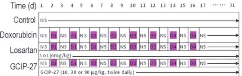
Pharmacy Co., Shenzhen, China) was dissolved in normal saline and administered intraperito-neally at a dose of 1 mg/kg on the 2ndand 4thdays, 2 mg/kg on the 6thand 8thdays, 3 mg/kg on the 10thand 12thdays, and 4 mg/kg on the 14thand 16thdays [21]. An accumulated dose of 20 mg/kg Dox was administered to all of the animals, except those in the control group, at the scheduled time (Fig. 1). The rats in the GCIP-27-treated groups were injected intraperitoneally with 10, 30 or 90μg/kg of GCIP-27 (purity>98%, synthesized by Scilight Biotechnology Co., Beijing, China) dissolved in normal saline (3 ml/kg) twice daily. The rats in the control group were injected with the identical volume of normal saline, and the rats in the losartan-treated group were given 6 mg/kg of losartan (Los, purity>98%, Kerui Pharmaceutical Co., Ltd., Chongqing, China) intragastrically once daily.
Humane end points were set according to the OECDGuidance Document on the Recogni-tion,Assessment,and Use of Clinical Signs as Humane End points for Experimental Animals Used in Safety Evaluation(https://www.aaalac.org/accreditation/RefResources/RR_
HumaneEndpoints.pdf). Specifically, as one was found showing body temperature drop, ap-pearance of hunched and starey coat, or decreased activity with no response to touch, the rat was sacrificed by overdose of pentobarbital. All of the animals survived were treated for 10 weeks and were weighed weekly, and the doses were adjusted accordingly.
Cell culture and treatment
Neonatal rats ventricular myocytes (NRVC) from 1–2 days old Sprague-Dawley rats were iso-lated and cultured as described previously [15,16]. NRVC were washed twice with D-Hanks' solution after being cultured in serum-free DMEM for 24 h, and incubated for 6 h in a
non-Fig 1. Drug administration schedule.NS, normal saline; Los, losartan (6 mg/kg, po., once daily); D1-D4, doxorubicin 1–4 mg/kg (ip., bid).
serum medium containing 0.1μmol/L DOX. Different concentrations of GCIP-27 (3, 10 and
30μg/L) or losartan (10μmol/L) were added to observe its effects. The cell size was determined
with fluorescent phalloidin staining and analyzed with ImageJ software (NIH Image, National Institutes of Health, Bethesda, MD; online at:http://rsbweb.nih.gov/ij/) [22]. And the protein content of cardiomyocytes were measured with the method of Lowry et al using bovine serum albumin as the standard [15] according to the manufacturer's guide.
Echocardiography
After 10 weeks of treatment (described above), all of the rats that survived were subjected to echocardiography measurements [17,23] under anesthesia with chloral hydrate (350 mg/kg, ip). Transthoracic two-dimensional and M-mode images were performed in animals using a Vivid 7 Echo machine with the 14 MHz probe (GE Medical Systems, Minneapolis, MN). M-mode measurements of the left ventricular internal end-diastolic diameter (diastolic LVID) and the posterior wall end-diastolic thickness (diastolic LVPW) were determined as suggested by the American Society of Echocardiography. Simultaneously, the LV diastolic volume (dia-stolic LVV) and function indexes (stroke volume, ejection fraction and fraction shortening) were calculated according to the results of the echocardiography [24]. An experienced sonogra-pher blinded to the treatment groups performed all of the studies. Two observers, blinded to the treatment assignments, analyzed the images.
Tissue Sampling
After recording the echocardiogram, the rats were sacrificed under anesthesia for sampling. The hearts were removed rapidly and washed with normal saline, dried with filter paper and weighed. The atria and free wall of right ventricles were removed, and the interventricular sep-tum was remained. The heart weight index and LV weight index were calculated as the ratio of the heart weight (HW) and left ventricle weight (LVW) to body weight (BW) or tibial length (TL), respectively [15,17,25]. The mid ventricle was fixed with a formalin neutral buffer solu-tion and embedded in paraffin. The apex of the ventricle was cryopreserved with liquid nitro-gen for future use.
Histopathological Analysis
Paraffin-embedded sections of the mid ventricle were stained with hematoxylin and eosin (H&E), as well as Masson’s trichrome [26]. Cross-sectional area (CSA) in each group was ac-quired with a microscopic digital camera system, Nikon ECLIPSE E100 (Nikon, Tokyo, Japan). The fibrosis level was evaluated with a 0–10 scale [27].
Plasma troponin I assay
The plasma cardiac troponin I (cTnI) levels were determined quantitatively using the immuno-chemiluminescence method (Architect i2000sr; Abbot Diagnostics, IL, USA) with an assay kit (Abbot Diagnostics) according to the manufacturer’s protocol.
Sarcoplasmic reticulum Ca
2+ATPase Measurements
manufacturer’s instructions. SERCA2a activity was normalized to the protein concentration [28].
Immunohistochemistry
The myocardial expression levels ofα- andβ-myosin heavy chain (MHC), Bax, as well as Bcl-2 were detected using immunohistochemistry techniques. Sections (10μm) from
paraffin-em-bedded tissues were stained using the simplified histostain SP-kit (Zymed Laboratories, USA) according to the manufacturer's instructions. After deparaffinization and rehydration and the inhibition of endogenous peroxidase, the sections were exposed to primary antibodies at 4°C overnight. After incubation with a secondary antibody at room temperature, the sections were incubated in 3,3 N-diaminobenzidine tetrahydrochloride (DAB) and counterstained with he-matoxylin. The following primary antibodies were used at a dilution of 1:150: anti-α-MHC, anti-β-MHC, anti-Bax and anti-Bcl-2 (Santa Cruz Biotechnology, CA, USA). The secondary antibody was goat-anti-mouse immunoglobulin conjugated with horseradish peroxidase (dilu-tion of 1:200). Digital images of the stained sec(dilu-tions were acquired using a Nikon ECLIPSE E100 microscope with a digital camera system. Unanimity regarding positive immunohisto-chemical staining in each preparation was reached by two blinded investigators using Image-Pro Plus 5.1 software (Media Cybernetics, Silver Spring, MD).
RNA preparation and real-time reverse transcription-PCR
Total RNA was isolated from the ventricular tissue using the Tripure reagent (Invitrogen, USA) according to the manufacturer’s instructions. The RNA samples were dissolved in nucle-ase-free water and treated with 5 U of DNase I (Takara, Shiga, Japan) for 30 min at 37°C. The reaction was stopped by the addition of 25 mmol/L EDTA and a 15-min incubation at 65°C. The total RNA concentration was quantified by measuring the absorbance at 260 nm. Total RNA (1μg) was reverse transcribed using AMV reverse transcriptase (TOYOBO, Osaka,
Japan) at 42°C for 1 h. The PCR primers used were designed by Premier 5.0 (PREMIER Biosoft International, Palo Alto, CA, USA) based on the published nucleotide sequences for ratα -MHC (forward: 5'-TAT GCT GGC ACC GTG GAC TA-3'; reverse: 5'-GAG TTT GAG GGA GGA CTT CTG G-3'),β-MHC (forward: 5'-GGG CAA AGG CAA AGC AAA GA-3’; reverse: 5'-AAA GTG AGG GTG CGT GGA GC-3'), andβ-actin (forward: 5'-CGT AAA GAC CTC TAT GCC AAC A-3’; reverse: 5'-TAG GAG CCA GGG CAG TAA TC-3'). Each real-time PCR reaction was performed in triplicate in a total volume of 25μl with QPK-201 SYBR Green PCR
Master Mix (TOYOBO, Osaka, Japan) using the following conditions: 5 min at 94°C, 38 cycles at 94°C for 30 s, annealing at 58°C for 30 s, 72°C for 45 s, and 82.5°C for 5 s (collecting fluores-cence) with the ABI Prism 7700 sequence detection system (ABI, Oyster Bay, NY, USA). After amplification, a melting curve analysis was performed by collecting fluorescence data while in-creasing the temperature from 72°C to 99°C over 135 s. The Ct (cycle threshold) values were normalized to theβ-actin expression levels.
Western blot assay
The myocardial expression levels of protein kinase (PK)-Cε, -βII and phosphor-extracellular signal regulated kinase (pERK)1/2 were assayed by Western blot. Total cellular homogenates were prepared, and equal amounts (20μg) of the denatured proteins were loaded and separated
HRP-conjugated anti-mouse IgG. Chemiluminescence was detected with an ECL western blot detec-tion kit (Millipore, Bedford, MA) according to the supplier’s recommendadetec-tions. The data were analyzed by an observer who was blind to the treatment given to the rats.
Statistical analysis
All values are expressed as the mean ± SEM. The differences between the groups were assessed by one-way ANOVA with least significant difference (LSD) post-hoc analyses. The differences were considered significant when P<0.05. All of the statistical analyses were performed using the Statistical Package for Social Sciences for Windows software, version 10.0J (SPSS Co., Inc., Chicago, IL).
Results
General condition and mortality
The animals were observed continuously for 10 weeks. All of the rats in the Dox group showed weight loss, lethargy and less activity. In contrast to those treated with Dox, the rats in the con-trol group, losartan group, and GCIP-27 groups were more healthy and active. During the en-tire experimental period, one rat in control group, 3 rats in Dox, Los and 10μg/kg GCIP-27
group, 2 rats in 30 and 90μg/kg GCIP-27 group, respectively, were sacrificed before the
hu-mane endpoints. There were no significant differences in the cumulative mortality between all the groups.
Echocardiography Measurements
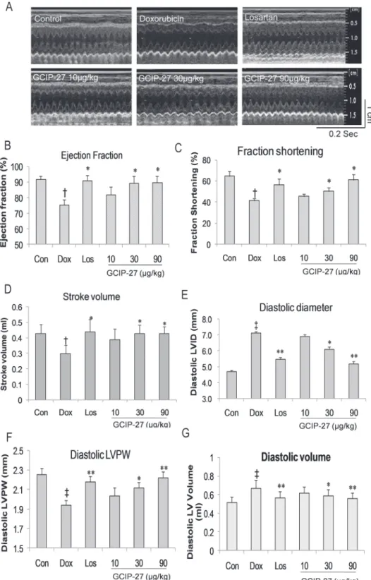
The structure and function of the left ventricles were measured using high frequency echocar-diography (Fig. 2A). The diastolic LVID and LVV in the Dox group increased significantly compared with that in the control group (Fig. 2E and G;P<0.01). The diastolic LVPW, left ventricular ejection fraction (EF), fraction shortening (FS) and stroke volume in the Dox group were markedly decreased compared with those in the control group (Fig. 2B, C, D, F;P<0.05 or 0.01). Doses of 30 and 90μg/kg of GCIP-27 and losartan, but not 10μg/kg of GCIP-27,
ame-liorated the aforementioned indexes compared with those in the Dox group (Fig. 2B-G,
P<0.05 or 0.01).
Heart indexes
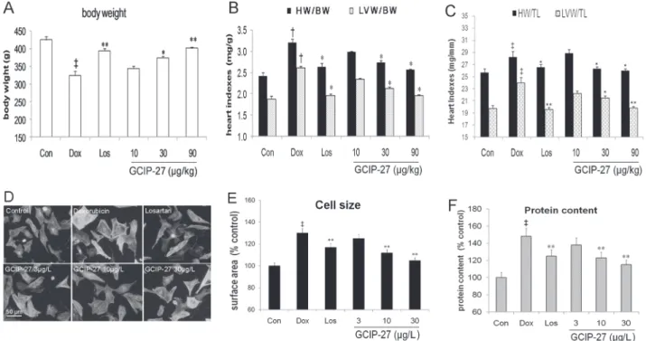
The heart index HW/BW and HW/TL, LV index LVW/BW and LVW/TL of the rats were measured to explore cardiac hypertrophy. Although Dox markedly decreased the body weight compared with that in the control group, the treatment with GCIP-27 (30 and 90μg/kg) and
losartan obviously ameliorated weight loss (P<0.01 or 0.05,Fig. 3A). Additionally, Dox in-creased heart index and LV index significantly (P<0.05,Fig. 3B and 3C). GCIP-27 (30 and 90μg/kg) and losartan remarkably decreased heart index and LV index compared with those
of the rats in the Dox group (P<0.01). There was no significant difference between the GCIP 10μg/kg group and the Dox group.
Cell size and protein content
Surface area and protein content of primary cardiomyocytes were measured to assess its hyper-trophic response induced by Dox. As shown inFig. 3D-F, at a dose of 0.1μmol/L, Dox
Fig 2. Cardiac function and structure were measured with echocardiography.(A). Ejection fraction (B), fraction shortening (C), stroke volume (D), left ventricular internal diastolic diameter (E), left ventricular posterior wall (LVPW) end diastolic thickness (F), and left ventricular end diastolic volume (G) were calculated with the formula stored in the echo machine by the manufacturer. The data are presented as the mean±SEM (n = 5–7 in each group). Scale for time = 0.2 sec; scale for length = 1cm.†P
<0.05,‡P<0.01 compared with the control group (Con);*P<0.05,**P<0.01 compared with the Dox group (one-way ANOVA).
Pathological Findings
The myofibrils were lined up without disruption, and the structure of the nuclei and cells were normal in the control group. In the Dox group, myocardial fiber disruption and disarray could be observed, and the hyperplastic cardiomyocytes were infiltrated with inflammatory cells. Ad-ditionally, CSA and fibrosis score increased significantly (P<0.01) compared with control group (Fig. 4). After treatment with 30 or 90μg/kg of GCIP-27, the injury and pathological
changes of the myocardium tissue improved markedly with less inflammatory cell infiltration and fiber disarray. After these treatments, CSA and fibrosis score decreased significantly (Fig. 4C and 4D).
Plasma cTnI concentration and cardiac SERCA2a activity
To evaluate the biochemical foundation of the systolic and diastolic function of the heart, plas-ma cTnI and ventricular SERCA2a activity were determined. The Dox treatment induced a marked increase in cTnI content compared with the control group (P<0.01) (Fig. 5A). Simul-taneously, the GCIP-27 (10, 30, 90μg/kg) and losartan treatments decreased the cTnI
concen-tration significantly compared with that of the Dox group (P<0.05 or 0.01). In sharp contrast to the increase of plasma cTnI, Dox obviously reduced the SERCA2a activity, and the GCIP-27 treatment elevated it significantly (Fig. 5B). The effects of GCIP-27 on the cTnI content and SERCA2a activity were obviously dose-dependent.
Fig 3. The effects of the GCIP-27 on hypertrophic response in cardiomyocytesin vitroand heart indexes in rats with heart failure induced by doxorubicin.Chronic heart failure was induced by a cumulative dose of 20 mg/kg doxorubicin (Dox). (A) body weight (n = 5–7 in each group); (B) Heart and left ventricular (LV) weight index to body weight; (C) Heart and LV indexes to tibia length; (D) Neonatal rats ventricular cells treated with Dox (0.1μmol/L) and Los or GCIP-27 for 6 h, stained with FITC-phalloidin (bar = 50μm); (E) cell size (n = 30 cells in each group); (F) protein content (n = 6 wells in each group)The data are presented as the mean±SEM.‡P
<0.01 compared with the control group (Con);*P<0.05,**P<0.01 compared with the Dox group (one-way ANOVA).
Myocardial expressions of
α
-MHC and
β
-MHC
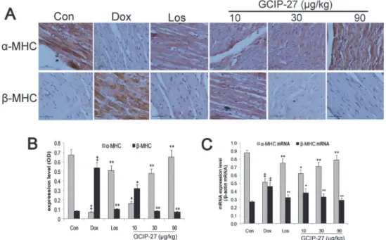
The baseline myocardial expression ofα-MHC andβ-MHC were shown as controls inFig. 6A. The expression level ofα-MHC in the Dox group was markedly decreased compared with that of the control group. The GCIP-27 (10, 30 and 90μg/kg) and losartan treatments significantly
elevated theα-MHC levels compared with those of the Dox group (Fig. 6A and 6B;P<0.01). The expression ofβ-MHC showed an opposite trend compared with that ofα-MHC. The ratio of the expression ofα-MHC to that ofβ-MHC had a similar change pattern as that shown forα-MHC; this change pattern was more significant. Treatment with GCIP-27 (10, 30 and 90μg/kg) and losartan noticeably elevated the myocardial expression ofα-MHC mRNA
Fig 4. Myocardial pathological changes after treatment with GCIP-27.Chronic heart failure was induced by a cumulative dose of 20 mg/kg doxorubicin (Dox). (A) H&E staining (bar = 25μm): (a) control, (b) Dox, (c) losartan (Los) 6mg/kg, (d) GCIP-27 10μg/kg, (e) GCIP-27 30μg/kg, (f) GCIP-27 90μg/kg; ip., bid; (B) Cross-sectional area; (C) Masson’s Trichrome staining (bar = 20μm), panel (a)-(f) represent same group as that in (A); (D) Fibrosis score. The data are presented as the mean±SEM (n = 5–7 in each group).‡P
<0.01 compared with the control group (Con);*P<0.05,**P<0.01 compared with the Dox group (one-way ANOVA).
doi:10.1371/journal.pone.0121007.g004
Fig 5. The effects of the GCIP-27 on serum cardiac troponin-I (cTnI) (A) and myocardial sarcoplasmic reticulum Ca2+ATPase (SERCA2a) activity (B) in rats with doxorubicin-induced heart failure.Chronic
heart failure was induced by a cumulative dose of 20 mg/kg doxorubicin (Dox). The rats were treated with normal saline, losartan (Los, 6 mg/kg, ig, once daily) or GCIP-27 (10, 30, 90μg/kg, ip, bid) for 10 weeks. The data are presented as the mean±SEM (n = 5–7 in each group).‡P<
0.01 compared with the control group (Con);*P<0.05,**P<0.01 compared with the Dox group (one-way ANOVA).
compared with that of the Dox group, whereas they decreased the expression ofβ-MHC mRNA (Fig. 6C;P<0.01 or 0.05).
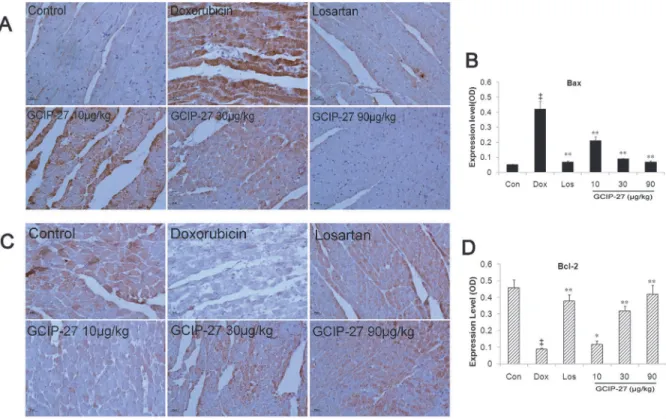
Expression of Bax and Bcl-2
Myocardial expression of Bax and Bcl-2 were measured to evaluate the effect of GCIP-27 on apoptosis induced by Dox. As shown inFig. 7, the expression of Bax increased and that of Bcl-2 reduced significantly in Dox group compared with controls. Treatment of Los and GCIP-Bcl-27 (10–90μg/kg) reduced myocardial Bax expression and elevated Bcl-2 expression significantly
(P<0.01 or 0.05).
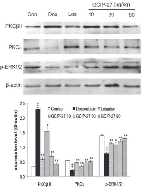
Expression of PKC
β
II, PKC and p-ERK1/2
Myocardial expression of PKCβII, PKC and p-ERK1/2 was detected by Western blotting (Fig. 8A). Compared with the control group, the expression of PKCβⅡincreased while the PKC
expression decreased significantly in the Dox group (Fig. 8B;P<0.01). Treatment with GCIP-27 (10, 30 and 90μg/kg) and losartan effectively ameliorated these abnormal expressions
com-pared with those in the rats in the Dox group (P<0.01 or 0.05). Simultaneously, Dox reduced the myocardial expression of p-ERK1/2 (P<0.01), and GCIP-27 (10, 30 and 90μg/kg) elevated its expression compared with the Dox group (P<0.05 or 0.01).
Discussion
Several studies have demonstrated a beneficial effect of the GCIP-27 polypeptide on cardiac hy-pertrophy induced by a variety of factors in vivo and in vitro [15–19]. Cardiac hyhy-pertrophy is an important factor in the development of CHF. Cardiac hypertrophy is typically believed to
Fig 6. The effects of GCIP-27 on the myocardial expression of myosin heavy chains (MHCs).(A) The expression levels ofα-MHC andβ-MHC were determined with immunohistochemistry (bar = 10μm). (B) Myocardial expression (mean±SEM) ofα- andβ-MHC. (C) The myocardial mRNA expression ofα- andβ -MHC measured with real-time PCR. The data are presented as the mean±SEM (n = 5–7 in each group).
‡P
<0.01 compared with the control group (Con);*P<0.05,**P<0.01 compared with the Dox group (one-way ANOVA).
be a compensatory mechanism of the heart in response to increased systemic demand for car-diac output. Given this premise, preventing or reversing ventricular remodeling might impair heart function during the compensatory stage and the heart failure stage. As GCIP-27 may re-duce cardiac hypertrophy, it becomes important to determine whether GCIP-27 is beneficial to heart functioning during CHF. In this study, GCIP-27 treatment was shown to markedly in-crease body weight, improve survival, and halt the process of hypertrophy and heart failure in doxorubicin-induced CHF rats, preventing adverse ventricular remodeling imposed by Dox.
Due to the cardiac toxicity and other adverse effects of doxorubicin, rats receiving Dox may eat less and gain less body weight [29]. Along with the decrease of the heart function, concen-tration of plasmic catecholamine, such as norepinephrine (NE) increase significantly [30], which can reduce appetite and food intake throughα1-adrenoceptor [31]. GCIP-27 is a syn-thetic peptide, which imitate the structure of carboxyl terminus of Gq proteinαsubunit and can inhibit the signal transduction of the Gq-coupled receptors includingα1-adrenoceptor.
Therefore, GCIP-27 not only ameliorated heart function but also increased food intake and body weight. Similarly, losartan can reduce afterload of the heart and improve heart function through blocking angiotensin II type 1 receptor, and subsequently decrease catecholamine and reduce body weight loss.
Additionally, it has been shown that GCIP-27 was superior at inhibiting ventricular remod-eling compared with losartan [17]. In the present study, B-mode and M-mode echocardiogra-phy revealed that GCIP-27 was able to improve heart function. These results indicated that this polypeptide drug produced favorable effects on doxorubicin-induced CHF in rats. Although
Fig 7. The effects of GCIP-27 on the myocardial expression of Bax and Bcl-2.The expression levels of Bax (A) and Bcl-2 (C) were determined with immunohistochemistry (bar = 10μm). Bar graphs representing the myocardial expression of Bax (B) and Bcl-2. (D). The data are presented as the mean±SEM (n = 5–7 in each group).‡P<0.01 compared with the control group (Con);
*P<0.05,**P<0.01 compared with the Dox group (one-way ANOVA).
GCIP-27could lower blood pressure in spontaneously hypertensive rats, the hypotensive effect of GCIP-27 is significant weaker than that of losartan. And due to compensation, the normo-tensive animals, such as rats, dogs and patients, are less sensitive to hyponormo-tensive agents, such as losartan and nitroprusside, etc., than hypertensive subjects [32,33]. We have also ever observed that GCIP-27 had no influence on the blood pressure in normotensive rats [17,19]. In doxoru-bicin-induced heart failure rats, blood pressure and hemodynamic parameters showed a slight decrease or no change [34,35]. Therefore, the hemodynamics factors contributed not as much as remodeling to the mechanism for GCIP-27-induced improvement of heart function.
In the current study, chloral hydrate was used to anesthetize the rats before measuring car-diac function and tissue collection. Although chloral hydrate has various adverse effects
Fig 8. The expression of protein kinase C (PKC)βII, PKCεand p-extracellular signal-regulated kinase (ERK).(A) Western blot; (B) Averaged myocardial expression (mean±the SEM) of the proteins mentioned above.†P
<0.05,‡P<0.01 compared with the control group;*P<0.05,**P<0.01 compared with the doxorubicin group (one-way ANOVA).
[36,37], such as not providing analgesia, prolonging recovery time after surgery, inducing mu-tagenesis and carcinogenesis, it is considered by some a good sedative-hypnotics for animals. Experimental anesthesia based on an intraperitoneal injection of chloral hydrate is believed to have minimal effects on cardiovascular function or reflexes, while ketamine and pentobarbital sodium can lead to a significant decrease in heart function indexes and survival rate [36,38]. Therefore, chloral hydrate is occasionally still used as an anesthetic agent in the laboratory [39–41]. Even though chloral hydrate has not been recommended for animal euthanasia in the CCAC guidelines, it was in line with the 10 general guiding principles listed in "CCAC guide-lines on:euthanasia of animals used in science" (http://www.ccac.ca/Documents/Standards/ Guidelines/Euthanasia.pdf), because it can result in rapid loss of consciousness and cause little distress and pain to the animals when used terminally. Meanwhile, gaseous anesthetics such as isoflurane may present health hazards to humans if not properly scavenged, for which special equipment is needed.
As the main contractile protein of myocardium, myoglobulin constitutes 60% of the mass of the entire heart and consists of one pair of MHCs and two pairs of MLCs. The two types of MHCs areα-MHC andβ-MHC. The myocardial MHC phenotype changes correspond to the different stages of growth and development [42]. Accumulating research has suggested that the essence of myocardial remodeling is the process of the transformation of the myocardial cell phenotypes [43]. The readjustment of the myocardial contractile proteins occurs in all types of emergency situations, and the change fromα-MHC toβ-MHC is regarded as a molecular marker for hypertrophied or failing myocardium [44,45]. In this research, we observed that GCIP-27 clearly increased the myocardial expression ofα-MHC and decreasedβ-MHC pression in rats with CHF. GCIP-27 is able to correct the imbalance of myosin heavy chain ex-pression and thereby enhance myocardial contractility.
Additionally, GCIP-27 treatment maintained the activity of the sarcoplasmic reticulum Ca2+ATPase (SERCA) 2a and reversed the intracellular calcium overload as well. In cardiac muscle, the sarcoplasmic reticulum plays an important role in excitation-contraction coupling through the regulation of intracellular free-Ca2+concentrations [46]. Muscle relaxation is initi-ated by Ca2+transport from the cytosol into the sarcoplasmic reticulum by cardiac SERCA2a. The downregulation of SERCA2a has been reported to be a sign for the transition from com-pensated hypertrophy to a decomcom-pensated stage of CHF [47]. Many observations have sug-gested that the downregulation of SERCA2 might occur through a protein kinase C (PKC)-related process [48–50].
The PKC family consists of 15 isoenzymes, including PKCα, PKCβ, PKCε, PKCδ, PKCη
and PKCμ. A study in a PKCε-knockout mouse model demonstrated that PKCεexpression is
not required for cardiac function under normal physiological conditions; however, PKCε acti-vation is necessary for cardioprotection in myocardial hypertrophy and heart failure [51]. PKCαand PKCβincrease their expression and thereby decrease the contractile ability of cardi-omyocytes during myocardial hypertrophy and heart failure [52–54]. It is reported that the postnatal cardiac-specific overexpression of the PKC-βisoform in transgenic mice caused car-diomyopathy with LV hypertrophy andin vivocardiac dysfunction [55]. All of the Gq-coupled receptors associated with remodeling in the myocardium, including endothelin ET1receptor,
type I angiotensin II receptor, and theα1adrenergic receptor, lead to the progression of
2 (ERK 1/2) might be concordantly regulated in the process of cardiac hypertrophy, extending to CHF [59]. It has been reported that the activation of ERK activity promotes a compensated form of hypertrophy [60,61]. In this study, we observed that GCIP-27 could obviously increase PKCεexpression in the rats with chronic heart failure, as well as reduce PKCβII expression. Si-multaneously, ERK1/2 was activated by GCIP-27.
Dox induced heart failure through increasing oxidative stress, inflammation and apoptosis of cardiomyocytes [62]. In this process, Gαq-PKCεsignaling is involved [55]. It has been re-ported that overexpression of Gαq resulted in obvious hypertrophic growth and apoptosis of cardiomyocytes and heart failure, and activating of PKCεwas able to blunt apoptosis and therefore heart failure [63,64]. As an imitation peptide of Gαq, GCIP-27 exerted anti-apopto-sis effects by elevating expression of Bcl-2 and reducing that of Bax.
As a peptide, transport across the cell membrane is critical for GCIP-27 to produce its ef-fects. In this systematic study, we found [16] that GCIP-27 could be transported through the plasmalemma in a time- and concentration-dependent manner, which was mediated by an en-ergy-dependent endocytosis process. This peptide could preferentially enter myocardial cells and VSMCs, which is especially beneficial for the treatment of cardiac hypertrophy and CHF.
In conclusion, GCIP-27 could beneficially influence heart function and delay the onset of doxorubicin-induced CHF in rats. The regulation of the PKCβII andεisoforms and ERK1/2 was involved in the intracellular signaling pathways leading to CHF. PKC–ERK1/2 signaling might represent the underlying mechanism responsible for the beneficial effect of GCIP-27.
Acknowledgments
This research was supported by grants from the National Key New Drug Development Project of China (2009ZX09103-052), the National Natural Science Foundation of China (No. 30973524), and the Basic and Frontier Research Program of Natural Science Foundation of Chongqing, China (No. CSTC2013jcyjA10094).
Author Contributions
Conceived and designed the experiments: HGZ. Performed the experiments: XLL YFT YL HY YLX GYZ. Analyzed the data: HGZ XHL. Contributed reagents/materials/analysis tools: HGZ XHL. Wrote the paper: XLL YL HGZ.
References
1. Zatoński WA; HEM project team. Epidemiological analysis of health situation development in Europe and its causes until 1990. (2011) Ann Agric Environ Med 18:194–202. PMID:22324071
2. Szuba A, Martynowicz H, Zatońska K, Ilow R, Regulska-Ilow B, Różańska D, et al. Prevalence of hyper-tension in a sample of Polish population—baseline assessment from the prospective cohort 'PONS' study. (2011) Ann Agric Environ Med 18:260–4. PMID:22216793
3. Cahalin LP. Heart failure. (1996) Phys Ther 76: 516–33. PMID:8637939
4. Dalal HM, Wingham J, Palmer J, Taylor R, Petre C, Lewin R, et al. Why do so few patients with heart failure participate in cardiac rehabilitation? A cross-sectional survey from England, Wales and Northern Ireland. (2012) BMJ Open 2:e000787. doi:10.1136/bmjopen-2011-000787PMID:22454188
5. Waagstein F, Caidahl K, Wallentin I, Bergh CH, Hjalmarson A. Long-term beta-blockade in dilated car-diomyopathy. Effects of short- and long-term metoprolol treatment followed by withdrawal and readmi-nistration of metoprolol. (1989) Circulation 80:551–63. PMID:2548768
6. Sütsch G, Bertel O, Kiowski W. Acute and short-term effects of the nonpeptide endothelin-1 receptor antagonist bosentan in humans. (1997) Cardiovasc Drugs Ther 10:717–25. PMID:9110115 7. Hunt SA, Abraham WT, Chin MH, Feldman AM, Francis GS, Ganiats TG, et al. 2009 Focused update
Lung Transplantation. (2009) J Am Coll Cardiol 53(15):e1–e90. doi:10.1016/j.jacc.2008.11.013PMID: 19358937
8. McMurray JJ, Adamopoulos S, Anker SD, Auricchio A, Böhm M, Dickstein K, et al.ESC Guidelines for the diagnosis and treatment of acute and chronic heart failure 2012: The Task Force for the Diagnosis and Treatment of Acute and Chronic Heart Failure 2012 of the European Society of Cardiology. Devel-oped in collaboration with the Heart Failure Association (HFA) of the ESC. (2012) Eur Heart J 33:1787–847. doi:10.1093/eurheartj/ehs104PMID:22611136
9. Neubauer S. The failing heart—an engine out of fuel. (2007) N Engl J Med 356:1140–51. PMID: 17360992
10. Oka T, Akazawa H, Naito AT, Komuro I. Angiogenesis and cardiac hypertrophy: maintenance of cardi-ac function and causative roles in heart failure. (2014) Circ Res 114:565–71. doi:10.1161/
CIRCRESAHA.114.300507PMID:24481846
11. Francis GS. Pathophysiology of chronic heart failure. (2001) Am J Med 110:37S–46S. PMID: 11334774
12. Hein S, Arnon E, Kostin S, Schönburg M, Elsässer A, Polyakova V, Bauer EP, Klövekorn WP, Schaper J. Progression from compensated hypertrophy to failure in the pressure-overloaded human heart: struc-tural deterioration and compensatory mechanisms. (2003) Circulation 107:984–91. PMID:12600911 13. Morisco C, Sadoshima J, Trimarco B, Arora R, Vatner DE, Vatner SF. Is treating cardiac hypertrophy
salutary or detrimental: the two faces of Janus. (2003) Am J Physiol Heart Circ Physiol 284:H1043–H7. PMID:12666659
14. Merlo M, Pivetta A, Pinamonti B, Stolfo D, Zecchin M, Barbati G, Di Lenarda A, Sinagra G. Long-term prognostic impact of therapeutic strategies in patients with idiopathic dilated cardiomyopathy: changing mortality over the last 30 years. (2013) Eur J Heart Fail Dec 14. doi:10.1002/ejhf.16[Epub ahead of print]
15. Zhang HG, Li XH, Zhou JZ, Liu Y, Jia Y, Yuan ZB. Gαq-Protein carboxyl terminus imitation polypeptide GCIP-27 attenuates cardiac hypertrophy in vitro and in vivo. (2007) Clin Exp Pharmacol Physiol 34:1276–81. PMID:17973867
16. Yang H, Liu Y, Lu XL, Li XH, Zhang HG. Transmembrane transport of the Gαq protein carboxyl terminus imitation polypeptide GCIP-27. (2013) Eur J Pharm Sci 49:791–9. doi:10.1016/j.ejps.2013.05.028 PMID:23748000
17. Wang XQ, Zhang HG, Cheng YQ, Li XH. Inhibition of left ventricular remodeling in spontaneously hy-pertensive rats by Gαq-protein carboxy terminus imitation polypeptide GCIP-27 is not entirely depen-dent on blood. (2008) Clin Exp Pharmacol Physiol 35:1215–21. doi:10.1111/j.1440-1681.2008.04981. xPMID:18518877
18. Yang DL, Zhang HG, Xu YL, Gao YH, Yang XJ, Hao XQ, Su M, Wang XQ, Li XH. Gαq-Protein carboxyl terminus imitation polypeptide (GCIP)-27 inhibits right ventricular hypertrophy induced by monocrota-line in rats. (2009) Biol Pharm Bull 32:376–81. PMID:19252281
19. Zhang HG, Cheng YQ, Liu Y, Zhou JZ, Jia Y, Wang XQ, Li XH. Gαq-protein carboxyl terminus imitation polypeptide GCIP-27 attenuates proliferation of vascular smooth muscle cells and vascular remodeling in spontaneously hypertensive rats. (2011) Biol Pharm Bull 34:1527–32. PMID:21963491
20. Sankar V, Nair RR, Harikrishnan VS, Fernandez AC, Kumar CS, Madhavachandran V. Cardoguard, an Ayurvedic antihypertensive formulation, prevents cardiac remodeling in spontaneously hypertensive rats by inhibition of ERK and PKCεsignaling pathways. (2012) Can J Physiol Pharmacol 90:627–35. doi:10.1139/y2012-047PMID:22550975
21. Wang HJ, Chen JS, Chen XM, Wu TM, Xu CS, Xie LD. Change of mysion heavy chain in heart failure rats induced by doxorbincin. (2001) Chin J Lab Anim Sci 11:202–5.
22. Spallarossa P, Altieri P, Aloi C, Garibaldi S, Barisione C, Ghigliotti G, Fugazza G, Barsotti A, Brunelli C. Doxorubicin induces senescence or apoptosis in rat neonatal cardiomyocytes by regulating the expres-sion levels of the telomere binding factors 1 and 2. (2009) Am J Physiol Heart Circ Physiol. 297: H2169–81. doi:10.1152/ajpheart.00068.2009PMID:19801496
23. Migrino RQ, Aggarwal D, Konorev E, Brahmbhatt T, Bright M, Kalyanaraman B. Early detection of doxo-rubicin cardiomyopathy using two-dimendional atrain echocardiography. (2008) Ultrasound in Med & Biol 34: 208–14.
24. Benavides-Vallve C, Corbacho D, Iglesias-Garcia O, Pelacho B, Albiasu E, Castaño S, Muñoz-Barrutia A, Prosper F, Ortiz-de-Solorzano C. New strategies for echocardiographic evaluation of left ventricular function in a mouse model of long-term myocardial infarction. (2012) PLoS One. 7(7):e41691. doi:10. 1371/journal.pone.0041691PMID:22848568
26. Chen X, Nakayama H, Zhang X, Ai X, Harris DM, Tang M, Zhang H, Szeto C, Stockbower K, Berretta RM, Eckhart AD, Koch WJ, Molkentin JD, Houser SR. Calcium influx through cav1.2 is a proximal sig-nal for pathological cardiomyocyte hypertrophy. (2011) J Mol Cell Cardiol 50:460–70 doi:10.1016/j. yjmcc.2010.11.012PMID:21111744
27. Dong R, Liu P, Wee L, Butany J, Sole MJ. Verapamil ameliorates the clinical and pathological course of murine myocarditis. The Journal of clinical investigation. 1992; 90:2022–30. PMID:1331179
28. Xin W, Lu X, Li X, Niu K, Cai J. Attenuation of endoplasmic reticulum stress–related myocardial apopto-sis by SERCA2a gene delivery in ischemic heart disease. (2011) Mol Med 17: 201–10. doi:10.2119/ molmed.2010.00197PMID:21152695
29. van Leeuwen BL, Kamps WA, Hartel RM, Veth RP, Sluiter WJ, Hoekstra HJ. Effect of single chemo-therapeutic agents on the growing skeleton of the rat. (2000) Ann Oncol. 11:1121–6. PMID:11061605 30. Kjaer A, Hesse B. Heart failure and neuroendocrine activation: diagnostic, prognostic and therapeutic
perspectives. (2001) Clin Physiol. 21:661–72. PMID:11722473
31. Wellman PJ. Norepinephrine and the control of food intake. (2000) Nutrition. 16:837–42. PMID: 11054588
32. Farah VM, Moreira ED, Ushizima M, Cestari IA, Irigoyen MC, Krieger EM. Acute AT1 receptor blockade does not improve the depressed baroreflex in rats with chronic renal hypertension. (2000) Braz J Med Biol Res. 33:1491–6. PMID:11105103
33. Ventura HO. Nitroprusside and the treatment of hypertension. (2011) Congest Heart Fail. 17:49. doi: 10.1111/j.1751-7133.2010.00197.xPMID:21272230
34. Rabelo E, De Angelis K, Bock P, Gatelli Fernandes T, Cervo F, Belló Klein A, Clausell N, Cláudia Irigo-yen M. Baroreflex sensitivity and oxidative stress in adriamycin-induced heart failure. (2001) Hyperten-sion. 38:576–80. PMID:11566934
35. Pacher P, Liaudet L, Bai P, Virag L, Mabley JG, Haskó G, Szabó C. Activation of poly(ADP-ribose) poly-merase contributes to development of doxorubicin-induced heart failure. (2002) J Pharmacol Exp Ther. 300:862–7. PMID:11861791
36. Maud P, Thavarak O, Cédrick L, Michèle B, Vincent B, Olivier P, Régis B. Evidence for the use of iso-flurane as a replacement for chloral hydrate anesthesia in experimental stroke: an ethical issue. (2014) Biomed Res Int. 2014:802539. doi:10.1155/2014/802539PMID:24719888
37. Baxter MG, Murphy KL, Taylor PM, Wolfensohn SE. Chloral hydrate is not acceptable for anesthesia or euthanasia of small animals. (2009) Anesthesiology. 111:209 doi:10.1097/ALN.0b013e3181a8617e PMID:19546703
38. Jiang X, Gao L, Zhang Y, Wang G, Liu Y, Yan C, Sun H. A comparison of the effects of ketamine, chlo-ral hydrate and pentobarbital sodium anesthesia on isolated rat hearts and cardiomyocytes. (2011) J Cardiovasc Med (Hagerstown). 12:732–5. doi:10.2459/JCM.0b013e32834a6697PMID:21873882 39. Monnerat-Cahli G, Alonso H, Gallego M, Alarcón ML, Bassani RA, Casis O, Medei E. Toll-like receptor
4 activation promotes cardiac arrhythmias by decreasing the transient outward potassium current (Ito) through an IRF3-dependent and MyD88-independent pathway. (2014) J Mol Cell Cardiol. 76:116–25. doi:10.1016/j.yjmcc.2014.08.012PMID:25169970
40. Liu J, Han P, Xiao Y, Liu J, Kang YJ. A novel knot method for individually measurable aortic constriction in rats. (2014) Am J Physiol Heart Circ Physiol. 307(7):H987–95. doi:10.1152/ajpheart.00990.2013 PMID:25108013
41. Hung KC, Huang HJ, Lin MW, Lei YP, Lin AM. Roles of autophagy in MPP+-induced neurotoxicity in vivo: the involvement of mitochondria andα-synuclein aggregation. (2014) PLoS One. 9:e91074. doi: 10.1371/journal.pone.0091074PMID:24646838
42. Cheng Y, Li W, McElfresh TA, Chen X, Berthiaume JM, Castel L, Yu X, Van Wagoner DR, Chandler MP. Changes in myofilament proteins, but not Ca2+regulation, are associated with a high-fat
diet-in-duced improvement in contractile function in heart failure. (2011) Am J Physiol Heart Circ Physiol 301: H1438–46. doi:10.1152/ajpheart.00440.2011PMID:21765056
43. Murray TV, Smyrnias I, Shah AM, Brewer AC. NADPH oxidase 4 regulates cardiomyocyte differentia-tion via redox activadifferentia-tion of c-Jun protein and the cis-reguladifferentia-tion of GATA-4 gene transcripdifferentia-tion. (2013) J Biol Chem 288:15745–59. doi:10.1074/jbc.M112.439844PMID:23589292
44. Hang CT, Yang J, Han P, Cheng HL, Shang C, Ashley E, Zhou B, Chang CP. Chromatin regulation by Brg1 underlies heart muscle development and disease. (2010) Nature 466:62–7. doi:10.1038/ nature09130PMID:20596014
46. Arai M, Matsui H, Periasamy M. Sarcoplasmic reticulum gene expression in cardiac hypertrophy and heart failure. (1994) Circ Res 74:555–64. PMID:8137493
47. Kiss E, Ball NA, Kranias EG, Walsh RA. Differential changes in cardiac phospholamban and sarcoplas-mic reticular Ca (2+)-ATPase protein levels. Effects on Ca2+transport and mechanics in compensated pressure-overload hypertrophy and congestive heart failure. (1995) Circ Res 77:759–64. PMID: 7554123
48. Hartong R, Villarreal FJ, Giordano F, Hilal-Dandan R, McDonough PM, Dillmann WH. Phorbol myristate acetate-induced hypertrophy of neonatal rat cardiac myocytes is associated with decreased sarcoplas-mic reticulum Ca2+ATPase (SERCA2) gene expression and calcium reuptake. (1996) J Mol Cell
Car-diol 28:2467–77. PMID:9004163
49. Rogers TB, Gaa ST, Massey C, Dösemeci A. Protein kinase C inhibits Ca2+accumulation in cardiac
sarcoplasmic reticulum. (1990) J Biol Chem 265:4302–8. PMID:2155221
50. Qi M, Bassani JW, Bers DM, Samarel AM. Phorbol 12-myristate 13-acetate alters SR Ca (2+)-ATPase gene expression in cultured neonatal rat heart cells. (1996) Am J Physiol 271:H1031–9. PMID: 8853338
51. Gray MO, Zhou HZ, Schafhalter-Zoppoth I, Zhu P, Mochly-Rosen D, Messing RO. Preservation of base-line hemodynamic function and loss of inducible cardioprotection in adult mice lacking protein ki-nase C epsilon. (2004) J Biol Chem 279:3596–604. PMID:14600145
52. Lei S, Li H, Xu J, Liu Y, Gao X, Wang J, et al. Hyperglycemia-induced protein kinase Cβ2 activation in-duces diastolic cardiac dysfunction in diabetic rats by impairing caveolin-3 expression and Akt/eNOS signaling. (2013) Diabetes 62:2318–28. doi:10.2337/db12-1391PMID:23474486
53. Braz JC, Bueno OF, De Windt LJ, Molkentin JD. PKCαregulates the hypertrophic growth of cardiomyo-cytes through extracellular signal-regulated kinase1/2(ERK1/2). (2002) J Cell Biol 156:905–19. PMID: 11864993
54. Pass JM, Gao J, Jones WK, Wead WB, Wu X, Zhang J, et al. Enhanced PKCβII translocation and PKCβII-RACK1 interactions in PKCε-induced heart failure: a role for RACK1. (2001) Am J Physiol Heart Circ Physiol 281:2500–10.
55. Wakasaki H, Koya D, Schoen FJ, Jirousek MR, Ways DK, Hoit BD, et al. Targeted overexpression of protein kinase C beta2 isoform in myocardium causes cardiomyopathy. (1997) Proc Natl Acad Sci USA 94:9320–5. PMID:9256480
56. D'Angelo DD, Sakata Y, Lorenz JN, Boivin GP, Walsh RA, Liggett SB, et al. Transgenic Galphaq over-expression induces cardiac contractile failure in mice. (1997) Proc Natl Acad Sci USA 94:8121–6. PMID:9223325
57. Braz JC, Bueno OF, De Windt LJ, Molkentin JD. PKC alpha regulates the hypertrophic growth of cardio-myocytes through extracellular signal-regulated kinase1/2 (ERK1/2). (2002) J Cell Biol 156:905–19. PMID:11864993
58. Vijayan K, Szotek EL, Martin JL, Samarel AM. Protein kinase C-alpha-induced hypertrophy of neonatal rat ventricular myocytes. (2004) Am J Physiol Heart Circ Physiol 287:H2777–89. PMID:15271671 59. Takahashi H, Takeishi Y, Miyamoto T, Shishido T, Arimoto T, Konta T, et al. Protein kinase C and
extra-cellular signal regulated kinase are involved in cardiac hypertrophy of rats with progressive renal injury. (2004) Eur J Clin Invest 34:85–93. PMID:14764070
60. Wang Y. Mitogen-activated protein kinases in heart development and diseases. (2007) Circulation 116:1413–23. PMID:17875982
61. Qin F, Liang MC, Liang CS. Progressive left ventricular remodeling, myocyte apoptosis, and protein sig-naling cascades after myocardial infarction in rabbits. (2005) Biochim Biophys Acta 1740:499–513. PMID:15949720
62. Singal PK, Li T, Kumar D, Danelisen I, Iliskovic N. Adriamycin-induced heart failure: mechanism and modulation. (2000) Mol Cell Biochem 207(1–2):77–86. PMID:10888239
63. Baines CP, Molkentin JD. STRESS signaling pathways that modulate cardiac myocyte apoptosis. 2005 J Mol Cell Cardiol 38:47–62. PMID:15623421