175
Magnetic resonance imaging in radiotherapy planning
Radiol Bras. 2010 Mai/Jun;43(3):175–178
Original Article • Artigo Original
Magnetic resonance imaging in the evaluation
of standard radiotherapy field borders in patients
with uterine cervix cancer*
Ressonância magnética para avaliação dos limites dos campos clássicos de radioterapia em pacientes portadoras de neoplasia maligna de colo uterino
Geison Moreira Freire1, Rodrigo Souza Dias2, Adelmo José Giordani3, Julisa Chamorro Lascasas Ribalta4, Helena Regina Comodo Segreto5, Roberto Araujo Segreto6
OBJECTIVE: To evaluate, by means of magnetic resonance imaging, the standardized field borders in radiotherapy for malignant neoplasm of uterine cervix, and to determine the role of this method in the reduction of possible planning errors related to the conventional technique. MATERIALS AND METHODS: Magnetic resonance imaging studies for planning of treatment of 51 patients with uterine cervix cancer were retrospectively analyzed. The parameters assessed were the anterior and posterior field borders on sagittal section. RESULTS: The anterior field border was inappropriate in 20 (39.2%) patients and geographic miss was observed in 37.3% of cases in the posterior border. The inappropriateness of both field borders did not correlate with clinical parameters such as patients’ age, tumor staging, histological type and degree. CONCLUSION: The evaluation of standardized field borders with the use of magnetic resonance imaging has demonstrated high indices of inappropriateness of the lateral field borders, as well as the relevant role of magnetic resonance imaging in the radiotherapy planning for patients with uterine cervix cancer with a view to reduce the occurrence of geographic miss of the target volume.
Keywords: Uterine cervix cancer; Radiotherapy planning; Magnetic resonance imaging; Geographic miss; Tumor volume.
OBJETIVO: Avaliar os limites de campo padronizados para radioterapia de neoplasia maligna de colo uterino com o uso de ressonância magnética e verificar a importância deste exame na redução de possíveis erros de planejamento com técnica convencional. MATERIAIS E MÉTODOS: Foram analisados, retrospectivamente, exames de ressonância magnética do planejamento de 51 pacientes tratadas devido a neoplasia de colo uterino. Os parâmetros estudados foram limites anterior e posterior no corte sagital. RESULTADOS: Observou-se, no corte sagital das ressonâncias magnéticas, que o limite de campo anterior apresentou-se inadequado em 20 (39,2%) pacientes e que houve perda geográfica em 37,3% dos casos no limite posterior. A inadequação de ambos os limites de campo não se relacionou com parâmetros clínicos como idade das pacientes, estadia-mento, tipo e grau histológico. CONCLUSÃO: A avaliação dos limites de campo padronizados pela literatura com o uso de ressonância magnética mostrou altos índices de inadequação dos limites do campo lateral, assim como a importância do uso deste exame no planejamento radioterápico de pacientes portadoras de câncer de colo uterino com a finalidade de reduzir a perda geográfica no volume alvo de tratamento.
Unitermos: Câncer de colo uterino; Radioterapia; Imagem por ressonância magnética; Perda geográfica; Volume tumoral.
Abstract
Resumo
* Study developed at the Unit of Radiotherapy – Universidade Federal de São Paulo (Unifesp), São Paulo, SP, Brazil.
1. Fellow Master degree, Unit of Radiotherapy – Universidade Federal de São Paulo (Unifesp), São Paulo, SP, Brazil.
2. Master, Physician Assistant, Unit of Radiotherapy – Univer-sidade Federal de São Paulo (Unifesp), São Paulo, SP, Brazil.
3. PhD, Physicist, Unit of Radiotherapy – Universidade Fede-ral de São Paulo (Unifesp), São Paulo, SP, Brazil.
4. Private Docent, Associate Professor, Department of Gyne-cology – Universidade Federal de São Paulo (Unifesp), São Paulo, SP, Brazil.
5. Post-doctorate degree, Associate Professor, Unit of Radio-therapy – Universidade Federal de São Paulo (Unifesp), São Paulo, SP, Brazil.
6. Private Docent, Associate Professor and Head for the Unit
countries and, in some of them, this is the type of cancer with highest incidence in the female population(1). The Brazilian Cancer Institute (Instituto Nacional de Câncer – INCA) has estimated the occurrence of 18,430 new cases of this disease in 2010, with an estimated risk of 18 cases in every 100,000 women(2).
Radiotherapy plays a significant role as a therapeutic strategy for this disease, with curative or even palliative purposes. Two
Freire GM, Dias RS, Giordani AJ, Ribalta JCL, Segreto HRC, Segreto RA. Magnetic resonance imaging in the evaluation of standard radiotherapy field borders in patients with uterine cervix cancer. Radiol Bras. 2010;43(3):175–178.
0100-3984 © Colégio Brasileiro de Radiologia e Diagnóstico por Imagem
of Radiotherapy – Universidade Federal de São Paulo (Unifesp), São Paulo, SP, Brazil.
Mailing address: Dr. Roberto Araujo Segreto. Rua Pascal, 778, ap. 102, Campo Belo. São Paulo, SP, Brazil, 04616-002. E-mail: segreto.dmed@epm.br
Received October 2, 2009. Accepted after revision April 16, 2010.
INTRODUCTION
176
Freire GM et al.
Radiol Bras. 2010 Mai/Jun;43(3):175–178 radiotherapy modalities are employed in
the management of this tumor, namely, tele-therapy and brachytele-therapy. Teletele-therapy is a technique where the radiation source is distant from the target lesion, and is utilized in the treatment of the whole pelvis, includ-ing the lesion, uterus, vagina, parametrium and the regional lymphatic system. In brachytherapy, the radiation source is placed in direct contact with the target vol-ume to be treated(3–5).
In teletherapy, the most frequently uti-lized technique includes four radiotherapy fields, namely, one anterior field, another posterior, and two latero-lateral fields, cov-ering the whole target volume to be treated(6).
Besides the staging methods recom-mended by International Federation of Gy-necology and Obstetrics (FIGO), magnetic resonance imaging (MRI) is a valuable method for women with this disease, since it allows the delineation of the typical fe-male pelvic anatomy and mainly the evalu-ation of the local tumor extent(7,8).
The present study was aimed at evalu-ating the standard radiotherapy field bor-ders by means of MRI in cases of uterine cervix cancer, , and to determine the role of this method in the reduction of possible planning errors related to the conventional technique.
MATERIALS AND METHODS
Before undergoing radiotherapy, 51 pa-tients were staged according to the FIGO criteria and submitted to radiotherapy plan-ning with orthogonal radiography and simulation of treatment fields, defining field borders based on bone references standardized by the literature.
Inclusion criteria were the following: patients aged ≥ 21 years, anatomopatho-logically confirmed diagnosis of uterine cervix neoplasm, patients with clinical stages IB to IVA (FIGO) and patients sub-mitted to pretreatment MRI. Exclusion cri-teria were the following: contraindication for MRI, patients previously submitted to therapy and presence of another concomi-tant or previously treated pelvic neoplasm. In the classical radiotherapy planning, the following parameters are utilized as limits for the anterior and posterior fields: the transition between the fourth and fifth
lumbar vertebrae, the lower border of the obturator foramen or pubis, depending on the vaginal involvement, and laterally, at 1.5 cm from the small pelvis margin. For the lateral fields, the anterior border (AB) is the pubic symphysis, and the posterior border (PB) is the transition between the second and third sacral vertebrae (S2-S3) or including the whole vertebra S3(4). All the patients underwent pelvic MRI before initiating the treatment. Sagittal T1- and T2-weighted sequences were performed in a Philips 1.0 tesla equipment.
With the assistance of a radiologist, the treatment volumes were delineated on MRI studies, considering the tumor and the uter-ine body as target volume, and the section demonstrating the volume with the largest anteroposterior diameter was selected. The MRI studies were retrospectively analyzed as regards the inclusion of the target vol-ume in the standard radiotherapy fields. The following parameters were evaluated: anterior and posterior borders on the sag-ittal section at the level of the median line. A 1.0 cm safety margin around the tumor and the uterine body was adopted.
The data obtained in the present study were submitted to statistical analysis.
Lo-gistic regression was the inferential analy-sis employed to either confirm or reject the evidences observed in the descriptive analysis. The significance level for null hy-pothesis rejection was p≤ 0.05.
Clinical data of the patients, such as age, histological type, grade, and FIGO staging are shown on Table 1.
RESULTS
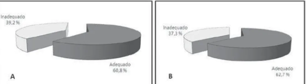
The evaluation of the appropriateness of radiotherapy field borders on sagittal MRI sections demonstrated inappropriate AB in 20 (39.2%) of the patients, and geographic miss in PB in 37.3% of cases (Figure 1).
No relation was observed between lat-eral field AB inappropriateness and clini-cal parameters such as patient’s age (p =
0.970), tumor staging (p > 0.999), type (p
= 0.162) and histological grade (p = 0.884).
Also, the PB inappropriateness was not related with the clinical parameters, age (p
= 0.726), tumor staging (p > 0.999), type
(p = 0.496) and histological grade (p =
0.813).
As regards staging, a high rate of inap-propriateness was observed in stages IIB (AB, 37.9% and PB, 34.4%) and IIIB (AB,
Table 1 Clinical characteristics of patients in the study sample.
Sample characteristics
Age (median in years)
Histological type
Histological grade
Staging (FIGO)
n
–
45 6
10 3 25 13
3 29 15 4
Rate
–
88.2% 11.8%
19.6% 5.9% 49.0% 25.5%
5.9% 56.9% 29.4% 7.8% 54
Spinocellular carcinoma Adenocarcinoma
X* 1 2 3
IB IIB IIIB IVA
* Grade X: Histological grade not reported by the pathologist.
177
Magnetic resonance imaging in radiotherapy planning
Radiol Bras. 2010 Mai/Jun;43(3):175–178 53.3% and PB, 46.6%), as the patients at each specific stage were analyzed. Such results demonstrate that no statistical sig-nificance was observed in relation to the evaluated parameters and, consequently, errors may occur with any staging or his-tological grade (Figure 2).
In the analysis of the appropriateness of each histological grade, inappropriateness was observed in 33.3% (AB) in histologi-cal grade 1, 36% (AB) and 40% (PB) in grade 2, and 46.2% (PB) in grade 3, also demonstrating a greater planning error in higher histological grades.
DISCUSSION
Radiotherapy is the main therapeutic modality for locally advanced uterine cer-vix tumors, and the radiotherapy planning is essential for guaranteeing the treatment appropriateness. Classically, bone refer-ences were employed to define standard radiotherapy field borders, without consid-ering the tumor characteristics and the patient’s individual data. In the present study, the anterior and posterior borders of the lateral field were analyzed by means of MRI to evaluate the planned radiotherapy fields appropriateness.
The present study results regarding pa-tients’ characteristics show a median age at the diagnosis of 54 years (22 to 77 years), with predominance of spinocellular carci-noma in 88.2% of cases and locally ad-vanced staging in 94.1%. This histological type of tumor is reported in the literature as the most frequent one(9–11). There was a predominance of the most undifferentiated tumors with histological grades 2 (49%) and 3 (25.5%). The median age observed was similar to the ones found in other stud-ies including patients with the same dis-ease(9,11). Thus the present results corrobo-rate the literature, except for those related
to predominant histological grades, since studies evaluating the utilization of MRI in radiotherapy planning have not taken this parameter into consideration(7,12,13).
As the appropriateness of the lateral field borders was evaluated, inappropriate-ness was very frequently observed, both for AB (39.2%) and for PB (37.3%). In a study evaluating 25 sagittal MRI sections of pa-tients with uterine cervix neoplasm, the presence of lesion beyond the PB was ob-served in six patients, demonstrating the relevance of the image in the evaluation of the lateral field borders(12).
Zunino et al. have applied the same method in the MRI analysis, utilizing a safety margin of 1.0 cm from the tumor and from the uterine fundus, and have found in-appropriateness of PB in 49% and of AB in 8.8% of cases(7).
In another Brazilian study, inappropri-ateness indices corresponding to 46% (AB) and 40% (PB) have been observed(14). Such results are similar to the ones observed in the present study. The same authors present an updating of these data with 80 patients, reporting 36% geographic miss in the AB, and 35% in the PB. In total, 56% of patients presented some kind of inappropriateness. In the present study, a significant relation was observed between field border inap-propriateness and anteroposterior tumor diameter, displacement of the anterior rec-tal wall, and tumor volume. The authors suggest that such factors can be utilized as risk predictors of geographic miss(13).
As regards staging according to the FIGO classification, no significant influ-ence from this factor on RT field borders appropriateness or inappropriateness was observed, maybe because of the absence of imaging studies such as computed tomog-raphy (CT) and MRI in this staging sys-tem(15). However, the FIGO staging allows a standardization of the pretreatment
as-sessment of patients in different regions of the world, even in locations where the ac-cess to more complex imaging methods is difficult, for comparison of therapeutic outcomes.
Another study has utilized the TNM staging system to evaluate the appropriate-ness of radiotherapy field borders, allow-ing the assessment of lymph nodes involve-ment. However, statistical analysis was not performed because of the low number of patients. Only a descriptive analysis was performed, demonstrating a high number of patients with staging T2B and absence of lymph nodes involvement(16).
As regards the analysis of each specific staging, the authors observed a high rate of inappropriateness in stages IIB (AB, 37.9% and PB, 34.4%) and IIIB (AB, 53.3% and PB, 46.6%), with no statistical signifi-cance. In the literature, Zunino et al. have observed staging inappropriateness in stages IB (AB, 0% and PB, 50%), IIA (AB, 0% and PB, 67%), IIB (AB, 5% and PB, 42%), IIIB (AB, 67% and PB, 33%) and IVA (AB, 0% and PB, 100%)(7).
Thus, the results of the present study demonstrate that the more advanced the staging, the more extensive the geographic miss. Maybe, a larger sample may provide a statistical evidence of these data.
As regards the histological grade that represents the tumor differentiation and plays a relevant role in the pretreatment evaluation of the disease, this parameter demonstrated a rate of inappropriateness of 33.3% (AB) and 0% (PB) in grade 1; 36% (AB) and 40% (PB) in grade 2; and 46.1% (AB) and 46.1% (PB) in grade 3. These results also demonstrate a greater planning error in higher histological grades, al-though this error is not significant. This finding may be correlated with the fact that higher histological grades are associated with more advanced or more voluminous Figure 2. Distribution of
178
Freire GM et al.
Radiol Bras. 2010 Mai/Jun;43(3):175–178 tumors. In the reviewed literature on
radio-therapy field borders appropriateness evaluation with MRI, the authors have not evaluated this factor in relation to geo-graphic miss(7,12,13).
Imaging methods such as CT and MRI would be indicated in the pretreatment evaluation for defining the tumor diameter and volume, and assessment of pelvic lymph nodes and parametrial invasion(17– 20). A meta-analyses of 57 studies
demon-strated the higher sensitivity of MRI as compared with CT in the evaluation of parametrial invasion(21). However, in a multicentric, prospective study with 208 patients with uterine cervix neoplasm sub-mitted to clinical evaluation according to FIGO, helical CT and MRI compared with surgical (pathological) findings, the au-thors have observed that CT and MRI pre-sented similar results in the evaluation of parametrial invasion, and have concluded that clinical examination was superior to other studies. The authors have suggested the imaging methods influence on the clini-cal staging, and that such imaging studies could improve the pretreatment staging of patients(22).
A recent study demonstrates the rel-evance of MRI as a prognostic factor in the analysis of the tumor volume and uterine invasion by the tumor, and of CT in the evaluation of positive pelvic lymph nodes(23).
The most recent National Comprehen-sive Cancer Network (NCCN) guidelines suggests the utilization of MRI and CT for defining the therapy planning, but these imaging methods do not influence the for-mal staging, and the FIGO staging is con-sidered as a standard for comparison be-tween treatments(24).
Thus, although the high cost of MRI for radiotherapy planning as a routine is one of the main factors which makes the imple-mentation of this method more difficult in poor and developing countries, the results of the present study, in conjunction with the literature review, suggest that MRI plays an extremely relevant role in the radiotherapy planning for patients with uterine cervix neoplasm, and this planning should be in-dividualized, considering the tumor vol-ume and anatomical variations(7,12,13,16). Uterine cervix cancer plays a relevant role
as the third major cause of death in women worldwide, with about 274,000 yearly deaths, 78% of such deaths occurring in developing countries(1). MRI can be benefi-cial to optimize the treatment of these pa-tients.
CONCLUSIONS
As a whole, the results of the present study demonstrate that MRI is a relevant tool for radiotherapy planning in patients with uterine cervix cancer, allowing higher accuracy in the definition of irradiation field borders, thus reducing the incidence of geographic miss. According to the present results, the patient’s age, tumor staging, histological type and grade are not related to the radiotherapy field borders inappropriateness.
REFERENCES
1. Parkin DM, Bray F, Ferlay J, et al. Global cancer statistics, 2002. CA Cancer J Clin. 2005;55:74– 108.
2. Brasil. Ministério da Saúde. Instituto Nacional de Câncer. Estimativa 2010: incidência de câncer no Brasil. Rio de Janeiro, RJ: Instituto Nacional de Câncer; 2009.
3. Perez CA, Kavanagh BD. Uterine cervix. In: Perez CA, Brady LW, Halperin EC, et al., editors. Principles and practice of radiation oncology. 4th ed. Philadelphia, PA: Lippincott-Raven; 2004.
4. Oliveira JP, Rosa LAR, Batista DVS, et al. Avalia-ção da dose no reto em pacientes submetidas a braquiterapia de alta taxa de dose para o tratamento do câncer do colo uterino. Radiol Bras. 2009;42: 83–8.
5. Guimarães RGR, Carvalho HA, Stuart SR, et al. Avaliação dosimétrica de uma combinação de aplicadores para braquiterapia de tumores do colo uterino com acometimento da porção distal da vagina. Radiol Bras. 2009;42:209–14. 6. Swift PS, Hsu CJ. Cancer of the uterine cervix.
In: Leibel SA, Philips TL, editors. Textbook of ra-diation oncology. 2nd ed. Philadelphia, PA: Saunders; 2004.
7. Zunino S, Rosato O, Lucino S, et al. Anatomic study of the pelvis in carcinoma of the uterine cervix as related to the box technique. Int J Radiat Oncol Biol Phys. 1999;44:53–9.
8. Mayr NA, Tali ET, Yuh WT, et al. Cervical can-cer: application of MR imaging in radiation therapy. Radiology. 1993;189:601–8. 9. Thomas G, Ali S, Hoebers FJ, et al. Phase III trial
to evaluate the efficacy of maintaining hemoglo-bin levels above 12.0 g/dL with erythropoietin vs above 10.0 g/dL without erythropoietin in anemic patients receiving concurrent radiation and cisplatin for cervical cancer. Gynecol Oncol. 2008;108:317–25.
10. Torres MA, Jhingran A, Thames HD Jr, et al. Comparison of treatment tolerance and outcomes in patients with cervical cancer treated with
con-current chemoradiotherapy in a prospective ran-domized trial or with standard treatment. Int J Radiat Oncol Biol Phys. 2008;70:118–25. 11. Huguet F, Cojocariu OM, Levy P, et al.
Preopera-tive concurrent radiation therapy and chemo-therapy for bulky stage IB2, IIA, and IIB carci-noma of the uterine cervix with proximal parame-trial invasion. Int J Radiat Oncol Biol Phys. 2008;72:1508–15.
12. Russell AH, Walter JP, Anderson MW, et al. Sag-ittal magnetic resonance imaging in the design of lateral radiation treatment portals for patients with locally advanced squamous cancer of the cervix. Int J Radiat Oncol Biol Phys. 1992;23:449–55. 13. Justino PB, Baroni R, Blasbalg R, et al. Clinical
tumor dimensions may be useful to prevent geo-graphic miss in conventional radiotherapy of uter-ine cervix cancer – a magnetic resonance imag-ing-based study. Int J Radiat Oncol Biol Phys. 2009;74:503–10.
14. Justino PB, Carvalho HA, Baroni R, et al. Valor da ressonância magnética no planejamento radio-terápico dos tumores de colo de útero: resultados preliminares. Radiol Bras. 2005;38:399–402.
15. Creasman WT. New gynecologic cancer staging. Gynecol Oncol. 1995;58:157–8.
16. Thomas L, Chacon B, Kind M, et al. Magnetic resonance imaging in the treatment planning of radiation therapy in carcinoma of the cervix treated with the four-field pelvic technique. Int J Radiat Oncol Biol Phys. 1997;37:827–32. 17. Greco A, Mason P, Leung AW, et al. Staging of
carcinoma of the uterine cervix: MRI-surgical correlation. Clin Radiol. 1989;40:401–5. 18. Hawnaur JM, Johnson RJ, Carrington BM, et al.
Predictive value of clinical examination, trans-rectal ultrasound and magnetic resonance imag-ing prior to radiotherapy in carcinoma of the cer-vix. Br J Radiol. 1998;71:819–27.
19. Toita T, Kakinohana Y, Shinzato S, et al. Tumor diameter/volume and pelvic node status assessed by magnetic resonance imaging (MRI) for uter-ine cervical cancer treated with irradiation. Int J Radiat Oncol Biol Phys. 1999;43:777–82.
20. Wagenaar HC, Trimbos JB, Postema S, et al. Tu-mor diameter and volume assessed by magnetic resonance imaging in the prediction of outcome for invasive cervical cancer. Gynecol Oncol. 2001;82:474–82.
21. Bipat S, Glas AS, van der Velden J, et al. Com-puted tomography and magnetic resonance imag-ing in stagimag-ing of uterine cervical carcinoma: a systematic review. Gynecol Oncol. 2003;91:59– 66.
22. Hricak H, Gatsonis C, Chi DS, et al. Role of im-aging in pretreatment evaluation of early invasive cervical cancer: results of the intergroup study American College of Radiology Imaging Network 6651-Gynecologic Oncology Group 183. J Clin Oncol. 2005;23:9329–37.
23. Kim H, Kim W, Lee M, et al. Tumor volume and uterine body invasion assessed by MRI for pre-diction of outcome in cervical carcinoma treated with concurrent chemotherapy and radiotherapy. Jpn J Clin Oncol. 2007;37:858–66.