Oligozoospermia and Sertoli-Cell-Only Syndrome
Frank Tu¨ttelmann1*, Manuela Simoni2, Sabine Kliesch3, Susanne Ledig1, Bernd Dworniczak1, Peter Wieacker1, Albrecht Ro¨pke1
1Institute of Human Genetics, University of Mu¨nster, Mu¨nster, Germany,2Department of Medicine, Endocrinology, Metabolism and Geriatrics, University of Modena and Reggio Emilia, Modena, Italy,3Department of Clinical Andrology, Centre of Reproductive Medicine and Andrology, University of Mu¨nster, Mu¨nster, Germany
Abstract
A genetic origin is estimated in 30% of infertile men with the common phenotypes of oligo- or azoospermia, but the pathogenesis of spermatogenic failure remains frequently obscure. To determine the involvement of Copy Number Variants (CNVs) in the origin of male infertility, patients with idiopathic severe oligozoospermia (N = 89), Sertoli-cell-only syndrome (SCOS, N = 37)) and controls with normozoospermia (N = 100) were analysed by array-CGH using the 244A/400K array sets (Agilent Technologies). The mean number of CNVs and the amount of DNA gain/loss were comparable between all groups. Ten recurring CNVs were only found in patients with severe oligozoospermia, three only in SCOS and one CNV in both groups with spermatogenic failure but not in normozoospermic men. Sex-chromosomal, mostly private CNVs were significantly overrepresented in patients with SCOS. CNVs found several times in all groups were analysed in a case-control design and four additional candidate genes and two regions without known genes were associated with SCOS (P,161023). In conclusion, by applying array-CGH to study male infertility for the first time, we provide a number of candidate genes possibly causing or being risk factors for the men’s spermatogenic failure. The recurring, patient-specific and private, sex-chromosomal CNVs as well as those associated with SCOS are candidates for further, larger case-control and re-sequencing studies.
Citation:Tu¨ttelmann F, Simoni M, Kliesch S, Ledig S, Dworniczak B, et al. (2011) Copy Number Variants in Patients with Severe Oligozoospermia and Sertoli-Cell-Only Syndrome. PLoS ONE 6(4): e19426. doi:10.1371/journal.pone.0019426
Editor:Laszlo Orban, Temasek Life Sciences Laboratory, Singapore
ReceivedNovember 23, 2010;AcceptedApril 3, 2011;PublishedApril 29, 2011
Copyright:ß2011 Tu¨ttelmann et al. This is an open-access article distributed under the terms of the Creative Commons Attribution License, which permits unrestricted use, distribution, and reproduction in any medium, provided the original author and source are credited.
Funding:The study was supported by the Deutsche Forschungsgemeinschaft Research Unit ‘Germ Cell Potential’ (FOR 1041, grant TU 298/1-1). The funders had no role in study design, data collection and analysis, decision to publish, or preparation of the manuscript.
Competing Interests:The authors have declared that no competing interests exist. * E-mail: frank.tuettelmann@ukmuenster.de
Introduction
Infertility, which affects 10–15% of all couples, is attributed to a male (co-)factor in around 50%. Male infertility is mostly caused by spermatogenic failure, clinically noted as oligo- or azoospermia. However, the reasons for the decreased sperm production remain largely unclear: After a full clinical workup around 30% of cases are considered ‘idiopathic infertile’ and an additional 40% have not sufficient/uncertain causes (e.g. varicocele, infections) [1,2]. On the other hand, it is estimated that about 30% of azoo-/ oligozoospermia are caused by chromosomal abnormalities or mutations of genes involved in germ cell production and function [3]. Thus, male infertility of genetic origin supposedly affects about 1 in 40 adult men and can be considered a common, complex disease.
The currently established genetic causes of male infertility comprise abnormalities on all genomic levels from chromosomal abnormalities, in particular Klinefelter syndrome, to Y-chromo-somal (azoospermia factor, AZF) deletions, to mutations of the cystic fibrosis transmembrane conductance regulator (CFTR, all gene information - name, location, IDs - available as Table S1) gene. However, although routinely performed in clinical workup [4] karyotyping, AZF deletion screening and CFTR sequencing elucidate the reason for infertility in only 5% of unselected and around 20% of azoospermic patients [1,2].
A multitude of up to 1,500 genes are thought to be involved in spermatogenesis of which 300–600 are specifically expressed in the male germline [5–7] and are therefore candidates for causing male infertility. In the last 5–10 years, many efforts have been undertaken with research focused on targeted re-sequencing of a number of these candidate genes. Nevertheless, up to date no additional genetic cause is recognised to be administrable in patient care, mostly because results have either not been replicated or might only explain minute effects [8–10].
oligo-and azoospermia. For comparison, normozoospermic controls were analysed, because the current databases of structural genomic variation do not provide the necessary information when evaluating spermatogenesis and fertility. We wanted to determine a) whether the number and/or pattern in selected men with spermatogenic failure differs from that in men with normal spermatogenesis; b) whether recurrent CNVs can be identified which might harbour genes involved in spermatogenesis; c) associations with reproductive parameters.
Materials and Methods
Study population
Caucasian patients of German origin with idiopathic infertility were selected from the clientele of the Department of Clinical Andrology of the Centre of Reproductive Medicine and Andrology, Mu¨nster, a tertiary-referral centre, using the Andro-baseß database [16]. All participants underwent a complete physical examination including ultrasonographic analysis of the Table 1.Comparison of clinical parameters between the study groups (mean6standard deviation and median, range (min-max) in brackets).
Normozoospermic controls (N = 100) Severe oligozoospermia (N = 89) Sertoli-cell-only syndrome (N = 37)
Age [y] 40.164.4 (40, 30–59) 38.765.8 (39, 23–58) 40.365.4 (40, 29–50)
Testicular volume [ml] 58618 (59, 22–111) 41613 (39, 12–89)** 29
611 (30, 10–56)**
LH [U/l] 3.161.2 (3.0, 0.7–7.1) 4.562.3**(4.0, 1.5–14.1) 7.8
63.2**(6.7, 3.1–15.6)
FSH [U/l] 3.762.0 (3.1, 1.3–9.4) 7.765.4**(5.7, 1.2–24.5) 15.3
64.9**(21.1, 8.5–37.9) Testosterone [nmol/l] 16.465.1 (15.9, 6.9–39.0) 15.364.9 (15.0, 4.6–31.3) 14.165.8 (12.4, 7.6–34.4)**
Abstinence [d] 3.961.6 (4, 2–7) 3.861.3 (4, 2–7) 4.261.2 (4, 2–10)
Sperm concentration [106/ml] 77.0
647.0 (62.0, 20.0–231.0) 1.461.2**(1.1, 0.1–5.0) 0**
Semen volume [ml] 3.961.5 (3.5, 2.0–9.0) 3.661.4 (3.4, 1.5–9.2) 4.161.8 (4.0, 1.0–10.0) Total sperm count [106] 287.3
6182.1 (269.5, 40.0–1083.5) 4.563.3**(3.8, 0.3–10.0) 0** a+b Motility [%] 53.765.9 (54, 36–75) 32.4613.0**(31, 4–60) -Normal morphology [%] 16.765.2 (16, 10–34) 6.065.6**(5, 1–24)
-**significantly different compared to controls (P,0.01 calculated by Mann-Whitney test). doi:10.1371/journal.pone.0019426.t001
scrotal content. Ejaculates were obtained by masturbation after 2– 7 days of sexual abstinence and all semen values were determined in accordance with the then current WHO criteria [17]. All men with known clinical (e.g. maldescended testes, varicocele, infec-tions) and genetic (karyotype anomalies, Y-chromosomal deleinfec-tions) causes of infertility were excluded. Of these men, one-hundred with $20610/ml sperm concentration, $406106 total sperm
count,$2 ml semen volume,$50% of a+b or$25% a motility, high percentage of normal forms ($10%) were selected as control population with normal spermatogenesis according to WHO criteria [17]. Twenty-one (21%) of these men had previously induced a spontaneous pregnancy in the current or a former relationship. The study populations comprised patients with a) severe oligozoospermia (N = 89 with #56106/ml sperm concen-tration and#106106total sperm count) and b) azoospermia and complete bilateral Sertoli-cell-only syndrome (SCOS, N = 37). The diagnosis of SCOS was established if only tubules with Sertoli-cells were detected in bilateral and multilocular (at least two sites per testis) testicular biopsies and the attempt of testicular sperm extraction (TESE) was unsuccessful.
Ethics statement
All participants gave written informed consent for evaluation of their clinical data and genetic analysis of their donated DNA samples according to a protocol approved by the Ethics Committee of the Medical Faculty in Mu¨nster and State Medical Board.
Array-CGH
Genomic DNA was extracted from peripheral blood by standard methods and analysed at first by the commercially available Human Genome CGH Microarray Kits 244A (Agilent Technologies, Santa Clara, California, USA). This array com-prises 236,381 60-mer oligonucleotide probes with a median probe spacing of 8.9 Kb. During the course of the study, the higher-resolution 400K microarray with 411,056 oligonucleotides and a median probe spacing of 5.3 Kb became available. We switched to using this array to be able to compare the impact of smaller CNVs detected with the higher-resolution on spermatogenic failure. Finally, 78 control men and 42 with severe oligozoospermia were analysed with the 244A arrays and 22 and 47 with the 400K arrays. All azoospermic patients with SCOS were analysed with the 400K arrays. Each patient’s DNA was compared to 10 pooled DNAs (Promega Human Genomic DNA: Male, Cat.-Nr. G1471). Labelling and hybridisation were performed according to the manufacturer’s protocol. In brief, 1mg of patients’ DNA and the
pooled control DNA were double-digested with AluI and RsaI
(Promega, Madison, Wisconsin, USA) and subsequently labelled with Cy5- and Cy3-dUTP using the Genomic DNA Enzymatic Labeling kit (Agilent Technologies), respectively. After purification of the labelled DNA by filtration (Microcon YM-30, Millipore, Bilerica, Massachusetts, USA), patient and control DNA were pooled and hybridised with 25mg of human COT DNA (ArrayGrade KREACot DNA, Kreatech, Amsterdam, The Netherlands) for 40 h at 65uC in the hybridisation oven (Agilent Technologies). After post-hybridisation washes, the arrays were scanned using a Microarray Scanner (G2565BA, Agilent Tech-nologies), and the spot intensities were measured by ‘Feature Extraction Software’ (version 10.7, Agilent Technologies). Anal-yses and visualisation were performed with ‘DNA Analytics’ (version 4.0.81, Agilent Technologies) with the following param-eters: aberration algorithm ADM-2, threshold 6.0, fuzzy zero, centralisation and moving average window 1 Mb. Aberrant signals including 4 or more adjacent probes were considered as genomic CNVs. The raw array-data acquired by the Feature Extraction Software is accessible through the Gene Expression Omnibus (GEO, http://www.ncbi.nlm.nih.gov/geo, series record GSE27965) and the abstracted CNVs were submitted to the Database of genomic structural variation (dbVar, http://www. ncbi.nlm.nih.gov/dbvar, study ID nstd53). CNVs found in normozoospermic controls are also available as Table S2. The Database of Genomic Variants (DGV, http://projects.tcag.ca/ variation/) was used to compare findings to previously reported studies. Coordinates of CNVs are based on the National Center for Biotechnology Information (NCBI) Human Genome Build 36 (hg18).
Statistical analysis
Comparisons between patients and controls were carried out using the two-sample t-tests if data were normally distributed (e.g. number of CNVs) and otherwise non-parametric Mann-Whitney test. Frequencies were compared by Fisher’s exact test. In principle,P-values less than 0.05 were considered statistically significant. To correct for multiple testing, P-values were adjusted according to the Bonferroni-Holm procedure, which is less conservative than the standard Bonferroni correction. As semen parameters were not normally distributed, correlations were calculated for log-transformed values. All calculations were performed with Stata/SE (StataCorp LP, version 9.1, College Station, Texas, USA) or GraphPad Prism version 5.00 for Windows (GraphPad Software, San Diego, California, USA).
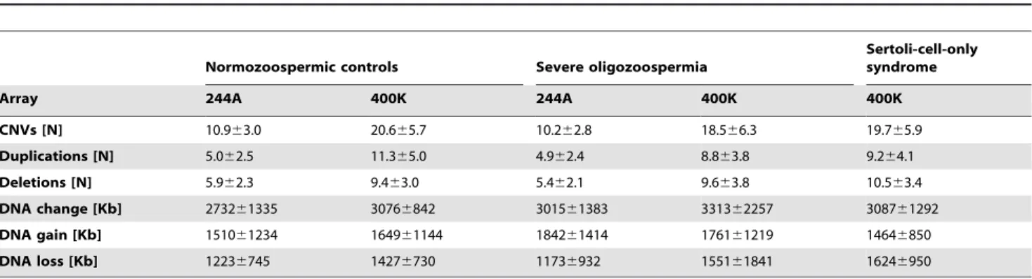
Table 2.Detected CNVs and amount of DNA change per man separately for each array resolution (mean6standard deviation).
Normozoospermic controls Severe oligozoospermia
Sertoli-cell-only syndrome
Array 244A 400K 244A 400K 400K
CNVs [N] 10.963.0 20.665.7 10.262.8 18.566.3 19.765.9
Duplications [N] 5.062.5 11.365.0 4.962.4 8.863.8 9.264.1
Deletions [N] 5.962.3 9.463.0 5.462.1 9.663.8 10.563.4
DNA change [Kb] 273261335 30766842 301561383 331362257 308761292
DNA gain [Kb] 151061234 164961144 184261414 176161219 14646850
DNA loss [Kb] 12236745 14276730 11736932 155161841 16246950
Results
Clinical data
The clinical parameters of the normozoospermic controls and the two study groups with severe oligozoospermia and azoosper-mia caused by SCOS are presented in Table 1. Testicular volume was lower and LH as well as FSH higher in the two groups with spermatogenic failure. Patients with SCOS also had lower testosterone serum levels. Duration of abstinence and semen volume were comparable between all groups. Sperm concentra-tion and count as well as percentage of sperm with normal morphology and progressive motility were significantly different by selection.
Comparison of CNVs between groups
In total, 1304 CNVs were detected in 100 controls, 1297 CNVs in oligozoospermic men (N = 89) and 728 CNVs in patients with SCOS (N = 37); the numbers and categorisations of CNVs are depicted in Figure 1. CNV size ranged from 1.9 Kb to 4.7 Mb with only 5% larger than 1 Mb. The number of detected CNVs and amount of DNA change per men is presented in Table 2 according to study group. As expected from the two-fold increased resolution, around twice as many CNVs and also duplications/ deletions separately were found with the higher-resolution 400K array (Fig. 2). The amount of DNA change did not increase likewise with the 400K arrays, as mostly smaller CNVs were additionally detected. Overall, no significant differences in number of CNVs (Fig. 2) or amount of DNA change were found between controls and the two study groups. In addition, particularly large (.500 Kb, .1 Mb) CNVs were analysed, but did not show different distributions between groups.
To analyse whether the chromosomal distribution of CNVs was different between the groups, the number of CNVs, and duplications/deletions separately, was calculated per chromosome and normalised to 100 men (Fig. 3). The overall distribution of duplications and deletions was found to be significantly different between all groups (P,0.01) and for both types of arrays. The post-hoc comparison of number of CNVs per single chromosome indicated significant differences between patients and controls for several chromosomes after Bonferroni-Holm correction. The detailed per-chromosome analysis revealed no specific hotspots associated with oligozoospermia or SCOS on the respective chromosomes. In contrast, the differences in the distribution were caused by recurring CNVs that were found more or less frequently in the respective study groups (see below).
In many cases, several differently sized CNVs spanned a common region but probably have different breakpoints. If either the gene content was identical or the CNVs spanned regions with the breakpoints distance610 Kb (roughly corresponding to one oligonucleotide and thereby the minimum array resolution), these CNVs were aggregated for statistical analyses. Duplications and deletions were considered as different variants because of their supposedly diverse impact. Of the 3329 CNVs, a majority of 2711 (81%) could thereby be summarised as 310 recurring variants (159 deletions and 151 duplications), of which the smallest common region is then reported. Some of these were only found in normozoospermic controls (N = 21), at most once per group (N = 58) or not in controls (N = 14, recurring, patient-specific CNVs).
The frequencies of the remaining 217 variants were compared between the study groups (Table S3 and Table S4). Plots of the 2log(10)P-values (Manhattan-plots) of case-control comparisons are presented in Fig. 4. To correct for multiple testing, theP-value for selection of candidate CNVs was set to 0.001 and these 6 are reported in Table 3. Except the decreasing frequency of a deletion on 4q13.1 including UGT2B17 and a small duplication on 14q32.33 without known genes, all differences remained signifi-cant also after Bonferroni-Holm correction. Ten recurring CNVs were only found in patients with severe oligozoospermia, while three were SCOS specific and one CNV (3p11.1 in an intron of
EPHA3) was found in both groups with spermatogenic failure but not in normozoospermic men (Table 4). Already from the chromosomal distribution it became clear, that sex-chromosomal CNVs were significantly overrepresented in patients with SCOS. These are all private variants summarised in Table 5. Both groups of patient-specific CNVs (Table 4 and 5) were marked as ‘likely pathogenic’ in dbVar. All genes in either CNVs with significantly different frequencies between patients and controls,
patient-Figure 2. Number of CNVs (A), duplications (B) and deletions (C) separated by array (left: 244A, right: 400K) and patient group (Normo = normozoospermia, Oligo = severe oligozoos-permia, SCOS = Sertoli-cell-only syndrome). Around twice as many CNVs (independent of type and fitting the roughly two-fold increased resolution) were detected with the 440K array and in general more duplications than deletions. No significant differences between patient groups were found.
specific CNVs or private, sex-chromosomal CNVs (Tables 3, 4, and 5) were checked through PUBMED (http://www.ncbi.nlm. nih.gov/pubmed) and OMIM (http://www.ncbi.nlm.nih.gov/ omim) searches. Those genes with known expression in the testis and/or function related to spermatogenesis are presented in Table 6.
Association of CNVs with semen parameters
Under the hypothesis that accumulating CNVs might lead to reduced sperm output, the number of total CNVs, duplications and deletions as well as amount of DNA change was correlated with sperm concentration and count. Because the number of deletions varies strongly with the array used, these were analysed separately. In the largest group, 78 normozoospermic men analysed by 244A arrays, a significant negative association (r =20.27, P= 0.017, Fig. 5) of number of deletions and total sperm count was found. Sperm concentration also showed a trend to be associated with number of deletions (r =20.22,P= 0.055). These correlations were strengthened by corrections for age and
abstinence time (P= 0.007 and P= 0.051) but could not be confirmed in the other, however smaller, groups of men analysed by 400K and/or oligozoospermia. Neither correlations with sperm motility nor morphology were found in any group.
Discussion
For the first time, we analysed 89 strictly selected patients with severe oligozoospermia, 37 with azoospermia due to SCOS and 100 with normal spermatogenesis as controls by array-CGH. Although interpretation of the results would have been more straightforward if only one type of microarray had been used and while knowing that amount of DNA available as well as funding would not permit us to repeat the analyses on the first set of samples, we favoured switching to the higher resolution array to detect smaller CNVs in almost half of our subjects, which in the eand appeared to be beneficial. As result, we report several genes and genomic regions on autosomes and more pronouncedly on the sex-chromosomes that might either be risk factors (also found in
Figure 3. Number of duplications (upwards) and deletions (downwards) per chromosome (normalised per 100 men) detected by 244A (A) and 400K (B) arrays for normozoospermic controls, oligozoospermic and SCOS patients (open, grey and black bars, respectively).Significantly different frequencies between the groups are marked with an asterisk.P-values calculated by Fisher’s exact test. To correct for multiple testing,P-level for significance was adjusted according to the Bonferroni-Holm procedure.
controls) or causative by themselves (not found in controls) for spermatogenic failure.
We hypothesised that an increased number or specific distribution of CNVs could result in defective recombination, meiotic and thereby spermatogenic failure. Structural chromo-somal aberrations are found more frequently in men with oligo-and azoospermia with an emphasis on autosomal translocations in the former (3–4% compared to 0.5–1.5% in controls) and sex-chromosomal aneuploidy in the latter (13–16% compared to 0.5– 1%) [18–20]. The causal relation between chromosomal rear-rangements and impaired sperm production has been suggested to be a structural effect related to alterations in the process of chromosome synapsis during meiosis [21], but whether submicro-scopic chromosomal rearrangements (CNVs) can result in meiotic recombination defects is not known. By comparing the number of all CNVs, duplications and deletions separately and amount of DNA change, gain and loss no significant differences were found between the groups analysed. The differences in chromosomal distribution of CNVs were attributed to single, recurring CNVs
(see below). Also particularly large (.1 Mb) variants were not found more frequently in oligozoospermia or SCOS. Therefore, only even larger variants of several megabases, microscopically detectable upon conventional karyotyping, might impair chromo-some synapsis and meiosis. Whether a size threshold for chromosomal aberrations having such a ‘‘direct’’, gene-indepen-dent impact on spermatogenesis exists cannot be concluded from the presented data. It seems, however, that structural variation below the detection limit of routine karyotyping should not be regarded, per se, as an obligate cause of spermatogenic failure. Considering that translocation carriers may have normal sper-matogenesis [18–20], the link between (large) structural chromo-somal variation and spermatogenesis remains to be elucidated.
In principle, CNVs may result in altered gene transcription/ protein function through different mechanisms: they might encompass dosage-sensitive genes, a deletion may demask a recessive mutation on the homologous chromosome, genes overlapped by structural variation may be disrupted directly or a CNV can exert position effects [22]. By comparing CNVs between
Figure 4. Plots of 2logP-values of frequency comparisons for all recurring CNVs found in patients and controls grouped by chromosome.P-values calculated by Fisher’s exact test. The figure does not include the Y chromosome because no recurring variants were found on it.
Table 3.The 6 recurring CNVs with significantly different (P,0.001, marked in bold) frequencies (number of cases/all cases*; percentage in brackets) between patients with oligozoospermia or SCOS and normozoospermia or between SCOS and both other groups combined.
Region Start End Size [Kb]
Gene
symbol(s) Type Normozoosp. Sev.
oligozoosp. P
Sertoli-cell-only
syndrome P
PSCOS vs. others DGV
4q13.2 69069451 69166014 96.6 UGT2B17 dup 10/100 (10%) 12/89 (13.5%) 0.50132 18/37 (48.6%) 0.00000 0.00000 yes 4q13.2 69069451 69166014 96.6 UGT2B17 del 28/100 (28%) 21/89 (23.6%) 0.51072 1/37 (2.7%) 0.00072 0.00086 yes 7q34 141413152 141438704 25.6 MGAM dup 1/22 (4.5%) 0/47 (0%) 0.31884 10/37 (27%) 0.04059 0.00009 yes 7q34 141413152 141438704 25.6 MGAM del 3/22 (13.6%) 8/47 (17%) 1.00000 4/37 (10.8%) 1.00000 0.56819 yes 12p13.31 9528390 9610254 81.9 dup 1/100 (1%) 9/89 (10.1%) 0.006830/37 (0%) 1.00000 0.37395 yes 12p13.31 9528390 9610254 81.9 del 4/100 (4%) 11/89 (12.4%) 0.05618 13/37 (35.1%) 0.00001 0.00005 yes 14q11.2 19268576 19490830 222.3 OR4Q3,
OR4M1, OR4N2,OR4K2, OR4K5,OR4K1
dup 10/100 (10%) 13/89 (14.6%) 0.37765 8/37 (21.6%) 0.08990 0.18733 yes
14q11.2 19268576 19490830 222.3 OR4Q3, OR4M1, OR4N2,OR4K2, OR4K5,OR4K1
del 36/100 (36%) 24/89 (27%) 0.21172 2/37 (5.4%) 0.00020 0.00049 yes
14q32.33 105602402 105630289 27.9 dup 8/100 (8%) 5/89 (5.6%) 0.57633 11/37 (29.7%) 0.00370 0.00029 yes 14q32.33 105602402 105630289 27.9 del 14/100 (14%) 12/89 (13.5%) 1.00000 7/37 (18.9%) 0.59345 0.44578 yes 17q21.31 41521344 41566740 45.4 KIAA1267 dup 10/100 (10%) 11/89 (12.4%) 0.64844 1/37 (2.7%) 0.28787 0.13883 yes 17q21.31 41521344 41566740 45.4 KIAA1267 del 6/100 (6%) 9/89 (10.1%) 0.41980 16/37 (43.2%) 0.00000 0.00000 yes
For comparison, the corresponding duplication/deletion (if present in any group) is included independent ofP-value. CNVs were checked for occurrence in the Database of Genomic Variants (DGV).
*If CNVs were only found by higher-resolution 400K-array, the number of all cases is reduced to 22 for normozoospermic controls and to 47 for patients with severe oligozoospermia (see methods).
SCOS = Sertoli-cell-only syndrome.P-values calculated by Fisher’s exact test. To correct for multiple testing,P-level for significance was adjusted according to the Bonferroni-Holm procedure. Gene information - name, location, IDs - available as Suppl. Table S1.
doi:10.1371/journal.pone.0019426.t003
Table 4.The 11 and 4 recurring, patient-specific CNVs not found in normozoospermic controls with number of cases, type (dup = duplication, del = deletion), gene content and whether the CNV was described in the Database of Genomic Variants (DGV).
Group Region Start End Size (Kb) Number, type Gene symbol(s) DGV
Oligozoospermia 2p11.2 89635198 89902565 267.0 2xdel - yes
3p11.1 89476719 89499633 22.0 4xdel EPHA3 yes
4p16.1 8235974 8261720 25.7 2xdup SH3TC1 yes
6p21.31 35143115 35184210 41.1 2xdup ANKS1A no
10q23.1 84138134 84171245 33.1 2xdel NRG3 no
10q23.33 96497202 96536412 39.2 2xdel CYP2C19 no
12q13.3 55866674 55896055 29.4 2xdup LRP1,MIR1228 no
16q22.1 66942648 66967713 25.1 2xdup PRMT7,SMPD3 no
17q12 30624580 30787596 163.0 2xdel SLFN11,SLFN12,
SLFN13
no
18q23 75746093 75779459 33.0 1xdup, 2xdel KCNG2,PQLC1 no
Xq26.3 134120502 134157976 37.5 2xdup CXorf48 yes
SCOS 3p11.1 89476719 89499633 22.9 3xdel EPHA3 yes
8q24.3 145061948 145093349 31.4 1xdup, 1xdel PLEC,MIR661 yes
12p11.21 31132516 31223665 91.1 2xdel DDX11,OVOS2 yes
12q23.1 98491661 98519308 27.6 2xdel ANKS1B no
the controls and the two patient groups, 11 and 4 CNVs specific for severe oligozoospermia and SCOS, respectively, were identified in more than one patient each (Table 4). In addition, 12 and 14 private, sex-chromosomal CNVs were listed (Table 5) because these may have a functional consequence of the naturally haploid genes contained therein. Also private, autosomal CNVs currently found in one patient each may contribute to the infertility phenotype, but their significance may only be evaluated by much larger studies. According to our moderately conservative approach, six CNVs were found in highly significantly different frequencies in patients with SCOS but also in controls and may be new risk factors associated with male infertility. All of these CNVs remained significant also after Bonferroni-correction. Interesting-ly, only one CNV in 12p13.31 without known genes was associated with severe oligozoospermia. The 81.9 Kb deletion was found with increasing frequency from controls (4%) to oligozoospermic men (12%) to men with SCOS (35%) as the most severe phenotype. However, the common deletion polymorphism
in UGT2B17 was found in decreasing frequency while a duplication of the same locus was found in increasing frequency. These glucoronidase variants determine urinary excretion of testosterone [23,24], but have not been studied in male infertility. Genes may be prioritised according to a known function or indirectly through their expression in the testis. Therefore, the 4 autosomal and 8 X-chromosomal genes presented in Table 6 are the distilled outcome of the current study with respect to identification of new, promising candidate genes. Most of these were shown to be expressed in Sertoli or germ cells, but their functions are mostly unknown. EPHA3, ANKS1A and ANKS1B
were mentioned because the Anks proteins were found to modulate degradation of EphA receptors in mice [25] and therefore the three genes might represent members of a functional unity. CNVs altering one of these genes were found in 6 men (7%) with severe oligozoospermia and 5 (14%) with SCOS.
In two recent studies, our group analysed patients with POF and XY gonadal dysgenesis by array-CGH [13,14]. Interestingly, the Table 5.The 12 and 14 private, sex-chromosomal, patient-specific CNVs not found in normozoospermic controls with type (dup = duplication, del = deletion), gene content and whether the CNV was described in the Database of Genomic Variants (DGV).
Group Region Start End Size (Kb) Type Gene symbol(s) DGV
Oligozoospermia Xp21.3 28162190 28214748 52.0 dup - yes
Xp11.4 38376283 38513841 137.6 dup TSPAN7 no
Xp11.22 52657689 52978139 320.5 dup SSX7,SSX2,SPANXN5,
XAGE5,XAGE3,FAM156A, FAM156B
yes
Xp11.22 52842080 52909890 67.8 dup SPANXN5,XAGE5,XAGE3no
Xq22.1 102134796 102496321 361.5 dup BEX1,NXF3,BEX4,
TCEAL8,TCEAL5,BEX2, TCEAL7
no
Xq22.2 103066101 103190187 124.0 dup TMSB15B,H2BFXP,
H2BFWT,H2BFM
yes
Xq22.3 105010614 105561054 550.4 dup NRK,SERPINA7,MUM1L1yes
Xq22.3 110238448 110260226 21.0 dup PAK3 no
Xq23 111598447 111621531 23.0 del - no
Xq25 123911267 124039708 128.4 del ODZ1 no
Xq27.1 139706586 139904507 197.9 dup MIR320D2 no
Xq28 154044877 154079019 34.0 del - no
SCOS Xp22.33 2711073 2814530 103.5 del XG,GYG2 no
Xp22.2 16688233 16707403 19.2 dup SYAP1 no
Xp21.3 25568263 25583583 15.3 del - no
Xp11.3 44067590 44084085 16.5 dup EFHC2 no
Xq11.1 64806000 64854709 48.7 dup MSN no
Xq12 65385501 65413711 28.2 dup HEPH no
Xq22.3–q23 110226892 110965127 738.2 dup PAK3,CAPN6,DCX, ALG13,TRPC5
no
Xq24 118780844 118798128 17.3 dup - no
Xq25 122920543 123009115 88.6 dup STAG2 no
Xq26.2 131413847 131439663 25.8 del MBNL3 no
Xq26.3 134600709 134628136 27.4 dup - yes
Yp11.2 7348864 7491480 142.6 dup - no
Y11.223 21964794 22058959 94.2 dup RBMY2EP (AZFb/bc) yes
Y11.23 26870161 27073218 203.1 dup - yes
putative causal genes identified were enriched for genes also implied in spermatogenesis. When comparing results of these and the current study, CNVs in thePLEC, TSPAN7, PAK3, TRCP5,
H2BFWTloci were found not only in men with SCOS, but also in either patients with POF or XY gonadal dysgenesis (personal communication). These cover 5 of 11 genes identified in the current study which might hint to a common genetic origin of loss of spermatogonia in the male and loss of oogonia in the female resulting in SCOS, XY gonadal dysgenesis and POF, respectively. With sperm counts ranging from zero to hundreds of millions per ejaculate, sperm output may be viewed as a quantitative trait and male infertility is sometimes postulated as a polygenic disease [26]. Some evidence for this hypothesis may be gained from the significant negative correlation between sperm counts and number of deletions described in our 78 normozoospermic men analysed by 244A array. Especially as hundreds of genes are implicated in spermatogenesis, it is plausible that more deletions also more often involve spermatogenesis-relevant genes. Therefore, a man bearing more deletions may have a less efficient spermatogenesis and therefore lower sperm output. We could, however, not detect a comparable correlation in the, albeit at most half as large, groups of normozoospermic men analysed by 400K arrays or in oligozoospermic patients (irrespective of array used).
While on the one hand the selection of our control group from the patient clientele avoids population stratification, on the other Table 6.Candidate genes with proposed function.
Group Phenotype Region Gene symbol OMIM Function
Risk factor Normo, Oligoz., SCOS 4q13.2 UGT2B17 601903 glucuronidase essential for urinary testosterone excretion [23,24] Recurring, patient-specific
CNVs
4xOligoz./3xSCOS 3p11.1 EPHA3 179611 Anks proteins involved in modulating degradation of EphA receptors [25]
2xOligoz. 6p21.31 ANKS1A 608994
2xSCOS 12q23.1 ANKS1B 607815
2xSCOS 8q24.3 PLEC 601282 Plectin in Sertoli cells concentrated
at intercellular junctions and nuclear surface [29]
2xOligoz. 16q22.1 PRMT7 610087 protein methyltransferase,
cooperates with the testis-specific factor CTCFL [30]
Private, sex-chromosomal, patient-specific CNVs
1xOligoz. Xp11.4 TSPAN7 300096 interaction with SPAG11B (sperm
associated antigen) isoform D, associated with spermatozoa [31]
1xOligoz. Xp11.22 SSX7 300542 cancer-testis antigen, expressed in
normal testis tissue [32,33] 1xOligoz. Xp11.22 SPANXN5 300668 cancer-testis antigen, expressed in
post-meiotic spermatids [34]
1xOligoz. Xq22.1 BEX1 300690 in mice: expression in pachytene
spermatocytes and spermatids [35]
1xOligoz. Xq22.1 NXF3 300316 belongs to a family of nuclear RNA
export factors (NXF), Nxf2 plays a role in spermatogenesis (meiosis and maintenance) [36,37] 1xOligoz. Xq22.2 H2BFWT 300507 testis-specific histone, SNP in 59
untranslated region associated with oligozoospermia [38–40]
1xOligoz./1xSCOS Xq22.3 PAK3 300142 one isoform specifically expressed in testis [41]
1xSCOS Xq23 TRPC5 300334 interacts and co-localises with
Enkurin in sperm [42]
OMIM = Online Mendelian Inheritance in Man (http://www.ncbi.nlm.nih.gov/omim); Oligoz. = Oligozoospermia; SCOS = Sertoli-cell-only syndrome. Gene information - name, location, IDs - available as Table S1.
doi:10.1371/journal.pone.0019426.t006
Figure 5. Significant negative correlation (r =20.270, P,0.05) of total sperm count with number of deletions in 78 normozoospermic men analysed by 244A array.P-value calcu-lated on log-transformed total sperm counts.
hand the conclusions drawn primarily remain limited to the phenotype of spermatogenic failure and cannot be readily extended to fertility. For this purpose, a group of proven fertile men (fathers) would be needed as additional controls. However, at least one-fifth of our normozoospermic controls had fathered a child before then presenting with either secondary infertility or infertility in a new relationship. Contrariwise, the usually utilised Database of Genomic Variants (DGV) of ‘healthy’ controls is not amenable to be used with respect to the phenotype of spermatogenic impairment, as the fertility status (let alone spermatogenesis) is unknown. Thus, as a larger group of proven fertile men was neither available to us nor has - to our knowledge - been analysed anywhere else yet, our control group may well be used to study spermatogenesis. The CNV data of the 100 controls is provided as supplement as well as accessible through dbVar and will be valuable for other studies on genetics of male infertility.
In contrast to many candidate-gene approaches, only one recently published study analysed 80 men with normozoosper-mia, 52 with OAT, and 40 with non-obstructive azoospermia (histology not mentioned) using whole genome SNP arrays [27]. Twenty-one SNPs were found significantly associated with azoo-or oligozoospermia, but only four could be replicated in a larger follow-up study [28]. We applied a more stringent patient selection especially for the group of azoospermia as the known histology of SCOS leads to one of the most homogeneous phenotypes possible in male infertility. Accordingly, we could identify a larger number of putative causal CNVs/genes in SCOS than in oligozoospermia. All the recurring, patient-specific and private, sex-chromosomal CNVs as well as the CNVs associated with SCOS described herein are candidates deserving further detailed analyses in larger patient and control groups as well as other populations. Especially with respect to the sex-chromo-somal CNVs, analyses of trios would be helpful to determine their relevance. It should be considered, however, that involving the parents of infertile patients is very difficult in comparison to other diseases.
In conclusion, by the first CNV study in male infertility, we provide evidence that CNVs contribute to the complex origin of male infertility and present a number of candidate genes possibly causing or being risk factors for spermatogenic failure.
Supporting Information
Table S1 Information about mentioned genes (OMI-M = Online (OMI-Mendelian Inheritance in (OMI-Man, http://www. ncbi.nlm.nih.gov/omim).
(XLS)
Table S2 CNVs found in 100 normozoospermic con-trols.
(XLS)
Table S3 All recurring deletions with comparison of frequency (number of cases/all cases; percentage in brackets) between groups. If CNVs were only found by higher-resolution 400K-array, the number of all cases is reduced to 22 for normozoospermic controls and to 47 for patients with severe oligozoospermia (see methods). P-values calculated by Fisher’s exact test. SCOS = Sertoli-cell-only syndrome.
(XLS)
Table S4 All recurring duplications with comparison of frequency (number of cases/all cases; percentage in brackets) between groups. If CNVs were only found by higher-resolution 400K-array, the number of all cases is reduced to 22 for normozoospermic controls and to 47 for patients with severe oligozoospermia (see methods). P-values calculated by Fisher’s exact test. SCOS = Sertoli-cell-only syndrome.
(XLS)
Acknowledgments
The authors thank the patients and physicians who took care of them at the Centre of Reproductive Medicine and Prof. Jo¨rg Gromoll for general support. The technical assistance of Mandy Hoffmann, Katja Hagen, Nilusha Sivapalan and Gerrit Randau is gratefully acknowledged. We thank Prof. So¨ren W. Perrey, University of Applied Sciences, Gelsen-kirchen, for the development of a tool for automated DGV queries.
Author Contributions
Conceived and designed the experiments: FT MS BD PW. Performed the experiments: FT SL AR. Analyzed the data: FT AR. Contributed reagents/materials/analysis tools: FT SK PW AR. Wrote the paper: FT AR. Characterised and selected the subjects: FT MS SK. Approved the manuscript: FT MS SK SL BD PW AR.
References
1. Tu¨ttelmann F, Nieschlag E (2010) Classification of andrological disorders. In: Nieschlag E, Behre HM, Nieschlag S, eds. Andrology: Male Reproductive Health and Dysfunction. Heidelberg: Springer. pp 87–92.
2. Tu¨ttelmann F, Werny F, Cooper TG, Kliesch S, Simoni M, et al. (2010) Clinical experience with azoospermia: Aetiology and chances for spermatozoa detection upon biopsy. Int J Androl Jun 28: [Epub ahead of print].
3. Huang WJ, Yen PH (2008) Genetics of spermatogenic failure. Sex Dev 2: 251–259. 4. McLachlan RI, O’Bryan MK (2010) State of the art for genetic testing of
infertile men. J Clin Endocrinol Metab 95: 1013–1024.
5. Schultz N, Hamra FK, Garbers DL (2003) A multitude of genes expressed solely in meiotic or postmeiotic spermatogenic cells offers a myriad of contraceptive targets. Proc Natl Acad Sci U S A 100: 12201–12206.
6. Matzuk MM, Lamb DJ (2008) The biology of infertility: Research advances and clinical challenges. Nat Med 14: 1197–1213.
7. Yan W (2009) Male infertility caused by spermiogenic defects: Lessons from gene knockouts. Mol Cell Endocrinol 306: 24–32.
8. Tu¨ttelmann F, Rajpert-De Meyts E, Nieschlag E, Simoni M (2007) Gene polymorphisms and male infertility - a meta-analysis and literature review. Reprod Biomed Online 15: 643–658.
9. Ferlin A, Raicu F, Gatta V, Zuccarello D, Palka G, et al. (2007) Male infertility: Role of genetic background. Reprod Biomed Online 14: 734–745.
10. Nuti F, Krausz C (2008) Gene polymorphisms/mutations relevant to abnormal spermatogenesis. Reprod Biomed Online 16: 504–513.
11. Fanciulli M, Petretto E, Aitman TJ (2010) Gene copy number variation and common human disease. Clin Genet 77: 201–213.
12. Redon R, Ishikawa S, Fitch KR, Feuk L, Perry GH, et al. (2006) Global variation in copy number in the human genome. Nature 444: 444–454.
13. Ledig S, Ro¨pke A, Wieacker P (2010) Copy number variants in premature ovarian failure and ovarian dysgenesis. Sex Dev 4: 225–232.
14. Ledig S, Hiort O, Scherer G, Hoffmann M, Wolff G, et al. (2010) Array-CGH analysis in patients with syndromic and non-syndromic XY gonadal dysgenesis: Evaluation of array CGH as diagnostic tool and search for new candidate loci. Hum Reprod 25: 2637–2646.
15. Ledig S, Schippert C, Strick R, Beckmann MW, Oppelt PG, et al. (2010) Recurrent aberrations identified by array-CGH in patients with mayer-rokitansky-ku¨ster-hauser syndrome. Fertil Steril 4: 213–224.
16. Tu¨ttelmann F, Luetjens CM, Nieschlag E (2006) Optimising workflow in andrology: A new electronic patient record and database. Asian J Androl 8: 235–241.
17. World Health Organization (1999) WHO laboratory manual for the examination of human semen and sperm-cervical mucus interaction. Cam-bridge: Cambridge University Press.
18. Van Assche E, Bonduelle M, Tournaye H, Joris H, Verheyen G, et al. (1996) Cytogenetics of infertile men. Hum Reprod 11 Suppl 4: 1–24.
19. Vincent MC, Daudin M, De MP, Massat G, Mieusset R, et al. (2002) Cytogenetic investigations of infertile men with low sperm counts: A 25-year experience. J Androl 23: 18–22; discussion 44–5.
20. Foresta C, Garolla A, Bartoloni L, Bettella A, Ferlin A (2005) Genetic abnormalities among severely oligospermic men who are candidates for intracytoplasmic sperm injection. J Clin Endocrinol Metab 90: 152–156. 21. Burgoyne PS, Baker TG (1984) Meiotic pairing and gametogenic failure. Symp
Soc Exp Biol 38: 349–362.
23. Jakobsson J, Ekstrom L, Inotsume N, Garle M, Lorentzon M, et al. (2006) Large differences in testosterone excretion in korean and swedish men are strongly associated with a UDP-glucuronosyl transferase 2B17 polymorphism. J Clin Endocrinol Metab 91: 687–693.
24. Juul A, Sorensen K, Aksglaede L, Garn I, Rajpert-De Meyts E, et al. (2009) A common deletion in the uridine diphosphate glucuronyltransferase (UGT) 2B17 gene is a strong determinant of androgen excretion in healthy pubertal boys. J Clin Endocrinol Metab 94: 1005–1011.
25. Kim J, Lee H, Kim Y, Yoo S, Park E, et al. (2010) The SAM domains of anks family proteins are critically involved in modulating the degradation of EphA receptors. Mol Cell Biol 30: 1582–1592.
26. Nishimune Y, Tanaka H (2006) Infertility caused by polymorphisms or mutations in spermatogenesis-specific genes. J Androl 27: 326–334. 27. Aston KI, Carrell DT (2009) Genome-wide study of single-nucleotide
polymorphisms associated with azoospermia and severe oligozoospermia. J Androl 30: 711–725.
28. Aston KI, Krausz C, Laface I, Ruiz-Castane E, Carrell DT (2010) Evaluation of 172 candidate polymorphisms for association with oligozoospermia or azoospermia in a large cohort of men of european descent. Hum Reprod 25: 1383–1397.
29. Guttman JA, Mulholland DJ, Vogl AW (1999) Plectin is concentrated at intercellular junctions and at the nuclear surface in morphologically differen-tiated rat sertoli cells. Anat Rec 254: 418–428.
30. Jelinic P, Stehle JC, Shaw P (2006) The testis-specific factor CTCFL cooperates with the protein methyltransferase PRMT7 in H19 imprinting control region methylation. PLoS Biol 4: e355.
31. Radhakrishnan Y, Hamil KG, Tan JA, Grossman G, Petrusz P, et al. (2009) Novel partners of SPAG11B isoform D in the human male reproductive tract. Biol Reprod 81: 647–656.
32. Gure AO, Wei IJ, Old LJ, Chen YT (2002) The SSX gene family: Characterization of 9 complete genes. Int J Cancer 101: 448–453.
33. Chen YT, Alpen B, Ono T, Gure AO, Scanlan MA, et al. (2003) Identification and characterization of mouse SSX genes: A multigene family on the X chromosome with restricted cancer/testis expression. Genomics 82: 628–636. 34. Kouprina N, Noskov VN, Pavlicek A, Collins NK, Schoppee Bortz PD, et al.
(2007) Evolutionary diversification of SPANX-N sperm protein gene structure and expression. PLoS One 2: e359.
35. Yang QS, Xia F, Gu SH, Yuan HL, Chen JZ, et al. (2002) Cloning and expression pattern of a spermatogenesis-related gene, BEX1, mapped to chromosome Xq22. Biochem Genet 40: 1–12.
36. Sasaki M, Takeda E, Takano K, Yomogida K, Katahira J, et al. (2005) Molecular cloning and functional characterization of mouse nxf family gene products. Genomics 85: 641–653.
37. Pan J, Eckardt S, Leu NA, Buffone MG, Zhou J, et al. (2009) Inactivation of Nxf2 causes defects in male meiosis and age-dependent depletion of spermatogonia. Dev Biol 330: 167–174.
38. Churikov D, Siino J, Svetlova M, Zhang K, Gineitis A, et al. (2004) Novel human testis-specific histone H2B encoded by the interrupted gene on the X chromosome. Genomics 84: 745–756.
39. Boulard M, Gautier T, Mbele GO, Gerson V, Hamiche A, et al. (2006) The NH2 tail of the novel histone variant H2BFWT exhibits properties distinct from conventional H2B with respect to the assembly of mitotic chromosomes. Mol Cell Biol 26: 1518–1526.
40. Lee J, Park HS, Kim HH, Yun YJ, Lee DR, et al. (2009) Functional polymorphism in H2BFWT-59UTR is associated with susceptibility to male infertility. J Cell Mol Med 13: 1942–1951.
41. Kohn M, Steinbach P, Hameister H, Kehrer-Sawatzki H (2004) A comparative expression analysis of four MRX genes regulating intracellular signalling via small GTPases. Eur J Hum Genet 12: 29–37.