UNIVERSIDADE FEDERAL DE MINAS GERAIS
TÉCNICAS PARA SEXAGEM
PRECOCE EM PACAMÃ
(Lophiosilurus alexandri, STEINDACHNER, 1876)
REINALDO MELILLO FILHO
Belo Horizonte
REINALDO MELILLO FILHO
TÉCNICAS PARA SEXAGEM PRECOCE EM PACAMÃ
(Lophiosilurus alexandri, STEINDACHNER, 1876)
Belo Horizonte
Escola de Veterinária - UFMG
2016
“A realidade toda de um objeto não pode ser captada por um investigador
humano, quiçá, nem todos juntos alcançarão um dia desvendar todo o
mistério do objeto investigado. Isto, porém não invalida o esforço
humano na busca da verdade, na procura incansável de decifrar os
enigmas do universo.”
DEDICATÓRIA
Agradecimentos
Aos meus pais, minha família e amigos por todo o apoio com que sempre pude contar;
À Cristina, pelo amor e companheirismo;
Ao Dr. Roberto Melado e toda a equipe Fênix, pela amizade;
Ao professor Ronald Kennedy luz, pela orientação, amizade e confiança no meu
trabalho;
Ao professor Valentim Arabicano Gheller, pela co-orientação e amizade;
Ao colega Glauco Vinício Chaves, pelo repasse de técnicas e pela participação direta
nos trabalhos;
A todos os amigos do LAQUA que de alguma forma colaboraram para a realização dos
experimentos;
A CAPES pela concessão da bolsa de Mestrado;
Ao CNPq e FAPEMIG pelo apoio financeiro;
SUMÁRIO
1. INTRODUÇÃO... 11
2. REVISÃO DE LITERATURA... 11
2.1. COLORAÇÃO POR ACETATO-CARMIM... 11
2.2. AVALIAÇÃO DE CARACTERES SEXUAIS SECUNDÁRIOS... 12
2.3. ULTRASSONOGRAFIA... 13
2.4. SEXAGEM POR ANÁLISES LABORATORIAIS... 14
2.5. ENDOSCOPIA... 14
2.6. O PACAMÃ... 16
2.7. REFERÊNCIAS BIBLIOGRÁFICAS... 17
3. OBJETIVOS... 21
3.1. OBJETIVO GERAL... 21
3.2. OBJETIVOS ESPECÍFICOS... 21
4. ARTIGO... 22
4.1. ABSTRACT... 23
4.2. INTRODUCTION... 24
4.3. MATERIALS AND METHODS……… 25
4.4. EXPERIMENT 1 –COELIOSCOPY………. 25
4.5. EXPERIMENT 2 –COELIOTOMY……….. 29
4.6. EXPERIMENT 3 - SEXING BY CANNULATION………. 30
4.7. BIOMETRIC INDEX……….. 30
4.8. STATISTICAL ANALYSIS………... 30
4.9. RESULTS……… 31
4.9.1.. EXPERIMENT 1………... 31
4.9.2. EXPERIMENT 2……… 33
4.9.3. EXPERIMENT 3……… 33
4.10. DISCUSSION……….. 35
4.11. CONCLUSION………... 37
4.12. ACKNOWLEDGEMENTS………. 38
4.13. REFERENCES... 38
LISTA DE TABELAS
Tabela 1- Values for sexing accuracy, weight, length, anesthesia
induction time, procedure time, recovery time, gonadosomatic index
(GSI), viscerosomatic fat index (VFI) and intestinal coefficient (IC) of
Lophiosilurus alexandri submitted to sexing by coelioscopic
technique………... 32
Tabela 2- Values for sexing accuracy, weight, length, anesthesia
induction time, procedure time, recovery time), gonadosomatic index
(GSI), viscerosomatic fat index (VFI), and intestinal coefficient (IC) of
Lophiosilurus alexandri submitted to sexing by coeliotomy
LISTA DE FIGURAS
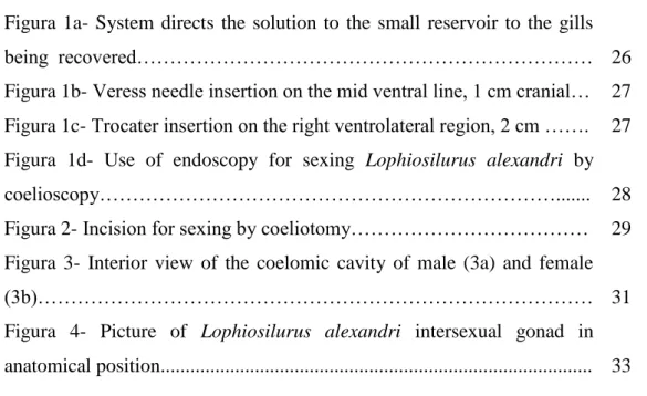
Figura 1a- System directs the solution to the small reservoir to the gills
being recovered……… 26
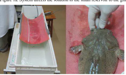
Figura 1b- Veress needle insertion on the mid ventral line, 1 cm cranial… 27 Figura 1c- Trocater insertion on the right ventrolateral region, 2 cm ……. 27
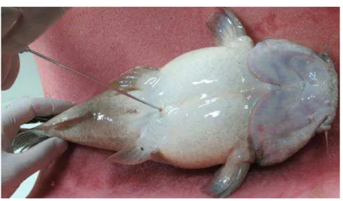
Figura 1d- Use of endoscopy for sexing Lophiosilurus alexandri by
coelioscopy………... 28
Figura 2- Incision for sexing by coeliotomy……… 29
Figura 3- Interior view of the coelomic cavity of male (3a) and female
(3b)……… 31
Figura 4- Picture of Lophiosilurus alexandri intersexual gonad in
RESUMO
O objetivo deste estudo foi avaliar técnicas de sexagem em juvenis de
Lophiosilurus alexandri. Foram testadas as técnicas de celioscopia, que é realizada com
o uso de equipamento para vídeo cirurgia, celiotomia que consistiu em procedimento
cirúrgico para visualização direta das gônadas e sexagem por sonda uretral, que se
baseia na identificação do sexo pela comparação entre as papilas genitais. Utilizando a
técnica de celioscopia, a sobrevivência 30 dias após os procedimentos foi de 100%. Os
peixes voltaram a se alimentar 10 dias após a cirurgia. A técnica apresentou 100% de
confirmação para os indivíduos identificados como fêmeas, enquanto que para aqueles
identificados como machos a confirmação foi de 66,6%. Não foi verificada diferença
significativa para tempo de indução à anestesia e tempo de recuperação para machos e
fêmeas. Contudo, o tempo de procedimento foi maior para os machos devido a
dificuldade em observar as gônadas, fato que pode ser atribuído a grande quantidade de
gordura visceral nos machos e ao coeficiente intestinal maior que 1, apesar de ser um
peixe carnívoro. O índice gonadossomático foi maior nas fêmeas. Para a técnica de
celiotomia, após 30 dias da cirurgia, a sobrevivência também foi de 100%. Foram
verificados dois animais interssexo. A técnica foi eficiente para a sexagem com 96,3%
de confirmação para machos e 93,9% para fêmeas. Os peixes voltaram a se alimentar
entre 10 a 14 dias após a cirurgia. Não foi verificado diferenças significativas para
tempo de indução anestésica, procedimento cirúrgico para visualização das gônadas e
tempo de retorno da anestesia, entre machos e fêmeas (P>0,05). A técnica de sonda
uretral foi menos eficiente, com 67,8 e 81,8% de confirmação para machos e fêmeas,
respectivamente. Concluímos que a técnica de celiotomia é recomendada para sexagem
de ambos os sexos e a celioscopia para identificar fêmeas juvenis de L. alexandri.
ABSTRACT
This study aimed to evaluate sexing techniques for juvenile Lophiosilurus
alexandri. The following techniques were tested: Coelioscopy, performed with the use
of video surgery equipment; coeliotomy, consisting of a surgical procedure for direct
visualisation of the gonads, and sex determination by a urethral probe, based on sex
identification by the comparison between the genital papillae. Using coelioscopy,
survival was 100%, 30 days after the procedure. The fish restarted eating 10 days after
surgery. This technique presented 100% confirmation for individuals identified as
females, while for those identified as males confirmation was 66.6%. There was no
significant difference between males and females for anesthesia induction and recovery
time. However, the procedure took longer for males due to the difficulty in observing
the gonads, which can be attributed to the large amount of visceral fat in males and to an
intestinal coefficient larger than 1, despite it being a carnivorous fish. The
gonadosomatic index was higher in females. As for the coeliotomy technique, 30 days
after surgery, the survival rate was also 100%. Two intersex animals were found. This
technique was efficient for sexing males with 96.3% accuracy and females with 93.9%
accuracy. The fish restarted eating between 10 and 14 days after surgery. Significant
differences between males and females were not found for anesthesia induction time,
duration of the surgical procedure to visualise the gonads and anesthesia recovery time
(p > 0.05). The urethral probe technique was less efficient, with 67.8 and 81.8%
accuracy for males and females, respectively. We conclude that coeliotomy is
recommended for sexing L. alexandri juvenile and coelioscopy, to identify females of L.
alexandri juvenile.
11
1. INTRODUÇÃO
A identificação do sexo em peixes é fundamental para o desenvolvimento de
estratégias de manejo nos programas de reprodução na aquicultura (Rota e Marques,
2007). O dimorfismo sexual é facilmente identificado em algumas espécies de peixes
como em lebiste Poecilia reticulata, na qual os machos apresentam tamanho menor e
coloração mais intensa do que as fêmeas (Barreiro-Buceta, 2013). Entretanto, várias
espécies de importância na aquicultura não apresentam dimorfismo sexual, ou este só
aparece de forma secundária, ou seja, durante a maturidade sexual, como é o caso do
padrão de coloração em pirarucu Arapaima gigas (Monteiro et al., 2010) e diferenças na
anatomia da papila genital em tilápia Oreochromis niloticus (Yasui et al., 2006). Há
espécies que atingem a maturidade sexual após alcançarem um grande tamanho
corporal, nesse caso, os custos para se formar e manter o plantel de reprodutores é alto
(Godinho, 2007). Metodologias como técnicas ultrassonográficas e cirúrgicas são
utilizadas como alternativas para identificação sexual precoce em peixes, o que pode
gerar economia para o produtor (Masoudifard et al., 2011; Chaves et al., 2015). Desta
maneira, quando a seleção de reprodutores é realizada em peixes juvenis, torna possível
adiantar a seleção dos indivíduos que apresentam desempenho zootécnico superior,
minimizando a ocupação das estruturas de cultivo, o uso de mão de obra e os custos
com alimentação.
2. REVISÃO DE LITERATURA
A revisão a seguir aborda técnicas utilizadas para sexagem em peixes, tanto para
monitoramento da eficiência de inversão sexual, quanto aquelas utilizadas para segregar
machos e fêmeas tendo em vista o manejo reprodutivo.
2.1. Coloração por acetato-carmim
Esta técnica é muito utilizada para a tilápia-do-nilo (Oreochromis niloticus).
Pode ser empregada com maior acurácia em peixes com comprimento mínimo de 3 cm
e 0,5 g de peso médio (Makino et al., 2009). Consiste na retirada de uma amostra
representativa de animais em cada lote. Depois de abatidos, o par de gônadas é
identificado. Em seguida, algumas gotas de solução de Bouin são aplicadas diretamente
12
Então, se estende as gônadas sobre uma lâmina de vidro onde são instiladas algumas
gotas da solução de acetato-carmim. Em seguida, uma lamínula é colocada sobre este
material fazendo-se uma leve pressão e é realizado exame ao microscópio de luz. O
tecido gonadal dos machos é caracterizado pela presença de cistos que contêm
espermatogônias e espermatócitos, enquanto que, nas gônadas das fêmeas, é verificada
presença de ovócitos no estádio perinucleolar (Wassermann e Afonso, 2002; Makino et
al., 2009).
A tilápia-do-nilo (O. niloticus) é uma das espécies mais cultivadas no Brasil,
recomendando-se o cultivo de lotes monossexo, devido ao fato de o macho apresentar
maior crescimento e melhor conversão alimentar do que a fêmea, como também para
evitar problemas como cópula e desova que podem resultar em excesso populacional
nos viveiros (Dias-Koberstein et al., 2007). A masculinização é realizada com o uso de
17α-metiltestosterona adicionado à ração, na fase de alevinagem (Gayão et al., 2013). A eficiência deste processo de inversão sexual é avaliada através do método de coloração
por acetato-carmim. Contudo, por ser necessário o sacrifício dos animais, é uma técnica
que não poderia ser usada para formar planteis de reprodutores.
2.2. Avaliação de caracteres sexuais secundários
Em peixes, os caracteres sexuais secundários são variados: ex.: coloração,
tubérculos na cabeça, glândulas cutâneas e outros (Godinho, 2007). Estas características
são utilizadas para diferenciação sexual de algumas espécies de grande interesse para
aquicultura brasileira, como o pirarucu (A. gigas) e a tilápia-do-nilo (O. niloticus).
O manejo reprodutivo do pirarucu (A. gigas) esbarra na falta de estudos para
melhor compreensão de sua complexa estratégia reprodutiva. Na época de
acasalamento, os machos exibem uma cor vermelha intensa em escamas abdominais,
enquanto que, a cor vermelha é menos intensa nas fêmeas (Chu-Koo et al., 2008). Lopes
e Queiroz (2009) analisaram a área e a intensidade de coloração, mas não conseguiram
identificar o sexo corretamente nessa espécie. Desta forma, esta característica não é
confiável para diferenciar os sexos (Carreiro et al., 2011). Contudo, Monteiro et al.
(2010) relataram sucesso na identificação sexual de pirarucu em período reprodutivo
(animais acima de 3 anos), através de uma mancha alaranjada na região inferior da
cabeça, presente somente nos machos.
As tilápias apresentam dimorfismo sexual secundário permanente, representado
13
reprodutivo desta espécie. Na fêmea, a papila apresenta duas aberturas distintas, o
orifício urinário e a saída do oviduto. Nos machos, existe somente uma abertura, que
servirá para a liberação de sêmen e a excreção da urina (Yasui et al., 2006). Makino et
al. (2009) utilizando Azul de Metileno a 1% para evidenciar a papila, conseguiram
realizar a sexagem de forma eficaz em peixes com comprimento acima de 5 cm, o que
ocorre entre 60 e 90 dias de idade.
2.3. Ultrassonografia
A ultrassonografia é uma técnica que permite identificar estruturas moles em
diversas profundidades do organismo de maneira não invasiva. Essa técnica baseia-se na
captação dos sons refletidos (ecos) ao passarem por tecidos de composições diferentes.
Esses ecos são transformados em imagens, que aparecem na tela do aparelho de
ultrassom como uma variação de sombras em cinza, preto e branco e podem ser
facilmente interpretadas (Crepaldi et al., 2006).
Para espécies de peixes que não apresentam dimorfismo sexual secundário, a
ultrassonografia está se destacando como alternativa aos métodos mais invasivos e
complexos, como as técnicas baseadas em cirurgia, que podem comprometer a saúde
destes animais e seu sucesso reprodutivo (Rota e Marques, 2007). Nos países onde a
piscicultura já é consagrada como uma atividade empresarial, a utilização do ultrassom
é rotineira em centros de pesquisas, principalmente na reprodução e permite a
visualização das estruturas reprodutivas sem retirar o peixe da água, o que é uma grande
vantagem, uma vez que se elimina qualquer interferência do ar na imagem gerada,
proporcionando uma maior nitidez (Crepaldi et al., 2006).
Masoudifard et al. (2011) relataram a identificação sexual em esturjão (Huso
huso) com exatidão acima de 96%, tanto para machos quanto para fêmeas utilizando o
ultrassom. Entretanto, a presença da bexiga natatória e gases intestinais podem obstruir
a visualização ultrassonográfica em outra espécie de esturjão (Scaphirhynchus sp.)
(Divers et al., 2009). Na sexagem da tuvira (Gymnotus sp.) o uso do ultrassom
apresentou 98% de acerto, com custo unitário inferior a R$ 0,70 por peixe (Rota e
Marques, 2007). Contudo, o uso do ultrassom requer experiência do operador para
analisar as imagens, sendo que em peixes mais jovens, a acurácia diminui como
verificado em esturjão russo (Acipenser gueldenstaedtii) (Hurvitz et al., 2007). Além
disso, em arenque (Clupea harengus pallasi), a sexagem e a classificação do estádio
14
das gônadas, ou seja, quando estas apresentaram aumento de volume devido à
proximidade do período reprodutivo, o que facilitou sua visualização (Bonar et al.,
1989).
2.4. Sexagem por análises laboratoriais
Técnicas genéticas e moleculares estão sendo desenvolvidas para identificação
do sexo em peixes. Uma das metodologias utilizadas é a identificação de marcadores
moleculares associados aos cromossomos sexuais, através da técnica de Reação em
Cadeia da Polimerase (Vale et al., 2014).
Porém, esta técnica não se aplica a todas as espécies. O pirarucu (A. gigas) por
exemplo, não apresenta diferenciação cromossômica entre machos e fêmeas,
apresentando um número diplóide 2n = 56 em ambos os sexos (Porto et al., 1992;
Marques et al., 2006). Esturjões (H. huso) jovens não contem cromossomos sexuais,
tornando impossível a identificação genética dos sexos numa fase precoce da vida
(Wuertz et al., 2006; McCormick et al., 2008).
Como alternativa, Chu-Koo et al. (2008) buscaram determinar o sexo em
pirarucu (A. gigas) por meio de detecção da proteína vitelogenina (Vtg) e mensuração
dos níveis de esteróides sexuais 17β – estradiol (E2) e 11 – Ketotesterona (11KT), em amostras de sangue. Os resultados mostraram que é possível distinguir machos de
fêmeas com seis anos de idade por meio da concentração de Vtg (100% de eficácia) e da
proporção dos hormônios 11KT/E2 (95% de eficácia). Os autores afirmaram, ainda, que
essa metodologia também se aplica em peixes com três anos de idade. No entanto, ainda
não existe uma metodologia que permita distinguir machos e fêmeas antes de atingirem
a primeira maturação gonadal.
2.5. Endoscopia
O endoscópio é constituído por um tubo fino contendo fibras ópticas para
transmitir a luz de uma fonte externa, para iluminação de órgãos internos que são vistos
através uma microcâmera de vídeo ou ocular ligado ao tubo (Swenson et al., 2007).
A identificação do sexo de peixes por endoscopia, também conhecida como
celioscopia, se baseia na diferenciação macroscópica das estruturas reprodutivas, que
são visualizadas inserindo-se o endoscópio através de uma pequena incisão abdominal,
o que permite uma precisa avaliação tecidual e a classificação das gônadas como
15
Endoscópios têm sido utilizados com sucesso para esta finalidade em diversas
espécies de peixe, incluindo truta (Salvelinus fontinalis) (Swenson et al., 2007), pirarucu
(A. gigas) (Carreiro et al., 2011), esturjão (H. huso) (Falahatkar et al., 2011), enguia
europeia (Anguilla Anguilla) (Macrì et al., 2014) e surubim (Pseudoplatystoma sp.)
(Chaves et al., 2015), com índices de acerto que variam de 94% a 100%.
A preparação para a cirurgia com antissepsia de pele, feita como em mamíferos,
não é recomendada para peixes, devido aos efeitos prejudiciais que a retirada do muco,
rico em imunoglobulinas, pode causar (Stetter, 2010). Entretanto, para realização da
cirurgia é necessário submeter o animal à anestesia. A indução anestésica é realizada
por dissolução do princípio ativo escolhido na água, o que promove a absorção
branquial do anestésico e sedação do peixe, que então pode ser retirado da água para
realização do procedimento cirúrgico, que dura em média de 3 a 4 minutos (Falahatkar
et al., 2011; Macrì et al., 2014; Chaves et al., 2015). Diversas substâncias podem ser
utilizadas, sendo as mais frequentemente escolhidas a tricaína metanossulfonato e o
eugenol (Murray, 2002). A manutenção da anestesia e a manutenção da respiração
podem ser realizadas através de uma mangueira inserida na boca do peixe, promovendo
a irrigação dos arcos branquiais com água e anestésico, conforme realizado por Macrì et
al. (2014) e Chaves et al. (2015).
Os ovários se apresentam em formato sacular, alongados, de coloração rosada a
amarelada, variando em função do status reprodutivo da fêmea, enquanto que, o
formato dos testículos é variável em função da espécie e a coloração é esbranquiçada
(Falahatkar et al., 2011). Estas características podem limitar a capacidade de distinção
entre a gordura visceral e os testículos, o que dificulta a identificação do sexo em peixes
que possuem muita gordura na cavidade celomática (Swenson et al., 2007; Falahatkar et
al., 2011).
Para a sutura, após o procedimento, são empregados um ou dois pontos em
padrão simples, sendo recomendada a utilização de fio cirúrgico monofilamento, que
pode ser constituído por nylon, dentre outros materiais de uso comum em cirurgias e a
retirada dos pontos não é necessária, pois estes são naturalmente expulsos pelo
organismo (Murray, 2002). A recuperação é realizada retornando com o peixe para a
água em um tanque provido de oxigenação suplementar. Neste momento é recomendado
realizar manipulação do animal para estimular a passagem de água através das
16
Os índices de mortalidade relatados para esta técnica foram de 3,3% em truta
(Salvelinus fontinalis) (Swenson et al., 2007) e 3,8% em pintado (Pseudoplatystoma sp.)
(Chaves et al., 2015). Já, pra pirarucu (A. gigas) (Carreiro et al., 2011), esturjão (H.
huso) (Falahatkar et al., 2011) e enguia europeia (A. Anguilla) (Macrì et al., 2014) não
foi observada mortalidade associada ao procedimento.
2.6. O pacamã
O Lophiosilurus alexandri (Steindachner, 1876) é um peixe conhecido na bacia
do rio São Francisco como pacamã, pacumã, pacamão ou niquin (Campeche et al.
2011). Pertence à ordem Siluriforme, é carnívoro, apresenta carne de excelente sabor,
sem espinhos intramusculares e também vem despertando interesse do mercado de
peixes ornamentais, o que tem incentivado sua introdução na aquicultura (Ribeiro et al.,
2013, Lucanus & Mischook, 1997). Em sua região de ocorrência é muito cobiçado, o
que gera forte pressão sobre os estoques naturais e o coloca na lista oficial de espécies
em risco de extinção (Brasil, 2014). Foi introduzido na bacia do rio Doce, mas seu
efeito sobre as espécies nativas não foi estudado (Barros et al., 2007).
Esta espécie apresenta desova parcelada e comportamento sedentário (Travassos,
1959). Libera seus ovos no substrato arenoso, onde o macho é geralmente o responsável
pelo cuidado parental (Sato et al., 2003). Macroscopicamente, as gônadas de pacamã
são pares e franjadas nos machos e pares e saculiformes nas fêmeas (Barros et al.,
2007). A idade de primeira maturação gonadal nesta espécie ainda não foi descrita. Sua
larvicultura já é realizada com sucesso através do emprego de alimento vivo (Melillo
Filho et al., 2014).
Alguns autores afirmam que pacamã não apresenta dimorfismo sexual
(Travassos, 1959; Sato et al., 2003 ) entretanto, Lopes et al. (2013) descreveram
diferenças sutis entre as papilas urogenitais de machos e fêmeas adultos, que podem ser
observadas a partir da inserção de uma sonda uretral na papila, com o intuito de
17
2.7. Referências bibliográficas
BARREIRO-BUCETA P. Selección sexual en Poecilia reticulata (Guppys). Anales
Universitarios de Etología, v.7, p.62- 68, 2013.
BARROS MDM, CRUZ RJG, JÚNIOR VCV, SANTOS JE. Reproductive apparatus
and gametogenesis of Lophiosilurus alexandri Steindachner (Pisces, teleostei,
Siluriformes) Rev. Bras. Zool., v. 24 (1), p. 213-221, 2007.
BONAR SA, THOMAS BP, PAULEY GB, MARTIN RW. Use of ultrasonic images
for rapid no lethal determination of sex and maturity of Pacific Herring. Aquacult. Res.,
v.32, p.113-120, 1989.
BRASIL. Ministério do Meio Ambiente. Portaria MMA n◦ 445, de 17 de dezembro de 2014. Lista Nacional Oficial de Espécies da Fauna Ameaçadas de Extinção – Peixes e Invertebrados Aquáticos.
CAMPECHE DFB, BALZANA L, FIGUEIREDO RCR. Peixes nativos do Rio São
Francisco adaptados para cultivo Petrolina: Embrapa Semiárido, v. 244, p. 1-20, 2011.
CARREIRO CRP, FURTADO-NETO MAA, MESQUITA PEC, BEZERRA TA. Se
determination in the giant fish of Amazon Basin, Arapaima gigas (Osteoglossiformes,
Arapaimatidae), using laparoscopy. Acta Amazon., v.41, n.3, p.415 -420, 2011.
CHAVES GV, GHELLER VA, TEIXEIRA EA, LUZ RK. Coelioscopy technique for
gender in surubim hybrid Pseudoplatystoma corruscans x Pseudoplatystoma
reticulatum. Aquacult. Res., v.46, p.1007-1012, 2015.
CHU-KOO F, DUGUÉ R, AGUILAR MA. et al. Gender determination in the Paiche or
Pirarucu (Arapaima gigas) using plasma vitellogenin, 17b-estradiol, and
11-ketotestosterone levels. Fish Physiol. Biochem., v.35, p.125-136, 2008.
CREPALDI DV, TEIXEIRA EA, FARIA PM et al. A ultrassonografia na piscicultura.
18
DIAS-KOBERSTEIN TCR, NETO AG, STÉFANI MV et al. Reversão sexual de larvas
de tilapia do nilo (Oreochromis niloticus) por meio de banhos de imersão em diferentes
dosagens hormonais. Rev. Acad., v.5, n.4, p.391-395, 2007.
DIVERS SJ, BOONE S S, HOOVER J J et al. Field endoscopy for identifying gender,
reproductive stage and gonadal anomalies in free-ranging sturgeon (Scaphirhynchus sp.)
from the lower Mississippi River. J. Appl. Ichthyol., v.25, Supl. 2, p.68-74, 2009.
FALAHATKAR A, GILANI MHT, FALAHATKAR S, ABBASALIZADEH A.
Laparoscopy, a minimally-invasive technique for sex identification in cultured great
sturgeon Huso huso. Aquaculture, v.321, p.273-279, 2011.
GAYÃO ALBA, BUZOLLO H, FÁVERO GC et al. Histologia hepática e produção
em tanques-rede de tilápia-do-nilo masculinizada hormonalmente ou não masculinizada.
Pesq. Agropec. Bras., v.48, n.8, p.991-997, 2013.
GODINHO HP. Estratégias reprodutivas de peixes aplicadas à aqüicultura: bases para o
desenvolvimento de tecnologias de produção. Rev. Bras. Reprod. Anim., v.31, n.3,
p.351-360, 2007.
HURVITZ A, JACKSON K, DEGANI G et al. Use of endoscopy for gender and
ovarian stage determinations in Russian sturgeon (Acipenser gueldenstaedtii) grown in
aquaculture. Aquaculture, v.270, p.158-166, 2007.
LOPES JP, FRANÇA FL, NETO MAS. O domínio na produção de alevinos de pacamã
– Propagação na Chesf permite repovoamento no rio São Francisco. Pan. da Aquic., v.23, nº136, pg 24-29, 2013
LOPES K, QUEIROZ HL. Avaliação do conhecimento tradicional dos pescadores da
RDSM. Uakari, v.5, n.2, p.59-66, 2009.
LUCANUS O, MISCHOOK SN. Interesting imports. Trop. Fish Hob. Nep., v.45,
19
MACRÌ F, RAPISARDA G, STEFANO C et al. Coelioscopic investigation in european
eels (Anguilla anguilla). J. Exot. Pet Med., v.23, p.147-151, 2014.
MAKINO LC, NAKAGHI LSO, PAES MCF et al. Efetividade de métodos de
identificação sexual em tilápias-do-nilo (Oreochromis niloticus) revertidas sexualmente
com hormônio em ração com diferentes granulometrias. Biosci. J., v.25, n.2, p.112-121,
2009.
MARQUES DK, VENERE PC, GALETTI PM. Chromosomal characterization of the
bonytongue Arapaima gigas (Osteoglossiformes: Arapaimidae), Neotrop. Ichthyol., v.4,
p.215-218, 2006.
MASOUDIFARD MAR, VAJHI M, MOGHIM RM et al. High validity sex
determination of three years old cultured Beluga Sturgeon (Huso huso) using
ultrasonography. J. Appl. Ichthyol., v.27, p.643-647, 2011.
McCORMICK CR, BOS DH, DEWOODY JA. Multiple molecular approaches yield no
evidence for sex-determining genes in lake sturgeon (Acipenser fulvescens). J. Appl.
Ichthyol., v.24, p.643-645, 2008.
MELILLO FILHO R, TAKATA R, SANTOS AEH et al. Draining system and feeding
rate during the initial development of Lophiosilurus alexandri (Steindachner, 1877), a
carnivorous freshwater fish. Aquacult. Res., v.45, p.1913-1920, 2014.
MONTEIRO LBB, SOARES MCF, CATANHO MTJ, HONCZARYK A. Aspectos
reprodutivos e perfil hormonal dos esteróides sexuais do pirarucu, Arapaima gigas
(Schinz, 1822), em condições de cativeiro. Acta Amaz., v.40, p.435-450, 2010.
MURRAY MJ. Fish Surgery. Semin. Avian. Exotic. Pet Med., v.11, n.4, p.246-257,
2002.
PORTO JIR, FELDBERG E, NAKAYAMA CM, FALCÃO JN. A checklist of
chromosome numbers and karyotypes of Amazonian freshwater fishes. Rev. Hydrobiol.
20
RIBEIRO PAP, MIRANDA FILHO KC, MELILLO FILHO R et al. Efeito anestésico
do eugenol em juvenis de pacamã. Pesq. Agropec. Bras. v.48, p.1136-1139, 2013.
ROTA MA, MARQUES DKS. Uso da ultra-sonografia na determinação do sexo de
peixes nativos. 2007. Artigo em Hypertexto. Disponível em:
<http://www.infobibos.com/Artigos/2007_4/ultrassonografia/index.htm>. Acesso
em: 9/2/2015
SATO Y, FENRICH-VERANI N, NUÑER APO et al.Padrões reprodutivos de peixes
da bacia do São Francisco. Em: Godinho, H.P.; Godinho, A.L. Águas e peixes e
pescadores do São Francisco das Minas Gerais. Editora PUC Minas, Belo Horizonte, p.
229-274, 2003.
STETTER M. Laparoscopic reproductive sterilization of waterfowl. Proceedings of the
AAZV, AWV, AZA/NAG Joint Conference. Tampa, FL, p.290-293, 2006.
STETTER M. Minimally invasive surgical techniques in bony fish (Osteichthyes). Vet.
Clin. Exot. Anim., v.13, p.291-299, 2010.
SWENSON EA, ROSENBERGER AE, HOWELL PJ. Validation of endoscopy for
determination of maturity in small salmonids and sex of mature individuals. Trans. Am.
Fish. Soc., v.136 p.994-998, 2007.
TRAVASSOS H. Nótula sobre o pacamã Lophiosilurus alexandri Steindachner, 1876.
Atlas da Sociedade de Biologia v.4, p.1-2, 1959.
VALE L, DIEGUEZ R, SÁNCHEZ L et al. A sex-associated sequence identified by
RAPD screening in gynogenetic individuals of turbot (Scophthalmus maximus). Mol.
Biol. Rep., v.41, p.1501-1509, 2014.
YASUI GS, SANTOS LC, FILHO OPR et al. Cultivo monossexual de tilápias:
importância e obtenção por sexagem e inversão sexual. Cad. Téc. Vet. Zootec., v.51, p.
21
WASSERMANN GJ, AFONSO LOB. Validation of the aceto-carmine technique for
evaluating phenotypic sex in Nile Tilapia (Oreochromis niloticus) fry. Cienc. Rural,
v.32, n.1, p.133-139, 2002.
WUERTZ S, GAILLARD S, BARBISAN F et al. Extensive screening of sturgeon
genomes by random screening techniques revealed no sex-specific marker. Aquaculture,
v.258, p.685-688, 2006.
3. OBJETIVOS
3.1. Objetivo geral
Realizar a sexagem em juvenis de pacamã Lophiosilurus alexandri.
3.2. Objetivos específicos
- Realizar a sexagem em pacamã pela técnica de celoscopia;
- Realizar a sexagem em pacamã pela a técnica de celiotomia;
- Realizar a sexagem em pacamã pela técnica de diferenciação de papila
urogenital com o uso de sonda uretral;
-Avaliar o retorno à alimentação e sobrevivência como parâmetros para
22
4. ARTIGO
Early sexing techniques in Lophiosilurus alexandri (Steindachner, 1876), a freshwater carnivorous catfish
Reinaldo Melillo Filhoa, Valentim Arabicano Ghellerb, Glauco Vinício Chavesc,
Walisson de Souza e Silvaa, Deliane Cristina Costaa, Luis Gustavo Figueiredoa, Gustavo
Soares da Costa Julioa, Ronald Kennedy Luza*
a
Universidade Federal de Minas Gerais, Departamento de Zootecnia, Laboratório de
Aquacultura, Belo Horizonte, Minas Gerais, Brazil.
b
Universidade Federal de Minas Gerais, Laboratório de Obstetrícia Veterinária, Belo
Horizonte, Minas Gerais, Brazil.
c
Centro Universitário de Formiga, UNIFOR –MG, Formiga, Minas Gerais, Brazil.
*Author's address - Ronald K. Luz, Universidade Federal de Minas Gerais,
Departamento, Departamento de Zootecnia, Laboratório de Aquacultura. Avenida
Antônio Carlos, 6627, CEP-30161-970, Belo Horizonte, MG, Brasil. E-mail address:
23
4.1. Abstract
This study aimed to evaluate sexing techniques for juvenile Lophiosilurus
alexandri. The following techniques were tested: Coelioscopy, performed with the use
of video surgery equipment; coeliotomy, consisting of a surgical procedure for direct
visualisation of the gonads, and sex determination by a urethral probe, based on sex
identification by the comparison between the genital papillae. Using coelioscopy,
survival was 100%, 30 days after the procedure. The fish restarted eating 10 days after
surgery. This technique presented 100% confirmation for individuals identified as
females, while for those identified as males confirmation was 66.6%. There was no
significant difference between males and females for anesthesia induction and recovery
time. However, the procedure took longer for males due to the difficulty in observing
the gonads, which can be attributed to the large amount of visceral fat in males and to an
intestinal coefficient larger than 1, despite it being a carnivorous fish. The
gonadosomatic index was higher in females. As for the coeliotomy technique, 30 days
after surgery, the survival rate was also 100%. Two intersex animals were found. This
technique was efficient for sexing males with 96.3% accuracy and females with 93.9%
accuracy. The fish restarted eating between 10 and 14 days after surgery. Significant
differences between males and females were not found for anesthesia induction time,
duration of the surgical procedure to visualise the gonads and anesthesia recovery time
(p > 0.05). The urethral probe technique was less efficient, with 67.8 and 81.8%
accuracy for males and females, respectively. We conclude that coeliotomy is
recommended for sexing L. alexandri juvenile and coelioscopy, to identify females of L.
alexandri juvenile.
24
4.2. Introduction
The "pacamã" Lophiosilurus alexandri, a carnivorous fish belonging to the
Siluriformes order, has high market value due to the flesh having no intramuscular
bones and a flavour that is appreciated by the consumer. This species is also becoming
popular in the ornamental fish market (Lucanus & Mischook, 1997). It is an important
species of the São Francisco River Basin, both socially and ecologically. Its natural
stocks are decreasing, and due to the fact that it has been considered an endangered
species, it is commonly used in restocking programmes (Lins et al., 1997; Brazil, 2014).
It presents a dorsoventrally flattened body shape, partial spawning, sedentary behaviour,
a preference for lentic environments (Travassos, 1959), and parental care, with its eggs
released on a sandy substrate (Sato et al., 2003). The larviculture of this species is
already successful using different breeding systems (Tenório et al., 2006; Luz & Santos,
2008; Santos & Luz, 2008; Pedreira et al., 2009; Takata et al., 2014; Melillo Filho et al.,
2014).
Sexual dimorphism in the urogenital papillae may be detected in L. alexandri
adults from the third year of age (Lopes et al., 2013). However, an early sexing
technique, performed on eight-month old fish, can generate savings for the breeder of
USD 5.00 per fish, as it is not possible to perform sexing by secondary features, until
fish reach sexual maturity (Burtle et al., 2003).
The assisted reproduction of a species is of fundamental importance and is
hindered by sexual differentiation. In order to know the sex, it is usually necessary to
kill the fish, which is prohibited for rare or endangered species (Wildhaber et al., 2005).
Non-lethal methods for determining sex include sex hormone detection by
radioimmunoassay of blood samples, ultrasound, and endoscopy (Ortenburger et al.,
1996 ; Chu-Koo et al., 2009; Arantes et al., 2010). The detection of sex hormones is
extremely expensive and cannot be carried out in a standard laboratory. The ultrasound
technique has methodological limitations for its use in the field. The use of ultrasound
requires a degree of expertise to analyse the images, and in young fish, accuracy
decreases (Hurvitz et al., 2007). Furthermore, the presence of the swim bladder and
intestinal gases can obstruct the view (Divers et al., 2009). The coelioscopy/endoscopy
technique, however, was successfully performed in Acipenser gueldenstaedtii (Hurvitz
et al., 2007), Salmonids (Swenson et al., 2007), and female of hybrids catfish (Chaves et
25
The aim of this study was to perform sexing in juveniles of Lophiosilurus
alexandri using different methodologies.
4.3. Materials and Methods
The studies were conducted in the Aquaculture Laboratory of the Department of
Zootechnics, Veterinary School of the Federal University of Minas Gerais, Brazil
(LAQUA/UFMG), with support from the Department of Veterinary Surgery and
Clinical Care-UFMG and they were divided into three experiments following the
methodology approved by the Ethics Committee on Animal Use (CEUA /UFMG),
protocol numbers. 153/2010 and 181/2014.
For the experiments, the animals produced at LAQUA were placed in circular
tanks with a 400 L working volume, with supplemental aeration (dissolved oxygen > 5
mg/L), temperature of 28.0 ± 1.0 ºC and heated water recirculation system. They were
fed twice a day with commercial diet for carnivorous fish (40% crude protein).
4.4. Experiment 1 - Coelioscopy
This experiment was conducted in July 2013 with 34 specimens of L. alexandri
with 36.3 ± 3.1 cm and 697.8 ± 205.0 g of length and weight, respectively.
Previous to the procedures, the fish were fasted for 24 hours. The fish were individually
placed in a tank for anesthesia with eugenol dissolved in the water at a dilution of 150
mg/L. During anesthesia induction and for animal identification, Partners & Quality
Technology microchips were inserted, lateral to the dorsal fin. The animals were placed
in dorsal decubitus on a padded surface on top of a supporting cast on a small water
tank with anaesthetic. A recirculated water system with anaesthetic (100 mg of
eugenol/L) was placed so the gills were constantly irrigated, providing oxygenation and
maintaining anesthesia. The system, through an aquarium pump, directs the solution to
the small reservoir to the gills being recovered (Figure 1a). To minimise irritation to the
26
Figure 1a. System directs the solution to the small reservoir to the gills being recovered
The equipment for coelioscopy consisted of a tower with a 14-inch Sony
monitor, a Karl Storz electronic insufflator, a Telecam DX microcamera, a xenon light
source with Karl Storz fibre optic cable, a CO2 cylinder, and a JVC digital camcorder
with cable to attach to the monitor. Video surgery equipment included a Veress needle
to inflate the abdominal cavity, a 3-mm trocater specially made for the procedure; a 2.7
mm diameter rigid endoscope with 0° viewing angle. A small incision (2 mm) in the
skin, craniocaudalis, was made between the pelvic fins of the fish, 1 cm cranial to the
urogenital pore, for insertion of the Veress needle and insufflation with CO2 (Figure
1b).The 3-mm trocater was inserted 2 cm cranially to the right pelvic fin (Figure 1c).
The endoscope was directed to the chip reader for recording the identification of the
animal and then inserted into the coelomic cavity, and after a brief general exam, the
gonads were viewed (Figure 1d). The viewed structures were classified as male or
female, and were recorded along with the number of the chip. The endoscope was then
removed, followed by withdrawal of the Veress needle, and lastly the trocater. Before
removing the trocater, a slight pressure was applied to the coelomic cavity, so that the
maximum amount of CO2 gas was expelled to return the fish to its normal density, thus
avoiding excess buoyancy. After removing the trocater, the incision was sutured in
standard single discontinuous with Nylon 3-0 (Shalom). Between each procedure, the
27
Figure1b. Veress needle insertion on the mid ventral line, 1 cm cranial
28
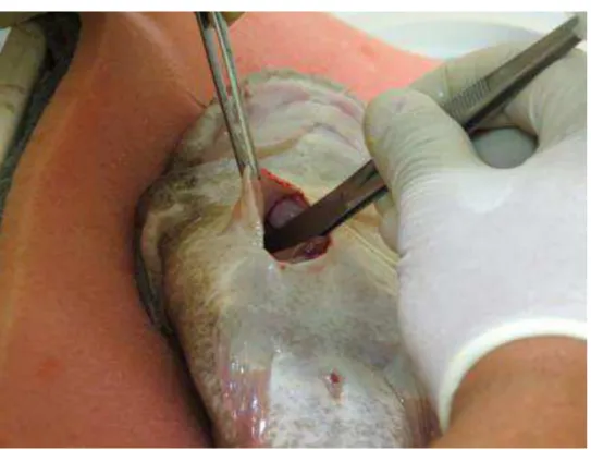
Figure 1d. Use of endoscopy for sexing Lophiosilurus alexandri by coelioscopy
For recovery from the procedure and anesthesia, the fish were placed in another
tank with aeration and heating. In order to recover from anesthesia, the fish was
manipulated and held against a flow of water to ensure ventilation, until they could
move forward (Chaves et al., 2015). They were then placed in the original tanks.
The times for the induction of anesthesia, the procedure (which includes surgery up to
the moment when the animals returned to the water), as well as recovery, when the
animals presented hydrostatic equilibrium, were recorded individually.
After release in the tanks, the animals were fed daily for 30 days with 2% of the
biomass divided into two meals at 8:00 am and 5:00 pm. We recorded when the animals
29
4.5. Experiment 2 - Coeliotomy
For this experiment, carried out in September 2014, 63 animals were used with
an average weight of 369.6 ± 126.7 g and average length of 29.3 ± 3.1 cm. The
procedures of fasting and anesthesia were the same as in experiment 1.
For the surgical procedures, routine instruments for coelomic cavity synthesis
and diaeresis were used. These include a disposable scalpel blade nº 10, a Mayo-Hegar
needle holder, and a toothless anatomical clamp. A 4 to 5 cm long "L" incision was
made in the ventral region of the animal to facilitate viewing the surgical field (Figure
2). A 2 to 3 cm incision was made along the middle line of the animal, for 2 to 3 cm,
and another 2 cm on the lower part of "L" to the right. When the coelomic cavity was
accessed, anatomical forceps were used to remove the internal organs, allowing direct
observation and classification of the gonads as testes or ovaries. After the observation,
the synthesis of the cavity using a 3-0 (Shalon®) nylon thread and a continuous simple
suture pattern in a single suture plan involving skin and muscles was done.
Figure 2. Incision for sexing by coeliotomy
Between each procedure, the surgical instruments were disinfected with
30
therapy, consisting of sulphadoxine 70 mg/kg associated with trimethoprim-14 mg/kg
(Borgal®), was injected intramuscularly into the dorsal region of the animals.
The times for the induction of anesthesia, the procedure and recovery was
recorded as for Experiment 1. Additionally, similar procedures to the first experiment
were adopted for the recovery of post-surgery animals, feeding, and observations
4.6. Experiment 3 - Sexing by cannulation
For this experiment, the animals of experiment 2 were used, in order to avoid
killing other animals. After 30 days of observation, the 63 specimens of L. alexandri
used in experiment 2 were euthanized by immersion in a eugenol 285 mg/L solution
(396/2012 CETEA protocol) for sexing, using a urethral probe with 2 mm gauge. When
this probe is inserted in the urogenital orifice of adult animals, it is possible to confirm
three orifices in females (anus, urethra and oviduct) and two in males (anus and the
urogenital hole), as described by Lopes et al. (2013). This sexing procedure was
performed on euthanized animals, but this did not affect the results, which would
otherwise have been performed on anaesthetised animals. In addition, this type of
sexing is commonly used in fish farms, and it is known that it does not cause mortality.
After that, the microchip was read for animal identification.
4.7. Biometric index
After euthanasia, the animals used in experiment 1 and experiment 2-3 were
weighed and their length was measured. After the microchip identification, the
macroscopic confirmation of sex by the gonads was performed. In experiments 1 and
2-3, the weight of gonads and visceral fat were registered. For the animals of experiment
1, the intestine length was also measured. With these data, we calculated: the GSI, from
the expression GSI = 100 (WG/W), where W is the total weight of the animal and WG
is the mass of the gonads; the viscerosomatic fat index (VFI) from the expression VFI =
100 (Wfv / W) where W represents the total weight of the animal and Wfv is the mass
of visceral fat, and the intestinal coefficient (IC) from IC ratio = intestine length/total
length of the animal.
4.8. Statistical analysis
In order to compare the effectiveness of sexing by the different methods,
31
viscerosomatic fat index, anesthesia time, procedure time, and recovery time were
compared by ANOVA. For the statistical analysis, the anesthesia induction time was
square-root transformed and recovery time was log10 transformed, prior to analysis.
4.9. Results
4.9.1. Experiment 1
Survival was 100% after 30 days from the procedure and the fish restarted eating
10 days after the procedure. Macroscopically, the gonads of pacamã are paired and
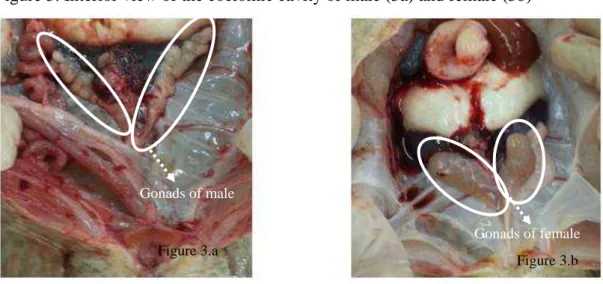
fringed in males and paired and bag-shaped in females (Figure 3).
Figure 3. Interior view of the coelomic cavity of male (3a) and female (3b)
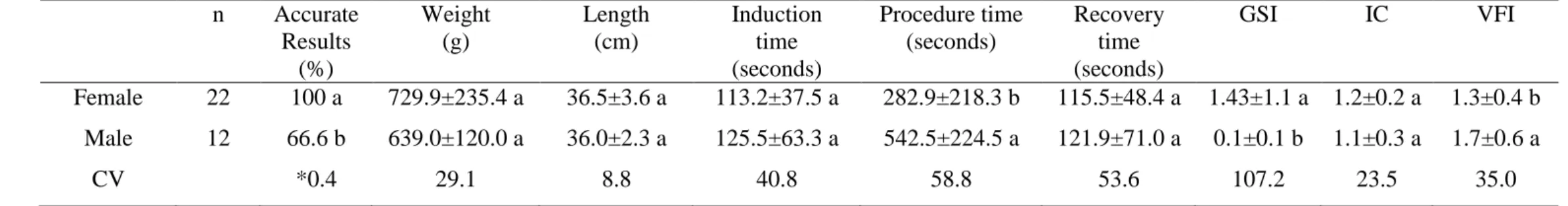
Table 1 shows the biometric and confirmation data of the coelioscopic
technique. The technique is more efficient to identify females (100%) than males. No
significant difference was observed for weight, length, anesthesia induction time,
recovery time, and intestinal coefficient between males and females. The procedure
time and the VFI were higher for males, whereas the GSI was higher in females. Figure 3.a
Figure 3.b
Gonads of male
32
Table 1. Values for sexing accuracy, weight, length, anesthesia induction time, procedure time, recovery time, gonadosomatic index (GSI),
viscerosomatic fat index (VFI) and intestinal coefficient (IC) of Lophiosilurus alexandri submitted to sexing by coelioscopic technique
Within a column, superscripts indicate significant differences between means (p < 0.05).
*Contingency coefficient
n Accurate
Results (%)
Weight (g)
Length (cm)
Induction time (seconds)
Procedure time (seconds)
Recovery time (seconds)
GSI IC VFI
Female 22 100 a 729.9±235.4 a 36.5±3.6 a 113.2±37.5 a 282.9±218.3 b 115.5±48.4 a 1.43±1.1 a 1.2±0.2 a 1.3±0.4 b
Male 12 66.6 b 639.0±120.0 a 36.0±2.3 a 125.5±63.3 a 542.5±224.5 a 121.9±71.0 a 0.1±0.1 b 1.1±0.3 a 1.7±0.6 a
33
4.9.2. Experiment 2
There was no mortality during the 30 days of observation after surgery. The
animals restarted eating 10 to 14 days after the procedure.
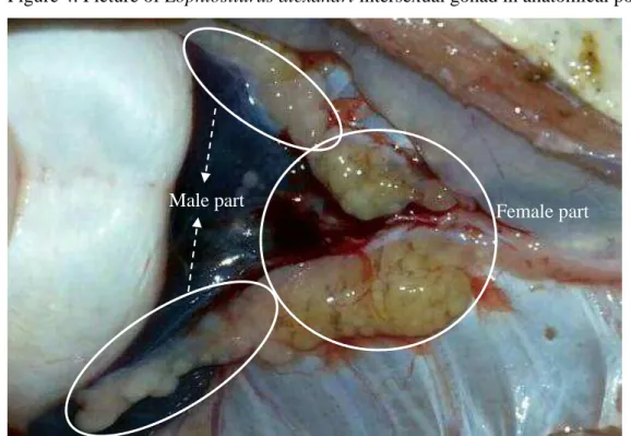
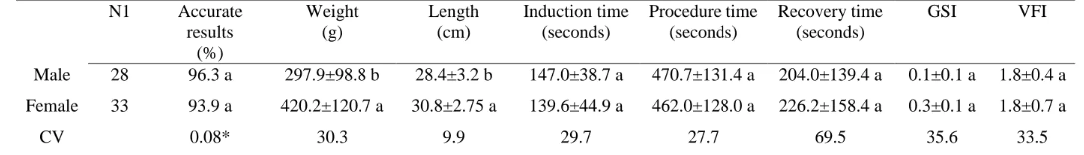
The coeliotomy sexing technique was efficient for sexing with a 96.3% accuracy
for males and 93.9% for females (Table 2). When the animals were killed for
macroscopic confirmation, two intersex individuals were found (Figure 4). No
significant differences between males and females were observed for anesthesia
induction time, surgical procedure time for viewing the gonads, and anesthesia recovery
time. The weight and length of females were greater than the males. Nevertheless, there
were no differences (p > 0.05) for the gonadossomatic and viscerosomatic fat indices.
Figure 4. Picture of Lophiosilurus alexandri intersexual gonad in anatomical position
4.9.3. Experiment 3
The urethral probe technique, despite being faster and less invasive, was less
efficient, with 67.8 and 81.8% accuracy for males and females, respectively.
34
Table 2. Values for sexing accuracy, weight, length, anesthesia induction time, procedure time, recovery time), gonadosomatic index (GSI),
viscerosomatic fat index (VFI), and intestinal coefficient (IC) of Lophiosilurus alexandri submitted to sexing by coeliotomy technique
1 N total = 63 animals, of which 28 males, 33 females and two intersex individuals not considered for statistics
Sexing technique performed following the methodology described by Lopes et al. (2013) on euthanized animals with 285 mg/L eugenol.
*Contingency coefficient
Within a column, superscripts indicate significant differences between means (p < 0.05).
N1 Accurate
results (%)
Weight (g)
Length (cm)
Induction time (seconds)
Procedure time (seconds)
Recovery time (seconds)
GSI VFI
Male 28 96.3 a 297.9±98.8 b 28.4±3.2 b 147.0±38.7 a 470.7±131.4 a 204.0±139.4 a 0.1±0.1 a 1.8±0.4 a
Female 33 93.9 a 420.2±120.7 a 30.8±2.75 a 139.6±44.9 a 462.0±128.0 a 226.2±158.4 a 0.3±0.1 a 1.8±0.7 a
35
4.10. Discussion
Studies related to L. alexandri reproduction are few and have been performed on
captured animals based on the reproduction of adult individuals (Santos et al. 2013;
Costa et al. 2015) Macroscopically, the pacamã gonads are paired and fringed in males
and paired and bag-shaped in females, as described by Barros et al. (2007). To date, the
age and size of the first gonadal maturation of this species are not known.
The coeliotomy technique showed high efficiency for sexing, with 96.3%
accuracy for males and 93.9% for females. This result was similar to that obtained by
other authors for the use of endoscopy for Acipenser gueldenstaedtii (Hurvitz et al.,
2007), Salmonids (Swenson et al., 2007), and Huso huso (Falahatkar et al., 2011).
Despite being more invasive, coeliotomy does not require the use of sophisticated
devices and offers a clear and direct view of the tissue. An L-shaped incision allows for
a greater operating field compared to a straight incision of the same size, facilitating
visceral manipulation and allowing the removal of structures blocking the view of the
gonads, such as the intestine and intracoelomic fat.
Coelioscopy presented 100% confirmation for individuals identified as females,
while for those identified as males confirmation was 66.6%. Therefore, this technique
was less effective in males compared to coeliotomy in juvenile L. alexandri. This result
can be explained by the lower GSI in males, a great amount of intracoelomic fat, a long
and coiled intestine (despite the carnivorous feeding habits), and the flattened shape of
the body, which provides little space for intracoelomic handling of the endoscope.
These characteristics of the species made it difficult to visualise the male gonads. This
difficulty was reflected in the procedure time (540 seconds on average for males), which
was longer than the average for females (280 seconds). The magnification of the image
achieved by the endoscope lenses also increases the movement in the generated image.
It requires great care in handling the device because any abrupt movement causes
oscillation in the image thus affecting observation. There was no significant difference
between males and females for weight, length, and anesthesia induction time.
The urethral probe technique was less efficient than the coelioscopy and
celiotomy, with 67.8 and 81.8% accuracy for males and females, respectively. These
results are lower than what have been found for adult animals of the same species, from
the third year of life, with 100% correct identification (Lopes et al., 2013). This
suggests that it is still difficult to view the urogenital papilla in juveniles with
36
Survival 30 days after the procedures was 100% for both surgical techniques
(coelioscopy and coeliotomy), equivalent to the results described for Huso huso
(Falahatkar et al., 2011) and higher than those for catfish, 6.2 % mortality (Chaves et
al., 2013), both using coelioscopy (endoscopy). This suggests that L. alexandri juveniles
are resistant to anesthesia and surgical procedures. This resistance is confirmed since
the time to restart eating was short. It was longer (14 days) for the coeliotomy
technique, compared to the coelioscopy technique (10 days). The difference can be
attributed to the more invasive nature of coeliotomy. The use of endoscopes allows
gonad observation through a much smaller incision (2 to 3 mm), while the coeliotomy
incision size (4 to 5 cm) causes more extensive tissue injury, requiring more time to
complete healing. Furthermore, smaller animals were used for coeliotomy, which
highlighted the difference of the incision size between the two techniques, in relation to
the body size of the animals.
For the surgical procedures, eugenol proved to be an effective anaesthetic. This
anaesthetic has been used successfully on L. alexandri juveniles smaller than the ones
used in this study with different size classes and doses ranging from 20 to 120 mg/L
(Ribeiro et al., 2013). Eugenol requires no "grace period", which is the waiting time
after using the active ingredient before slaughtering the animal, in order to avoid the
presence of residues in the flesh. This product is not carcinogenic or mutagenic, it is
affordable, easily found on the market, has good efficiency at low dosages, besides
being safe for the handler and the environment (Munday and Wilson, 1997; Iversen et
al. 2003 ; Charoendat et al., 2009 ; Roubach et al., 2005). The anaesthetic procedures
were safe and efficient, keeping anesthesia and induction times within the recommended
range, which is: induction in 3 minutes or less, complete recovery in 10 minutes or less,
and no mortality after 15 minutes (Schoettger and Julin 1967; Gilderhus and Marking
1987; Small, 2003). No differences in the anesthesia and recovery times between sexes
were recorded.
The sex ratio in this study was 35.3% males and 64.7% females in experiment 1
and 45.9% males and 54.1% females in experiment 2. A predominance of females has
also been recorded in fish of this species in a natural environment, with the proportion
of 62.8% females and 37.2% males (Barros et al., 2007). In experiment 1, females and
males had similar weight and length, while in experiment 2, females were larger than
37
In the literature, there are no data for this species relating to the size at first maturity.
However, females in reproductive stage are reported to be between three to five years
(Lopes et al., 2013). In another study with animals collected in nature, vitellogenic
oocytes were found in fish weighing 0.6 to 3.5 kg, and smaller females had a lower GSI
(Barros et al., 2007). Females of this species suitable for breeding and weighing 3.1 ±
0.9 kg present a GSI ranging from 1.5 to 2.9 (Sato, 1999). In this study, experiment 1
was conducted with larger animals than those used in experiment 2, so animals used in
experiment 1 macroscopically presented more developed female gonads, with higher a
GSI than males. In experiment 2, on the other hand, there was no GSI difference
between males and females, suggesting little gonadal development in this weight range.
This reinforces the importance of the coeliotomy technique for early sexing.
The VFI in experiment 1 (larger animals) was lower in females with a higher
GSI, while in experiment 2, the VFI and GSI were the same for males and females. This
result may be related to the fact that visceral fat and hepatic reserves of individuals are
used during the gonadal development process, as observed in various fish species
(Chellappa et al., 1995; Huntingford et al., 2001; Lima-Junior et al., 2002; Gurgel et al.
2008; Silva et al. 2008).
After the animals were killed for macroscopic confirmation, two intersex
individuals were found. This was the first report of intersex L. alexandri individuals.
The existence of intersex animals is normal in some fish species that present
hermaphroditism, while all fish in the Cyprinidae family undergo an intersex stage
before developing ovaries and, later, testes (Takahashi and Shimizu, 1983). However,
several authors have reported that estrogenic substances found in wastewater entering
rivers, either treated or untreated, cause intersexuality in fish (Purdom et al., 1994;
Jobling and Sumpter, 1993; Desbrow et al., 1998; Tyler and Routledge, 1998). In this
study, however, all animals were kept in the laboratory from birth, under the same
conditions, and they were kept in a water recirculation system. This rules out the
possibility that intersexuality occurred due to environmental contamination, and studies
are needed to clarify this sort of occurrence.
4.11. Conclusion
Coeliotomy and coelioscopy are recommended for sexing juvenile L. alexandri.
The coelioscopy technique, however, must be performed with great care to identify
38
4.12. Acknowledgements
We thank the Conselho Nacional de Desenvolvimento Científico e Tecnológico
(CNPq-Brasil), Coordenação de Aperfeiçoamento de Pessoal de Nível Superior
(CAPES-Brasil), Fundação de Amparo à Pesquisa do Estado de Minas Gerais
(FAPEMIG-Brasil) and Pró-Reitoria de Pesquisa (PRPq-UFMG-Brasil). LUZ, R.K.
received a research grant and a research fellowship from the Conselho Nacional de
Desenvolvimento Científico e Tecnológico (CNPq No. 305913/2012-3) and from the
Fundação de Amparo à Pesquisa do Estado de Minas Gerais (FAPEMIG No.
PPM-00403/13)
4.13. References
ARANTES PA, SANTOS HB, RIZZO E et al. Profiles of sex steroids, fecundity, and
spawning of the curimatã-pacu Prochilodus argenteus in the São Francisco River,
downstream from the Três Marias Dam, Southeastern Brazil. Anim. Reprod. Sci., v.118,
p.330-336, 2010.
BARROS MDM, CRUZ RJG, JÚNIOR VCV, SANTOS JE. Reproductive apparatus
and gametogenesis of Lophiosilurus alexandri Steindachner (Pisces, Teleostei,
Siluriformes). Rev. Bras. Zool., v.24, p.213-221, 2007.
BRASIL. Ministério do Meio Ambiente. Portaria MMA nº 445, de 17 de dezembro de
2014. "Lista Nacional Oficial de Espécies da Fauna Ameaçadas de Extinção - Peixes e
Invertebrados Aquáticos".
BURTLE GJ, NEWTON GL, LEWIS GW, JACOBS, J. Ultrasound for sex
determination of catfish, 2003. Available in
http://www.caes.uga.edu/commodities/animals/aquaculture/catfish/ultrasound.html.
Acess:21/05/2014
CHAROENDAT U, AREECHON N, SRISAPOOME P, CHANTASART D. The
efficacy of synthetic eugenol as an anesthetic for tilapia (Oreochromis niloticus).
39
CHAVES GV, GHELLER VA, TEIXEIRA EA, LUZ RK. Coelioscopy technique for
gender in surubim hybrid Pseudoplatystoma corruscans x Pseudoplatystoma
reticulatum. Aquacult Res., v.46, p.1007-1012, 2015.
CHELLAPPA S, HUNTINGFORD FA, STRANG RHC, THOMSON RY. Condition
factor and hepatosomatic index as estimates of energy status in male three-spined
stickleback. J. Fish Biol., v.47, p.775-787, 1995.
CHU-KOO F, DUGUÉ R, ALVÁN AM et al. Gender determination in the Paiche or
Pirarucu (Arapaima gigas) using plasma vitellogenin, 17beta-estradiol, and
11-ketotestosterone levels. Fish Physiol Biochem, v.35, p.125-36, 2009.
COSTA DC, SILVA WS, MELILLO FILHO R et al. Capture, adaptation and artificial
control of reproduction of Lophiosilurus alexandri: A carnivorous freshwater species.
Anim. Reprod. Sci., v.159, p.148-154, 2015.
.
DESBROW C, ROUTLEDGE EJ, BRIGHTY GC et al. Identification of estrogenic
chemicals in STW effluent. 1. Chemical fractionation and in vitro biological screening.
Environ. Sci. Technol., v.32, p.1549-1558, 1998.
DIVERS SJ, BOONE SS, HOOVER JJ et al. Field endoscopy for identifying gender,
reproductive stage and gonadal anomalies in free-ranging sturgeon (Scaphirhynchus sp.)
from the lower Mississippi River. J. Appl. Ichthyol., v.25, p.68-74, 2009.
FALAHATKAR B, GILANI MHT, FALAHATKAR S, ABBASALIZADEH A.
Laparoscopy, a minimally-invasive technique for sex identification in cultured great
sturgeon Huso huso. Aquaculture, v.321, p.273-279, 2011.
GILDERHUS PA, MARKING LL. Comparative efficacy of 16 anaesthetic chemicals
on rainbow trout. N. Am. J. Fish. Manag., v.7, p.288-292, 1987.
GURGEL HDCB, ALBUQUERQUE CQ, LIMA DDS, BARBIERI G. Aspectos da
biologia pesqueira em fêmeas de Cathrops spixii do estuário do rio Potengi, Natal/RN,
40
HUNTINGFORD FA, CHELLAPPA S, TAYLOR AC, STRANG RHC. Energy
reserves and reproductive investment in male three spined sticklebacks, Gasterosteus
aculeatus. Ecol. Freshwat. Fish, v.10, p.111-117, 2001.
HURVITZ A, JACKSON K, DEGANI G, LEVAVI-SIVAN B Use of endoscopy for
gender and ovarian stage determinations in Russian sturgeon (Acipenser
gueldenstaedtii) grown in aquaculture. Aquaculture, v.270, p.158-166, 2007.
IVERSEN M, FINSTADA B, MCKINLEYC RS, ELIASSEN RA The efficacy of
metomidate, clove oil, Aqui-S and Benzoak as anesthetics in Atlantic salmon (Salmo
salar ) smolts, and their potential stress reducing capacity. Aquaculture, v.221,
p.549-566, 2003.
JOBLING S, SUMPTER JJ. Detergent components in sewage effluent are weakly
estrogenic to fish: An in vitro study using rainbow trout (Onchorhynchus mykiss)
hepatocytes. Aquat. Toxicol., v.l27, p.361-372, 1993.
LIMA-JUNIOR SE, CARDONE IB, GOITEIN R. Determination of a method for
calculation of allometric condition factor of fish. Acta Sci., v.24, p.397-400, 2002.
LINS LV, MACHADO ABM, COSTA CMR, HERRMANN G. Roteiro metodológico
para elaboração da lista de espécies ameaçadas de extinção: contendo a lista oficial da
fauna ameaçada de extinção em Minas Gerais. Publicações Avulsas da Fundação
Biodiversitas, v.1, p.1-50, 1997.
LOPES JP, FRANÇA FL, NETO MAS. O domínio na produção de alevinos de pacamã
– Propagação na Chesf permite repovoamento no rio São Francisco. Pan. da Aquicult., v.23, p.24-29, 2013.
LUCANUS O, MISCHOOK SN. Interesting imports. Trop. Fish Hob. Nep., v.45,
41
LUZ RK, SANTOS JCE. Densidade de estocagem e salinidade da água na larvicultura
do pacamã. Pesq. Agropec. Bras., v.43, p.903-909, 2009.
MELILLO FILHO R, TAKATA R, SANTOS AEH et al. Draining system and feeding
rate during the initial development of Lophiosilurus alexandri (Steindachner, 1877), a
carnivorous freshwater fish. Aquacult. Res., v.45, p.1913-1920, 2014.
MUNDAY L, WILSON SK. Comparative efficacy of clove oil and other chemicals in
anaesthetization of Pomacentrus amboinensis, a coral reef fish. J. Fish Biol., v.51,
p.931-938, 1997.
ORTENBURGER A, JANSEN ME, WHYTE SK. Nonsurgical videolaproscopy for
determination of reproductive status of the Arctic charr. Can. Vet. J., v.37, p.96-100,
1996.
PEDREIRA MM, LUZ RK, SANTOS JCE et al. Biofiltração da água e tipos de
substrato na larvicultura do pacamã. Pesq. Agropec. Bras., v.44, p.511-518, 2009.
PURDOM C, HARDIMAN P, BYE V et al. Estrogenic effects of effluents from sewage
treatment works. Chem. Ecol., v.8, p.275-285, 1994.
RIBEIRO PAP, MIRANDA FILHO KC, MELILLO FILHO R et al. Efeito anestésico
do eugenol em juvenis de pacamã. Pesq. Agropec. Bras., v.48, p.1136-1139, 2013.
ROUBACH R, GOMES LC, FONSECA FAL, VAL AL. Eugenol as na efficacious
anaesthetic for tambaqui Colossoma macropomum (Cuvier). Aquacult. Res., v.36,
p.1056-1061, 2005.
SANTOS HB, SAMPAIO EV, ARANTES FP, SATO Y. Induced spawning and
reproductive variables of the catfish Lophiosilurus alexandri Steindachner, 1876
42
SANTOS JCE, LUZ RK. Effect of salinity and prey concentrations on
Pseudoplatystoma corruscans, Prochilodus costatus and Lophiosilurus alexandri
larviculture. Aquaculture, v.87, p.324-328, 2009.
SATO Y. Reprodução de peixes da bacia do rio São Francisco: indução e caracterização
de padrões – Doctoral Thesis, Universidade Federal de São Carlos; 1999.
SATO Y, FENERICH-VERANI N, NUNER APON et al. Padrões reprodutivos de
peixes da bacia do São Francisco. Em: Godinho HP, Godinho AL, editors. Águas e
peixes e pescadores do São Francisco das Minas Gerais. Editora PUC Minas, Belo
Horizonte; p. 229-274, 2003.
SCHOETTGER RA, JULIN AM. Efficacy of MS-222 as an anesthetic on four
salmonids. United States Department of Interior Resource Publication 19, Bureau of
Sport Fisheries and Wildlife, Washington, D.C 1967.
SILVA GC, CASTRO ACL, GUBIANI EA. Estrutura populacional e indicadores
reprodutivos de Scomberomorus brasiliensis Collette, Russo e Zavala-Camin, 1978
(Perciformes: Scombridae) no litoral ocidental maranhense. Acta. Sci., v.27, p.383-389,
2008.
SMALL BC. Anesthetic efficacy of metomidate and comparison of plasma cortisol
responses to tricaine methanesulfonate, quinaldine and clove oil anesthetized channel
catfish Ictalurus punctatus. Aquaculture, v.218, p.177-185, 2003.
SWENSON EA, ROSENBERGER AE, HOWELL PJ. Validation of endoscopy for
determination of maturity in small salmonids and sex of mature individuals. Trans. Am.
Fish Soc., v.136, p.994-998, 2007.
TAKAHASHI H, SHIMIZU M. Juvenile intersexuality in a cyprinid fish, the Sumatra
barb, Barbus tetrazona. Bull Faculty Fish Hokkaido University v.34, p.69-78, 1983.
TAKATA R, SILVA WS, COSTA DC et al. Effect of water temperature and prey