the Regulation of Circadian
Per1
Gene
Jungtae Na1, Kwanghyun Lee1, Hwan-Gon Kim1, Jee-Yoon Shin1, Wonho Na1, Hayan Jeong1, Jong-Woo Lee1, Sehyung Cho2, Won-Sun Kim1, Bong-Gun Ju1*
1Department of Life Science, Sogang University, Seoul, Korea,2Department of Physiology, Kyung Hee University School of Medicine, Seoul, Korea
Abstract
Circadian clocks are the endogenous oscillators that regulate rhythmic physiological and behavioral changes to correspond to daily light-dark cycles. Molecular dissections have revealed that transcriptional feedback loops of the circadian clock genes drive the molecular oscillation, in which PER/CRY complexes inhibit the transcriptional activity of the CLOCK/BMAL1 heterodimer to constitute a negative feedback loop. In this study, we identified the type II protein arginine methyltransferase 5 (PRMT5) as an interacting molecule of CRY1. Although thePrmt5gene was constitutively expressed, increased interaction of PRMT5 with CRY1 was observed when thePer1gene was repressed both in synchronized mouse liver and NIH3T3 cells. Moreover, rhythmic recruitment of PRMT5 and CRY1 to thePer1gene promoter was found to be associated with an increased level of histone H4R3 dimethylation andPer1gene repression. Consistently, decreased histone H4R3 dimethylation and altered rhythmicPer1gene expression were observed in Prmt5-depleted cells. Taken together, these findings provide an insight into the link between histone arginine methylation by PRMT5 and transcriptional regulation of the circadianPer1gene.
Citation:Na J, Lee K, Kim H-G, Shin J-Y, Na W, et al. (2012) Role of Type II Protein Arginine Methyltransferase 5 in the Regulation of CircadianPer1Gene. PLoS ONE 7(10): e48152. doi:10.1371/journal.pone.0048152
Editor:Nicholas S. Foulkes, Karlsruhe Institute of Technology, Germany
ReceivedMarch 26, 2012;AcceptedSeptember 20, 2012;PublishedOctober 25, 2012
Copyright:ß2012 Na et al. This is an open-access article distributed under the terms of the Creative Commons Attribution License, which permits unrestricted use, distribution, and reproduction in any medium, provided the original author and source are credited.
Funding:This work was supported by the Brain Research Center of the 21st Century Frontier Research Program (2010K000824 to B.J.) and Basic Science Research Program (20110000930 to B.J.) through the National Research Foundation of Korea funded by the Ministry of Education, Science and Technology, the Republic of Korea. The funders had no role in study design, data collection and analysis, decision to publish, or preparation of the manuscript.
Competing Interests:The authors have declared that no competing interests exist. * E-mail: bgju@sogang.ac.kr
Introduction
Circadian clocks are the endogenous oscillators that drive metabolic, physiological, and behavioral rhythms with an intrinsic period of approximately 24 hours [1]. The circadian clocks are entrained to day-night cycles generated by the rotation of the Earth. In mammals, the suprachiasmatic nucleus (SCN) in the anterior hypothalamus functions as a central clock, which orchestrates peripheral clocks present in almost every tissue, even in cultured cells [2,3,4,5]. At the molecular level, transcriptional/ translational feedback loops underlie the mammalian circadian clocks that give rise to molecular oscillation through the action of transcriptional factors such as CLOCK/BMAL1 transcriptional activators and PER/CRY transcriptional repressors [6,7]. The CLOCK/BMAL1 heterodimer transactivates clock genes in-cludingPers(Per1, Per2, Per3) andCrys(Cry1, Cry2) via binding to the E-box on their promoter region. The resulting PER/CRY complexes translocate into the nucleus with a timed delay, and then inhibit the CLOCK/BMAL1-mediated transcriptional acti-vation.
CRYs are flavin/pterin-containing proteins and were initially identified as plant blue-light receptors due to their structural similarity to photolyase, which repairs DNA photoproducts in response to UV irradiation [8]. Although mammalian CRYs have been reported to have no direct DNA repair activity, it has been suggested that mammalian CRYs participate in DNA repair through transcriptional regulation of theXpagene, a component of
nucleotide excision repair, in a circadian rhythm-dependent manner [9]. Genetically modified mice with twoCrysinactivated are completely arrhythmic, indicating that CRYs are critical components of the central circadian pacemaker [10,11].
Accumulating evidence suggests that post-transcriptional mod-ifications of CRY play an important role in circadian rhythm regulation. For example, casein kinase I phosphorylates PERs, which associates with CRYs, leading to translocation of the PERs/ CRYs complexes and inhibition of CLOCK/BMAL1-driven transcription [12,13,14,15]. CRY1 undergoes ubiquitination by F-box and leucine-rich repeat protein 3 (FBXL3), which results in its subsequent degradation [16,17,18]. Recent study suggests that adenosine monophosphate-activated protein kinase (AMPK) phosphorylates CRY1 and destabilizes it in response to nutrient signals in the mouse liver [19]. It has also been reported that CRY1 inhibits the CLOCK/BMAL1-mediated transcriptional activation through regulation of histone modifications. In fact, it has been shown that CRY1 negatively regulates Per1 gene expression by recruiting histone deacetylases (HDACs) and mSin3B [20]. Dimethylation of histone H3K9 and recruitment of HP1ato the Dbp (albumin D site-binding protein) gene promoter might be the general mechanism for CRY-mediated transcription repression [21].
NIH3T3 cells. In contrast to rhythmic expression ofCry1andPer1
genes, non-rhythmic expression of thePrmt5gene was observed. However, rhythmic recruitment of CRY1 and PRMT5 to thePer1
gene promoter coincides with the rhythmic dimethylation of histone H4R3 and Per1gene repression. Consistently, we found decreased H4R3 dimethylation and alteration of rhythmic Per1
gene expression inPrmt5-depleted cells. Results
CRY1 interacts with PRMT5
To identify possible histone-modifying enzymes involved in CRY1-mediated circadian clock gene regulation, we first im-munoprecipitated CRY1-associated proteins from 293T cells overexpressing Flag-tagged CRY1 and performed liquid chroma-tography-tandem mass spectrometry (Table S1). Interestingly, type II protein arginine methyltransferase 5 (PRMT5) was identified as a CRY1-interacting partner. To verify the interaction between CRY1 and PRMT5, we overexpressed Flag-tagged CRY1 and HA-tagged PRMT5 in 293T cells. Cell extracts were immunoprecipitated with the anti-Flag antibody, followed by Western blot analysis with the anti-HA antibody. While immu-noprecipitation with control IgG showed no specific PRMT5 interaction, the overexpressed CRY1 clearly interacted with PRMT5in vivo(Figure 1A). When cell extracts were reciprocally immunoprecipitated with the anti-HA antibody and Western blot analysis with the anti-Flag antibody was performed, a significant interaction of PRMT5 with CRY1 was observed (Figure 1B). To eliminate the possibility that overexpressed proteins are non-specifically immunoprecipitated by Flag or HA anti-bodies, we performed immunoprecipitation/Western blot analysis using overexpressed untagged PRMT5 and CRY1 in 293T cells (Figures 1A, 1B, S1A, and S1B). We also tested the interaction between CRY1 and NF-kB (p65) as a negative control (Figure S1C).
We further investigated the interaction region of CRY1 with PRMT5 using a variety of deletion mutants, i.e., Flag-tagged CRY1 (aa 1 to 374), tagged CRY1 (aa 370 to 470), and Flag-tagged CRY1 (aa 471 to 586) (Figure 1C). HA-Flag-tagged PRMT5 equally interacted with Flag-tagged CRY1 (aa 370 to 470) and Flag-tagged CRY1 (aa 471 to 586), respectively (Figure 1D). However, our results suggest that CRY1 (aa 471 to 586) was less strongly associated with PRMT5, since expression of Flag-tagged CRY1 (aa 471 to 586) was weak or unstable compared to that of Flag-tagged CRY1 (aa 370 to 470) in 293T cells (Figures 1D, S1D, and S1E). Interestingly, we detected each homodimeric form of Flag-tagged CRY1 (aa 370 to 470) and Flag-tagged CRY1 (aa 471 to 586) in the Western blot (Figures 1D, S1D, and S1E). This observation was supported by the dissociation of monomer forms when the cell extract preparation and SDS-PAGE were performed in the presence of 4 M urea (Figure S1E).
PRMT5 acts as a transcriptional repressor of thePer1 gene
PRMT5 is known to be a transcriptional corepressor due to its activity on the dimethylation of histone H3R8 and H4R3 [22]. In addition, we isolated PRMT5 as an associated protein of the CRY1 transcriptional repressor. Therefore, we tested whether it has a transcriptional inhibitory effect on CLOCK/BMAL1-driven circadian clock gene regulation using the Per1 gene promoter luciferase-reporter containing E-box (Figure 2). While Per1 pro-moter activity increased in CLOCK/BMAL1 co-expressed 293T cells, overexpression of PRMT5 augmented the transcriptional repression activity of CRY1. Depletion of Prmt5 by shRNA transfection abolished CRY1-mediated inhibition ofPer1promoter activity. Significant inhibition of PRMT5-mediated promoter activity was not observed when the Per1 promoter luciferase-reporter containing mutated E-box was used as a negative control (Figure 2). These results suggest that PRMT5 may act as
Figure 1. CRY1 interacts with PRMT5.(A, B) The exogenously expressed HA-tagged PRMT5 interacts with Flag-tagged CRY1 in 293T cells (left panel). To avoid nonspecific immunoprecipitation by anti-Flag or anti-HA antibodies, immunoprecipitation/Western blot analysis was performed using overexpressed untagged PRMT5 and CRY1 (right panel). (A) The lysates were immunoprecipitated with the anti-Flag antibody and immunoblotted with the anti-HA antibody or (B) immunoprecipitated with the anti-HA antibody and immunoblotted with the anti-Flag antibody. (C) Schematic diagram showing the full-length CRY1 and its deletion mutants. (D) PRMT5 interacts with CRY1 (aa 370 to 470) and CRY1 (aa 471 to 586), respectively. The lysates from the 293T cell overexpressing deletion fragments of CRY1 and HA-tagged PRMT5 were immunoprecipitated with the anti-Flag antibody and immunoblotted with the anti-HA antibody. IgG was used as the negative control.
a transcriptional corepressor of CRY1 in CLOCK/BMAL1-mediated circadian clock gene regulation.
PRMT5 is nonrhythmically expressed but rhythmically interacts with CRY1
In order to investigate whether thePrmt5gene is rhythmically expressed, we performed quantitative PCR using mouse liver samples harvested during the circadian cycle. The expression level of theCry1gene peaked at CT (circadian time) 21, whereas that of the Per1gene peaked at CT 13, indicating that these genes are rhythmically expressed in the mouse liver (Figure 3A). However, we did not observe clear rhythmic Prmt5 gene expression (Figure 3A). To further confirm this result, we also examined the expression pattern of the Prmt5 gene using a synchronized NIH3T3 cell model treated with dexamethasone. Similarly, nonrhythmic expression of thePrmt5gene was evident, while the
Cry1 and Per1 genes were expressed rhythmically in the synchronized NIH3T3 cells (Figure 3B).
Given that thePrmt5gene was not rhythmically expressed, we investigated whether PRMT5 interacts rhythmically with CRY1 whenPer1gene expression was either activated or repressed in the mouse liver. We performed immunoprecipitation/Western blot analysis using nuclear extracts with the anti-PRMT5 and CRY1 antibodies, which were confirmed their specificity (Figures S2A and S2B). The endogenous interaction of PRMT5 with CRY1 increased when Per1 gene expression was repressed (CT 21) (Figures 3A and 4A). In contrast, whenPer1gene expression was activated (CT 9), a weak interaction of PRMT5 with CRY1 was
observed. To confirm rhythmic interaction of PRMT5 and CRY1 in the synchronized NIH3T3 cells, we performed the same immunoprecipitation/Western blot analysis. As expected, the interaction of PRMT5 with CRY1 increased when the expression level of thePer1gene was low at 36 hours after synchronization (Figures 3B and 4B). When the gene expression level of Per1
reached a peak at 48 hours after synchronization, the interaction of PRMT5 with CRY1 significantly decreased. The specificity in the interaction was further confirmed by immunoprecipitation/ Western blot analysis using Cry1-depleted or Prmt5-depleted NIH3T3 cells (Figures S1F and S1G). In light of the fact that FBXL3-mediated degradation of CRY1 occurred when thePer1
gene was activated [16], our results suggest that rhythmic interaction between PRMT5 and CRY1 could be the product of rhythmic degradation of CRY1 in the synchronized mouse liver and NIH3T3 cells.
Rhythmic recruitment of PRMT5 to thePer1 gene promoter
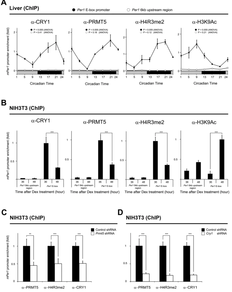
CRY1 binds to the CLOCK/BMAL1 heterodimer and represses Per1 gene expression. Therefore, we next investigated the association of PRMT5 and CRY1 with thePer1gene promoter in the mouse liver samples harvested during the circadian cycle. ChIP assay revealed that binding of CRY1 and PRMT5 to the
Per1 gene promoter increased when Per1 gene expression was repressed at night (Figures 3A and 5A). However, CRY1 and PRMT5 dissociated from thePer1gene promoter in the activation phase of gene expression in the daytime. Collectively, the rhythmic Figure 2. PRMT5 acts as a transcriptional repressor of thePer1promoter-reporter gene containing the E-box.Overexpression of PRMT5 enhances CRY1-mediated repression of the E-box containing reporter gene. NIH3T3 cells were transiently transfected with thePer1gene promoter-driven firefly luciferase reporter vector (20 ng) in conjunction with a controlthymidine kinasepromoter-driven Renilla luciferase vector. Expression vectors for Myc-tagged CLOCK (200 ng), Myc-tagged BMAL1 (200 ng), Flag-tagged CRY1 (10 ng), HA-tagged PRMT5 (100, 200, 400 ng), PRMT5 shRNA (100, 200, 400 ng), and control shRNA (100, 200, 400 ng) were transfected in combination. As a negative control, thePer1promoter containing the mutated E-box-driven firefly luciferase reporter vector was used. Data are presented as mean6S.E.M. of the three biological replicates with two cell plates analyzed for each group. A paired t-test was used for two group comparison. When luciferase activity of the wt E-box reporter was compared to that of the mut E-box reporter, significance values were *P#0.05, **P#0.01, ***P#0.005.#
P#0.05,##
P#0.01, and###
P#0.005 compared to the third column in the group of the wt E-box reporters.
Figure 3.Prmt5is non-rhythmically expressed.At the indicated times, total RNA was isolated from (A) mouse livers harvested during the circadian cycle or (B) NIH3T3 cells that were synchronized with 100 nM dexamethasone for 2 hours. Transcripts of Cry1, Per1, andPrmt5 were determined by quantitative PCR. TheGapdhtranscript was used for normalization. Data are presented as mean6S.E.M. of the three biological replicates with two mice or two cell plates analyzed for each group. Analysis of variance (ANOVA) was used for multiple comparisons.
doi:10.1371/journal.pone.0048152.g003
Figure 4. Rhythmic interaction of PRMT5 with CRY1.At the indicated times, nuclear extracts from (A) mouse livers harvested during the circadian cycle or (B) synchronized NIH3T3 cells were immunoprecipitated with the anti-PRMT5 antibody and immunoblotted with the anti-CRY1 or anti-PRMT5 antibody. IgG was used as the negative control.
recruitment of CRY1 and PRMT5 to the Per1 gene promoter occurred in an anti-phasic manner with rhythmicPer1expression. Since PRMT5 catalyzes the dimethylation of Arg 3 on histone H4 (H4R3me2), which is generally known as a transcription repressive mark, we investigated whether rhythmic PRMT5 recruitment to thePer1gene promoter coincides with enrichment of dimethylated H4R3. We found that dimethylation of H4R3 at the Per1 gene promoter fluctuated in phase with the rhythmic recruitment of PRMT5 (Figures 3A and 5A). As expected, the acetylated histone H3K9 activation mark (H3K9Ac) anti-phasically oscillated with H4R3 dimethylation (Figure 5A). However, we were unable to detect any significant rhythmic enrichment of PRMT5, CRY1, H4R3me2, and H3K9Ac at the upstream genomic region, which is 6 kb away from the E-box of thePer1gene promoter. To further confirm our results, we performed the same ChIP assay whenPer1
gene expression was activated or repressed using a synchronized NIH3T3 cell model. As shown in Figure 5B, we again found that increased recruitment of PRMT5 and CRY1 coincided with increased H4R3 dimethylation and decreased H3K9 acetylation at 36 hours after synchronization when thePer1gene expression was repressed. In contrast, we observed that decreased binding of PRMT5 and CRY1 to thePer1gene promoter was associated with decreased H4R3 dimethylation and increased H3K9 acetylation at 48 hours after synchronization when thePer1gene expression was activated (Figures 3B and 5B). We also did not observe significant rhythmic enrichment of PRMT5, CRY1, H4R3me2, and H3K9Ac at the upstream genomic region, which was used as a negative control (Figure 5B). Collectively, these data suggest that rhythmic recruitment of PRMT5 and CRY to the Per1 gene promoter is associated with the rhythmic dimethylation of H4R3 andPer1gene expression.
In order to examine whether depletion ofPrmt5is paralleled by decreased dimethylated H4R3, we also performed the ChIP assay when PRMT5 and CRY1 were bound strongly to the Per1
promoter at 36 hours after synchronization (Figure 5C). We observed that the dimethylation level of H4R3 at the promoter significantly decreased. Interestingly, recruitment of CRY1 to the
Per1gene promoter also significantly decreased (Figure 5C). We further tested whether depletion ofCry1affects the recruitment of PRMT5 by ChIP assay at 36 hours after synchronization (Figure 5D). Consistent with up-regulation ofPer1gene expression in Cry1-depleted cells, we observed significantly decreased enrichment of PRMT5 and H4R3me2 at the promoter (Figures 5D and S3A). These results indicate that both PRMT5 and CRY1 may be required for the repression of thePer1gene. WhenCry2was depleted by shRNA transfection, we observed less pronounced up-regulation of Per1 gene expression and less decreased enrichment of PRMT5 and H4R3me2 at the promoter compared toCry1depletion (Figure S3).
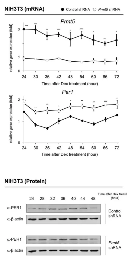
Depletion ofPrmt5causes altered rhythmic expression of thePer1gene
Next, we hypothesized that the depletion ofPrmt5deregulates expression of the Per1 gene in the synchronized NIH3T3 cells. After transfection ofPrmt5shRNA, NIH3T3 cells were synchro-nized with dexamethasone treatment. As shown in Figure 6A, the
Per1 gene expression was significantly up-regulated. Moreover, rhythmic expression of the Per1 gene was abolished in Prmt5 -depleted cells. We further observed up-regulation of PER1 at the protein level without rhythmicity in Prmt5-depleted cells (Figure 6B).
In conclusion, our results consistently support the hypothesis that PRMT5 functions as a transcriptional corepressor in
CRY-mediated circadianPer1gene regulation through the regulation of H4R3 dimethylation at the promoter.
Discussion
Regulation of histone modifications such as acetylation and methylation on specific lysine residues plays a crucial role in the gene expression that underlies circadian rhythm. In fact, rhythmic p300-mediated H3 acetylation has been reported at various circadian genes [23]. CRY1 negatively regulates Per1 gene expression by recruiting histone deacetylases (HDACs) [20]. Rhythmic enrichment of trimethylation of H3K4 and dimethyla-tion of H3K9 were observed at theRev-erbagene promoter [24]. Dimethylation of histone H3K9 and recruitment of HP1ato the
Dbp (albumin D site-binding protein) gene promoter might be the general mechanism for CRY-mediated transcription repression [21]. In addition, Polycomb group EZH2 regulates di- and tri-methylation of H3K27 at the Per1 and Per2 promoters [25]. Recently, it has been proposed that H4K4 trimethylation by MLL1 methyltransferase, which forms complex with CLOCK-BMAL1 is required for circadian transcription [26]. Histone demethylase JARID1a also plays an important role in circadian transcription by inhibiting HDAC1 [27]. In this study, we suggest that PRMT5 interacts with CRY1 and is involved in circadian
Per1gene expression through the rhythmic arginine dimethylation of H4R3.
PRMT5 is a type II arginine methyltransferase that catalyzes the monomethylation and symmetric dimethylation of arginine residues [28,29]. PRMT5 has multiple functions including transcriptional regulation, alternative splicing, apoptosis, and ribosome biogenesis. As a splicing regulator, PRMT5 mediates RNA splicing by methylation of Sm proteins, components of the spliceosome [30]. TheArabidopsis homolog of PRMT5 has been found to be important in regulating pre-mRNA splicing [31]. In addition, PRMT5 functions as a transcriptional regulator in a variety of mechanisms. For example, PRMT5-mediated methylation of transcriptional elongation factor SPT5 results in alteration of RNA polymerase II-dependent gene expression [32]. PRMT5 has also been demonstrated to act as a transcriptional corepressor based on its dimethylation activity on H3R8 and H4R3 [33,34,35,36,37,38,39,40]. Recently, two reports demon-strated that PRMT5 is involved in Arabidopsis and Drosophila
circadian clock regulation. Hong et al. found that PRMT5 is a critical determinant of period length in Arabidopsis [41]. Moreover, Sanchez et al. found that PRMT5 is required for the control of alternative splicing of clock genes such as PRR9 in
Arabidopsis [42]. They further showed that a Drosophila PRMT5
homologue (dart5-1) is associated with alterations in splicing of the
periodand several clock-associated genes, indicating that PRMT5 links the circadian clock to the regulation of alternative splicing.
Although we cannot exclude the possibility that mammalian PRMT5 also regulates alternative splicing of circadian clock genes, our results indicate that mammalian PRMT5 may act as a transcriptional corepressor in circadian Per1 gene regulation. First, we found that PRMT5 interacts with the CRY1 transcrip-tional repressor. In addition, overexpression of PRMT5 augment-ed the transcriptional repression activity of CRY1 in the reporter assay. Consistent with our finding, the presence of an unknown transcriptional corepressor that binds to the C-terminal region of CRY1 had been postulated because its deletion abolished target gene repression [43]. Second, rhythmic association of PRMT5 with CRY1 at thePer1gene promoter coincided with rhythmic dimethylation of H4R3 and transcriptional repression of thePer1
results in a significant reduction of H4R3 dimethylation and alteration of rhythmicPer1gene expression.
Although more studies are required to discover species-specific roles of PRMT5, we expect that our observations may have resulted from the different structures of CRYs, since the amino acid composition and length of the C-terminal region of mammalian CRY are quite different from that ofArabidopsisand
Drosophila CRY [44,45]. In addition, some species-specific mechanisms of circadian clock regulation by CRY have been previously suggested [46]. For example,ArabidopsisandDrosophila
CRY is a major circadian photoreceptor for light entrainment, while mammalian CRY is not required for photo-entrainment.
In this study, we also observed decreased enrichment of CRY1 at the Per1gene promoter in Prmt5-depleted cells. These results imply that PRMT5 may regulate CRY1 translocation to the nucleus and/or its degradation. In fact, there is speculation that PRMT5 may regulate the nuclear cytoplasmic shuttling of several proteins such as NF-AT family members [47]. Moreover, it has recently been reported that methylation of RAF proteins by PRMT5 is involved in the degradation of RAF proteins in the RAS signaling pathway [48]. Similarly, we observed up-regulation ofPer1gene expression and decreased enrichment of PRMT5 and H4R3me2 at the promoter inCry1-depleted cells. In contrast, a less pronounced effect ofCry2depletion onPer1gene regulation was observed, implying that CRY1 may have a major role in PRMT5-mediated Per1 gene repression. Supporting our observations, several reports indicate that CRY1 may function as the main transcriptional repressor in CLOCK/BMAL1-mediated transcrip-tion [49,50,51]. However, we do not exclude the possibility that both CRY1 and CRY2 may be required for the full repression of thePer1gene.
When we overexpressed Flag-tagged CRY1 (aa 370 to 470) or Flag-tagged CRY1 (aa 471 to 586) in 293T cells, each of the CRY1 fragments formed a homodimer (Figures S1D and S1E). Based on our observation, there is a possibility that PRMT5 interacts with CRY1 when it forms a homodimer. However, we did not detect homodimerization of full-length CRY1 in standard Western blot analysis. Although it is unclear whether homodimer-ization of each of CRY1 fragment in vitro has a physiological impact on PRMT5 interaction or circadian rhythm, it has been previously reported that homodimerization of the N-terminal of the Arabidopsis cryptochrome photoreceptor plays an important role in light signaling [52].
In summary, our study demonstrates that PRMT5 interacts with CRY1in vivo. Although non-rhythmic expression of thePrmt5
gene was observed, rhythmic recruitment of CRY1 and PRMT5 to thePer1gene promoter coincides with the rhythmic dimethyla-tion of histone H4R3 andPer1gene repression. Consistently, we found decreased H4R3 dimethylation and alteration of rhythmic
Per1gene expression inPrmt5-depleted cells. Our study provides an
insight into the link between histone modification by PRMT5 and mammalian circadian clock genePer1regulation.
Materials and Methods
Cell culture and animal handling
NIH3T3 and 293T cells were maintained in DMEM supple-mented with 10% fetal bovine serum and antibiotics. To synchronize NIH3T3 cells, they were kept for 2–3 days until the cells reached confluence, and then were treated with 100 nM dexamethasone. Two hours after treatment, the medium was replaced with DMEM. At the indicated times, the cells were washed twice with ice-cold phosphate-buffered saline (PBS) and harvested for RNA and protein extraction. For the ChIP assay, the cells were fixed with formaldehyde in PBS (1% final concentration) for 15 min. The committee for experimental animal research at Sogang University approved the animal experiments. Male BALB/c mice (Samtaco, Osan, Korea) were maintained for 2 weeks on a 12 hr: 12 hr light-dark cycle and then released to constant darkness. On the third day in constant darkness, the livers were harvested at the indicated times and kept frozen at280C until use. For the ChIP assay, the livers were chopped into small pieces and fixed with formaldehyde in PBS (1% final concentra-tion) for 15 min at room temperature. To stop the crosslinking, 1.25 M glycine was added (1% final concentration) for 5 min. Tissues were stored in liquid N2until use.
Antibodies
The following commercially available antibodies were used: anti-CRY1 antibody (sc-33177, Santa Cruz Biotechnology, Santa Cruz, CA, USA; ab54649, Abcam, Cambridge, MA, USA), PRMT5 antibody (07–405, Millipore, Temecula, CA, USA), anti-dimethyl H4R3 antibody (ab5823, Abcam), anti-acetylated H3K9 antibody (ab4441, Abcam), anti-H4 antibody (05–858, Millipore), anti-b actin antibody (A5471, Sigma-Aldrich, St. Louis, MO, USA), anti-Flag antibody (F1804, Sigma-Aldrich), and anti-HA antibody (MMS101P, Covance, Berkeley, CA, USA). Normal IgG (sc-2027, Santa Cruz Biotechnology) was used as a control.
Transfection and immunoprecipitation
NIH3T3 cells were transfected using a Lipofectamine 2000 (Invitrogen, Carlsbad, CA, USA) or a Vivamagic transfection reagent (Vivagen, Seongnam, Korea). The 293T cells were transfected using a calcium phosphate method. For immunopre-cipitation, cells or tissues were rinsed in PBS, harvested, and sonicated in an IP150 buffer (10% glycerol, 0.5 mM EDTA, 0.1% NP40, 150 mM NaCl, 1 mM DTT, 25 mM Tris, pH 8.0) in the presence of complete protease inhibitors (Roche, Mannheim, Germany) and 1 mM phenylmethylsulphonylfluoride. To isolate the nucleus, cells or tissues were lysed in a nuclei buffer (1.5 mM a negative control, the upstream genomic region which is 6 kb away from the E-box of thePer1gene promoter was amplified (Per16kb upstream region). Data are presented as mean6S.E.M. of the three biological replicates with two mice analyzed for each group. Analysis of variance (ANOVA) was used for multiple comparisons. (B) NIH3T3 cells that were synchronized with dexamethasone using the anti-CRY1 antibody, anti-PRMT5 antibody, anti-dimethylated H4R3 antibody, and anti-acetylated H4K9 antibody at the indicated times. Data are presented as mean6S.E.M. of the three biological replicates with two cell plates analyzed for each group. A paired t-test was used for two group comparison. Significance value was *** P#0.005. (C) Knock-down ofPrmt5results in decreased dimethylation of H4R3 and recruitment of CRY1 to thePer1gene promoter. A ChIP assay was performed using the anti-PRMT5 antibody, anti-CRY1 antibody, and anti-dimethylated H4R3 antibody at 36 hours after synchronization in NIH3T3 cells that were transfected withPrmt5shRNA. Data are presented as mean6S.E.M. of the three biological replicates with two cell plates analyzed for each group. A paired t-test was used for two group comparison. Significance values were **P#0.01 and ***P#0.005. (D) Knock-down ofCry1results in decreased recruitment of PRMT5 and dimethylation of H4R3 to thePer1gene promoter. A ChIP assay was performed using the anti-CRY1 antibody, anti-PRMT5 antibody, and anti-dimethylated H4R3 antibody at 36 hours after synchronization in NIH3T3 cells that were transfected withCry1shRNA. Data are presented as mean6S.E.M. of the three biological replicates with two cell plates analyzed for each group. A paired t-test was used for two group comparison. Significance value was ***P#0.005.
MgCl2, 10 mM KCl, 10 mM HEPES-KOH, pH 7.9). After
centrifugation, the nuclear pellet was lysed in an IP150 buffer using sonication. After clearing by centrifugation, the extracts were incubated with the specific antibody overnight at 4uC, followed by incubation with protein A/G agarose beads (Sigma-Aldrich), washed extensively and dissolved in an SDS sample buffer. Western blotting was carried out using standard procedures. Protein concentration was determined by the Lowry method. To detect the monomeric form of CRY1 fragments, proteins were extracted with an SDS sample buffer containing 4 M urea and electrophoresis was performed using SDS-polyacrylamide gel containing 4 M urea.
Luciferase activity assay
NIH3T3 cells were transfected with various plasmid constructs such as 1.8 kb mousePer1gene promoter-driven firefly luciferase,
thymidine kinasepromoter-driven renilla luciferase,Clock, Myc-Bmal1, Flag-Cry1, and HA-Prmt5. After 48 hours, cells were harvested for luciferase activity using the Dual-Luciferase Assay System (Promega, Madison, WI, USA) with a Lumat BL 9507 luminometer (Berthold technologies, Bad Wildbad, Germany). As a negative control, thePer1promoter luciferase-reporter contain-ing mutated E-box was used [53,54]. Renilla luciferase activity served as an internal control for normalizing firefly luciferase activity of thePer1gene promoter. All constructs were confirmed by DNA sequencing.
RNA isolation and quantitative PCR analysis
Total RNA was extracted from cells or tissues using the Nucleospin RNA isolation kit (Macherey-Nagel, Du¨ren, Ger-many). First-strand cDNA synthesis from the total RNA template was performed with the PrimeScript II 1st strand cDNA Synthesis Kit (Takara, Japan). The resulting cDNAs were subjected to real-time PCR with a Stratagene Mx3000P (Agilent Technologies, Waldbronn, Germany) using the SYBR Green I Mix (Takara, Japan). PCR conditions used to amplify all genes were 30 s at 95uC, 40 cycles of 95uC for 5 s, and 60uC for 34 s. Expression data were calculated from the cycle threshold (Ct) value using the DCt method of quantification.GAPDHmRNA levels were used for normalization [55]. Amplification was performed using the following primers: 59-CTGCAACATTCCCAGTACAAC-CAAGCG-39 and 59-ATCAGAGGCTGAAGAGGCAGTGT-39 for Per1, 59-GTCTTCTCGCCTCGGTCCCTTCTAACT-39 and 59-CCATTCCCGCTGCTGCTACAAC-39 for mouse Cry 1, 59-TGTGGTGGCATAACTTTCGGACTCTGT-39 and 59 -TGGGGAGAATGGCTGCTTTGAT-39 for mouse Prmt5, and 59-AGGTCGGTGTGAACGGATTTG-39 and 59-TGTAGAC-CATGTAGTTGAGGTCA-39for mouseGapdh. For validation of all PCR primers, quantitative PCR amplification plots and dissociation curves were analyzed and a single band of the expected size was confirmed after PCR by agarose gel analysis.
Chromatin immunoprecipitation (ChIP)
NIH3T3 cells or tissues were collected and crosslinked using 1% formaldehyde in PBS for 15 min at room temperature. To stop the crosslinking, 1.25 M glycine was added (1% final concentration). After washing with ice-cold PBS, the cells in a SDS lysis buffer (1% SDS, 10 mM EDTA, 50 mM Tris-HCl, pH 8.1) were sonicated until the DNA fragments were 300–500 bp in size. The extracts were subsequently centrifuged, and the resulting soluble chromatin solutions were diluted ten fold with a ChIP dilution buffer (0.01% SDS, 1.1% Triton X-100, 1.2 mM EDTA, 167 mM NaCl, 16.7 mM Tris-HCl, pH 8.1). The specific antibodies or IgG were added to a soluble chromatin solution overnight at 4uC. Protein
A-sepharose beads (Sigma-Aldrich) were further added to the solution in order to precipitate the DNA-protein complexes for 2 hr. The beads were extensively washed with a low salt wash buffer (0.1% SDS, 1% Triton X-100, 2 mM EDTA, 150 mM NaCl, 20 mM Tris-HCl, pH 8.1), a high salt wash buffer (0.1% SDS, 1% Triton X-100, 2 mM EDTA, 500 mM NaCl, 20 mM Tris-HCl, pH 8.1), an LiCl wash buffer (0.25 M LiCl, 1% NP-40, 1% deoxycholate, 1 mM EDTA, 10 mM Tris-HCl, pH 8.1), and finally with a TE buffer. After elution of the DNA/protein with 1% SDS, crosslinking was reversed for 6 hr at 65uC. The DNA was recovered using the QIAquick spin column (Qiagen, Valencia, CA, USA). Real time-PCR was performed with a Stratagene Mx3000P using primers that cover the mousePer1promoter (59
-ACGGTGTGAGACATCCTGATCGCATTG-39 and
59-TCTTCCTGGCATCTGATTGGCTACTGG -39). As a negative control, the upstream genomic region which is 6 kb away from the E-box of the mouse Per1 gene promoter, was amplified using primers (59- TCCCGAACTCTTGAACTG-39and 59- GCAAC-CAGAAATGCTACC-39). Briefly, 1–2ml of immunoprecipitated DNA was used as a template in 20ml reactions containing 16 SYBR Green Master Mix (Takara).
All PCR reactions were performed in triplicate and included negative controls (IgG) as well as positive controls (genomic DNA). The relative proportions of immunoprecipitated fragments were determined using the DCt comparative method, based on the threshold cycle (Ct) value for each PCR reaction, and normalized to input genomic DNA. For validation of all PCR primers, quantitative PCR amplification plots and dissociation curves were analyzed, and a single band of the expected size was confirmed after PCR by agarose gel analysis.
RNA interference (siRNA/shRNA)
NIH3T3 cells were transfected with siRNA (M-042281-01-0005, Dharmacon, Lafayette, CO, USA) or shRNA (TRCN0000181891, Sigma-Aldrich) against Prmt5. To knock down Cry1 and Cry2, shRNAs (TRCN0000173481 and TRCN0000194121, Sigma-Aldrich) were used, respectively. For a control, siRNA (sc37007, Santa Cruz Biotechnology) or shRNA (SHC002, Sigma-Aldrich) was used. The efficiency of the knock down of specific genes was confirmed with real time-PCR.
Supporting Information
Figure S1 Confirmation of the interaction between
overexpressed in 293T cells, the cell extracts preparation and SDS-PAGE were performed in the presence of 4 M urea. Western blot analysis was performed using anti-Flag (left) or anti-CRY1 antibodies (right). Note that the expected molecular weight of CRY1 fragments was observed. (F) The specificity of interaction between CRY1 and PRMT5 was confirmed usingCry1-depleted cells. The lysates from Cry1shRNA-transfected NIH3T3 cells at 36 hours after synchronization were immunoprecipitated with the anti-PRMT5 antibody and immunoblotted with the anti-CRY1 and anti-PRMT5 antibodies. IgG was used as the negative control. (G) The specificity of interaction between CRY1 and PRMT5 was confirmed using Prmt5-depleted cells. The lysates from Prmt5
shRNA-transfected NIH3T3 cells at 36 hours after synchroniza-tion were immunoprecipitated with the anti-PRMT5 antibody and immunoblotted with the anti-PRMT5 and anti-CRY1 antibodies. IgG was used as the negative control.
(TIF)
Figure S2 Confirmation of antibody specificity. (A) The lysates from control shRNA orCry1shRNA-transfected NIH3T3 cells were immunoblotted with the CRY1 antibody. The anti-b actin antibody was used as a loading control. (B) The lysates from control shRNA orPrmt5shRNA-transfected NIH3T3 cells were immunoblotted with the PRMT5 antibody. The anti-b actin antibody was used as a loading control. (C) The lysates from control shRNA orPrmt5shRNA-transfected NIH3T3 cells were immunoblotted with the H4R3me2 antibody. The anti-histone H4 antibody was used as a loading control.
(TIF)
Figure S3 Depletion of Cryderegulates rhythmic Per1
gene expression.(A) Knock-down ofCry1induces up-regulated and non-rhythmic Per1 gene expression. NIH3T3 cells were transfected with Cry1 shRNA and then cells were synchronized with 100 nM dexamethasone for 2 hours. Transcripts of Per1, Cry1, and Gapdh were measured using quantitative PCR at 36 hours after synchronization. Data are presented as mean 6 S.E.M. of the three biological replicates with two cell plates analyzed for each group. A paired t-test was used for two group comparison. Significance value was ***P#0.005. (B) Knock-down of Cry1results in significantly decreased enrichment of PRMT5 and H4R3me2 at the Per1 gene promoter. A ChIP assay was performed using the anti-CRY1 antibody, anti-PRMT5 antibody,
and anti-dimethylated H4R3 antibody at 36 hours after synchro-nization in NIH3T3 cells that were transfected withCry1shRNA. Data are presented as mean 6 S.E.M. of the three biological replicates with two cell plates analyzed for each group. A paired t-test was used for two group comparison. Significance value was ***
P#0.005. (C) Knock-down of Cry2shows less pronounced effect on thePer1gene expression. NIH3T3 cells were transfected with
Cry2 shRNA and then cells were synchronized with 100 nM dexamethasone for 2 hours. Transcripts ofPer1, Cry2, andGapdh
were measured using quantitative PCR at 36 hours after synchronization. Data are presented as mean 6 S.E.M. of the three biological replicates with two cell plates analyzed for each group. A paired t-test was used for two group comparison. Significance value was *** P#0.005. (D) Knock-down of Cry2
results in less decreased enrichment of PRMT5 and H4R3me2 at thePer1gene promoter. A ChIP assay was performed using the anti-CRY1 antibody, anti-PRMT5 antibody, and anti-dimethy-lated H4R3 antibody at 36 hours after synchronization in NIH3T3 cells that were transfected withCry2shRNA. Data are presented as mean6S.E.M. of the three biological replicates with two cell plates analyzed for each group. A paired t-test was used for two group comparison. Significance values were *P#0.05, **
P#0.01, and *** P#0.005. Figure 5D was designated as Figure S3B to more conveniently compare the effect ofCry1and
Cry2depletion onPer1gene expression. (TIF)
Table S1 Proteins interacting with CRY1 by mass
spectrometry.
(DOCX)
Acknowledgments
We thank Drs. Eun Young Kim, Gi Hoon Son, Kwang-Hwan Jung, and Kyungjin Kim for valuable discussions and advices.
Author Contributions
Conceived and designed the experiments: JN JL SC WK BJ. Performed the experiments: JN KL HK WN HJ. Analyzed the data: JN JS JL SC WK BJ. Contributed reagents/materials/analysis tools: JS SC. Wrote the paper: JL BJ.
References
1. Schibler U (2005) The daily rhythms of genes, cells and organs. Biological clocks and circadian timing in cells. EMBO Rep 6 Spec No: S9–13.
2. Stephan FK, Zucker I (1972) Circadian rhythms in drinking behavior and locomotor activity of rats are eliminated by hypothalamic lesions. Proc Natl Acad Sci U S A 69: 1583–1586.
3. Balsalobre A, Damiola F, Schibler U (1998) A serum shock induces circadian gene expression in mammalian tissue culture cells. Cell 93: 929–937. 4. Yamazaki S, Numano R, Abe M, Hida A, Takahashi R, et al. (2000) Resetting
central and peripheral circadian oscillators in transgenic rats. Science 288: 682– 685.
5. Yagita K, Tamanini F, van Der Horst GT, Okamura H (2001) Molecular mechanisms of the biological clock in cultured fibroblasts. Science 292: 278–281. 6. Reppert SM, Weaver DR (2002) Coordination of circadian timing in mammals.
Nature 418: 935–941.
7. Lowrey PL, Takahashi JS (2004) Mammalian circadian biology: elucidating genome-wide levels of temporal organization. Annu Rev Genomics Hum Genet 5: 407–441.
8. Cashmore AR (2003) Cryptochromes: enabling plants and animals to determine circadian time. Cell 114: 537–543.
9. Kang TH, Lindsey-Boltz LA, Reardon JT, Sancar A (2010) Circadian control of XPA and excision repair of cisplatin-DNA damage by cryptochrome and HERC2 ubiquitin ligase. Proc Natl Acad Sci U S A 107: 4890–4895. 10. van der Horst GT, Muijtjens M, Kobayashi K, Takano R, Kanno S, et al. (1999)
Mammalian Cry1 and Cry2 are essential for maintenance of circadian rhythms. Nature 398: 627–630.
11. Vitaterna MH, Selby CP, Todo T, Niwa H, Thompson C, et al. (1999) Differential regulation of mammalian period genes and circadian rhythmicity by cryptochromes 1 and 2. Proc Natl Acad Sci U S A 96: 12114–12119. 12. Vielhaber E, Eide E, Rivers A, Gao ZH, Virshup DM (2000) Nuclear entry of
the circadian regulator mPER1 is controlled by mammalian casein kinase I epsilon. Mol Cell Biol 20: 4888–4899.
13. Camacho F, Cilio M, Guo Y, Virshup DM, Patel K, et al. (2001) Human casein kinase Idelta phosphorylation of human circadian clock proteins period 1 and 2. FEBS Lett 489: 159–165.
14. Akashi M, Tsuchiya Y, Yoshino T, Nishida E (2002) Control of intracellular dynamics of mammalian period proteins by casein kinase I epsilon (CKIepsilon) and CKIdelta in cultured cells. Mol Cell Biol 22: 1693–1703.
15. Blau J (2003) A new role for an old kinase: CK2 and the circadian clock. Nat Neurosci 6: 208–210.
16. Busino L, Bassermann F, Maiolica A, Lee C, Nolan PM, et al. (2007) SCFFbxl3 controls the oscillation of the circadian clock by directing the degradation of cryptochrome proteins. Science 316: 900–904.
17. Godinho SI, Maywood ES, Shaw L, Tucci V, Barnard AR, et al. (2007) The after-hours mutant reveals a role for Fbxl3 in determining mammalian circadian period. Science 316: 897–900.
18. Siepka SM, Yoo SH, Park J, Song W, Kumar V, et al. (2007) Circadian mutant Overtime reveals F-box protein FBXL3 regulation of cryptochrome and period gene expression. Cell 129: 1011–1023.
20. Naruse Y, Oh-hashi K, Iijima N, Naruse M, Yoshioka H, et al. (2004) Circadian and light-induced transcription of clock gene Per1 depends on histone acetylation and deacetylation. Mol Cell Biol 24: 6278–6287.
21. Ripperger JA, Schibler U (2006) Rhythmic CLOCK-BMAL1 binding to multiple E-box motifs drives circadian Dbp transcription and chromatin transitions. Nat Genet 38: 369–374.
22. Wysocka J, Allis CD, Coonrod S (2006) Histone arginine methylation and its dynamic regulation. Front Biosci 11: 344–355.
23. Etchegaray JP, Lee C, Wade PA, Reppert SM (2005) Rhythmic histone acetylation underlies transcription in the mammalian circadian clock. Nature 421: 177–182.
24. Brown SA, Ripperger J, Kadener S, Fleury-Olela F, Vilbois F, et al. (2005) PERIOD1-associated proteins modulate the negative limb of the mammalian circadian oscillator. Science 308: 693–696.
25. Etchegaray JP, Yang X, DeBruyne JP, Peters AH, Weaver DR, et al. (2006) The polycomb group protein EZH2 is required for mammalian circadian clock function. J Biol Chem 281: 21209–21215.
26. Katada S, Sassone-Corsi P (2010) The histone methyltransferase MLL1 permits the oscillation of circadian gene expression. Nat Struct Mol Biol 17: 1414–1421. 27. DiTacchio L, Le HD, Vollmers C, Hatori M, Witcher M, et al. (2011) Histone lysine demethylase JARID1a activates CLOCK-BMAL1 and influences the circadian clock. Science 333: 1881–1885.
28. Bedford MT, Richard S (2005) Arginine methylation an emerging regulator of protein function. Mol Cell 18: 263–272.
29. Branscombe TL, Frankel A, Lee JH, Cook JR, Yang Z, et al. (2001) PRMT5 (Janus kinase-binding protein 1) catalyzes the formation of symmetric dimethylarginine residues in proteins. J Biol Chem 276: 32971–32976. 30. Meister G, Eggert C, Buhler D, Brahms H, Kambach C, et al. (2001)
Methylation of Sm proteins by a complex containing PRMT5 and the putative U snRNP assembly factor pICln. Curr Biol 11: 1990–1994.
31. Deng X, Gu L, Liu C, Lu T, Lu F, et al. (2010) Arginine methylation mediated by the Arabidopsis homolog of PRMT5 is essential for proper pre-mRNA splicing. Proc Natl Acad Sci U S A 107: 19114–19119.
32. Kwak YT, Guo J, Prajapati S, Park KJ, Surabhi RM, et al. (2003) Methylation of SPT5 regulates its interaction with RNA polymerase II and transcriptional elongation properties. Mol Cell 11: 1055–1066.
33. Fabbrizio E, El Messaoudi S, Polanowska J, Paul C, Cook JR, et al. (2002) Negative regulation of transcription by the type II arginine methyltransferase PRMT5. EMBO Rep 3: 641–645.
34. Pal S, Yun R, Datta A, Lacomis L, Erdjument-Bromage H, et al. (2003) mSin3A/histone deacetylase 2- and PRMT5-containing Brg1 complex is involved in transcriptional repression of the Myc target gene cad. Mol Cell Biol 23: 7475–7487.
35. Pal S, Vishwanath SN, Erdjument-Bromage H, Tempst P, Sif S (2004) Human SWI/SNF-associated PRMT5 methylates histone H3 arginine 8 and negatively regulates expression of ST7 and NM23 tumor suppressor genes. Mol Cell Biol 24: 9630–9645.
36. Tae S, Karkhanis V, Velasco K, Yaneva M, Erdjument-Bromage H, et al. (2011) Bromodomain protein 7 interacts with PRMT5 and PRC2, and is involved in transcriptional repression of their target genes. Nucleic Acids Res 39: 5424–5438.
37. Lacroix M, El Messaoudi S, Rodier G, Le Cam A, Sardet C, et al. (2008) The histone-binding protein COPR5 is required for nuclear functions of the protein arginine methyltransferase PRMT5. EMBO Rep 9: 452–458.
38. Cesaro E, De Cegli R, Medugno L, Florio F, Grosso M, et al. (2009) The Kruppel-like zinc finger protein ZNF224 recruits the arginine methyltransferase
PRMT5 on the transcriptional repressor complex of the aldolase A gene. J Biol Chem 284: 32321–32330.
39. Majumder S, Alinari L, Roy S, Miller T, Datta J, et al. (2010) Methylation of histone H3 and H4 by PRMT5 regulates ribosomal RNA gene transcription. J Cell Biochem 109: 553–563.
40. Rank G, Cerruti L, Simpson RJ, Moritz RL, Jane SM, et al. (2010) Identification of a PRMT5-dependent repressor complex linked to silencing of human fetal globin gene expression. Blood 116: 1585–1592.
41. Hong S, Song HR, Lutz K, Kerstetter RA, Michael TP, et al. (2010) Type II protein arginine methyltransferase 5 (PRMT5) is required for circadian period determination in Arabidopsis thaliana. Proc Natl Acad Sci U S A 107: 21211– 21216.
42. Sanchez SE, Petrillo E, Beckwith EJ, Zhang X, Rugnone ML, et al. (2010) A methyl transferase links the circadian clock to the regulation of alternative splicing. Nature 468: 112–116.
43. Chaves I, Yagita K, Barnhoorn S, Okamura H, van der Horst GT, et al. (2006) Functional evolution of the photolyase/cryptochrome protein family: impor-tance of the C terminus of mammalian CRY1 for circadian core oscillator performance. Mol Cell Biol 26: 1743–1753.
44. Todo T, Ryo H, Yamamoto K, Toh H, Inui T, et al. (1996) Similarity among the Drosophila (6–4)photolyase, a human photolyase homolog, and the DNA photolyase-blue-light photoreceptor family. Science 272: 109–112.
45. van der Spek PJ, Kobayashi K, Bootsma D, Takao M, Eker AP, et al. (1996) Cloning, tissue expression, and mapping of a human photolyase homolog with similarity to plant blue-light receptors. Genomics 37: 177–182.
46. Wijnen H, Young MW (2006) Interplay of circadian clocks and metabolic rhythms. Annu Rev Genet 40: 409–448.
47. Richard S, Morel M, Cle´roux P (2005) Arginine methylation regulates IL-2 gene expression: a role for protein arginine methyltransferase 5 (PRMT5). Biochem J 388: 379–386.
48. Andreu-Pe´rez P, Esteve-Puig R, de Torre-Minguela C, Lo´pez-Fauqued M, Bech-Serra JJ, et al. (2011) Protein arginine methyltransferase 5 regulates ERK1/2 signal transduction amplitude and cell fate through CRAF. Sci Signal 4: ra58.
49. Kiyohara YB, Tagao S, Tamanini F, Morita A, Sugisawa Y, et al. (2006) The BMAL1 C terminus regulates the circadian transcription feedback loop. Proc Natl Acad Sci U S A. 103: 10074–10079.
50. Sato TK, Yamada RG, Ukai H, Baggs JE, Miraglia LJ, et al. (2006) Feedback repression is required for mammalian circadian clock function. Nat Genet 38: 312–319.
51. Langmesser S, Tallone T, Bordon A, Rusconi S, Albrecht U (2008) Interaction of circadian clock proteins PER2 and CRY with BMAL1 and CLOCK. BMC Mol Biol 9: 41.
52. Sang Y, Li QH, Rubio V, Zhang YC, Mao J, et al. (2005) N-terminal domain-mediated homodimerization is required for photoreceptor activity of Arabidopsis CRYPTOCHROME 1. Plant Cell 17: 1569–1584.
53. Hida A, Koike N, Hirose M, Hattori M, Sakaki Y, et al. (2000) The human and mouse Period1 genes: five well-conserved E-boxes additively contribute to the enhancement of mPer1 transcription. Genomics 65: 224–233.
54. Ueda HR, Hayashi S, Chen W, Sano M, Machida M, et al. (2005) System-level identification of transcriptional circuits underlying mammalian circadian clocks. Nat Genet 37: 187–192.