Submitted15 August 2016 Accepted 19 December 2016 Published24 January 2017
Corresponding author Andreas H. Laustsen, [email protected]
Academic editor J. Thomas Sanderson
Additional Information and Declarations can be found on page 13
DOI10.7717/peerj.2924
Copyright 2017 Laustsen et al.
Distributed under
Creative Commons CC-BY 4.0
OPEN ACCESS
Exploration of immunoglobulin
transcriptomes from mice immunized
with three-finger toxins and
phospholipases A
2
from the Central
American coral snake,
Micrurus
nigrocinctus
Andreas H. Laustsen1, Mikael Engmark1,2, Christopher Clouser3,4, Sonia
Timberlake5, Francois Vigneault3,4, José María Gutiérrez6and Bruno Lomonte6
1Department of Biotechnology and Biomedicine, Technical University of Denmark, Kgs. Lyngby, Denmark 2Department of Bio and Health Informatics, Technical University of Denmark, Kgs. Lyngby, Denmark 3Juno Therapeutics, Seattle, WA, United States of America
4AbVitro, Boston, MA, United States of America
5Finch Therapeutics, Somerville, MA, United States of America
6Instituto Clodomiro Picado, Universidad de Costa Rica, San José, Costa Rica
ABSTRACT
Snakebite envenomings represent a neglected public health issue in many parts of the rural tropical world. Animal-derived antivenoms have existed for more than a hundred years and are effective in neutralizing snake venom toxins when timely administered. However, the low immunogenicity of many small but potent snake venom toxins represents a challenge for obtaining a balanced immune response against the medically relevant components of the venom. Here, we employ high-throughput sequencing of the immunoglobulin (Ig) transcriptome of mice immunized with a three-finger toxin and a phospholipase A2from the venom of the Central American coral snake,Micrurus
nigrocinctus.Although exploratory in nature, our indicate results showed that only low frequencies of mRNA encoding IgG isotypes, the most relevant isotype for therapeutic purposes, were present in splenocytes of five mice immunized with 6 doses of the two types of toxins over 90 days. Furthermore, analysis of Ig heavy chain transcripts showed that no particular combination of variable (V) and joining (J) gene segments had been selected in the immunization process, as would be expected after a strong humoral immune response to a single antigen. Combined with the titration of toxin-specific antibodies in the sera of immunized mice, these data support the low immunogenicity of three-finger toxins and phospholipases A2found inM. nigrocinctusvenoms, and
highlight the need for future studies analyzing the complexity of antibody responses to toxins at the molecular level.
SubjectsBioinformatics, Biotechnology, Toxicology, Immunology, Synthetic Biology
INTRODUCTION
Snakebite envenomings represent a major public health concern in tropical regions of the world (Williams et al., 2011). Despite emerging discoveries that may one day pave the way for novel biotechnology-based antivenoms (reviewed by Laustsen et al., 2016a;Laustsen et al., submitted), animal serum-derived antivenoms remain the cornerstone of snakebite envenoming treatment (Gutiérrez et al., 2011). Production of antivenom is challenged by a large variation in immunogenicity of many key snake venom toxins resulting in unpredictable immune responses in production animals (Cook et al., 2010;Guidolin et al., 2010). It has been shown that many of the immunogenic venom components are in fact not important for toxicity (Antúnez et al., 2010;Gutiérrez et al., 2009;Laustsen et al., 2015), and conversely, that some highly toxic venom components, such asα-neurotoxins (both
short and long neurotoxins), phospholipases A2, and P-I snake venom metalloproteinases
may be poorly immunogenic (Schottler, 1951;Gutiérrez et al., 2009;Chotwiwatthanakun et al., 2001;Ownby & Colberg, 1990;Judge et al., 2006;Wong, Tan & Tan, 2016;Tan et al., 2016;Leong et al., 2015;Tan et al., 2015). Combined, this creates a challenge for antivenom production, since the goal of obtaining an antivenom with a strong, yet balanced response against all the medically relevant toxins becomes a complex endeavor.
Coral snakes (generaMicrurus,Leptomicrurus, andMicruroides) are, together with the sea snakeHydrophis(Pelamis)platura, the representatives of the snake family Elapidae in the Americas, comprising approximately 85 species (Campbell & Lamar, 2004; the Reptile Database—www.reptile-database.org). AlthoughMicrurusspecies are only responsible for about 1-2% of snakebite cases in this continent, roughly corresponding to 750 to 1000 cases per year, envenomings by these snakes can be fatal if not treated properly and timely (Warrell, 2004;Gutiérrez et al., 2016;Bucaretchi et al., 2016). Envenomings resulting from coral snakebites are predominantly associated with descending neuromuscular paralysis, which may end in respiratory arrest (Warrell, 2004;Bucaretchi et al., 2016).
Production of antivenoms againstMicrurussnakes is particularly challenging, as (a) it is very difficult to maintain coral snakes in captivity (Chacón et al., 2012); (b) the majority ofMicrurusspecies provide a very low yield of venom, implying that the collection of the quantities of venom required for horse immunization and quality control testing demands the ‘milking’ of many specimens (Chacón et al., 2012;Bolaños, 1972); and (c) there is a variable extent of immunological cross-recognition between venoms from coral snakes of different species; hence, antivenoms raised against some species are not always effective in the neutralization of venoms of other species (Bolaños, Cerdas & Abalos, 1978;Tanaka et al., 2016). As a result, only a few laboratories manufactureMicrurusantivenoms, and several countries where these snakes inhabit completely lack this therapeutic resource, e.g., Venezuela, Ecuador, Peru, Bolivia, the Guyanas, and Paraguay, which severely limits the clinical management of these accidents.
A2(PLA2s) (Fernández et al., 2015). In addition to these two main protein families, other
minor components of these venoms include L-amino acid oxidases, serine proteinases, metalloproteinases, nerve growth factor, C-type lectin-like proteins, Kunitz-type inhibitors, among others (Fernández et al., 2011; Fernández et al., 2015;Corrêa-Netto et al., 2011;
Lomonte et al., 2016a;Sanz et al., 2016;Rey-Suárez et al., 2011;Rey-Suárez et al., 2016). In some cases, the toxins playing the main role in overall toxicity have been identified, these being 3FTxs and PLA2s (Rey-Suárez et al., 2012;Vergara et al., 2014;Fernández et al., 2015;
Castro et al., 2015;Ramos et al., 2016).
The limited immunogenicity of the highly toxic PLA2s and 3FTxs (Fernández et
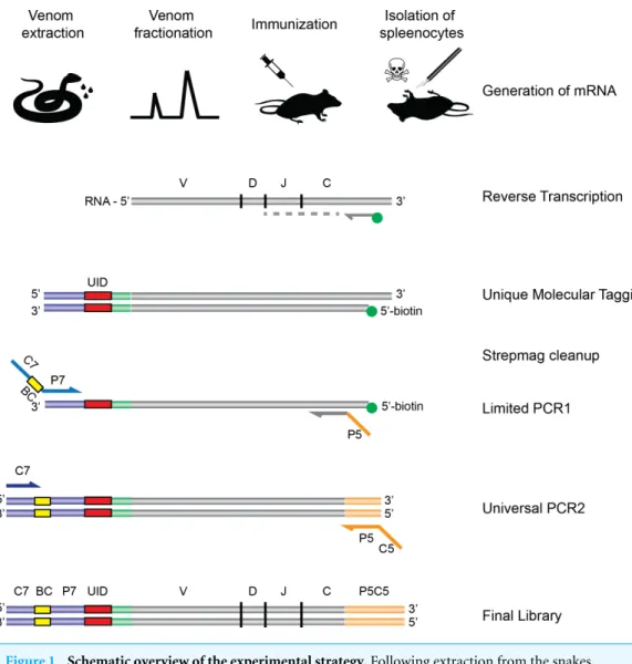
al., 2011; Rosso et al., 1996; Alape-Girón et al., 1996) represents another difficulty in production ofMicrurusantivenom, since it thwarts the goal of raising a balanced immune response against these medically relevant toxins. In order to further explore how these toxins interact with the mammalian immune system, we chose a mouse model and employed an NGS approach using the AbSeqTMtechnology developed by AbVitro (now Juno Therapeutics,https://www.junotherapeutics.com), based on Illumina sequencing (Fig. 1). The methodology was utilized to sequence immunoglobulin (Ig) encoding mRNA transcripts from splenic B-lymphocytes in mice subjected to immunization with either a 3FTx or a PLA2 toxin from the venom of M. nigrocinctus (Central American coral
snake). By this approach, the transcription levels of different immunoglobulin isotypes and dominant clones of B-lymphocytes with a particular usage of V (variable) and J (joining) gene segments can be determined for Ig heavy chain transcripts. This methodology has previously been employed for investigating B-cell populations in autoimmune (Stern et al., 2014) or infectious diseases (Tsioris et al., 2015;Di Niro et al., 2015), for example. By employing the AbSeqTMhigh-throughput approach, we explore, for the first time, the Ig transcriptome including VJ usage patterns in individual animals subjected to immunization with two relevant toxin classes of elapid snakes. Although exploratory in its nature and somewhat limited by a small sample size, this study thus provides novel insight into the humoral response of mice immunized with 3FTx or PLA2toxins and highlights important
challenges of raising antibodies against poorly immunogenic toxins.
MATERIALS AND METHODS
Snake venom and toxins
Venom fromM. nigrocinctuswas obtained from a pool of more than 50 adult specimens collected in the Central Pacific region of Costa Rica, kept at the serpentarium of Instituto Clodomiro Picado, Universidad de Costa Rica. The snakes were not collected for this study, but belong to Instituto Clodomiro Picado, where their venoms are routinely used for the production of antivenom, wherefore a field permit was not necessary. Venom extraction is performed every four months (Chacón et al., 2012). The venom was lyophilized and stored at−20 ◦C.
Fractionation of the venom was performed by RP-HPLC on a C18 column (4.6×250
mm, 5µm particle diameter; Supelco) as previously described (Fernández et al., 2011).
Figure 1 Schematic overview of the experimental strategy.Following extraction from the snakes, venom is fractionated by HPLC, and the fractions of interest are used for immunization of rodents. Upon completion of the immunization protocol, the rodents are sacrificed and RNA is extracted and subjected to the AbSeq protocol (see ‘Materials and Methods’), whereby the RNA is reverse transcribed and barcoded, allowing for correct pairing of VHand VLchains after DNA sequencing.
acid (TFA; solution A) were separated at 1 mL/min in an Agilent 1200 chromatograph monitored at 215 nm, applying a gradient towards solution B (acetonitrile, containing 0.1% TFA): 0% B for 5 min, 0–15% B over 10 min, 15–45% B over 60 min, 45–70% B over 10 min, and 70% B over 9 min. Fractions of interest (the major components of the venom, belonging to the 3FTx (lethal) and PLA2 (myotoxic) protein families) were collected
Immunization of mice
Three female CD-1 mice (16–18 g) from Instituto Clodomiro Picado were immunized with a three-finger toxin (3FTx), and two with a phospholipase A2(PLA2), respectively.
These correspond to fractions #3 (∼P80548) and #30 (∼P81166/P81167) described in the previous proteomic characterization of this venom (Fernández et al., 2011). All toxin doses were injected by the intraperitoneal route. The priming dose was 1µg emulsified in
Freund’s complete adjuvant, followed by five booster doses injected in physiological saline without adjuvant, at days 15 (1µg), 43 (2µg) 63 (4µg), and 83 (6µg for the 3FTx and 8µg
for the PLA2). At day 90, after obtaining a blood sample for monitoring of the antibody
response by enzyme-immunoassay, mice were euthanized by CO2inhalation. Their spleens
were immediately removed, cut in small pieces, and disaggregated over a stainless steel mesh to obtain splenocytes. These cell suspensions were aliquoted in RNAlaterR solution
(Thermo) and shipped within 24 h to AbVitro, at room temperature, for subsequent molecular studies. The use of animals for these experiments followed the ethical guidelines of theComité Institucional para el Uso y Cuido de Animales(CICUA), Universidad de Costa Rica, with the approval number 82-08.
Enzyme-immunoassay (ELISA)
In order to evaluate the individual antibody responses of the mice, wells in MaxiSorp 96-well plates (NUNC, Roskilde, Denmark) were coated overnight with 1 µg of either
3FTx or PLA2, dissolved in 100µL PBS (0.12 M NaCl, 0.04 M sodium phosphate, pH 7.2).
Wells were washed five times with PBS and blocked by adding 100µL PBS containing 2%
(w:v) bovine serum albumin (BSA; Sigma), and incubated at room temperature for 1 h. Plates were then washed five times with PBS. Serial dilutions of serum from each mouse were prepared in PBS+2% BSA and 100µL was added to each well, in triplicates, and
incubated overnight at 4 ◦C. Normal mouse serum, run simultaneously under identical
conditions was used as a control for background. Plates were then washed five times with PBS, followed by the addition of 100µL of a 1:3,000 dilution of anti-mouse IgG (whole
molecule) antibodies conjugated to alkaline phosphatase, in PBS+1% BSA. The plates were incubated for 2 h, and then washed five times with FALC buffer (0.05 M Tris, 0.15 M NaCl, 20µM ZnCl2, 1 mM MgCl2, pH 7.4). Development of color was attained by addition
of 100µLp-nitrophenyl phosphate (1 mg/mL in 9.7% v/v diethanolamine buffer, pH 9.8)
and absorbances at 405 nm were recorded (Multiskan FC; Thermo Scientific).
Assessment of mRNA quality
Assessment of RNA quality was performed using Agilent’s TapeStation according to the manufacturers protocol and algorithm to calculate RINescores (http://www.agilent.com/ cs/library/technicaloverviews/public/5990-9613EN.pdf).
Library preparation and high-throughput sequencing of B-cell receptors
added to the 3′end of all cDNA, which contains the Illumina P7 universal priming site and a 17-nucleotide unique molecular identifier (UMI). Products were purified using streptavidin-coated magnetic beads followed by a primary PCR reaction using a pool of primers targeting the IGHA, IGHD, IGHE, IGHG, IGHM, IGKC and IGLC regions, as well as a sample-indexed Illumina P7C7 primer. The immunoglobulin-specific primers contained tails corresponding to the Illumina P5 sequence. PCR products were then purified using AMPure XP beads. A secondary PCR was then performed to add the Illumina C5 clustering sequence to the end of the molecule containing the constant region. The number of secondary PCR cycles was tailored to each sample to avoid entering plateau phase, as judged by a prior quantitative PCR analysis. The final products (reverse-transcribed UTR
+VDJ+partial—Cexon segments of the transcripts of the immunoglobulin chains, plus molecular barcode, Illumina multiplexing barcode, and Illumina sequencing adapters) were purified, quantified with Agilent TapeStation and pooled in equimolar proportions, followed by high-throughput paired-end sequencing on the Illumina MiSeq platform. For sequencing, the Illumina 600 cycle kit was used with the modifications that 325 cycles was used for read 1, 6 cycles for the index reads, 300 cycles for read 2 and a 10% PhiX spike-in to increase sequence diversity.
VJ repertoire sequencing data analysis
most expanded binding motifs. To examine preferences for V–J usage common across mice exposed to the same toxin, clones were grouped into larger bins that each encompass a single V gene-J gene combination, but all isotypes and CDR3s.
In silicoepitope predictions
The sequences of selected toxin components in the two venom fractions used for immunization were obtained from the UniProtKB database (http://www.uniprot.org) and linear parts of B-cell epitopes were predicted using the Bepi-Pred 1.0 server (Larsen, Lund & Nielsen, 2006) using 0.9 as Threshold to obtain a sensitivity of 0.25 and Specificity of 0.91. As no experimental structures were available, homology models were build based CPHmodels 3.2 (Nielsen et al., 2010) and the pdb-files were submitted to DiscoTope 2.0 (Kringelum et al., 2012) using 0.5 as Threshold to obtain a sensitivity of 0.23 and Specificity of 0.90.
RESULTS AND DISCUSSION
Three-finger toxins (3FTx) and phospholipase A2s (PLA2) are the two most abundant
toxin families in the venom ofM. nigrocinctus(Fernández et al., 2011), and generally they are the two snake toxin families which have been most investigated (Laustsen et al., 2016a). In the venom ofM. nigrocinctusthese toxins cause neuromuscular paralysis, owing to a combination of pre- and post-synaptic actions, and myotoxicity, providing the venom with its high toxicity (Rosso et al., 1996;Alape-Girón et al., 1996). In previous studies it was observed that 3FTxs and PLA2s were recognized more weakly than larger proteins from
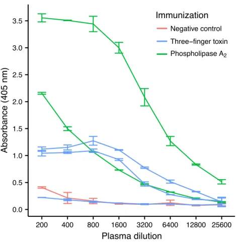
this venom, by a therapeutic equine antivenom (Fernández et al., 2011). Despite their low immunogenicity, it was possible to raise an antibody response against both toxins in four out of five mice, although high variation in the antibody titer was observed (Fig. 2). Mice immunized with PLA2had a higher antibody titer than mice immunized with the 3FTx, in
agreement with the higher molecular mass of the former.
Assessment of the mRNA from harvested mouse splenocytes indicated that it was of sufficient quality to proceed to sequencing (RINe scores between 5.2 and 6.4). A high-throughput sequencing approach (AbSeqTM) was employed to investigate transcription
levels of Ig isotypes and the usage of V and J gene segments for heavy chain assembly in mice that were immunized with a 3FTx or a PLA2. Investigation of the 50 most frequent
VJ combinations for the immunized mice did not, however, result in identification of a dominant combination, as the VJ usage was found to be similar across all samples (Fig. 3). This finding suggests that the generated antibody responses might be diverse and that multiple specific antibodies with low abundance are generated in each mouse.
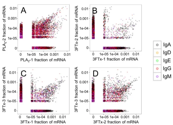
Looking at the sequences of all mRNA transcripts encoding heavy chain variable domain (VH) clones across each sample, we were able to find shared VH clones with
similar relative abundances in either the PLA2-immunized or the 3FTx-immunized mice
(Fig. 4). In comparison, almost no VHclones were shared between mice immunized with
0.0 0.5 1.0 1.5 2.0 2.5 3.0 3.5
200 400 800 1600 3200 6400 12800 25600
Plasma dilution
Absorbance (405 nm)
Immunization Negative control
Three−finger toxin
PhospholipaseA2
Figure 2 ELISA titrations of serum antibodies againstM. nigrocinctusPLA2or 3FTx in mice following
a 90-day immunization protocol. Two mice were immunized with PLA2, and three were immunized with 3FTx. Plates were coated with either PLA2or 3FTx, and antibodies were detected as described in ‘Materials and Methods’.
for immunization. The relatively high number of VHclones found in both of the PLA2
-immunized mice (Fig. 4A) compared to lower number of VHclones found across the three
3FTx-immunized mice (Figs. 4B–4D) further indicate that immunization with PLA2s is
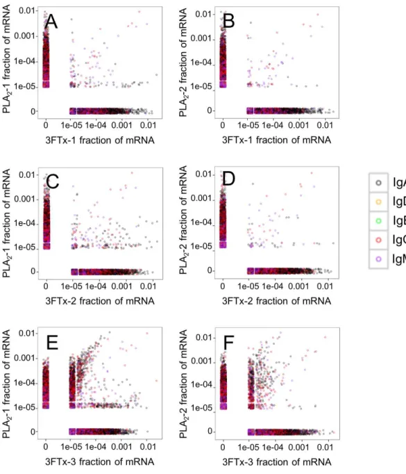
more prone to give rise to antibodies transcribed in similar quantities. Also, an intermediate number of similar VHclones was found in both the PLA2-1 and 3FTx-3 samples (Fig. 5E),
even though the correlation in relative abundance was not equally pronounced. This is likely explained by the fact that the majority of VHclones found in both PLA2-immunized
mice are not expected to be specific towards the toxins, but instead are likely to be directed against other (environmental) antigens that the mice have encountered throughout their life.
The AbSeqTMantibody sequencing methodology is capable of determining the Ig isotype
of the identified VHclones. The coloring of the VH clones inFigs. 4and5reveals that a
large number of the most abundant VH clones present in the mice are of the IgA isotype,
Figure 3 Comparison of the relative abundance of mRNA for the 50 most abundant VJ combinations for the mouse 3FTx-3 and mouse PLA2-2 showing VJ usage to be similar across samples.Similar VJ us-age patterns were observed for other pairs of immunized mice.
Figure 5 Relative abundance of unique VHclone transcripts compared between samples. Only few VH transcripts are found in similar abundance in more than one mouse, when mice immunized with differ-ent toxins are compared. (A) Comparison between mouse PLA2-1 and 3FTx-1, (B) Comparison between mouse PLA2-2 and 3FTx-1, (C) Comparison between mouse PLA2-1 and 3FTx-2, (D) Comparison be-tween mouse PLA2-2 and 3FTx-2, (E) Comparison bebe-tween mouse PLA2-1 and 3FTx-3, (F) Comparison between mouse PLA2-2 and 3FTx-3.
Figure 6 Overview of total mRNA transcripts encoding different immunoglobulin isotypes from the immunized mice (normalized).Numbers above each bar represents the mRNA count in each sample.
from the immunized mice revealed only a low percentage of IgG transcripts, as compared to the transcripts for other Igs (Figs. 4–6). With the acknowledgement of the small sample size employed in this study, this finding may indicate a difficulty in raising potent IgG antibodies against both 3FTxs and PLA2s in rodents. If similar difficulty is present in
horses, this may therefore have implications for antivenom production. Our results may further suggest that the immune response is slightly lower against 3FTxs than for PLA2s
based on the lower abundance of IgG transcripts in mice immunized with 3FTx (Fig. 6). Taken together with the results from the ELISA assay (Fig. 2) and the observation that immunization with PLA2s is more prone to give rise to similar Ig transcripts (Fig. 4Avs. Figs. 4B–4D), we suggest that the PLA2toxins may possibly be slightly more immunogenic
than the 3FTx, although neither toxin seems to have high immunogenicity. The underlying reason for this could possibly be due to the smaller molecular size of 3FTx compared to PLA2s, or that PLA2s may contain distinct epitopes better capable of eliciting an adaptive
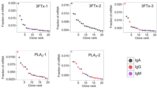
immune response than 3FTxs. Predictions of possible B-cell epitopes using the protein sequences and BepiPred 1.0 (Larsen, Lund & Nielsen, 2006) or DiscoTope 2.0 (Kringelum et al., 2012) with structural models of the investigated toxins as input do not indicate a major difference in the bare number of possible epitopes. However, the suggested difference in immunogenicity is further indicated by the fact that only two IgG-encoding mRNA transcripts are found in the top 20 most abundant Ig-encoding mRNA transcripts for only one out of three of the 3FTx-immunized mice. In comparison, six and nine of the top 20 mRNA transcripts for mice immunized with PLA2s encode the IgG isotype (Fig. 7). It
Figure 7 The 20 most abundant VHclone transcripts and their corresponding isotypes in each
immu-nized mouse based on their fraction of total immunoglobulin mRNA.
venom toxins is also characterized by a low proportion of IgG—a finding that would have evident implications for antivenom manufacture, as IgG has been shown to be the antibody isotype of therapeutic value (Fernandes et al., 2000). However, this is beyond the scope of this exploratory study.
CONCLUDING REMARKS AND OUTLOOK
In addition to demonstrating the utility of high-throughput sequencing technology, AbSeqTM, for investigation of immune responses in animals immunized with snake
venom toxins, the exploratory findings presented here may indicate possible difficulties in obtaining an IgG response against the medically important toxins of the 3FTx and PLA2 families from M. nigrocinctus. Given that these proteins play key toxic roles
in envenomings by elapid snakes, this underlines a drawback of current antivenom production based on immunized animal serum, since IgG has been shown to be the antibody isotype of therapeutic value (Fernandes et al., 2000). Although based on a small exploratory sample size, these findings therefore contribute to the understanding of snake toxin immunogenicity and indicate the possible difficulty in obtaining balanced immune responses in animals during the immunization process.
ACKNOWLEDGEMENTS
ADDITIONAL INFORMATION AND DECLARATIONS
Funding
The following institutions and foundations supported the research: Juno Therapeutics Inc., Instituto Clodomiro Picado, Universidad de Costa Rica, and the Novo Nordisk Foundation (NNF13OC0005613 and NNF16OC0019248). The funders had no role in study design, data collection and analysis, decision to publish, or preparation of the manuscript.
Grant Disclosures
The following grant information was disclosed by the authors:
Juno Therapeutics Inc., Instituto Clodomiro Picado, Universidad de Costa Rica. Novo Nordisk Foundation: NNF13OC0005613, NNF16OC0019248.
Competing Interests
Bruno Lomonte is an Academic Editor for PeerJ. Christopher Clouser and Francois Vigneault are employees of Juno Therapeutics, Seattle, Washington, United States of America, and AbVitro, Boston, United States of America. Sonia Timberlake is an employee of Finch Therapeutics, Somerville, Massachusetts, United States of America. The authors declare no other competing interests.
Author Contributions
• Andreas H. Laustsen conceived and designed the experiments, analyzed the data, wrote the paper, prepared figures and/or tables, reviewed drafts of the paper.
• Mikael Engmark analyzed the data, wrote the paper, prepared figures and/or tables, reviewed drafts of the paper.
• Christopher Clouser and Sonia Timberlake performed the experiments, analyzed the data, contributed reagents/materials/analysis tools, prepared figures and/or tables, reviewed drafts of the paper.
• Francois Vigneault and José María Gutiérrez conceived and designed the experiments, analyzed the data, contributed reagents/materials/analysis tools, reviewed drafts of the paper.
• Bruno Lomonte conceived and designed the experiments, performed the experiments, analyzed the data, contributed reagents/materials/analysis tools, prepared figures and/or tables, reviewed drafts of the paper.
Animal Ethics
The following information was supplied relating to ethical approvals (i.e., approving body and any reference numbers):
Comité Institucional para el Uso y Cuido de Animales (CICUA), Universidad de Costa Rica.
Data Availability
The following information was supplied regarding data availability:
The raw sequencing data is irrelevant for the reader as this is only used to isotype immunoglobulins—not to deduct any information about specific sequences.
REFERENCES
Alape-Girón A, Stiles B, Schmidt J, Girón-Cortés M, Thelestam M, Jörnvall H,
Bergman T. 1996.Characterization of multiple nicotinic acetylcholine
receptor-binding proteins and phospholipases A2, from the venom of the coral snakeMicrurus
nigrocinctus nigrocinctus.FEBS Letters380:29–32DOI 10.1016/0014-5793(95)01543-4.
Antúnez J, Fernández J, Lomonte B, Angulo Y, Sanz L, Pérez A, Calvete JJ, Gutiérrez
JM. 2010.Antivenomics ofAtropoides mexicanusandAtropoides picadoisnake
venoms: relationship to the neutralization of toxic and enzymatic activities.Journal of Venom Research1:8–17.
Bolaños R, Cerdas L, Abalos JW. 1978.Venoms of coral snakes (Micrurusspp.): report
on a multivalent antivenin for the Americas.Bulletin of the Pan American Health Organization12:23–27.
Bolaños R. 1972.Toxicity of Costa Rican snake venoms for the white mouse.The
American Journal of Tropical Medicine and Hygiene21:360–363.
Bucaretchi F, De Capitani EM, Vieira RJ, Rodrigues CK, Zannin M, Da Silva Jr NJ,
Casais-e-Silva LL, Hyslop S. 2016.Coral snake bites (Micrurusspp.) in Brazil: a
re-view of literature reports.Clinical Toxicology 54:222–234
DOI 10.3109/15563650.2015.1135337.
Campbell JA, Lamar WW. 2004.The venomous reptiles of the Western Hemisphere. Vol. II.
Ithaca: Comstock Publishing Associates, Cornell University Press.
Castro KL, Duarte CG, Ramos HR, De Avila RAM, Schneider FS, Oliveira D, Freitas
CF, Kalapothakis E, Ho PL, Chávez-Olortegui C. 2015.Identification and
charac-terization of B-cell epitopes of 3FTx and PLA2toxins fromMicrurus corallinussnake
venom.Toxicon93:51–60DOI 10.1016/j.toxicon.2014.10.015.
Chacón D, Arias J, Solano G, Bonilla F, Gómez A. 2012.Maintaining Coral Snakes
(Micrurus nigrocinctus, Serpentes: Elapidae) for venom production on an alternative fish-based diet.Toxicon60:249–253DOI 10.1016/j.toxicon.2012.04.332.
Chotwiwatthanakun C, Ronachai P, Akesowan S, Sriprapat S, Ratanabanangkoon K.
2001.Production of potent polyvalent antivenom against three elapid venoms using
a low dose, low volume, multi-site immunization protocol.Toxicon39:1487–1494
DOI 10.1016/S0041-0101(01)00108-8.
Cook DAN, Owen T, Wagstaff SC, Kinne J, Wernery U, Harrison RA. 2010.Analysis of
camelid IgG for antivenom development: serological responses of venom-immunised camels to prepare either monospecific or polyspecific antivenoms for West Africa.
Toxicon56:363–372DOI 10.1016/j.toxicon.2010.03.025.
Corrêa-Netto C, Junqueira-de-Azevedo IL, Silva DA, Ho PL, Leit¯ao-de-Araújo M, Alves
gland transcriptomic analysis of Brazilian coral snakes,Micrurus altirostrisandM. corallinus.Journal of Proteomics74:1795–1809DOI 10.1016/j.jprot.2011.04.003.
Di Niro R, Lee SJ, Vander Heiden JA, Elsner RA, Trivedi N, Bannock JM, Gupta
NT, Kleinstein SH, Vigneault F, Gilbert TJ, Meffre E. 2015.Salmonellainfection
drives promiscuous B cell activation followed by extrafollicular affinity maturation.
Immunity 43:120–131DOI 10.1016/j.immuni.2015.06.013.
Edgar RC. 2004.MUSCLE: multiple sequence alignment with high accuracy and high
throughput.Nucleic Acids Research32:1792–1797DOI 10.1093/nar/gkh340.
Fernández J, Alape-Girón A, Angulo Y, Sanz L, Gutiérrez JM, Calvete JJ, Lomonte
B. 2011.Venomic and antivenomic analyses of the Central American coral snake,
Micrurus nigrocinctus(Elapidae).Journal of Proteome Research10:1816–1827
DOI 10.1021/pr101091a.
Fernandes I, Lima EX, Takehara HA, Moura-da-Silva AM, Tanjoni I, Gutiérrez
JM. 2000.Horse IgG isotypes and cross-neutralization of two snake antivenoms
produced in Brazil and Costa Rica.Toxicon38:633–644
DOI 10.1016/S0041-0101(99)00177-4.
Fernández J, Vargas N, Pla D, Sasa M, Rey-Suárez P, Sanz L, Gutiérrez JM, Calvete JJ,
Lomonte B. 2015.Snake venomics ofMicrurus alleniandMicrurus mosquitensisfrom
the Caribbean region of Costa Rica reveals two divergent compositional patterns in New World elapids.Toxicon107:217–233DOI 10.1016/j.toxicon.2015.08.016.
Guidolin RG, Marcelino RM, Gondo HH, Morais JF, Ferreira RA, Silva CL, Kipnis TL,
Silva JA, Fafetine J, Da Silva WD. 2010.Polyvalent horse F(ab’)2, snake antivenom:
development of process to produce polyvalent horse F(ab’)2antibodies anti-african
snake venom.African Journal of Biotechnology9:2446–2455.
Gutiérrez JM, León G, Lomonte B, Angulo Y. 2011.Antivenoms for snakebite
envenom-ings.Inflammation & Allergy Drug Targets10:369–380
DOI 10.2174/187152811797200669.
Gutiérrez JM, Lomonte B, Aird SD, Da Silva Jr NJ. 2016. Mecanismo de a¸cao dos
venenos de cobras corais. In: Da Silva Jr NJ, ed.As Cobras Corais do Brasil: Biologia, Taxonomia, Venenos e Envenenamientos. GO: Editora PUC Goiás, 415 pp.
Gutiérrez JM, Sanz L, Flores-Díaz M, Figueroa L, Madrigal M, Herrera M, Villalta M,
León G, Estrada R, Borges A, Alape-Girón A, Calvete JJ. 2009.Impact of regional
variation inBothrops aspervenom on the design of antivenoms: integrating antive-nomics and neutralization approaches.Journal of Proteome Research9:564–577
DOI 10.1021/pr9009518.
Judge RK, Henry PJ, Mirtschin P, Jelinek G, Wilce JA. 2006.Toxins not neutralized
by brown snake antivenom.Toxicology and Applied Pharmacology 213:117–125
DOI 10.1016/j.taap.2005.09.010.
Kringelum JV, Lundegaard C, Lund O, Nielsen M. 2012.Reliable B cell epitope
predictions: impacts of method development and improved benchmarking.PLOS Computational Biology8(12):e1002829DOI 10.1371/journal.pcbi.1002829.
Larsen JE, Lund O, Nielsen M. 2006.Improved method for predicting linear B-cell
Laustsen AH, Engmark M, Milbo C, Johannesen J, Lomonte B, Gutiérrez JM, Lohse B.
2016a.From fangs to pharmacology: the future of snakebite envenoming therapy.
Current Pharmaceutical Design22:5270–5293
DOI 10.2174/1381612822666160623073438.
Laustsen AH, Lomonte B, Lohse B, Fernández J, Gutiérrez JM. 2015.Unveiling the
nature of black mamba (Dendroaspis polylepis) venom through venomics and antivenom immunoprofiling: identification of key toxin targets for antivenom development.Journal of Proteomics119:126–142DOI 10.1016/j.jprot.2015.02.002.
Laustsen AH, Solà M, Jappe EC, Oscoz S, Lauridsen LP, Engmark M. 2016b.
Biotech-nological trends in spider and scorpion antivenom development.Toxins8:1–33
DOI 10.3923/rjt.2016.1.7.
Leong PK, Fung SY, Tan CH, Sim SM, Tan NH. 2015.Immunological cross-reactivity
and neutralization of the principal toxins ofNaja sumatranaand related cobra venoms by a Thai polyvalent antivenom (Neuro Polyvalent Snake Antivenom).Acta Tropica149:86–93DOI 10.1016/j.actatropica.2015.05.020.
Lomonte B, Rey-Suárez P, Fernández J, Sasa M, Pla D, Vargas N, Bénard-Valle M, Sanz L, Corrêa-Netto C, Núñez V, Alape-Girón A, Alagón A, Gutiérrez JM,
Calvete JJ. 2016b.Venoms ofMicruruscoral snakes: evolutionary trends in
compositional patterns emerging from proteomic analyses.Toxicon122:7–25
DOI 10.1016/j.toxicon.2016.09.008.
Lomonte B, Sasa M, Rey-Suárez P, Bryan W, Gutiérrez JM. 2016a.Venom of
the coral snakeMicrurus clarki: proteomic profile, toxicity, immunological cross-neutralization, and characterization of a three-finger toxin.Toxins8:138
DOI 10.3390/toxins8050138.
Nielsen M, Lundegaard C, Lund O, Petersen TN. 2010.CPHmodels-3.0—remote
homology modeling using structure-guided sequence profiles.Nucleic Acids Research
38(suppl 2):W576–W581DOI 10.1093/nar/gkq535.
Ownby C, Colberg T. 1990.Comparison of the immunogenicity and antigenic
composi-tion of several venoms of snakes in the family Crotalidae.Toxicon1990(28):189–199.
R Core Team. 2014.R: a language and environment for statistical computing. Vienna: R
Foundation for Statistical Computing.Available athttp:// www.R-project.org/.
Ramos HR, Junqueira-de-Azevedo ILM, Novo JB, Castro K, Duarte CG,
Machado-de-Avila RA, Chavez-Olortegui C, Ho PL. 2016.A heterologous multiepitope DNA
prime/recombinant protein boost immunisation strategy for the development of an antiserum againstMicrurus corallinus(coral snake) venom.PLOS Neglected Tropical Diseases10:e0004484DOI 10.1371/journal.pntd.0004484.
Rey-Suárez P, Núñez V, Fernández J, Lomonte B. 2016.Integrative characterization
of the venom of the coral snakeMicrurus dumerilii(Elapidae) from Colombia: proteome, toxicity, and cross-neutralization by antivenom.Journal of Proteomics
136:262–273DOI 10.1016/j.jprot.2016.02.006.
Rey-Suárez P, Núñez V, Gutiérrez JM, Lomonte B. 2011.Proteomic and biological
(Elapidae), from Colombia and Costa Rica.Journal of Proteomics75:655–667
DOI 10.1016/j.jprot.2011.09.003.
Rey-Suárez P, Stuani-Floriano R, Rostelato-Ferreira S, Saldarriaga M, Núñez V,
Rodrigues-Simioni L, Lomonte B. 2012.Mipartoxin-I, a novel three-finger toxin,
is the major neurotoxic component in the venom of the redtail coral snakeMicrurus mipartitus(Elapidae).Toxicon60:851–863DOI 10.1016/j.toxicon.2012.05.023.
Rosso JP, Vargas-Rosso O, Gutiérrez JM, Rochat H, Bougis PE. 1996.Characterization
ofα-neurotoxin and phospholipase A2activities fromMicrurusvenoms.European
Journal of Biochemistry238:231–239DOI 10.1111/j.1432-1033.1996.0231q.x.
Sanz L, Pla D, Pérez A, Rodríguez Y, Zavaleta-Martínez A, Salas M, Lomonte B, Calvete
JJ. 2016.Venomic analysis of the poorly studied desert coral snake,Micrurus tschudii
tschudii, supports the 3FTx/PLA2dichotomy acrossMicrurusvenoms.Toxins8:178 DOI 10.3390/toxins8060178.
Schottler WH. 1951.Antigen-antibody relations in the present antivenin production of
Brazil.American Journal of Tropical Medicine and Hygiene31:500–509.
Stern JN, Yaari G, Vander Heiden JA, Church G, Donahue WF, Hintzen RQ, Huttner
AJ, Laman JD, Nagra RM, Nylander A, Pitt D. 2014.B cells populating the multiple
sclerosis brain mature in the draining cervical lymph nodes.Science Translational Medicine6: 248ra107DOI 10.1126/scitranslmed.3008879.
Tan KY, Tan CH, Fung SY, Tan NH. 2016.Neutralization of the principal toxins from
the venoms of ThaiNaja kaouthiaand MalaysianHydrophis schistosus: insights into toxin-specific neutralization by two different antivenoms.Toxins8:1–17
DOI 10.3923/rjt.2016.1.7.
Tan CH, Tan KY, Lim SE, Tan NH. 2015.Venomics of the beaked sea snake,Hydrophis
schistosus: a minimalist toxin arsenal and its cross-neutralization by heterologous antivenoms.Journal of Proteomics126:121–130DOI 10.1016/j.jprot.2015.05.035.
Tanaka GD, Sant’Anna OA, Marcelino JR, Da Luz ACL, Da Rocha MMT, Tambourgi
DV. 2016.Micrurussnake species: venom immunogenicity, antiserum
cross-reactivity and neutralization potential.Toxicon117:59–68
DOI 10.1016/j.toxicon.2016.03.020.
Tsioris K, Gupta NT, Ogunniyi AO, Zimnisky RM, Qian F, Yao Y, Wang X, Stern
JN, Chari R, Briggs AW, Clouser CR. 2015.Neutralizing antibodies against West
Nile virus identified directly from human B cells by single-cell analysis and next generation sequencing.Integrative Biology7:1587–1597DOI 10.1039/C5IB00169B.
Vander Heiden JA, Yaari G, Uduman M, Stern JN, O’Connor KC, Hafler DA, Vigneault
F, Kleinstein SH. 2014.pRESTO: a toolkit for processing high-throughput
sequenc-ing raw reads of lymphocyte receptor repertoires.Bioinformatics30:1930–1932
DOI 10.1093/bioinformatics/btu138.
Vergara I, Pedraza-Escalona M, Paniagua D, Restano-Cassulini R, Zamudio F, Batista
CV, Possani L, Alagón A. 2014.Eastern coral snakeMicrurus fulviusvenom toxicity
in mice is mainly determined by neurotoxic phospholipases A2.Journal of Proteomics
Walsh G. 2014.Biopharmaceutical benchmarks 2014.Nature Biotechnology32:992–1000
DOI 10.1038/nbt.3040.
Warrell DA. 2004. Snakebites in Central and South America: epidemiology, clinical
features and clinical management. In: Campbell JA, Lamar WW, eds.The venomous reptiles of the western hemisphere. Vol. II. Ithaca: Comstock Publishing Associates, Cornell University Press, 709–761.
Williams DJ, Gutiérrez JM, Calvete JJ, Wüster W, Ratanabanangkoon K, Paiva O, Brown NI, Casewell NR, Harrison RA, Rowley PD, O’Shea M. 2011.
Ending the drought: new strategies for improving the flow of affordable, ef-fective antivenoms in Asia and Africa.Journal of Proteomics74:1735–1767
DOI 10.1016/j.jprot.2011.05.027.
Wong KY, Tan CH, Tan NH. 2016.Venom and purified toxins of the spectacled
cobra (Naja naja) from Pakistan: insights into toxicity and antivenom neutral-ization.The American Journal of Tropical Medicine and Hygiene94:1392–1399
DOI 10.4269/ajtmh.15-0871.
Ye J, Ma N, Madden TL, Ostell JM. 2013.IgBLAST: an immunoglobulin
vari-able domain sequence analysis tool.Nucleic Acids Research41:W34–W40