BIOINFORMATICS
UDC 577.152:611.576.31
Flexible 3D structure of
Bos taurus
tyrosyl-tRNA
synthetase suggests the existence of the hinge mechanism
provided by conservative Gly353 at interdomain linker
N. A. Pydiura, A. I. Kornelyuk
Institute of Molecular Biology and Genetics, NAS of Ukraine 150, Akademika Zabolotnoho Str., Kyiv, Ukraine, 03680 [email protected]
Mammalian tyrosyl-tRNA synthetase is composed of two structural modules: N-terminal catalytic miniTyrRS and non-catalytic cytokine-like C-terminal module connected by a flexible peptide linker. Till now, the 3D structure of any full-length mammalian TyrRS has not been solved by X-ray crystallography. Theaimof this work was a ho-mology modeling of 3D structure of full-lehgth B.taurus tyrosyl-tRNA synthetase.Methods. Homology modeling of TyrRS was performed by Modeller 9.1 package. Quality of the models was assessed using Biotech Validation Suite web-server.Results. Our BLAST search identified 34% sequence homology between interdomain linker of TyrRS and linker of human c-Abl tyrosine kinase. In order to model the full-length TyrRS structure we as-sembled the models of three parts of the protein (N- and C- terminal domains and the linker) using Modeller 9.1 software. The best Abl-17 model structure was refined by energy minimization.Conclusions. High flexibility of the interdomain linker can generate multiple conformations of TyrRS. The hinge mechanism at interdomain linker may be provided by conservative Gly353. It is proposed, that due to the linker flexibility an open extended conformation of TyrRS could transform into closed conformations in the enzyme-substrate complexes.
Keywords: tyrosyl-tRNA synthetase, homology modeling, interdomain linker, c-Abl tyrosine kinase, EMAP II, tRNATyr
.
Introduction. Tyrosyl-tRNA synthetase (TyrRS, EC 6.1.1.1) is one of the key enzymes of protein
biosyn-thesis in both pro- and eukaryotes [1]. Bovine (B.
tau-rus) cytoplasmic TyrRS is one of the best studied
mam-malian aminoacyl-tRNA synthetases. This enzyme forms a homodimer of two 59.2 kDa subunits, each of 528 amino acid (aa) residues. N- and C-terminal do-mains of the enzyme subunit are connected by a long
disordered 17 aa linker (Fig. 1,a) [1]. The NH2
-termi-nal catalytic domain comprises a «minimal» 39 kDa
TyrRS and has full catalytic activityin vitro[1, 2]. The
C-terminal domain formed by aa residues Val363-Ser528 is 166 aa long [3] and reveals the 52.7 % iden-tity to the mammalian cytokine endothelial monocyte-activating polypeptide II (EMAPII) [4, 5], which activa-tes monocyactiva-tes and endothelial cells – an effect first dis-covered at cancerogenesis induced with chemicals [6, 7].
A multiple alignment of C-domains guided by pre-dicted secondary structure revealed two independent
sub-domains (folds): ab-pleated Myf domain (OB-fold,
re-sidues Val363-Lys470) anda-helical sub-domain
(re-sidues Gly471-Ser528) [3]. Myf domain anda-helical
sub-domain form the RNA binding surface. A lysine-rich cluster KPKKK located within the sub-domain may play a role of a nuclear localization signal [8]. Several organisms posess C-terminal domain homologous to that of TyrRS. There are experimental data showing
involvement of Arc1p (G4p1) fromSaccharomyces
ce-revisiae (55.3 % identity) [10], human p43
(pro-EMAPII) (62.7 %) [3], and ARCE from Euplotes
octocarinatus(52 %) [11] in non-specific tRNA bin-ding. These proteins direct tRNA to the active sites of corresponding aminoacyl-tRNA synthetases [10, 12]. It is possible, that during the evolution C-terminal do-main was transferred to several diverse proteins invol-ved in translation (such as TyrRS, MetRS, p43, and
Arc1p) to enable their proper functioning in higher eu-karyotes [3]. Bovine C-domain contributes about 50 % of TyrRS affinity to ribosomal RNAs. RNA binding has a certain specificity: among others, poly(G) has the most
inhibitory effect in the reaction of tRNATyr
amino-acylation [13].
At present there are more than 40 three-dimensional structures of diverse aminoacyl-tRNA synthetases depo-sited in PDB. Unfortunately, no structure of full-length mammalian TyrRS has been solved by experimental means. The difficulties of obtaining crystals can be cau-sed by the presence of transiently disordered labile 17-aa long linker between N- and C-terminal domains. A large size of the protein makes NMR approach to the structure determination difficult, if not altogether im-practical. Mobility and flexibility of this linker may be required for the adaptable orientation of domains neces-sary for aminoacylation reaction. It is noteworthy that the linker is accessible to specific proteinases and con-tains a putative proteolytic PEST sequence [8]. Despite all the experimental information gathered up to date, there is no clear understanding either C-domain or inter-domain linker role and mode of action. The
significan-ce of linker relation to both cytokine motives and tRNA-binding domains is unclear and needs to be explained. The absence of experimentally derived full-length struc-ture and importance of understanding a role of these two eukaryotic cytokine motives justify computational approach to the study of mammalian cytoplasmic TyrRS. An accurate model will allow further investigation of TyrRS properties, suggest biochemical, biophysical and computational experiments and may lead to elucidation of its mechanism of action.
Materials and methods. The amino acid sequence of bovine TyrRS was reported earlier [3, 9] (Entrez (http://www.ncbi.nlm.nih.gov/entrez/), accession num-ber Q29465). The sequence has been analyzed for pos-sible intrinsically disordered regions by DISPROT predictor VSL2B (http://www.ist.temple.edu/disprot/ predictorVSL2.php) [14] and IUPPRED (http://iupred. enzim.hu/) [15] web-servers.
We used Internet web-servers such as BLAST (http: //www.ncbi.nim.nih.gov/BLAST) and PDB-BLAST (http://www.ebi.ac.uk/pdb) to search for homologous sequences. Three-dimensional coordinates of the pro-tein structural templates were downloaded from Propro-tein
Rossman fold a-helical domain OB-fold domain A-subdomain 528 a.a.
N-terminal module Linker C-terminal module a
b
c
d
Data Bank (PDB) (http://www.pdb.org/pdb) [16]. A rigid alignment of TyrRSs 3D structures was carried out
using function «Fit/Iterative Magic Fit/Caatoms only»
of Swiss-PdbViewer 4.0.1 (http://expasy.org/spdbv/) [17]. Images were prepared with PyMOL software [18] and POV-Ray [19]. Electrostatic potentials were cal-culated using APBS [20, 21].
To build 3D models of full-length bovine TyrRS we used homology modeling techniques [22]. Multiple alignments were done using ClustalW server (http:// www2.ebi.ac.uk/clustalw/) [23, 24]. A search for homo-logues was performed using above described tools and the resulting templates were used to model TyrRS by Modeller 9.1 package [25]. Quality of the models was as-sessed using Biotech Validation Suite web-server (http: //biotech.ebi.ac.uk). Ramachandran plots were built using PROCHECK [26].
Our approach to full-length TyrRS modeling can be outlined as follows: separate prediction of N- and C-do-mains 3D structures based on experimentally solved highly homologous structures, prediction of interdo-main linker followed by the assembly of full-length pro-tein models.
Both domains of bovine TyrRS are well studied and their sequences bear high degree of homology to corres-ponding human TyrRS domains. We used two crystallo-graphic structures deposited in PDB (codes 1N3L and 1NTG, chain A) as templates for homology modeling of the bovine N- and C-terminal domains correspon-dingly [27, 28]. These pairs of sequences are highly ho-mologues and their modeling causes no significant pro-blem. An alignment of bovine N-terminal domain to
hu-man 1N3L is shown on Fig. 1,b. The homology between
the two domains is 95 %.
A crystal structure of the human N-terminal domain reported as 1N3L lacks 7 aa of the catalytic loop. We
op-ted not to close this loopde novo, but instead searched
for homologues in the PDB. The PDB was queried by a catalytic loop sequence plus 5-aa overhangs at each side: GLTGSKMSSSEEESKID. The search resulted in about 100 sequences with 70 unique ones. The sequen-ces with low homology to catalytic loop itself were dis-carded. The best homologues were selected for further analysis (PDB codes 2BBO (chain A, human Nbd1 with Phe508), 2DBG (chain A, pyrin Paad-Dapin domain), 1PS9 (chain A), 1POY (chain 1, spermidine-putresci-ne-binding protein), 1X7F (chain A, an uncharacteri-zed Bacillus cereus protein)). Two structures, 1X7F and 1POY, containing turns in their loops, were consi-dered. The 41 % homologous 1X7F [29] was selected for actual modeling due to gapless alignment with
bo-vine TyrRS linker (Fig. 1, c). For the N-domain we
generated 10 models and selected a model number 8 as having the optimal Modeller objective function score and the best Biotech Validation Suite score (objective = = 1490, Biotech = 1.38).
The C-terminal domain was aligned with
cytokine-like human 1NTG_A (Fig. 2,a) with 92 % identity. Ten
slightly different models of each domain were obtai-ned. Selection of the models for future use was based on Biotech scores and Modeller objective function. A model number 5 was selected (objective = 934, Bio-tech = 1.28).
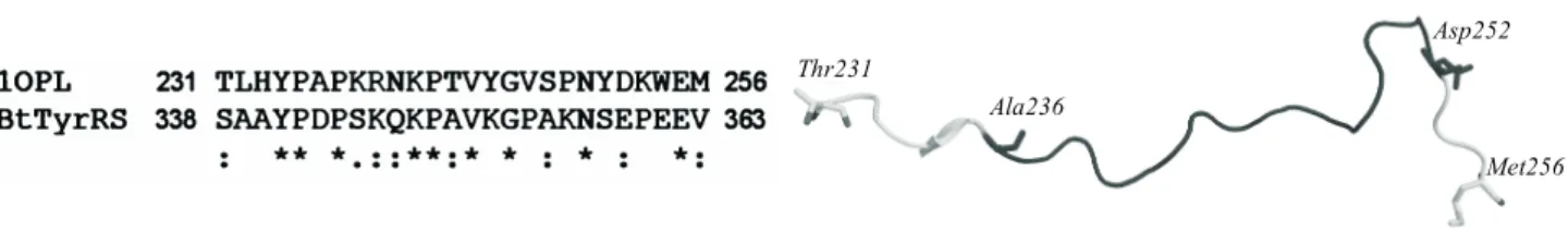
Modeling the interdomain linker of 17 aa was much more problematic. To model its structure we used two different approaches. A straightforward template-based approach commenced with BLAST search of all known linkers and identified a fragment of an auto-inhibitor of human C-Abl tyrosine kinase as the best template (PDB code 1OPL, chain A) [30]. To enable the modeling we added 5-aa overlaps to each side of the linker sequence. Resulting homology between the two sequences was
Thr231
Ala236
Asp252
Met256
a b
only 34 % (Fig. 1,d). The first model was selected from the ten models obtained, with objective = 3700, Bio-tech = 1.20. The C-Abl kinase linker structure is shown
in Fig. 2,b.
In order to model the full-length TyrRS protein we assembled all three parts (C-, and N-domains and lin-ker) using Modeller 9.1. To get a realistic domain orientation we strived to obtain a maximum overlaps of
the linker terminal amino acids backbone j and y
torsion angles with corresponding amino acids of do-mains being attached. All models were refined by ener-gy minimization in Swiss-PDB Viewer [17] until their potential energy converged to an average value of ap-proximately –23700 kJ/mol.
Results and discussion. The failed efforts to crys-tallize full-length bovine TyrRS, as well as preliminary NMR data obtained in our laboratory (unpublished) made us suspect that the protein has intrinsically or tran-siently disordered regions. We have carried out bioin-formatics analysis of potential TyrRS disorder. The da-ta are analogous to those obda-tained for human TyrRS [31] and are shown in Fig. 3. The main unfolded regions correspond to the N- and C-termini, catalytic loop (Pro 216-Glu229) and interdomain linker (Pro342-Glu362) of the protein. The linker flexibility may be necessary for correct mutual orientation of N- and C- domains during
tRNATyr
binding and recognition. We decided to build an ensemble of possible linker conformations based on the homology with human C-Abl kinase linker (homo-logy models will be further designated as Abl-X).
All homology models of full-length bovine TyrRS were generated in Modeller 9.1 from four components: N-terminal domain, catalytic loop, linker structure and the C-domain as described in Materials and methods. A selected model was refined by minimization in Swiss-PDB Viewer 4.0.1 (GROMOS96 43B1 parameter set) until its free energy reached a plateau. An average final energy for selected Abl-17 model was –23700 kJ/mol. This model is characterized as extended («open») structu-re. It is possible that the solvated linker makes positive con-tribution to the overall structure free energy. The mutual orientation of two domains in Abl-17 is shown in Fig. 4.
Earlier, some putative tRNATyr
binding residues of
the C-domain were predicted from the tRNAPhe
and
tRNATyr
modification protection experiments [32]. Structural analysis of the tRNA-C-domain complex re-vealed a potential tRNA binding surface which consists
of b'1-b'2 hairpin (Leu426-Gly433, Lys435-Gln437)
Residue position
D
is
o
rd
er
te
nde
nc
y
0 100 200 300 400 500 1
0 0.2 0.4 0.6 0.8
1
0 0.2 0.4 0.6 0.8
D
is
o
rd
er
pr
ob
abi
li
ty
Residue position 0 100 200 300 400 500
DISPROT – disorder prediction results
a b
Fig. 3. Results of the bovine TyrRS protein intrinsically disordered region prediction by IUPRED [14] (a) and by VSL2B program of DISPROT server [15] (b). The values above a middle line correspond to the intrinsically disordered regions of the protein
C-module
N-module
Reconstructed catalytic loop Gly353
Interdomain linker
inserted into OB fold, interdomain region (Glu480-Leu481), KPKKK lysine-rich cluster and
Glu489-Lys490 residues of thea-helix of A-sub-domain.
N-Domain is responsible for the specific recognition and binding of L-tyrosine (residues Tyr39, Tyr166, Gln170, Asp173, and Gln188). The contact surface of the
N-mo-dule with tRNATyr
is formed by 14 aa, which are homo-logous to the corresponding residues in the yeast TyrRS (His158, Lys246-Pro252, Trp283, His305-Asp308, and
Lys310) (Fig. 5, a). An evolutionary conservative
Trp283 residue should form a stacking interaction with G34 of the tRNA, while a conservative Asp308 – hydro-gen bonds with the same nucleotide.
We have superimposed the selected models on the crystallographic structure of the human TyrRS N-do-main dimer 1N3L with the N-doN-do-main structure as a re-ference. Conformations of the side chains of initial mo-del, which made clashes on the interface of N-modules in the dimer were optimized by Swiss-PdbViewer «si-mulated annealing» procedure. It was found that the Abl-17 structure had C-domain in the proximity with RNA binding surface of N-domain. The «open» structu-re of Abl-17 can form a large positively charged
conti-nuous RNA binding surface (Fig. 5,b).
Since the flexibility of the linker is able to rotate around pivotal glycine-353 we do not consider the mu-tual domain orientation to be fixed. We have analyzed TyrRS interdomain linker sequences from 22 species ofMetazoa(Fig. 6). In allChordatathe glycine posi-tion at the linker is absolutely conserved, which points
out to its functional importance. OtherMetazoalinkers
also contain glycine, several amino acids shifted up- or downwards from the homologues of the Gly353. The shorter linker length in insects probably reflects a more
ancient variant, while acquired prolines and lysines and linker extension in chordates result in more flexible linker backbone, probably needed for more adaptable
in-teraction with different structural elements of tRNATyr
. It is possible that flexibility of the linker plays an important role in the interdomain communications and dynamic coupling/uncoupling between the N- and C-domains, analogously to the effect observed in Csk ki-nase [33]. Class Ic of aminoacyl-tRNA synthetases re-cognizes the tRNA anticodon by one subunit and this «signal» has to be somehow transferred to the other subunit. There should be a mechanism of adaptable dy-namic juxtaposition of the two subunits and the struc-tural analysis of the interdomain linker gives us a hint at possible mechanism. The presence of four exposed lysine moieties in the linker of «open» conformation allows us to hypothesize a non-specific tRNA binding by the linker. On the other hand, the interdomain linker contains a proPEST motive and can be cleaved by pro-teases to release a cytokine-like C-domain. The flexibi-lity of the linker may modulate accessibiflexibi-lity and expo-sure of this proteolysis site.
The full-length model of bovine TyrRS, as well as both its domains separately, can be used for further ana-lysis and computational experiments, such as
molecu-lar docking with tRNATyr, molecular dynamics
simu-lations etc. Recently, we have performed an analysis of the YCD2 fragment of this model, comprised of the
a-helical part of N-domain, the linker and the
C-do-main. The results reported in [34], revealed a specific behavior of the linker in ten-nanosecond time-frame. It changes conformation from extended and disordered to
more compact one with short transienta-helical
struc-tures, supporting the currently proposed general model
a b
of the interdomain linker role in modulation of the enzy-me activity [35].
Future study on the full-length model of TyrRS mo-lecules by molecular dynamics, with obtained structu-res as starting points, will hopefully allow us to suggest a mechanism of tyrosyl-tRNA synthetase action.
Acknowledgements. This work was supported by the National Academy of Sciences of Ukraine within the project «Dynamic aspects of functioning of
eukaryotic tyrosyl-tRNA synthetase» (registration
number 0107 U004938).
Ì. Î. Ïèäþðà, Î. ². Êîðíåëþê
Ãíó÷êà ïðîñòîðîâà ñòðóêòóðàBos taurusòèðîçèë-òÐÍÊ ñèíòåòàçè ïðèïóñêຠ³ñíóâàííÿ øàðí³ðíîãî ìåõàí³çìó, ÿêèé
çàáåçïå÷óºòüñÿ êîíñåðâàòèâíèì Gly353 ó ì³æäîìåííîìó ë³íêåð³ Ðåçþìå
Òèðîçèë-òÐÍÊ ñèíòåòàçà (TyrRS) ññàâö³â ñêëàäàºòüñÿ ç äâîõ ñòðóêòóðíèõ ìîäóë³â: N-ê³íöåâîãî êàòàë³òè÷íîãî ìîäóëÿ (mini TyrRS) ³ íåêàòàë³òè÷íîãî öèòîê³í-ïîä³áíîãî C-ê³íöåâîãî ìîäóëÿ, ç’ºäíàíèõ ãíó÷êèì ïåïòèäíèì ë³íêåðîì. Äî ñüîãîäí³ ïðîñòîðîâó ñòðóêòóðó ïîâíîðîçì³ðíî¿ TyrRS ññàâö³â íå âèð³øåíî ìåòîäîì ðåíòãåí³âñüêî¿ êðèñòàëîãðàô³¿.Ìåòàö³º¿ ðîáîòè ïîëÿãàëà â ìî-äåëþâàíí³ çà ãîìîëî㳺þ ïðîñòîðîâî¿ ñòðóêòóðè ïîâíîðîçì³ðíî¿ TyrRS B. taurus.Ìåòîäè. Ìîäåëþâàííÿ çà ãîìîëî㳺þ TyrRS ïðî-âåäåíî ç âèêîðèñòàííÿì Modeller 9.1. ßê³ñòü ìîäåëåé îö³íþâàëè çà äîïîìîãîþ Biotech Validation Suite web-ñåðâåðà.Ðåçóëüòàòè. Çà äàíèìè BLAST-ïîøóêó âèçíà÷åíî 34 %-âó ãîìîëîã³þ ïîñë³äîâ-íîñò³ ì³æäîìåííîãî ë³íêåðà TyrRS ³ ë³íêåðà c-Abl òèðîçèíê³íàçè ëþäèíè. Äëÿ ìîäåëþâàííÿ ñòðóêòóðè ïîâíîðîçì³ðíî¿ TyrRS ìè ç³áðàëè ìîäåëü ç òðüîõ ôðàãìåíò³â á³ëêà (N-³ Ñ-ê³íöåâèõ äîìåí³â ³ ì³æäîìåííîãî ë³íêåðà), âèêîðèñòîâóþ÷è Modeller 9.1. Êðàùó ìî-äåëü ñòðóêòóðè Abl-17 óòî÷íåíî ìåòîäîì ì³í³ì³çàö³¿ åíåð㳿.
Âèñíîâêè. Âèñîêà ãíó÷ê³ñòü ì³æäîìåííîãî ë³íêåðà ìîæå ïðèçâî-äèòè äî ôîðìóâàííÿ ìíîæèííèõ êîíôîðìàö³é TyrRS. Øàðí³ðíèé ìåõàí³çì ó ì³æäîìåííîìó ë³íêåð³, â³ðîã³äíî, çàáåçïå÷óºòüñÿ êîí-ñåðâàòèâíèì çàëèøêîì Gly353. Ïåðåäáà÷àºòüñÿ, ùî çàâäÿêè âè-ñîê³é ãíó÷êîñò³ ì³æäîìåííîãî ë³íêåðà â³äêðèòà êîíôîðìàö³ÿ TyrRS ìîæå ïåðåõîäèòè â çàêðèòó êîíôîðìàö³þ ó ôåðìåíòíî-ñóáñòðàòíèõ êîìïëåêñàõ.
Êëþ÷îâ³ ñëîâà: òèðîçèë-òÐÍÊ ñèíòåòàçà, ìîäåëþâàííÿ çà ãîìîëî㳺þ, ì³æäîìåííèé ë³íêåð, c-Abl òèðîçèíê³íàçà, EMAP II, òÐÍÊTyr
.
Í. À. Ïèäþðà, À. È. Êîðíåëþê
Ãèáêàÿ ïðîñòðàíñòâåííàÿ ñòðóêòóðà Bos taurusòèðîçèë-òÐÍÊ ñèíòåòàçû ïðåäïîëàãàåò ñóùåñòâîâàíèå øàðíèðíîãî ìåõàíèçìà, îáåñïå÷èâàåìîãî êîíñåðâàòèâíûì Gly353 â ìåæäîìåííîì ëèíêåðå Ðåçþìå
Òèðîçèë-òÐÍÊ ñèíòåòàçà (TyrRS) ìëåêîïèòàþùèõ ñîñòîèò èç äâóõ ñòðóêòóðíûõ ìîäóëåé: N-êîíöåâîãî êàòàëèòè÷åñêîãî ìîäó-ëÿ (miniTyrRS) è íåêàòàëèòè÷åñêîãî öèòîêèí-ïîäîáíîãî C-êîíöå-âîãî ìîäóëÿ, ñîåäèíåííûõ ãèáêèì ïåïòèäíûì ëèíêåðîì. Äî íàñòî-ÿùåãî âðåìåíè ïðîñòðàíñòâåííàÿ ñòðóêòóðà ïîëíîðàçìåðíîé TyrRS ìëåêîïèòàþùèõ íå ðåøåíà ìåòîäîì ðåíòãåíîâñêîé êðèñ-òàëëîãðàôèè.Öåëüäàííîé ðàáîòû ñîñòîÿëà â ìîäåëèðîâàíèè ïî ãîìîëîãèè ïðîñòðàíñòâåííîé ñòðóêòóðû ïîëíîðàçìåðíîé TyrRS B. taurus.Ìåòîäû. Ìîäåëèðîâàíèå ïî ãîìîëîãèè TyrRS âûïîëíå-íî ñ ïîìîùüþ ïàêåòà Modeller 9.1. Êà÷åñòâî ìîäåëåé îöåíèâàëè ñ ïîìîùüþ Biotech Validation Suite web-ñåðâåðà.Ðåçóëüòàòû. Ïî äàííûì BLAST-ïîèñêà îïðåäåëåíà 34 %-ÿ ãîìîëîãèÿ ïîñëåäîâà-òåëüíîñòè ìåæäîìåííîãî ëèíêåðà TyrRS è ëèíêåðà c-Abl òèðî-çèíêèíàçû ÷åëîâåêà. Äëÿ ìîäåëèðîâàíèÿ ñòðóêòóðû ïîëíîðàç-ìåðíîé TyrRS, ìû ñîáðàëè ìîäåëü èç òðåõ ôðàãìåíòîâ áåëêà (N-è Ñ-êîíöåâûõ äîìåíîâ (N-è ìåæäîìåííîãî ë(N-èíêåðà), (N-èñïîëüçóÿ Mo-deller 9.1. Ëó÷øàÿ ìîäåëü ñòðóêòóðû Abl-17 óòî÷íåíà ìåòîäîì ìèíèìèçàöèè ýíåðãèè.Âûâîäû. Âûñîêàÿ ãèáêîñòü ìåæäîìåííî-ãî ëèíêåðà ìîæåò ïðèâîäèòü ê ôîðìèðîâàíèþ ìíîæåñòâåííûõ êîíôîðìàöèé TyrRS. Øàðíèðíûé ìåõàíèçì â ìåæäîìåííîì ëèí-êåðå, âåðîÿòíî, îáåñïå÷èâàåòñÿ êîíñåðâàòèâíûì îñòàòêîì Gly 353. Ïðåäïîëàãàåòñÿ, ÷òî èç-çà âûñîêîé ãèáêîñòè ìåæäîìåííî-ãî ëèíêåðà îòêðûòàÿ êîíôîðìàöèÿ TyrRS ìîæåò ïåðåõîäèòü â çàêðûòóþ êîíôîðìàöèþ â ôåðìåíòíî-ñóáñòðàòíûõ êîìïëåêñàõ. Êëþ÷åâûå ñëîâà: òèðîçèë-òÐÍÊ ñèíòåòàçà, ìîäåëèðîâàíèå ïî ãîìîëîãèè, ìåæäîìåííûé ëèíêåð, c-Abl òèðîçèíêèíàçà, EMAP II, òÐÍÊTyr
.
REFERENCES
1.Kornelyuk A. I.Structural and functional investigation of mam-malian tyrosyl-tRNA synthetase // Biopolym. Cell.–1998.–14, N 4.–P. 349–359.
ve proteolytically modified form of tyrosyl-tRNA synthetase from bovine liver // Ukr. Biokhim. Zh.–1991.–63, N 4.–P. 61–67. 3.Levanets O. V., Naidenov V. G., Odynets K. A., Woodmaska M. I., Matsuka G. Kh., Kornelyuk A. I.Homology of C-terminal non-catalytic domain of mammalian tyrosyl-tRNA synthetase with cytokine EMAP II and non-catalytic domains of methionyl- and phenylalanyl-tRNA synthetases // Biopolym. Cell.–1997.–13, N 6.–P. 474–478.
4.Kleeman T. A., Wei D., Simpson K. L., First E. A.Human tyro-syl-tRNA synthetase shares amino acid sequence homology with a putative cytokine // J. Biol. Chem.–1997.–272, N 22.–P. 14420– 14425.
5.Kao J., Ryan J., Brett G., Chen J., Shen H., Fan Y. G., Godman G., Familletti P. C., Wang F., Pan Y. C., Stern D., Clauss M. Endo-thelial monocyte-activating polypeptide II. A novel tumor-deri-ved polypeptide that activates host-response mechanisms // J. Biol. Chem.–1992.–267, N 28.–P. 20239–20247.
6.Kao J., Houck K., Fan Y., Haehnel I., Libutti S. K., Kayton M. L., Grikscheit T., Chabot J., Nowygrod R., Greenberg S., Kuang W.-J., Leung D., Hayward J. R., Kisiel W., Heath M., Brett J., Stern D. M.Characterization of a novel tumor-derived cytokine. Endothelial monocyte-activating polypeptide II // J. Biol. Chem.– 1994.–269, N 40.–P. 25106–25119.
7.Tas M. P., Murray J. C.Endothelial monocyte-activating poly-peptide II // Int. J. Biochem. Cell. Biol.–1996.–28, N 8.–P. 837– 841.
8.Ivakhno S. S., Kornelyuk O. I.Cytokine activities of some amino-acyl-tRNA synthetases and auxiliary cofactors of aminoacyla-tion reacaminoacyla-tion // Exp. Oncol.–2004.–26, N 4.–P. 250–255. 9.Levanets O. V., Naidenov V. G., Woodmaska M. I., Matsuka G. H.,
Kornelyuk A. I.Cloning of cDNA encoding C-terminal part of mammalian tyrosyl-tRNA synthetase using of PCR-amplified ra-dioactive probe // Biopolym. Cell.–1997.–13, N 2.–P. 121–126. 10.Simos G., Sauer A., Fasiolo F., Hurt E. C.A conserved domain
within Arc1p delivers tRNA to aminoacyl-tRNA synthetases // Mol. Cell.–1998.–1, N 2.–P. 235–242.
11.Tan M., Heckmann K., Brunen-Nieweler C.The micronuclear gene encoding a putative aminoacyl-tRNA synthetase cofactor of the ciliateEuplotes octocarinatusis interrupted by two sequen-ces that are removed during macronuclear development // Gene.– 1999.–233, N 1–2.–P. 131–140.
12.Simos G., Segref A., Fasiolo F., Hellmuth K., Shevchenko A., Mann M., Hurt E.C. The yeast protein Arc1p binds to tRNA and functions as a cofactor for the methionyl- and glutamyl-tRNA synthetases // EMBO J.–1996.–15, N 19.–P. 5437–5448. 13.Kurochkin I. V., Kornelyuk A. I., Matsuka G. Kh.Interaction of
eukaryotic tyrosyl-tRNA synthetases with high molecular weight RNA // Mol. Biol. (Moscow).–1991.–25, N 3.–P. 779–786. 14.Peng K., Radivojac P., Vucetic S., Dunker A. K., Obradovic Z.
Length-dependent prediction of protein intrinsic disorder // BMC Bioinformatics.–2006.–7.–P. 208.
15.Dosztanyi Z., Csizmok V., Tompa P., Simon I.Web server for the prediction of intrinsically unstructured regions of proteins based on estimated energy content // Bioinformatics.–2005.–21, N 16.– P. 3433–3434.
16.Berman H. M., Westbrook J., Feng Z., Gilliland G., Bhat T. N., Weissig H., Shindyalov I. N., Bourne P. E. The protein data bank // Nucleic Acids Res.–2000.–28, N 1.–P. 235–242.
17.Guex N., Peitsch M. C.SWISS-MODEL and the Swiss-PdbVie-wer: an environment for comparative protein modeling // Elect-rophoresis.–1997.–18, N 15.–P. 2714–2723.
18.DeLano W. L. The PyMOL Molecular Graphics System.–San Carlos: DeLano Scientific, 2002.
19.Persistenceof Vision Pty. Ltd, Persistence of Vision Raytracer (Version 3.6).–2004.
20.Baker N. A., Sept D., Joseph S., Holst M. J., McCammon J. A. Electrostatics of nanosystems: application to microtubules and the ribosome // Proc. Natl Acad. Sci. USA.–2001.–98, N 18.– P. 10037–10041.
21.Holst M., Saied F.Numerical solution of the nonlinear Poisson-Boltzmann equation: Developing more robust and efficient methods // J. Comput. Chem.–1995.–16, N 3.–P. 337–364. 22.Elofsson A., Fischer D., Rice D. W., Le Grand S. M., Eisenberg
D.A study of combined structure/sequence profiles // Fold Des.– 1996.–1, N 6.–P. 451–461.
23.Larkin M. A., Blackshields G., Brown N. P., Chenna R., McGet-tigan P. A., McWilliam H., Valentin F., Wallace I. M., Wilm A., Lopez R., Thompson J. D., Gibson T. J., Higgins, D. G.Clustal W and Clustal X version 2 // Bioinformatics.–2007.–23, N 21.– P. 2947–2948.
24.Goujon M., McWilliam H., Li W., Valentin F., Squizzato S., Paern J., Lopez R.A new bioinformatics analysis tools framework at EMBL-EBI // Nucleic Acids Res.–2010.–38(Web Server issue).– W695–699.
25.Fiser A., Sali A.Modeller: generation and refinement of homolo-gy-based protein structure models // Methods Enzymol.–2003.– 374.–P. 461–491.
26.Laskowski R. A., MacArthur M. W., Moss D. S., Thornton J. M. PROCHECK: a program to check the stereochemical quality of protein structures // J. Appl. Cryst.–1993.–26, N 2.–P. 283–291. 27.Yang X. L., Skene R. J., McRee D. E., Schimmel P.Crystal struc-ture of a human aminoacyl-tRNA synthetase cytokine // Proc. Natl Acad. Sci. USA.–2002.–99, N 24.–P. 15369–15374. 28.Yang X. L., Liu J., Skene R. J., McRee D. E., Schimmel P.Crystal
structure of an EMAP-II-like cytokine released from a human tRNA synthetase // Helv. Chim. Acta.–2003.–86, N 4.–P. 1246– 1257.
29.Minasov G., Shuvalova L., Brunzelle J. S., Collart F. R., Ander-son W. F., Mcsg. Crystal structure of an uncharacterizedB. ce-reusprotein // PDB: 1X7F.
30.Nagar B., Hantschel O., Young M. A., Scheffzek K., Veach D., Bornmann W., Clarkson B., Superti-Furga G., Kuriyan J. Struc-tural basis for the autoinhibition of c-Abl tyrosine kinase // Cell.– 2003.–112, N 6.–P. 859–871 .
31.Odynets K. A., Kornelyuk A. I.Analysis of unstructured regions of human cytoplasmic tyrosyl-tRNAsynthetase by methods of bioinformatics // Biopolym. Cell.–2005.–21, N 5.–P. 445–452. 32.Olszak K., Solecka K., Odynets K. A., Przykorska A., Kornelyuk
A. I. The analysis of the complex between cytokine-like COOH-terminal module of mammalian tyrosyl-tRNA synthetase and tRNA // Abstrs of the 29th
Meet. of FEBS.–Warsaw, 2004.–P. 54. 33.Wong L., Lieser S., Chie-Leon B., Miyashita O., Aubol B.,
Shaf-fer J., Onuchic J. N., Jennings P. A., Woods V. L. Jr., Adams J. A.Dynamic coupling between the SH2 domain and active site of the COOH terminal Src kinase, Csk // J. Mol. Biol.–2004.–341, N 1.–P. 93–106.
34.Pydiura N. A., Tereshchenko F. A., Kornelyuk A. I. Conforma-tional flexibility of interdomain linker in bovine tyrosyl-tRNA synthetase studied by molecular dynamics simulation // Biopo-lym. Cell.–2006.–22, N 6.–P. 433–438.
35.Wriggers W., Chakravarty S., Jennings P. A.Control of protein functional dynamics by peptide linkers // Biopolymers.–2005.– 80, N 6.–P. 736–746.

![Fig. 3. Results of the bovine TyrRS protein intrinsically disordered region prediction by IUPRED [14] (a) and by VSL2B program of DISPROT server [15] (b)](https://thumb-eu.123doks.com/thumbv2/123dok_br/18169869.329813/4.892.86.813.157.346/results-protein-intrinsically-disordered-prediction-iupred-program-disprot.webp)

