FACULDADE DE MEDICINA
CURSO DE PÓS GRADUAÇÃO EM PEDIATRIA E SAÚDE DA CRIANÇA DOUTORADO EM MEDICINA/PEDIATRIA
A PROTEÍNA DE LIGAÇÃO DO VÍRUS SINCICIAL RESPIRATÓRIO INIBE CITOCINAS INFLAMATÓRIAS
DA RESPOSTA IMUNE EM UM MODELO DE SEPSE INDUZIDO POR LIPOPOLISSACARÍDEOS
CHARLES ORNELAS BRUM
FACULDADE DE MEDICINA
CURSO DE PÓS GRADUAÇÃO EM PEDIATRIA E SAÚDE DA CRIANÇA DOUTORADO EM MEDICINA/PEDIATRIA
A PROTEÍNA DE LIGAÇÃO DO VÍRUS SINCICIAL RESPIRATÓRIO INIBE CITOCINAS INFLAMATÓRIAS DA RESPOSTA IMUNE EM UM MODELO DE
SEPSE INDUZIDO POR LIPOPOLISSACARÍDEOS
CHARLES ORNELAS BRUM
Tese apresentada como requisito para obtenção do grau de doutor em Medicina, área de concentração em Pediatria, pelo programa de Pós graduação em Pediatria e Saúde da Criança da Faculdade de Medicina da Pontifícia Universidade Católica do Rio Grande do Sul.
Orientador PUCRS: Paulo Márcio Condessa Pitrez Orientador Vanderbilt University: Fernando P. Polack
FICHA CATALOGRÁFICA
Rosária Maria Lúcia Prenna Geremia Bibliotecária CRB 10/196
B893p Brum, Charles Ornelas
A proteína de ligação do vírus sincicial respiratório inibe citocinas inflamatórias da resposta imune em um modelo de sepse induzido por lipossacarídeos / Charles Ornelas Brum. Porto Alegre: PUCRS, 2012. 64 f.: gráf. il. Inclui um artigo científico submetido à publicação. Orientadores: Prof. Dr. Paulo Márcio Condessa Pitrez.
Prof. MD. Fernando P. Polack (Vanderbilt University)
Tese (Doutorado) – Pontifícia Universidade Católica do Rio Grande do Sul. Faculdade de Medicina. Curso de Pós Graduação em Pediatria e Saúde da Criança. Doutorado em Medicina/Pediatria.
1. VÍRUS SINCICIAIS RESPIRATÓRIOS. 2. INFECÇÕES POR VÍRUS RESPIRATÓRIO SINCICIAL. 3. CITOCINAS. 4. PROTEÍNAS DE TRANSPORTE. 5.
LIPOPOLISSACARÍDEOS. 6. IMUNIDADE INATA. 7. RECEPTOR 4 TOLL LIKE. 8.
EPIDEMIOLOGIA EXPERIMENTAL. 9. CAMUNDONGOS KNOCKOUT. 10.
MURIDAE. 11. SEPSE. I.Pitrez, Paulo Márcio Condessa.II.Polack, Fernando P. III.Título.
Charles Ornelas Brum
Endereço: Rua Riachuelo, 1280/206 Porto Alegre / RS – CEP.: 90010 273 e mail: brumcobrum@ibest.com.br
Telefone: (53) 3247 6946 / (51) 8179 4551
ÓRGÃO FINANCIADOR: Coordenação de Aperfeiçoamento de Pessoal de Nível Superior (CAPES)
AGRADECIMENTOS
Ao Dr. Paulo Márcio Condessa Pitrez, por sua receptividade, bom senso, orientação e amizade.
Ao Dr. Renato T. Stein, por sua inestimável colaboração para o êxito do programa de doutorado PDEE/Capes na Vanderbilt University, Tn.
Ao Dr. Fernando P. Polack, por oferecer uma incrível oportunidade fazer parte de seu grupo de pesquisa na Vanderbilt University, Tn.
Ao Dr. Dr. James E. Crowe Jr, por manter as pontuais discussões científicas de seu grupo de imunologia virologia, as quais tomei parte, assim como por fornecer grande parte dos meios (equipamentos) para trabalho em laboratório.
Ao Dr. John T. Bates, por sua grande contribuição ao estudos de virologia e rotinas em laboratório, assim como sua disponibilidade.
Ao Dr. Ray S Peebles Jr, pela disponibilidade em reunir se para discussão da metodologia científica, assim como ceder seu laboratório para experimentos.
Ao Dr. Diego R. Hijano, pelo trabalho em conjunto no laboratório nos últimos meses de minha estada em Nashville.
À Gay Nell Krauss, pela assessoria dentro do Vanderbilt Vaccine Center, assim como pela amizade.
Ao Dr. Richard Feldman, pela amizade e ajuda na ambientação em Nashville.
Ao Dr. Russel Ries, pela amizade e por possibilitar minha participação nas reuniões de casos clínicos da cirurgia plástica facial e otorrinolaringologia.
Aos companheiros e estagiários das instalações do Crowe Lab, no Medical Center North.
À CAPES, pelo financiamento dos meus estudos na pós graduação.
Aqueles que, apesar de não estarem aqui citados, foram indispensáveis na elaboração deste trabalho.
RESUMO
Sepse é uma desordem inflamatória sistêmica e sua progressão para o choque séptico caracteriza um sério problema clínico, apresentando altas taxas de mortalidade. Apesar dos significativos avanços no tratamento intensivo, os mesmos não são capazes de reverter a resposta inflamatória sistêmica, assim como suas conseqüências. A sepse por bactérias gram negativas é desencadeada pela exposição a um componente da membrana destas bactérias, conhecido como lipopolissacarídeo (LPS), o que leva a uma super produção de citocinas inflamatórias no hospedeiro, incluindo o fator de necrose tumoral (TNF α), a interleucina 1 (IL 1) e a interleucina 6 (IL 6), por sua vez provindas de células do sistema imune, como os monócitos. Estudos recentes demonstraram que, de forma similar ao LPS, o vírus sincicial respiratório (RSV) – principal causa de infecção respiratória baixa em crianças e recém natos utiliza o receptor toll like 4 (TLR4) para sinalização celular. Estudos demonstraram que a glicoproteína de ligação do vírus sincicial respiratório (RSV G) age como imunomoduladora na produção de citocinas e quimiocinas por monócitos, inibindo a resposta inflamatória estimulada por LPS. Neste artigo, nós utilizamos monócitos monócitos murinos de diferentes camundongos knockout para demonstrar que RSV G pode inibir a produção de citocinas na resposta imune contra lipopolissacarídeos em um modelo de sepse. Demonstra se também a modulação das citocinas IL 1β, IL 6, IL 10 e TNF α por RSV G em monócitos estimulados por LPS. O artigo ainda considera de forma destacada o efeito de subunidades (peptídeos) de RSV G, bem como destaca perspectivas para o futuro uso clínico de glicoproteínas na modulação da resposta inflamatória mediada por LPS.
ABSTRACT
Sepsis is a systemic inflammatory disorder, and its progression to septic shock is a serious clinical problem associated with a high mortality rate. Despite significant advances in critical care, treatments do not reverse the systemic inflammatory response and its consequences. Gram negative sepsis is initiated by exposure to a component of gram negative bacterial membrane, lipopolysaccharide (LPS), and induces overproduction of host inflammatory cytokines, including tumor necrosis factor (TNF α), interleukin 1 (IL 1) and interleukin 6 (IL 6) from immunocytes such as monocytes. Recent studies have shown that similar to LPS, the Respiratory syncytial virus leading cause of severe lower respiratory tract infections in infants and young children – requires the toll like receptor 4 (TLR4) for signaling. Studies have shown that the respiratory syncytial virus attachment glycoprotein (RSV G) modulates cytokine and chemokine production in monocytes, inhibiting the inflammatory response elicited by LPS stimulation. In this report, we use murine monocytes from different knockout mice to show that RSV G can inhibit the release of cytokines in the immune response against lipoplysaccharide induced sepsis. We demonstrate the modulation of IL 1β, IL 6, IL 10 and TNF α by RSV G in monocytes LPS stimulated. Importantly, we also considered the effect of RSV G subunits (peptides) and future directions for the clinical use of the glycoprotein on LPS mediated inflammation.
LISTA DE ILUSTRAÇÕES
FIGURA 1.ESTRUTURA DA MOLÉCULA DE LPS. ...3
FIGURA 2.ESQUEMA DE LIGANTES DA MOLÉCULA DE LPS...8
FIGURA 3.ESQUEMA DA PROVÁVEL INTERAÇÃO ENTRE OS RECEPTORES TLR4 E RP105..12
FIGURA 1.EFEITO DE DOSES CRESCENTES DE LPS EM MONÓCITOS...28
FIGURE1.EFFECT OF RSVG ON TLR4/ ,RP105/ AND C57BL/6 MOUSE MONOCYTES.39 FIGURE2.EFFECT OF INCREASING CONCENTRATIONS OF PURIFIED RSVG PROTEIN...41
LISTA DE ABREVIATURAS
CBA Cytometric Beading Array CD Cluster of Differentiation
ELISA Enzyme linked Immunosorbent Assay
GCRR Glycoprotein conserved Cysteine Rich Region HEK Human embryonic kidney
IL Interleucina LPS Lipopolissacarídeo
LBP Lipopolysaccharide binding protein mRNA Messenger RNA
MD Myeloid Differentiation Protein
PAMP Pathogen Associated Molecular Patterns RP Radiation Protection
RSV Vírus Sincicial Respiratório Th Linfócito T helper
SUMÁRIO
CAPÍTULO I ...1
1. REFERENCIAL TEÓRICO ...2
1.1.INTRODUÇÃO ...2
1.2.OLIPOPOLISSACARÍDEO...3
1.3.ARESPOSTADOHOSPEDEIROAOLPS...5
1.4.RECEPTORESDELIPOPOLISSACARÍDEOS ... ...7
1.5.OSRECEPTORESTLR4ERP105 ...10
1.6.TOLL LIKE4/MD 2ERP105/MD 1 ...11
1.7.OVÍRUSSINCICIALRESPIRATÓRIO...12
1.8.APROTEÍNAGDOVÍRUSSINCICIALRESPIRATÓRIO...13
2. JUSTIFICATIVA ...16
3. OBJETIVOS ...17
4. REFERÊNCIAS ...18
CAPÍTULO II ...26
5. MATERIAIS E MÉTODOS ...27
5.1.ANIMAISECONSIDERAÇÕESÉTICAS ...27
5.2.ISOLAMENTODEMONÓCITOS...27
5.3.CLONAGEMEPRODUÇÃODERSVG ...28
5.4.ESTIMULAÇÃODEMONÓCITOSNOMODELOMURINO ...28
5.5.EXPERIMENTOPARAORECEPTORTOLL LIKE4... …………29
5.6.ESTUDODESOBREVIVÊNCIA ...30
5.7.ANÁLISEESTATÍSTICA ...30
CAPÍTULO III ...31
ARTIGO ORIGINAL ...32
1. REFERENCIAL TEÓRICO
1.1. INTRODUÇÃO
A sepse e o choque séptico causados por bactérias gram negativas ou gram positivas, fungos, vírus e parasitas têm se tornado mais importantes nas últimas décadas (1). Nos Estados Unidos, as taxas de septicemia quase triplicaram entre 1979 e 1987, causando 250.000 mortes anualmente (2, 3). Em três distintos estudos a proporção de infecção por bactérias gram negativas variou entre 30 e 80%, enquanto as gram positivas ficaram entre 6 e 24% do número total de casos de sepse (1).
Devido a diferentes definições de choque séptico, as taxas de mortalidade em pacientes com essa condição podem variar de 20 a 80% (4). Clinicamente, consideramos as seguintes definições: bacteremia culturas positivas no sangue; sepse evidência clínica de infecção, taquipnéia, taquicardia e hipertermia ou hipotermia; síndrome séptica – sepse com hipoxemia ou elevação do lactato no plasma ou oligúria; choque séptico – síndrome séptica com hipotensão, apesar da reposição de volume (4, 5).
1.2. O LIPOPOLISSACARÍDEO
Possivelmente o LPS é o mais potente estimulador microbiano do sistema imune, o que justifica que seja extensivamente usado no estudo dos mecanismos envolvidos na resposta imune do hospedeiro em infecções bacterianas, tanto “ ” como “ ” (8).
O LPS é o maior componente da parede celular das bactérias gram negativas, sendo o único constituinte lipídico da mesma. Uma única célula de contém aproximadamente 3,5 x 10⁶ moléculas de LPS (9). Os lipopolissacarídeo é um componente essencial da parede celular, sendo um pré requisito para a viabilidade bacteriana. A molécula de LPS não é tóxica quando está incorporada à membrana externa bacteriana, porém uma vez liberada da parede celular seu componente tóxico, o lipídio A, é exposto às células imunes dando início à resposta inflamatória. O LPS e outros componentes da parede celular são liberados quando há multiplicação, morte ou lise bacteriana (9, 10). Diversos fatores endógenos como o complemento, proteínas bactericidas ou certos antibióticos podem causar desintegração das bactérias, resultando em liberação de LPS (11, 12).
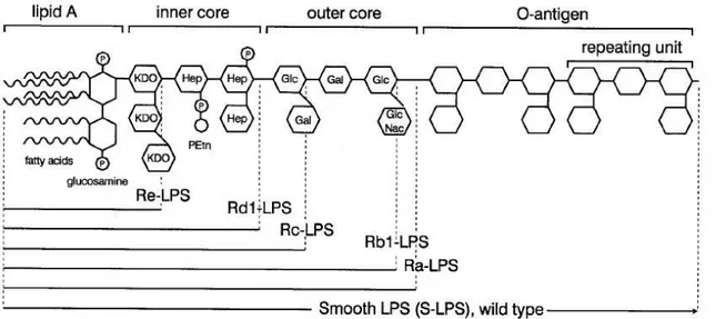
A molécula de LPS consiste de 4 partes diferentes (figura 1) (9, 13, 14). A primeira e mais importante é o lipídio A, que é a parte lipídica com ligação covalente do LPS. Seis ou mais resíduos de ácidos graxos são ligados a duas glicosaminas fosforiladas. Quatro destes ácidos graxos carregam uma hidroxila no terceiro carbono, enquanto os outros dois não são hidroxilados. Todas as espécies de bactérias carregam um LPS único e, algumas dessas variações são devidas à estrutura do lipídeo A: diferente acetilação, que é comumente assimétrica (4 + 2), ou configuração simétrica (3 + 3) como na ; comprimento da cadeia de resíduos de ácidos graxos ( tipicamente são 3 a 4 diferentes ácidos graxos, compostos de 10 a 16 átomos de carbono); a presença de 4 amino deoxi arabinose ou/e fosfoetanolamina ligadas aos grupos fosfo nas glicosaminas; e, finalmente, o número de ácidos graxos (a mais comum bactéria contém 6 resíduos de ácidos graxos). Experimentos com o lipídeo A sintético demonstraram que ele representa a porção tóxica da molécula de LPS (15).
A segunda parte da molécula de LPS é núcleo interno, que consiste de dois ou mais açucares 2 ceto 3 deoxioctonico (KDO) ligados a glicosamina do lipídeo A e duas ou três heptoses ligadas ao KDO. Os açucares são únicos para cada bactéria. A menor molécula de LPS produzida por bactérias gram negativas sob condições naturais é o Re LPS ( lipídeo A com 1 ou 2 KDO), ainda que moléculas longas sejam mais comuns. Os sorotipos Rd1 e Rd2 LPS contêm um núcleo interno completo e outro sem duas heptoses, respectivamente.
núcleo interno completo e um núcleo externo são chamados Ra LPS, enquanto os sorotipos Rb e Rc LPS contêm somente partes do núcleo externo.
A quarta parte da molécula de LPS é o antígeno “O”. Esta parte da molécula de LPS está ligada ao açúcar do núcleo externo e estende se da superfície da bactéria, sendo altamente imunogênico. É composto de unidades de açucares comuns com uma grande variação no tamanho e composição entre espécies e cepas. Em uma única preparação de LPS o tamanho do antígeno “O” pode variar de zero até 40 repetições. Cada unidade é composta por três açucares. As moléculas de LPS com o antígeno “O” são denominadas S LPS. Colônias de bactérias contendo o antígeno “O” têm aparência lisa na placa de cultura, enquanto a ausência deste confere a aparência rugosa.
1.3. RESPOSTA DO HOSPEDEIRO AO LPS
No nível celular, as células mononucleares (monócitos e macrófagos) e os neutrófilos são de grande significância, uma vez que estas células devem reconhecer, direta ou indiretamente, os constituintes da parede celular das bactérias, após a ação do complemento e de anticorpos. Em condições fisiológicas as células do sistema imune são continuamente expostas a baixas doses de LPS, derivadas das bactérias gastrointestinais, que entram no corpo via sistema porta. Este estímulo de LPS ocorre preferencialmente sobre macrófagos e será essencial para a manutenção no nível basal de ativação do sistema imune. Baixos níveis de LPS estão presentes em indivíduos saudáveis sem causar doença (16, 17).
características da expressão de genes das bactérias gram negativas de forma quase completa (18).
Os fagócitos (monócitos, macrófagos e polimorfonucleares são capazes de reconhecer componentes bacterianos opsonizados através de receptores de complemento e receptores Fc). Na resposta do hospedeiro às bactérias os monócitos e macrófagos são de maior importância. O reconhecimento de LPS ou outros componentes bacterianos por estas células inicia uma cascata de liberação de mediadores inflamatórios, alterações vasculares e fisiológicas, assim como o recrutamento de células imunes. Um macrófago é ativado por LPS e torna se metabolicamente ativo, produzindo estoques intracelulares de radicais livres de oxigênio e outros agentes bactericidas (lisozimas, proteínas catiônicas, hidrolases e lactoferrinas), secretando concomitantemente mediadores inflamatórios (19). Um mediador chave é o TNF α (20). Após a exposição ao LPS o TNF α é a primeira citocina a ser liberada pelos macrófagos. O TNF α mRNA é continuamente transcrito na células de kupffer, permitindo uma rápida resposta de TNF após o estímulo inflamatório (21). IL 1 e IL 6 não são constitutivamente expressas, entretanto mRNA destas citocinas juntamente com o mRNA do TNF α atingem o pico em macrófagos de fígados de comundongos 40 minutos após o estímulo (21).
complemento e receptores Fc, o que possibilita o reconhecimento e a fagocitose de LPS, fragmentos bacterianos e bactérias. Contudo o aumento da aderência de polimorfonucleares ao endotélio e a presença de altas concentrações de estimulantes pode também resultar na liberação de agentes microbicidas, que serão responsáveis por grande parte do dano endotelial (4). As células endoteliais respondem ao LPS (via CD14 solúvel) e às citocinas circulantes através da produção de IL 1, IL 6, eicosanóides, agentes vasoativos derivados do endotélio, endotelina 1, quimiocinas e fatores estimulantes de colônias (25).
A ação das células ativadas do sistema imune combinada com os efeitos dos mediadores inflamatórios produz sintomas como febre, dano endotelial, aumento da permeabilidade capilar, vasodilatação periférica, alterações na coagulação, microtrombos e depressão miocárdica. Estes fenômenos resultam em disfunção múltipla de órgãos, choque e morte (4).
1.4. RECEPTORES DE LIPOPOLISSACARÍDEOS
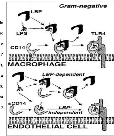
Figura 2. Esquema de ligantes da molécula de LPS na superfície celular em macrófagos e células endoteliais.
Proteína de Ligação de LPS A Proteína de ligação de LPS (LBP) é uma proteína de fase aguda (27) e é induzida por IL 6 e IL 1 (28). Os níveis basais de LBP no soro são baixos (1 a 15dg/ml), porém aumentam durante a infecção (29). Em humanos, durante a fase aguda do trauma ou sepse, os níveis de LBP atingem o máximo em 2 ou 3 dias.
O LBP liga se ao LPS, lipídio A e lipídio IVA. O C terminal, parte da molécula de
LBP, media a transferência de LPS para o CD14 (30). A ligação de LBP às bactérias mortas é mais intensa que a ligação com bactérias vivas (31).
O Receptor CD14
O receptor CD14 é expresso por células da linhagem mielóide (monócitos, macrófagos e polimorfonucleares), células B, células do parênquima hepático, fibroblastos e células de microgliais (35).
O mecanismo de ligação do LPS ao receptor CD14 é demonstrado na figura 2. Camundongos transgênicos para o receptor CD14 são três vezes mais sensitivos ao LPS. Ao passo que camundongos deficientes para CD14 são altamente resistentes ao estímulo com LPS (36). O receptor CD14 é um glicosil fosfatidil inusitol que não possui um domínio transmembrana, o que faz com que necessite de um receptor complementar para a transdução do sinal (37). O receptor complementar nesse caso é um TLR.
O Receptor Solúvel CD14 (sCD14)
1.5. OS RECEPTORES TLR 4 E RP 105
O reconhecimento é um elemento essencial para o início da resposta imune inata, como ocorre através da inflamação, sendo mediada por receptores específicos de reconhecimento capazes de identificar estruturas moleculares, as quais são amplamente compartilhadas por patógenos (PAMPs – Pathogen Associated Molecular Patterns) (41). Os receptores toll like foram os primeiros a serem identificados e caracterizados, assim como foram relacionados a uma ampla variedade de moléculas associadas a patógenos (42 45). Os receptores toll like são proteínas transmembrana tipo I e apresentam um ectodomínio com repetições ricas em leucina, que media o reconhecimento de PAMPs. Possuem uma região transmembrana e outra citosólica, com o domínio do receptor de interleucina 1 (TIR), que ativa a via intracelular. São reconhecidos 10 a 12 TLRs funcionais em humanos e camundongos, respectivamente (42).
Recentes estudos revelaram que características moleculares comuns de microorganismos, como LPS, são reconhecidas pelos receptores toll like (26, 41, 46). Dois conhecidos TLRs que mediam a sinalização do LPS são os receptores TLR4 e RP105 (CD 180). O receptor RP105 não está limitado às células B, mas também é expresso por monócitos e células dendríticas mielóides, as quais também apresentam consideravelmente mais expressão de receptores TLR4 que as células B (47).
sincicial respiratório. A seguir descreveremos os receptores toll like (TLR4 e RP 105) para que possamos entender melhor a interação entre LPS e RSV, bem como a possível modulação de LPS via receptores toll like.
1.6. TOLL LIKE 4/MD 2 E RP 105/MD 1
Os camundongos C3H/HeJ e C57BL10/ScCr são reconhecidos por sua hiporresponsividade ao LPS. Dois grupos independentes de pesquisadores identificaram uma mutação no gene Tlr4 (26) como o responsável. A estimulação do LPS é seguida por um aumento da proximidade física entre CD14 e TLR4, sugerindo a interação entre CD14 e TLR4 para a sinalização (49). A molécula MD 2 foi identificada como estando associada com a porção extra celular de TLR4 e melhora a responsividade ao LPS (50). Enquanto o receptor TLR4 é encontrado na superfície celular, o mesmo é encontrado no complexo de Golgi em células deficientes para MD 2, indicando que MD 2 é essencial para a distribuição intracelular de TLR4 (51).
dependem da co expressão e secreção extracelular da proteína MD 2, a expressão de RP 105 é dependente da proteína homóloga MD 1 (50).
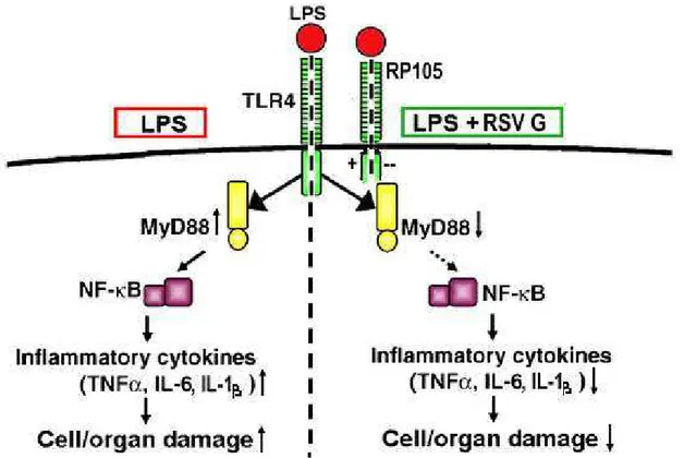
Figura 3. Ilustração da provável interação entre os receptores TLR4 e RP 105. O receptor RP 105 seria um controlador fisiológico de TLR4, assim células RP105/ seriam mais responsivas ao estímulo de ligantes (ex. LPS). A via da esquerda representa a ação de LPS, que ativa o fator MyD88 com conseqüente aumento de citocinas inflamatórias. À direita, o estímulo com LPS + proteína G do vírus sincicial respiratório interage com os receptores TLR4 e RP 105, diminuindo a ativação de MyD88, prevenindo o dano celular através da inibição de citocinas LPS dependentes.
1.7. O VÍRUS SINCICIAL RESPIRATÓRIO
infecção e mortalidade seja de 64 milhões e 160.000, respectivamente. Apesar do grande impacto da infecção por RSV, não há vacina disponível para a doença (55). Entretanto, os esforços para o desenvolvimento de uma vacina eficaz levaram à melhor caracterização das bases moleculares da doença.
O vírus sincicial respiratório humano, assim como o parainfluenza 1 4 e o metapneumovírus humano são exemplos de paramixovírus, os quais são vírus de RNA envelopados que apresentam, entre outras características, duas glicoproteínas: uma proteína de fusão (F) e uma proteína de ligação: hemaglutinina neuroaminidase (HN), hemoaglutinina (H) ou proteína G.
1.8. A PROTEÍNA G DO VÍRUS SINCICIAL RESPIRATÓRIO
Através da identificação destas glicoproteínas constituintes do vírus observou se que o RSV também pode ativar os receptores TLR4, através da interação com a glicoproteína viral de fusão (F) (48, 61). A proteína RSV F interage com o complexo CD14+/TLR4 nos monócitos e estimula a produção de citocinas pró inflamatórias, como IL 6, IL 1β e IL 8 (48), promovendo a translocação nuclear de NF κB (62). Comparativamente, a glicoproteína viral de ligação (RSV G) não é um forte agonista do complexo CD14+/TLR4 e, usualmente, desencadeia uma resposta Th2 às infecções virais por RSV (63, 64). Entretanto, experimentos com camundongos BALB/c e um RSV mutante, que não expressava a proteína G ou pequenos genes hidrofóbicos (SH), foi capaz de aumentar o número de linfócitos NK e neutrófilos circulantes nos pulmões, quando comparado ao controle infectado com vírus sincicial que expressava proteína G e SH (65). Esses achados, que sugerem um papel imunomodulador da proteína RSV G na resposta inflamatória, são reforçados pela apresentação de um efeito inibidor de células dentríticas humanas derivadas de monócitos pela glicoproteína G do metapneumovírus humano, através da via TLR4 dependente (66). Além disso, a glicoproteína RSV G antagoniza os efeitos pró inflamatórios da glicoproteína RSV F, assim como tem efeitos similares nos agonistas TLR4, como o LPS, regulando a resposta imune inata (67).
antigênicos A e B de RSV(68). Estudos prévios demonstram que essa região central altamente conservada da proteína RSV G é fundamental para que haja a modulação da resposta inflamatória desencadeada por agonistas TLR (67, 69).
2.JUSTIFICATIVA
Sepse é uma desordem inflamatória sistêmica e sua progressão para o choque séptico caracteriza um sério problema clínico, apresentando altas taxas de mortalidade. Apesar dos significativos avanços no tratamento intensivo, os mesmos não são capazes de reverter a resposta inflamatória sistêmica, assim como suas conseqüências. A sepse por bactérias gram negativas é desencadeada pela exposição a um componente da membrana destas bactérias, conhecido como lipopolissacarídeo (LPS), o que leva a uma super produção de citocinas inflamatórias no hospedeiro, incluindo o fator de necrose tumoral (TNF α), a interleucina 1 (IL 1) e a interleucina 6 (IL 6), por sua vez provindas de células do sistema imune, como monócitos. Estudos recentes demonstraram que, de forma similar ao LPS, o vírus sincicial respiratório (RSV) – principal causa de infecção respiratória baixa em crianças e recém natos
utiliza o receptor toll like 4 (TLR4) para sinalização celular.
3. OBJETIVOS
• Avaliar o efeito da proteína RSV G na inibição de citocinas LPS dependentes em monócitos, em um modelo “ ”;
• Demonstrar uma relação dose efeito da proteína RSV G, na inibição de citocinas inflamatórias em modelos murinos;
• Avaliar a inibição da resposta imune mediada pela proteína RSV G em um modelo murino de choque séptico induzido por LPS;
• Avaliar a produção/inibição de citocinas LPS dependentes em diferentes modelos murinos para expressão dos receptores TLR4 e RP105;
• Avaliar a suficiência do receptor Toll like 4 na inibição de citocinas LPS dependentes através da proteína RSV G.
4. REFERÊNCIAS
1. Glauser MP, Zanetti G, Baumgartner JD, Cohen J. Septic shock: pathogenesis. Lancet. 1991 Sep 21;338(8769):732 6.
2. Opal SM, Cohen J. Clinical gram positive sepsis: does it fundamentally differ from gram negative bacterial sepsis? Crit Care Med. 1999 Aug;27(8):1608 16.
3. Parrillo JE. Pathogenetic mechanisms of septic shock. N Engl J Med. 1993 May 20;328(20):1471 7.
4. Bone RC. The pathogenesis of sepsis. Ann Intern Med. 1991 Sep 15;115(6):457 69. 5. Bone RC. Why new definitions of sepsis and organ failure are needed. Am J Med. 1993 Oct;95(4):348 50.
6. Sriskandan S, Cohen J. Gram positive sepsis. Mechanisms and differences from gram negative sepsis. Infect Dis Clin North Am. 1999 Jun;13(2):397 412.
7. Bone RC. Gram positive organisms and sepsis. Arch Intern Med. 1994 Jan 10;154(1):26 34.
8. Ulevitch RJ, Tobias PS. Recognition of gram negative bacteria and endotoxin by the innate immune system. Curr Opin Immunol. 1999 Feb;11(1):19 22.
9. Rietschel ET, Kirikae T, Schade FU, Mamat U, Schmidt G, Loppnow H, et al. Bacterial endotoxin: molecular relationships of structure to activity and function. Faseb J. 1994 Feb;8(2):217 25.
12. Crosby HA, Bion JF, Penn CW, Elliott TS. Antibiotic induced release of endotoxin from bacteria in vitro. J Med Microbiol. 1994 Jan;40(1):23 30.
13. Raetz CR. Biochemistry of endotoxins. Annu Rev Biochem. 1990;59:129 70.
14. Lugtenberg B, Van Alphen L. Molecular architecture and functioning of the outer membrane of Escherichia coli and other gram negative bacteria. Biochim Biophys Acta. 1983 Mar 21;737(1):51 115.
15. Kotani S, Takada H, Tsujimoto M, Ogawa T, Takahashi I, Ikeda T, et al. Synthetic lipid A with endotoxic and related biological activities comparable to those of a natural lipid A from an Escherichia coli re mutant. Infect Immun. 1985 Jul;49(1):225 37.
16. Fox ES, Thomas P, Broitman SA. Comparative studies of endotoxin uptake by isolated rat Kupffer and peritoneal cells. Infect Immun. 1987 Dec;55(12):2962 6.
17. Takakuwa T, Endo S, Nakae H, Kikuchi M, Baba N, Inada K, et al. Blood cytokine and complement levels in patients with sepsis. Res Commun Chem Pathol Pharmacol. 1994 Jun;84(3):291 300.
18. Rosenberger CM, Scott MG, Gold MR, Hancock RE, Finlay BB. Salmonella typhimurium infection and lipopolysaccharide stimulation induce similar changes in macrophage gene expression. J Immunol. 2000 Jun 1;164(11):5894 904.
19. Mattsson E, Van Dijk H, Verhoef J, Norrby R, Rollof J. Supernatants from Staphylococcus epidermidis grown in the presence of different antibiotics induce differential release of tumor necrosis factor alpha from human monocytes. Infect Immun. 1996 Oct;64(10):4351 5.
21. Grube BJ, Cochane CG, Ye RD, Green CE, McPhail ME, Ulevitch RJ, et al. Lipopolysaccharide binding protein expression in primary human hepatocytes and HepG2 hepatoma cells. J Biol Chem. 1994 Mar 18;269(11):8477 82.
22. Hack CE, Aarden LA, Thijs LG. Role of cytokines in sepsis. Adv Immunol. 1997;66:101 95.
23. Qureshi N, Honovich JP, Hara H, Cotter RJ, Takayama K. Location of fatty acids in lipid A obtained from lipopolysaccharide of Rhodopseudomonas sphaeroides ATCC 17023. J Biol Chem. 1988 Apr 25;263(12):5502 4.
24. Mulligan MS, Smith CW, Anderson DC, Todd RF, 3rd, Miyasaka M, Tamatani T, et al. Role of leukocyte adhesion molecules in complement induced lung injury. J Immunol. 1993 Mar 15;150(6):2401 6.
25. Mahalingam S, Karupiah G. Chemokines and chemokine receptors in infectious diseases. Immunol Cell Biol. 1999 Dec;77(6):469 75.
26. Poltorak A, He X, Smirnova I, Liu MY, Van Huffel C, Du X, et al. Defective LPS signaling in C3H/HeJ and C57BL/10ScCr mice: mutations in Tlr4 gene. Science. 1998 Dec 11;282(5396):2085 8.
27. Schumann RR, Kirschning CJ, Unbehaun A, Aberle HP, Knope HP, Lamping N, et al. The lipopolysaccharide binding protein is a secretory class 1 acute phase protein whose gene is transcriptionally activated by APRF/STAT/3 and other cytokine inducible nuclear proteins. Mol Cell Biol. 1996 Jul;16(7):3490 503.
29. Schumann RR, Lamping N, Kirschning C, Knopf HP, Hoess A, Herrmann F. Lipopolysaccharide binding protein: its role and therapeutical potential in inflammation and sepsis. Biochem Soc Trans. 1994 Feb;22(1):80 2.
30. Han J, Mathison JC, Ulevitch RJ, Tobias PS. Lipopolysaccharide (LPS) binding protein, truncated at Ile 197, binds LPS but does not transfer LPS to CD14. J Biol Chem. 1994 Mar 18;269(11):8172 5.
31. Le Roy D, Di Padova F, Tees R, Lengacher S, Landmann R, Glauser MP, et al. Monoclonal antibodies to murine lipopolysaccharide (LPS) binding protein (LBP) protect mice from lethal endotoxemia by blocking either the binding of LPS to LBP or the presentation of LPS/LBP complexes to CD14. J Immunol. 1999 Jun 15;162(12):7454 60. 32. Schumann RR, Leong SR, Flaggs GW, Gray PW, Wright SD, Mathison JC, et al. Structure and function of lipopolysaccharide binding protein. Science. 1990 Sep 21;249(4975):1429 31.
33. Jack RS, Fan X, Bernheiden M, Rune G, Ehlers M, Weber A, et al. Lipopolysaccharide binding protein is required to combat a murine gram negative bacterial infection. Nature. 1997 Oct 16;389(6652):742 5.
34. Wurfel MM, Monks BG, Ingalls RR, Dedrick RL, Delude R, Zhou D, et al. Targeted deletion of the lipopolysaccharide (LPS) binding protein gene leads to profound suppression of LPS responses ex vivo, whereas in vivo responses remain intact. J Exp Med. 1997 Dec 15;186(12):2051 6.
37. Stefanova I, Horejsi V, Ansotegui IJ, Knapp W, Stockinger H. GPI anchored cell surface molecules complexed to protein tyrosine kinases. Science. 1991 Nov 15;254(5034):1016 9.
38. Liu S, Khemlani LS, Shapiro RA, Johnson ML, Liu K, Geller DA, et al. Expression of CD14 by hepatocytes: upregulation by cytokines during endotoxemia. Infect Immun. 1998 Nov;66(11):5089 98.
39. Landmann R, Zimmerli W, Sansano S, Link S, Hahn A, Glauser MP, et al. Increased circulating soluble CD14 is associated with high mortality in gram negative septic shock. J Infect Dis. 1995 Mar;171(3):639 44.
40. Haziot A, Rong GW, Bazil V, Silver J, Goyert SM. Recombinant soluble CD14 inhibits LPS induced tumor necrosis factor alpha production by cells in whole blood. J Immunol. 1994 Jun 15;152(12):5868 76.
41. Janeway CA, Jr. Approaching the asymptote? Evolution and revolution in immunology. Cold Spring Harb Symp Quant Biol. 1989;54 Pt 1:1 13.
42. Akira S, Uematsu S, Takeuchi O. Pathogen recognition and innate immunity. Cell. 2006 Feb 24;124(4):783 801.
43. Beutler BA. TLRs and innate immunity. Blood. 2009 Feb 12;113(7):1399 407.
44. Hoffmann JA. The immune response of Drosophila. Nature. 2003 Nov 6;426(6962):33 8.
45. Medzhitov R. Recognition of microorganisms and activation of the immune response. Nature. 2007 Oct 18;449(7164):819 26.
47. Divanovic S, Trompette A, Atabani SF, Madan R, Golenbock DT, Visintin A, et al. Negative regulation of Toll like receptor 4 signaling by the Toll like receptor homolog RP105. Nat Immunol. 2005 Jun;6(6):571 8.
48. Kurt Jones EA, Popova L, Kwinn L, Haynes LM, Jones LP, Tripp RA, et al. Pattern recognition receptors TLR4 and CD14 mediate response to respiratory syncytial virus. Nat Immunol. 2000 Nov;1(5):398 401.
49. da Silva Correia J, Soldau K, Christen U, Tobias PS, Ulevitch RJ. Lipopolysaccharide is in close proximity to each of the proteins in its membrane receptor complex. transfer from CD14 to TLR4 and MD 2. J Biol Chem. 2001 Jun 15;276(24):21129 35.
50. Shimazu R, Akashi S, Ogata H, Nagai Y, Fukudome K, Miyake K, et al. MD 2, a molecule that confers lipopolysaccharide responsiveness on Toll like receptor 4. J Exp Med. 1999 Jun 7;189(11):1777 82.
51. Nagai Y, Akashi S, Nagafuku M, Ogata M, Iwakura Y, Akira S, et al. Essential role of MD 2 in LPS responsiveness and TLR4 distribution. Nat Immunol. 2002 Jul;3(7):667 72. 52. Takeda K, Kaisho T, Akira S. Toll like receptors. Annu Rev Immunol. 2003;21:335 76.
53. Miyake K, Yamashita Y, Hitoshi Y, Takatsu K, Kimoto M. Murine B cell proliferation and protection from apoptosis with an antibody against a 105 kD molecule: unresponsiveness of X linked immunodeficient B cells. J Exp Med. 1994 Oct 1;180(4):1217 24.
54. Ogata H, Su I, Miyake K, Nagai Y, Akashi S, Mecklenbrauker I, et al. The toll like receptor protein RP105 regulates lipopolysaccharide signaling in B cells. J Exp Med. 2000 Jul 3;192(1):23 9.
56. Roberts SR, Lichtenstein D, Ball LA, Wertz GW. The membrane associated and secreted forms of the respiratory syncytial virus attachment glycoprotein G are synthesized from alternative initiation codons. J Virol. 1994 Jul;68(7):4538 46.
57. Levine S, Klaiber Franco R, Paradiso PR. Demonstration that glycoprotein G is the attachment protein of respiratory syncytial virus. J Gen Virol. 1987 Sep;68 ( Pt 9):2521 4. 58. Karron RA, Buonagurio DA, Georgiu AF, Whitehead SS, Adamus JE, Clements Mann ML, et al. Respiratory syncytial virus (RSV) SH and G proteins are not essential for viral replication in vitro: clinical evaluation and molecular characterization of a cold passaged, attenuated RSV subgroup B mutant. Proc Natl Acad Sci U S A. 1997 Dec 9;94(25):13961 6.
59. Johnson PR, Jr., Olmsted RA, Prince GA, Murphy BR, Alling DW, Walsh EE, et al. Antigenic relatedness between glycoproteins of human respiratory syncytial virus subgroups A and B: evaluation of the contributions of F and G glycoproteins to immunity. J Virol. 1987 Oct;61(10):3163 6.
60. Sullender WM, Anderson K, Wertz GW. The respiratory syncytial virus subgroup B attachment glycoprotein: analysis of sequence, expression from a recombinant vector, and evaluation as an immunogen against homologous and heterologous subgroup virus challenge. Virology. 1990 Sep;178(1):195 203.
61. Haynes LM, Moore DD, Kurt Jones EA, Finberg RW, Anderson LJ, Tripp RA. Involvement of toll like receptor 4 in innate immunity to respiratory syncytial virus. J Virol. 2001 Nov;75(22):10730 7.
63. Jackson M, Scott R. Different patterns of cytokine induction in cultures of respiratory syncytial (RS) virus specific human TH cell lines following stimulation with RS virus and RS virus proteins. J Med Virol. 1996 Jul;49(3):161 9.
64. Hancock GE, Scheuer CA, Sierzega R, Pryharski KS, McBride JT, Watelet LF, et al. Adaptive immune responses of patients with asthma to the attachment (G) glycoprotein of respiratory synctial virus. J Infect Dis. 2001 Dec 15;184(12):1589 93.
65. Tripp RA, Jones L, Anderson LJ. Respiratory syncytial virus G and/or SH glycoproteins modify CC and CXC chemokine mRNA expression in the BALB/c mouse. J Virol. 2000 Jul;74(13):6227 9.
66. Kolli D, Bao X, Liu T, Hong C, Wang T, Garofalo RP, et al. Human metapneumovirus glycoprotein G inhibits TLR4 dependent signaling in monocyte derived dendritic cells. J Immunol. Jul 1;187(1):47 54.
67. Polack FP, Irusta PM, Hoffman SJ, Schiatti MP, Melendi GA, Delgado MF, et al. The cysteine rich region of respiratory syncytial virus attachment protein inhibits innate immunity elicited by the virus and endotoxin. Proc Natl Acad Sci U S A. 2005 Jun 21;102(25):8996 9001.
68. Melero JA, Garcia Barreno B, Martinez I, Pringle CR, Cane PA. Antigenic structure, evolution and immunobiology of human respiratory syncytial virus attachment (G) protein. J Gen Virol. 1997 Oct;78 ( Pt 10):2411 8.
5. MATERIAIS E MÉTODOS
5.1. ANIMAIS E CONSIDERAÇÕES ÉTICAS
Todos experimentos foram realizados de acordo com o “guide for the Care and Use of Laboratory Animals” e aprovados pela intituição Vanderbilt University (TN, USA). Fêmeas entre 4 6 semanas de vida de camundongos C57BL/6J, C57BL/10ScCr (TLR4/) e RP105 knockout (RP105/) (Jackson Laboratories) foram utilizados para estes experimentos. Os camundongos foram mantidos em caixas micro isoladas, sob fluxo laminar, em um ambiente controlado e livre de patógenos no biotério, recebendo alimento e água .
5.2. ISOLAMENTO DE MONÓCITOS
5.3. CLONAGEM E PRODUÇÃO DE RSV G
Parte do DNA que codificava os aminoácidos 67 89 RSV A2 e uma 6x HIS tag foram sintetizados pela Genscript® e fusionados ao DNA. Todas as seqüências de DNA foram otimizadas para expressão em mamíferos antes da síntese. Foram usadas células 293 6E e, 4 5 dias após a transfecção, a porção secretória da proteína RSV G foi purificada em um His Trap HP columm (GE Biosciences). A análise da proteína purificada revelou uma larga banda variando de 49 a 98kD, o que é consistente com uma variável glicosilação da RSV G (experimento não mostrado). A conformação antigênica da proteína recombinante foi verificada através da ligação aos anticorpos monoclonais de camundongo específicos de RSV G, em ELISA.
5.4. ESTIMULAÇÃO DE MONÓCITOS NO MODELO MURINO
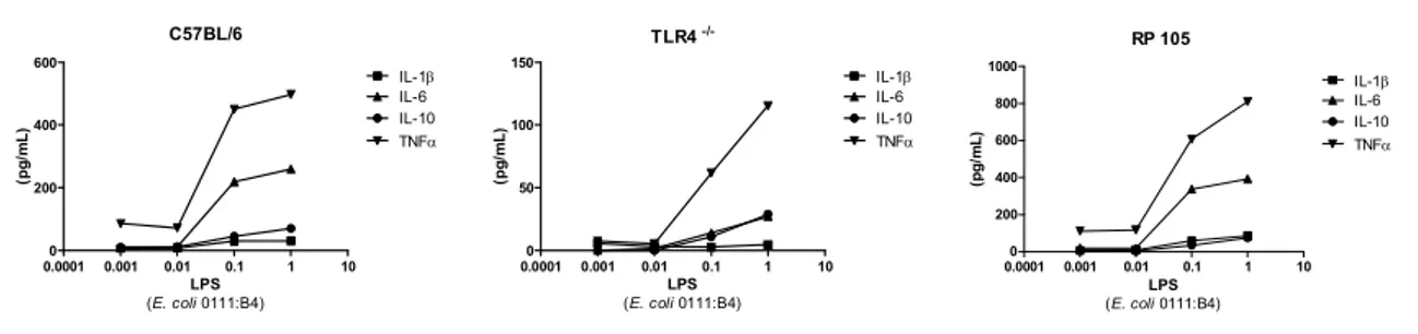
A concentração do lipopolissacarídeo a ser usada no modelo murino foi estabelecida através da curva de LPS, onde se estabeleceu uma escala logarítmica de diluições do LPS (
0111:B4) em cultura de monócitos de baço de camundongos C57BL/6J, TLR4/ e RP105 (Figura 1).
β
α
β
α
β
α
Monócitos purificados foram cultivados em 0,2 ml de meio de cultura (RPMI 1640) em uma placa de cultura de tecidos de 96 poços (flat bottom) (105 células/poço). Lipopolissacarídeos (Sigma L2630 LPS; 0111: B4) na dose de (1dg) ou proteína purificada RSV G (5 dg) foram utilizados para a estimulação. O efeito de diferentes doses de RSV G foi verificado através da estimulação das células com 2,5;5;10 e 20 dg de RSV G/poço, em um experimento independente. Os sobrenadantes foram usados 18h depois da estimulação para análise de citocinas, de acordo com as especificações do fabricante (CBA cytometric bead array from BD Biosciences).
5.5. EXPERIMENTO PARA AVALIAR A SUFICIÊNCIA DO RECEPTOR TOLL LIKE 4 INCUBADOS COM RSV G
5.6. ESTUDO DE SOBREVIVÊNCIA NO MODELO DE SEPSE POR LPS
Os camundongos C57BL/6j foram divididos em 2 grupos: ao grupo LPS foi administrado intraperitonealmente (i.p.) LPS (35 mg/kg de peso); ao grupo LPS + RSV G foram administrados i.p. com uma única dose LPS (35 mg/kg de peso) e duas doses de 300 dg de RSV G purificada, em um intervalo de 2h, iniciando se com a administração de LPS. Os grupos foram monitorados, pesados e clinicamente avaliados a cada 2 horas nas primeiras 24h e, a cada 4h, para as 24h seguintes. Após, avaliados uma vez ao dia até o fim do estudo, no dia 7. A correlação com uma tabela de sinais clínicos com valores pré definidos foi realizada apenas para as primeiras 24h de estudo. (0− Sem doença aparente, saudável e ativo; 1− Pêlo levemente ouriçado; 2− Pêlo ouriçado, mas ativo; 3− Pêlo ouriçado e inativo; 4− Pêlo ouriçado, inativo, curvado, emagrecido; 5− morto ou sacrificado). A análise dos valores foi expressa de forma descritiva.
5.7. ANÁLISE ESTATÍSTICA
ARTIGO ORIGINAL
The Respiratory syncytial virus attachment
protein inhibits inflammatory cytokines in
the immune response against
lipopolysaccharide induced sepsis
Authors
: Brum CO*†, Hijano DR†**, Bates JT†, Melendi
GA**, Pitrez PM*, Crowe JE Jr†, Polack FP†**
*
Instituto de Pesquisas Biomédicas, Pontifícia Universidade Católica do Rio Grande do Sul, Brazil.
†
Vanderbilt Vaccine Center, Medical Center North, Vanderbilt University, Nashville, Tennessee.
**
Abstract
Sepsis is a systemic inflammatory disorder, and its progression to septic shock is a serious clinical problem associated with a high mortality rate. Despite significant advances in critical care, treatments do not reverse the systemic inflammatory response and its consequences. Gram negative sepsis is initiated by exposure to a component of gram negative bacterial membrane, lipopolysaccharide (LPS), and induces overproduction of host inflammatory cytokines, including tumor necrosis factor (TNF α), interleukin 1 (IL 1) and interleukin 6 (IL 6) from immunocytes such as monocytes. Recent studies have shown that similar to LPS, the Respiratory syncytial virus leading cause of severe lower respiratory tract infections in infants and young children – requires the toll like receptor 4 (TLR4) for signaling. Studies have shown that the respiratory syncytial virus attachment glycoprotein (RSV G) modulates cytokine and chemokine production in monocytes, inhibiting the inflammatory response elicited by LPS stimulation. In this report, we use murine monocytes from different knockout mice to show that RSV G can inhibit the release of cytokines in the immune response against lipoplysaccharide induced sepsis. We demonstrate the modulation of IL 1β, IL 6, IL 10 and TNF α by RSV G in monocytes LPS stimulated. Importantly, we also considered the effect of RSV G subunits (peptides) and future directions for the clinical use of the glycoprotein on LPS mediated inflammation.
Introduction
Sepsis is a systemic inflammatory disorder, and its progression to septic shock is a serious clinical problem associated with a high mortality rate. The mortality rate of patients with septic shock is still 40–60% (1).Despite significant advances in critical care, treatments do not reverse the systemic inflammatory response and its consequences. Gram negative sepsis is initiated by exposure to a component of gram negative bacterial membrane, lipopolysaccharide (LPS), and induces overproduction of host inflammatory cytokines, including tumor necrosis factor (TNF α), interleukin 1 (IL 1) and interleukin 6 (IL 6) from immunocytes such as monocytes and macrophages – cells that are immunologically activated by endotoxin (2).
LPS is possibly the most potent microbial stimulator of the immune system and it has therefore been extensively used to study the mechanisms underlying host responses to infection both and (3).
Recent studies have revealed that common molecular patterns of microorganisms such as LPS are recognized by toll like receptors (TLRs). Two known TLRs that mediate LPS signaling are TLR4 and RP105 (CD180). The RP105 expression is not limited to B cells and was also expressed by monocytes as well as myeloid dendritic cells, which are cells that express considerably more TLR4 than do B cells (4). Furthermore, TLR4 signaling has been shown to play an important role in controlling paramyxovirus infection.
(HRSV), human parainfluenza virus types 1–4 and metapneumovirus (hMPV)(5). The paramyxoviruses encode two glycoproteins: a fusion (F) protein, and an attachment [HN (hemagglutinin neuraminidase), H (hemagglutinin), or G (glyco )] protein (6).
Respiratory syncytial virus is the leading cause of severe lower respiratory tract infections in infants and young children. The WHO estimated global annual infection and mortality figures for human RSV are 64 million and 160 000, respectively. Despite the importance of this disease, there currently is no licensed vaccine for RSV (7). However, these efforts have contributed to identify it molecular patterns which have offered alternative tools to modulate the innate immune response.
Respiratory syncytial virus (RSV) also can activate TLR4 through interaction with the viral fusion (F) glycoprotein (8, 9). The RSV F protein interacts with the CD14+/TLR4 complex in monocytes and stimulates production of proinflammatory cytokines, such as IL 6, IL 1β, and IL 8 (9), by promoting nuclear translocation of NF κB (10). Conversely, the RSV attachment glycoprotein (RSV G) is not a strong agonist of the CD14+/TLR4 complex, and triggers a Th2 response upon RSV infection (11, 12). However, the human metapneumovirus glycoprotein G inhibits human monocyte derived dendritic cell responses by affecting TLR4 dependent signaling(13). Furthermore the RSV G antagonizes the proinflammatory effect of RSV F as well as having similar effect on the unrelated TLR4 agonist LPS regulating the innate immune response (14).
bears two variable mucin like segments and a conserved central region rich in cysteine residues (G cysteine rich region or GCRR) (15). Within the GCRR, there are four cysteines at positions 173, 176, 182, and 186 that are highly conserved between RSV antigenic subgroups A and B (16). Recent studies have shown that the highly conserved region within RSV G is critical in the TLR(s) agonist immune response control (14, 17).
Our previous results highlighted the inhibition of RSV G on the RSV F glycoprotein. However, considering that similar to LPS, the primary ligand of TLR4, RSV F requires de presence of CD14/MD 2/TLR4 complex for signaling, and despite RSV G protein is not a strong agonist of the proinflammatory cytokines, these findings taken together led us to hypothesize that the RSV G protein inhibits not only RSV F pro inflammatory cytokines stimulated, but RSV G also can inhibit the release of cytokines in the immune response against lipoplysaccharide induced sepsis.
Methods Animals.
Splenocyte/Mouse Monocyte Isolation
The spleen was removed from the abdomen, ground, and passed through a 70 micron nylon cell strainer with 5 ml of RPMI 1640 medium (Life Technologies) supplemented with 10% fetal calf serum, 100U of penicillin/ml, and 100dg of streptomycin sulfate/ml. Cells were spun down and washed at 300g at 4°C for 10 minutes. Monocytes were isolated by using the CD 11b Microbeads (Miltenyi Biotec, CA, USA) with magnetic cell sorting LS separation columns. The procedure was performed following the manufacturer’s instructions.
Cloning and Production of RSV G
DNA encoding amino acids 67 98 RSV A2 and a 6x HIS tag were synthesized by Genscript and fused to DNA. All DNA sequences were optimized for mammalian expression before synthesis. We used 293 6E cells, and 4 5 days after transfection the secreted RSV G protein was purified on a His Trap HP column (GE Biosciences). Analysis of the purified protein revealed a large band ranging from ~49 to 98kD, which is consistent with variable glycosylation of RSV G (data not shown). The antigenic conformation of the recombinant protein was verified by binding to mouse RSV G specific monoclonal antibodies in ELISA.
Mouse Monocyte Stimulation
Toll Like Receptor 4 experiment
In order to assess the TLR4 effect we used HEK 293 cells. The cells (293mTLR4 MD2 CD14) (Invivogen, San Diego, CA) are designed for studying the stimulation of murine TLR4 (mTLR4). The 293mTLR4 cells were cultured in 0.2 ml of culture medium (RPMI 1640) of 96 well flat bottom tissue culture plates (105 cells/well). Lipopolysaccharide (LPS; E. coli 0111:B4) (1dg) or increasing doses of purified RSV G (0;2.5;5;10;20 dg) were used for stimulation. HEK 293 null cells were used as control. Supernatants were collected 18h after the stimulation for cytokine analysis according to manufacturer’s specifications using a cytometric bead array (BD Biosciences).
Survival Study
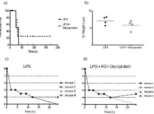
The C57BL/6j mice were divided into two groups: the LPS group, administered intraperitoneally (i.p.) with LPS (35 mg/kg body weight); and the LPS + RSV G group, administered i.p. with one single dose of LPS (35 mg/kg body weight) and two doses of 300dg of RSV G protein i.p. in an interval of 2h, starting along with the LPS administration. They were monitored, weighed, and clinically scored every 2 hours for the first 24 hours, every 4 hours for the next 24 hours, and once daily until the study end at day 7. Clinical score was performed for the first 24h of observation (0− No apparent illness, healthy, active; 1− Slightly Ruffled fur; 2− Ruffled fur but active; 3− Ruffled fur and inactive; 4− Ruffled fur, inactive, hunched posture, and gaunt; 5− Dead or Sac). Data were expressed as descriptive analysis.
Statistical analysis
cases. The Kruskal Wallis test was used for nonparametric comparisons where appropriate. In vivo model results were presented as descriptive data. Data are expressed as mean ± SEM. A p<0.05 was considered significant.
Results
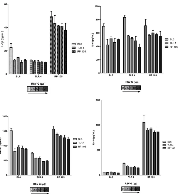
Monocytes from RP105 knockout mice stimulated with LPS (1dg) over express cytokines. Our results have shown that the release of IL 6, IL 1β and TNF α in the RP105/ mice was at least two fold higher than the C57BL/6. Similar proportions were observed for IL 10 between groups. These findings are in agreement with those of Divanovic S et. al., 2005(4), where it was shown that RP105 is a negative regulator of TLR4 signaling, and specifically inhibited TLR4 signaling. There was no difference in the cytokine expression between control groups for all measured cytokines.
ββββ
!
"
#αααα
cell production of the cytokines for the monocytes of C57BL/6 and RP105/ comparing the LPS stimulation to the LPS and LPS + RSV G. IL 10 was also decreased for TLR4/ comparing the LPS and LPS + RSV G. There was no difference between the control and G groups (p > 0.05). Results are given as mean ± SEM. * p < 0.001 and ** p < 0.01 relative to the LPS group. TNF α * p < 0.05.
The RSV G protein inhibits the immune response against LPS induced sepsis
The expression of IL 1β, IL 6 and TNF α from monocytes incubated with LPS (1dg) and RSV G protein (5dg) was inhibited in the wt C57BL/6 and RP105/(Figure 1). Despite the continuous inhibition of the inflammatory cytokines in the wt C57BL/6 and RP105/, there was no significant difference between the LPS and LPS + RSV G for TLR4/.
The RSV G inhibits the release of IL 10 by LPS stimulated monocytes (figure 1B). Rather than inhibiting the inflammatory cytokines, the RSV G protein has decreased the release of IL 10 in LPS stimulated monocytes. Unlike the previous results for inflammatory cytokines, the inhibition was significant not only for C57BL/6 and RP105/ but also included the TLR4/.
$
β
$
$
"
#αααα
$
Figure 2. Effect of increasing concentrations of purified RSV G protein (dg) on TLR4/ (horizontal striped bar), RP105/(checkered bar),and C57/bl6 (dotted bar) mouse monocytes. Spleen monocytes were incubated with LPS ( 0111:B4, 1dg), exposed to (2.5 5 10 and 20 dg) of RSV G protein, and compared to a control without RSV G. Monocytes decreased the release of IL 6 in a dose dependent fashion for C57BL/6 and TLR4/ . The RP105/ group presented the most constant decreasing effect for all cytokines. Results are given as mean ± SEM.
of RSV G protein (0;2.5;5;10 and 20dg). 293mTLR4 cells incubated with 20dg of RSV G decreased the IL 10 and IL 1β production (p < 0.001) relative to the respective 293/null control.
$
ββββ
%
$
% !
Figure 3. HEK 293 mTLR4 MD2 CD14 cells were stimulated to express TLR4 receptor, and incubated with LPS ( 0111:B4, 1dg) and purified RSV G protein (2.5 5 10 20dg). 293/null cells were used as control. Supernatants were collected 18h after addition of LPS. IL 10 and IL 1β presented a dose response inhibition to RSV G protein dependent on TLR4 expression. Results are given as mean ± SEM. * p < 0.05 and ** p < 0.001 relative to the respective 293/null control.
a) b)
c) d)
healthy, active; 1− Slightly Ruffled fur; 2− Ruffled fur but active; 3− Ruffled fur and inactive; 4− Ruffled fur, inactive, hunched posture, and gaunt; 5− Dead or Sac). Data were expressed as descriptive analysis.
experiment. In a preliminary and small experiment (n=4 per group), mice were treated with LPS (35 mg/kg body weight); and LPS + RSV G, administered i.p. with LPS (35 mg/kg body weight) and two doses of 300dg of purified RSV G protein i.p. in an interval of 2h. All groups had a non statistical decrease in mortality and weight loss. (Figure 4a, 4b). Initially both groups were equally ill. However, at 24 hours, mice in the treatment group were more active (Figure 4c, 4d).
Discussion
This study has identified that the RSV G protein inhibits the release of LPS mediated cytokines . The effect was dependent on the two known LPS receptors, TLR4 and RP105. Even though we are not able to quantify the role of the RP105 receptor, considering conflicting studies where the RP105 receptor is a negative regulator of TLR4 signaling and a physiological regulator of endotoxin driven TLR4(4) or those where RP105 pathway is independent of MyD88 expression(18).
Our group had demonstrated that a specific peptide derived from the secreted RSV G, the RSV G cysteine rich region (GCCR), inhibited proinflammatory cytokines responses in a dose dependent manner elicited by LPS stimulation in human monocytes (14). Conversely, this article has shown that the secreted RSV G inhibited cytokines LPS dependent.
TNF α production are increased in the LPS stimulated monocytes, and these effects taken together highlight the LPS mediated cytokine modulation by RSV G. Studies have shown that LPS and RSV G target the TLR4 dependent signaling(13, 17, 19), which led us to hypothesize that RSV G interacts with LPS before acting on the complex CD14+/TLR4. Binding of LPS to a cell does not result in an immediate response. A time lapse of 15 to 30 min between LPS binding and LPS induced responses such as cytokine release and adhesion was observed, which suggests that a time consuming process such as internalization is necessary to enable signaling (20). We are currently investigating the role of different timing administration of RSV G in LPS stimulated cells.
Recent studies have shown that the human metapneumovirus glycoprotein G modulates cytokine and chemokine secretion in monocyte derived dendritic cells by targeting the TLR4 dependent signaling pathway (13). Our findings using HEK 293mTLR4 cells demonstrated that the TLR4 is enough for the RSV G modulation of the LPS. However, LPS can stimulate a CD14 independent pathway in CD14 deficient mice (21), and similar to CD14 deficient mice, in TLR4 deficient mice, the expression of TLR4 is not required for the activation of the LPS pathway (21). In agreement with these findings our study has demonstrated that the RSV G elicited an inhibition of the IL 10 in the TLR4/ monocytes.
therapeutic effects on a single tissue of interest while avoiding potential detrimental systemic effects (25). Targeted administration of IL 10 to the thymus decreases apoptosis and improves mortality in sepsis (26). Nevertheless, lung specific administration of IL 10 improves the survival (27). Furthermore recombinant IL 10 potently suppresses LPS induced production of TNF α, and pretreatment with IL 10 inhibits TNF α activity and prevents LPS induced mortality in mice (28, 29).
In a preliminary and small experiment (n= 8), mice treated with RSV G had a non statistical decrease in mortality and weight loss. Despite the limitation of these results they would suggest a role of RSV G models of LPS sepsis. In addition, LPS plays a critical role in many illnesses, among others septic shock due to Gram negative bacteria and development of childhood asthma (30, 31). Studies with respiratory infections have demonstrated that RSV G inhibits production of inflammatory cytokines shortly after infection, thereby modulating the innate inflammatory response to the virus (14).
Limitations of the study are mainly focused on the difficulty to obtain RSV G protein enough. This drawback led us to use alternative forms of RSV G at the same time they are compared in functional equivalency. However, there are differences between and
models that do not allow the use of peptides experiments (7). The genetic polymorphisms in TLR4 must be considered, because it appears to affect severity of disease both in Gram negative sepsis and RSV infection (30, 33).
might offer future directions where the RSV G protein would play a role in the innate inflammatory response. Further studies ought to be taken toward the implications on adaptive immunity and its effects on the Th bias of the immune response that will clarify the RSV G cytokine inhibition and its clinical relationship.
REFERENCES
1. Ando H, Takamura T, Ota T, Nagai Y, Kobayashi K. Cerivastatin improves survival of mice with lipopolysaccharide induced sepsis. J Pharmacol Exp Ther. 2000 Sep;294(3):1043 6.
2. An N, Song Y, Zhang X, Ci X, Li H, Cao Y, et al. Pretreatment of mice with rifampicin prolongs survival of endotoxic shock by modulating the levels of inflammatory cytokines. Immunopharmacol Immunotoxicol. 2008;30(3):437 46.
3. Ulevitch RJ, Tobias PS. Recognition of gram negative bacteria and endotoxin by the innate immune system. Curr Opin Immunol. 1999 Feb;11(1):19 22.
4. Divanovic S, Trompette A, Atabani SF, Madan R, Golenbock DT, Visintin A, et al. Negative regulation of Toll like receptor 4 signaling by the Toll like receptor homolog RP105. Nat Immunol. 2005 Jun;6(6):571 8.
5. van den Hoogen BG, de Jong JC, Groen J, Kuiken T, de Groot R, Fouchier RA, et al. A newly discovered human pneumovirus isolated from young children with respiratory tract disease. Nat Med. 2001 Jun;7(6):719 24.
7. van Drunen Littel van den Hurk S, Mapletoft JW, Arsic N, Kovacs Nolan J. Immunopathology of RSV infection: prospects for developing vaccines without this complication. Rev Med Virol. 2007 Jan Feb;17(1):5 34.
8. Haynes LM, Moore DD, Kurt Jones EA, Finberg RW, Anderson LJ, Tripp RA. Involvement of toll like receptor 4 in innate immunity to respiratory syncytial virus. J Virol. 2001 Nov;75(22):10730 7.
9. Kurt Jones EA, Popova L, Kwinn L, Haynes LM, Jones LP, Tripp RA, et al. Pattern recognition receptors TLR4 and CD14 mediate response to respiratory syncytial virus. Nat Immunol. 2000 Nov;1(5):398 401.
10. Haeberle HA, Takizawa R, Casola A, Brasier AR, Dieterich HJ, Van Rooijen N, et al. Respiratory syncytial virus induced activation of nuclear factor kappaB in the lung involves alveolar macrophages and toll like receptor 4 dependent pathways. J Infect Dis. 2002 Nov 1;186(9):1199 206.
11. Jackson M, Scott R. Different patterns of cytokine induction in cultures of respiratory syncytial (RS) virus specific human TH cell lines following stimulation with RS virus and RS virus proteins. J Med Virol. 1996 Jul;49(3):161 9.
12. Hancock GE, Scheuer CA, Sierzega R, Pryharski KS, McBride JT, Watelet LF, et al. Adaptive immune responses of patients with asthma to the attachment (G) glycoprotein of respiratory synctial virus. J Infect Dis. 2001 Dec 15;184(12):1589 93.
13. Kolli D, Bao X, Liu T, Hong C, Wang T, Garofalo RP, et al. Human metapneumovirus glycoprotein G inhibits TLR4 dependent signaling in monocyte derived dendritic cells. J Immunol. Jul 1;187(1):47 54.
elicited by the virus and endotoxin. Proc Natl Acad Sci U S A. 2005 Jun 21;102(25):8996 9001.
15. Melero JA, Garcia Barreno B, Martinez I, Pringle CR, Cane PA. Antigenic structure, evolution and immunobiology of human respiratory syncytial virus attachment (G) protein. J Gen Virol. 1997 Oct;78 ( Pt 10):2411 8.
16. Martinez I, Dopazo J, Melero JA. Antigenic structure of the human respiratory syncytial virus G glycoprotein and relevance of hypermutation events for the generation of antigenic variants. J Gen Virol. 1997 Oct;78 ( Pt 10):2419 29.
17. Melendi GA, Bridget D, Monsalvo AC, Laham FF, Acosta P, Delgado MF, et al. Conserved cysteine residues within the attachment G glycoprotein of respiratory syncytial virus play a critical role in the enhancement of cytotoxic T lymphocyte responses. Virus Genes. Feb;42(1):46 54.
18. Yazawa N, Fujimoto M, Sato S, Miyake K, Asano N, Nagai Y, et al. CD19 regulates innate immunity by the toll like receptor RP105 signaling in B lymphocytes. Blood. 2003 Aug 15;102(4):1374 80.
19. Widjojoatmodjo MN, Boes J, van Bers M, van Remmerden Y, Roholl PJ, Luytjes W. A highly attenuated recombinant human respiratory syncytial virus lacking the G protein induces long lasting protection in cotton rats. Virol J.7:114.
20. Detmers PA, Thieblemont N, Vasselon T, Pironkova R, Miller DS, Wright SD. Potential role of membrane internalization and vesicle fusion in adhesion of neutrophils in response to lipopolysaccharide and TNF. J Immunol. 1996 Dec 15;157(12):5589 96.
22. van der Poll T, Marchant A, Buurman WA, Berman L, Keogh CV, Lazarus DD, et al. Endogenous IL 10 protects mice from death during septic peritonitis. J Immunol. 1995 Dec 1;155(11):5397 401.
23. Remick DG, Garg SJ, Newcomb DE, Wollenberg G, Huie TK, Bolgos GL. Exogenous interleukin 10 fails to decrease the mortality or morbidity of sepsis. Crit Care Med. 1998 May;26(5):895 904.
24. Murphey ED, Sherwood ER. Bacterial clearance and mortality are not improved by a combination of IL 10 neutralization and IFN gamma administration in a murine model of post CLP immunosuppression. Shock. 2006 Oct;26(4):417 24.
25. Scumpia PO, Moldawer LL. Biology of interleukin 10 and its regulatory roles in sepsis syndromes. Crit Care Med. 2005 Dec;33(12 Suppl):S468 71.
26. Oberholzer C, Oberholzer A, Bahjat FR, Minter RM, Tannahill CL, Abouhamze A, et al. Targeted adenovirus induced expression of IL 10 decreases thymic apoptosis and improves survival in murine sepsis. Proc Natl Acad Sci U S A. 2001 Sep 25;98(20):11503 8. 27. McAuliffe PF, Murday ME, Efron PA, Scumpia PO, Ungaro R, Abouhamze A, et al. Dose dependent improvements in outcome with adenoviral expression of interleukin 10 in a murine model of multisystem organ failure. Gene Ther. 2006 Feb;13(3):276 82.
28. Fiorentino DF, Zlotnik A, Mosmann TR, Howard M, O'Garra A. IL 10 inhibits cytokine production by activated macrophages. J Immunol. 1991 Dec 1;147(11):3815 22. 29. Gerard C, Bruyns C, Marchant A, Abramowicz D, Vandenabeele P, Delvaux A, et al. Interleukin 10 reduces the release of tumor necrosis factor and prevents lethality in experimental endotoxemia. J Exp Med. 1993 Feb 1;177(2):547 50.
31. Braun Fahrlander C, Riedler J, Herz U, Eder W, Waser M, Grize L, et al. Environmental exposure to endotoxin and its relation to asthma in school age children. N Engl J Med. 2002 Sep 19;347(12):869 77.
32. Roberts SR, Lichtenstein D, Ball LA, Wertz GW. The membrane associated and secreted forms of the respiratory syncytial virus attachment glycoprotein G are synthesized from alternative initiation codons. J Virol. 1994 Jul;68(7):4538 46.
3. CONCLUSÕES
O presente estudo demonstrou que a proteína RSV G é capaz de modular a produção de citocinas estimuladas por LPS “ ”. Houve inibição dose dependente de citocinas inflamatórias estimuladas por LPS, não constante, para a proteína RSV G.
Os camundongos knockout para o receptor RP 105 são mais suscetíveis à estimulação por LPS.
A presença do receptor toll like 4 em células HEK 293 foi suficiente para que a proteína RSV G produzisse a inibição dose dependente das citocinas LPS dependentes.
A dificuldade de se obter quantidades suficientes de proteína RSV G para o experimento reforça a necessidade de mais estudos com peptídeos capazes de produzir o mesmo efeito inibidor em citocinas inflamatórias, como apresentado no presente trabalho.