TLR4 Activation Promotes Bone Marrow MSC
Proliferation and Osteogenic Differentiation
via Wnt3a and Wnt5a Signaling
Xiaoqing He1,2, Hai Wang1,2, Tao Jin2, Yongqing Xu2*, Liangbin Mei2, Jun Yang2
1The Third Military Medical University, Chongqing, China,2Department of Orthopaedic Surgery, Kunming General Hospital of Chengdu Military Command, Kunming, China
Abstract
Mesenchymal stem cells (MSCs) from adult bone marrow maintain their self-renewal ability and the ability to differentiate into osteoblast. Thus, adult bone marrow MSCs play a key role in the regeneration of bone tissue. Previous studies indicated that TLR4 is expressed in MSCs and is critical in regulating the fate decision of MSCs. However, the exact functional role and underlying mechanisms of how TLR4 regulate bone marrow MSC proliferation and differentiation are unclear. Here, we found that activated TLR4 by its ligand LPS promoted the proliferation and osteogenic differentiation of MSCsin vitro. TLR4 activation by LPS
also increased cytokine IL-6 and IL-1βproduction in MSCs. In addition, LPS treatment has no effect on inducing cell death of MSCs. Deletion of TLR4 expression in MSCs completely eliminated the effects of LPS on MSC proliferation, osteogenic differentiation and cytokine production. We also found that the mRNA and protein expression of Wnt3a and Wnt5a, two important factors in regulating MSC fate decision, was upregulated in a TLR4-dependent manner. Silencing Wnt3a with specific siRNA remarkably inhibited TLR4-induced MSC pro-liferation, while Wnt5a specific siRNA treatment significantly antagonized TLR4-induced MSC osteogenic differentiation. These results together suggested that TLR4 regulates bone marrow MSC proliferation and osteogenic differentiation through Wnt3a and Wnt5a signaling. These finding provide new data to understand the role and the molecular mecha-nisms of TLR4 in regulating bone marrow MSC functions. These data also provide new insight in developing new therapy in bone regeneration using MSCs by modulating TLR4 and Wnt signaling activity.
Introduction
Adult bone marrow mesenchymal stem/stromal cells (MSCs) are multipotential stem cells, and have the ability to differentiate into osteoblasts, chondrocytes, tenocytes, neurons, adipocytes, and skeletal myocytes [1,2]. Due to the multipotent of MSCs, they have become a therapeutic option for several pathologies including osteogenesis imperfecta, myocardial infarction, and wound healing. MSCs play an important role in bone remodeling because they can be induced to differentiate into osteoblasts. Published data have revealed that bone marrow derived MSCs OPEN ACCESS
Citation:He X, Wang H, Jin T, Xu Y, Mei L, Yang J (2016) TLR4 Activation Promotes Bone Marrow MSC Proliferation and Osteogenic Differentiation via Wnt3a and Wnt5a Signaling. PLoS ONE 11(3): e0149876. doi:10.1371/journal.pone.0149876
Editor:Antonio Paolo Beltrami, University of Udine, ITALY
Received:October 11, 2015
Accepted:February 6, 2016
Published:March 1, 2016
Copyright:© 2016 He et al. This is an open access article distributed under the terms of theCreative Commons Attribution License, which permits unrestricted use, distribution, and reproduction in any medium, provided the original author and source are credited.
Data Availability Statement:All relevant data are within the paper and its Supporting Information files.
Funding:This work was funded by the National Natural Sciences Foundation of China (Grant No. 81472096). The funder had no role in study design, data collection and analysis, decision to publish, or preparation of the manuscript.
can repair bone defects in animal models [3,4]. In addition to their multipotential plasticity, MSCs play a critical role in regulating immune responses in a manner that depends on their state of activation [5,6]. It is also an important cell to form the critical microenvironment for bone regeneration after injury [7]. Therefore, it is important to reveal the molecular mecha-nisms that regulate the MSC function including survival, proliferation, differentiation and cytokine secretion.
Toll-like receptor 4 (TLR4) are the best studied immune sensors of invading microbes. It is broadly distributed on cells throughout the immune system. Activation of TLR4 is essential for inducing the immune responses, and enhances adaptive immunity against pathogens [8,9]. It has been revealed that MSCs derived from adult bone marrow also express functional TLR4 [10,11,12,13]. Activation of TLR4 signaling in MSCs may influence their survival, differentia-tion, proliferadifferentia-tion, migration and pro-inflammatory cytokine secretion [10,14,15]. TLR4 recog-nizes lipopolysaccharides (LPS) from gram-negative bacteria. It has been proved that LPS can protect MSCs from oxidative stress-induced apoptosis and enhance proliferation of MSCs via TLR4 and PI3K/Akt signaling [12]. LPS was also found to promote the osteogenic differentia-tion in adult human MSCs [14]. However, due to conflicting reports on various effects of TLR4 ligands on MSCs [12,13,14,16], further studies are still needed to explore the cellular biological changes of MSCs after TLR4 activation in different models. Importantly, the molecular mecha-nisms by which TLR4 regulating MSC proliferation and differentiation are largely unknown.
Wingless proteins (Wnt) are a family of cysteine-rich glycoproteins that regulate embryonic development, cell proliferation, migration, differentiation, and death [17,18]. Recently, Wnt signaling has been revealed to function in controlling the cell fate specification and differentia-tion of MSCs [19]. Previous researches have shown that Wnt signaling has the capacity to regu-late the proliferation and migration of MSCs [20,21]. Moreover, Wnt signaling has also been found involved in the osteogenic and adipogenic differentiation process in human MSCs [22]. Till now, exist evidence have identified that Wnt3a, Wnt5a, Wnt6, Wnt10a, and Wnt10b sig-naling are all involved in controlling the stem cell properties of MSCs [20,23,24,25]. Wnt5a has been shown to be involved in the induction of MSC osteogenesis and suppression of adipogen-esis [23]. Wnt3a promotes proliferation and suppresses osteogenic differentiation of adult human MSCs [20]. Wnt10b has been proposed to influence the decision of MSCs to give rise to either an adipocyte or an osteoblast. Knockdown of endogenous Wnt6 is associated with increasing preadipocyte differentiation and impaired osteoblastogenesis [25]. However, little is known about the interactions between TLR4 and Wnt signaling and their function in regulat-ing MSC proliferation and differentiation.
In the present work, we used TLR4 ligand LPS to activate TLR4 in bone marrow MSCs. We studied the effects of TLR4 activation on the survival, proliferation, osteogenic differentiation, and cytokine production of bone marrow originated MSCsin vitro. We found that TLR4 acti-vation promoted the proliferation and osteogenic differentiation of MSCs. In addition, we examined the involvement of Wnt signaling in this process, and found that Wnt3a and Wnt5a play critical role in TLR4-induced MSC proliferation and osteogenic differentiation. Our experiments provide new data to understanding the role and the molecular mechanisms of TLR4 in regulating bone marrow MSC functions.
Methods
Cell culture, identification, differentiation and LPS treatment
All the animal care and procedures were in accordance with the guidelines of the National Institutes of Health, and were approved by the Committee on Use of Live Animals for Research of the Third Military Medical University.
Bone marrow cells were obtained from femur and tibia of 6- to 8-week-old male C57BL/6J WT mice and TLR4-/-mice (The Jakson lab., Strain: B6.B10ScN-Tlr4lps-del/JthJ). The cells were filtered through 40μm nylon mesh, and centrifuged at 1000 rpm for 5 min. The buoyant adipo-cytes were removed by vacuum aspiration. The cell pellet was resuspended in Dulbecco’s modi-fied Eagle’s medium (DMEM) supplemented with 10% fetal bovine serum (FBS), 2 mM glutamine, and 1% penicillin and streptomycin (Invitrogen, USA). The cells were cultured in 10 cm dishes and nonadherent cells were removed after 24 hours. Culture medium was changed every 3 days. Upon confluence, the cells were dissociated using 0.25% trypsin (Invitro-gen, USA) and reseeded. For clone isolation, cultured cells were seeded in 96-well plates by lim-ited dilution. Individual clones were picked and expanded for further experiments. Cells were used with in the 10th passage [26].
The MSC phenotype was defined using flow cytometric analysis with the following mono-clonal antibodies: anti-CD31-PE (R&D, USA), anti-CD45-FITC (R&D, USA), anti-CD29-PE (Immunotech, France), and anti-CD44-PE (BD Biosciences, USA).
For osteogenic differentiation, cells were seeded into 24-well plates and allowed to attach overnight. The following day (day 0) growth medium was replaced with osteogenic differentia-tion medium, which is DMEM supplemented with 10% FBS, 100 nM dexamethasone (Sigma, USA), 10 mMβ-glycerophosphate (Sigma, USA) and 100 mM vitamin C. Medium was replaced every 3 or 4 days[27].
Lipopolysaccharide (LPS, Sigma, USA) was added to the culture medium at a final concen-tration of 0, 10, 100, 1000 ng/ml, and treated for indicated times.
Cell apoptosis measurement
MSCs were detached by trypsin treatment, washed, and then assessed for Annexin V (Invitro-gen) and Propidium Iodide (PI) staining. Flow cytometric analysis was used to detect early apoptotic cells Annexin positive/PI-negative cells and late apoptopic or necrotic Annexin V-positive/PI-positive cells.
Cell proliferation
The effect of LPS on the proliferation of MSCs was studied by a Colorimetric Cell-counting Kit (CCK-8, Dojindo, Japan) assay and EdU incorporation according to the manufacturer’s instruction. The EdU+cells were counted in 10 different fields of each condition by an indepen-dent observer in a blinded manner using a 20 x objective. Three indepenindepen-dent experiments were performed for every condition. The EdU+were expressed as a percentage of total cells deter-mined by Hoechst33342 staining [28].
Alkaline phosphatase activity and alizarin red staining
ALP activity was detected using an ALP kit according to the manufacturer's instructions (Sigma-Aldrich). ALP activity was normalized to protein concentration of the cell lysate. For evaluation of mineralization, cells were induced for 7, 15, 20 to 25 days, fixed with 4% parafor-maldehyde and stained with 2% Alizarin red (Sigma-Aldrich). For alizarin red quantification, the deposits were extracted with 10% acetic acid and 20% methanol solution. Light absorbance by the extracted dye was measured in 450 nm [29].
Real-time PCR
manufacturer's instructions. mRNA expression of the target genes was detected using the fol-lowing primers: TLR4 fwd 5’- gcaggtggaattgtatcgcc -3’and rev 5’—tgctcaggattcgaggcttt—3’, Wnt3a fwd 5’—ggctcctctcggatacctct—3’and rev 5’—gggcatgatctccacgtag—3’, Wnt5a fwd 5’—
tggagaaggtgcgaagacag—3’and rev 5’—cgtctctcggctgcctattt—3’, Wnt6 fwd 5’—ggtagtcctagaggg ccagg—3’and rev 5’—cactccgtcatgccttggta—3’, Wnt10a fwd 5’—catcgggctgaagtgactct—3’and rev 5’- ccggagcttctttttcccca -3’, Wnt10b fwd 5’—tcctccactacagcccagaa—3’and rev 5’- cctccc aagagcctgacaag—3’, IL-1βfwd 5’—gcaactgttcctgaactcaact—3’and rev 5’- atcttttggggtccgtcaact
—3’, IL-6 fwd 5’—tagtccttcctaccccaatttcc—3’and rev 5’—ttggtccttagccactccttc—3’, TNF-αfwd 5’—acggcatggatctcaaagac—3’and rev 5’—agatagcaaatcggctgacg—3’, and GAPDH fwd 5’—atac ggctacagcaacaggg- 3’and rev 5’—gcctctcttgctcagtgtcc—3’. Real-time PCR was performed on a CFX96TMreal-time system using SYBR Master Mix (Takara, Japan). All gene expression values were normalized to the housekeeping gene GAPDH. The relative expression levels of the target genes were calculated using the control as a reference.
Western blot analysis
Adherent cells were scraped from the culture dishes and total protein was extracted from the cell pellet with RIPA lysis buffer (Thermo Scientific, USA). Western blot analysis was per-formed and quantified with an Odyssey system (LI-COR, USA) as previously described [30]. The primary antibodies used for Western blot analysis were monoclonal mouse anti-TLR4 antibody (Santa Cruz Biotechnology, USA) and monoclonal mouse anti-GAPDH antibody (Abcam, USA) The secondary antibodies used were Odyssey-specific IRDye 680 or 800 donkey anti-mouse (1:5000; LI-COR, USA).
Enzyme-linked Immunosorbent Assay (ELISA)
MSCs were seeded in 24-well culture plates. After the cells were treated with LPS for the indi-cated time or pretreated with specific siRNAs as indiindi-cated, the culture supernatants were col-lected and stored at -80°C until used. The Wnt3a and Wnt5a levels in the supernatants were measured using a mouse Wnt3a and Wnt5a ELISA Kit following the manufacturer’s instruc-tions (Antibodies-online Inc. USA) [22]. For LPS-induced cytokines detection, MSCs were cul-ture in a 96 well plate and treated with or without LPS for 3 days. Then, the concentration of IL-1β, IL-6 and TNF-αin the culture medium was determined by ELISA according to the man-ufacturer’s instructions (R&D, USA).
RNA interference
siRNAs specific for mouse Wnt3a (sc-41109), Wnt5a (sc-41113) and control siRNA (sc-36869) with scrambled sequences were purchased from Santa Cruz. In each transfection, 0.1 to 0.2μM siRNAs were introduced into MSCs using the Lipofectamine 2000 kit (Invitrogen, USA). The efficiency of transduction was determined by detecting the rate of FITC-positive cells and the mRNA expression of specific genes.
Statistical analysis
All data were collected from three to four independent experiments unless otherwise stated, and were analyzed using the SPSS13.0 software package. A one-way ANOVA followed by Fish-er’s post hoc test were used for multiple-group comparisons. Student’s t-test was used when only two sets of data were compared. A statistically significant level was defined as p<0.05.
Error bars are reported as mean ± SEM.
Results
TLR4 ligand LPS treatment promotes bone marrow MSC proliferation
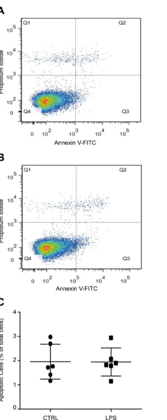
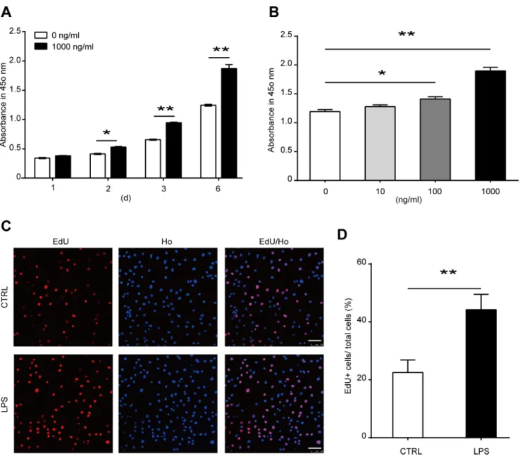
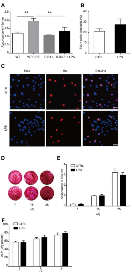
To study the role of TLR4 in bone marrow MSCs, we firstly cultured MSCs from wild type mouse bone marrow as previously described. The phenotype of the cultured cells was identified using flow cytometric analysis with CD29, CD44, CD31, and CD45 antibodies. Almost all the cells are CD29 and CD44 positive. The cells are also CD31 and CD45 negative (S1 Fig). LPS has been proved to be an effective ligand for TLR4. To activate TLR4, we treated the cells with 1000 ng/ml LPS. The concentration of LPS is widely used in previous studies [31,32]. No change of cell death was induced by 1000 ng/ml LPS as we detected 6 days after treatment (Fig 1A–1C). Then the time-dependent effects and dose-dependent effects of LPS on MSC prolifer-ation was examined using CCK-8 kit assay. MSCs were treated by 1000 ng/ml LPS for 1, 2, 3, and 6 days. LPS treatment significantly promoted MSC proliferation as detected 2 days after treatment, and the effects increased in a time-dependent manner (Fig 2A). Then the cells were treated with 0, 10, 100, and 1000 ng/ml LPS for 6 days. LPS remarkably enhanced MSC prolif-eration at a concentration of 100 ng/ml, and the effects increased significantly again at a con-centration of 1000 ng/ml (Fig 2B). These results were further confirmed by the results from EdU incorporation analysis. We found that the percentage of EdU positive (EdU+) cells in LPS treatment group was much higher than control group, indicating the ratio of proliferative cells in LPS treatment group is significantly higher than control group (Fig 2C and 2D). Taken together, these data suggested that TLR4 ligand LPS treatment promoted the proliferation of bone marrow MSCs.TLR4 activation by LPS enhances osteogenic differentiation of MSCs
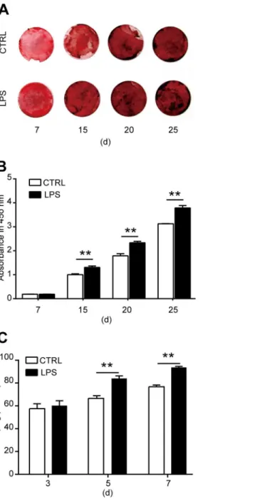
To reveal the role of TLR4 in the osteogenic differentiation of MSCs, we cultured the cells in an osteogenic differentiated medium. LPS was added to the cultured medium at a final concentra-tion of 1000 ng/ml to activated TLR4. The osteogenic differentiaconcentra-tion of MSCs was evaluated by calcium deposit and ALP activity in the differentiated cells. To detect calcium deposit, MSCs were differentiated in the presence of LPS for 7, 15, 20, and 25 days. Alizarin red staining was used to analysis the calcium deposit in the differentiated cells. We found that from the 15th day after LPS treatment, the calcium deposit was remarkably increased as compared with con-trol, and this effect increased in a time-dependent manner (Fig 3A and 3B). Then the ALP activity in the differentiated cells was detected 3, 5, and 7 days after LPS treatment. The ALP activity is enhanced notably at 5 and 7 days after LPS treatment (Fig 3C). From these data, we can get a conclusion that TLR4 activation by its ligand LPS enhanced the osteogenic differenti-ation of MSCs.LPS treatment increases cytokine production in MSCs
Fig 1. TLR4 ligand LPS treatment did not influence cell apoptosis of bone marrow derived MSCs.
MSCs were treated with 1000 ng/ml LPS for 6 days. Total apoptotic cells were detected using flow cytometric analysis with Annexin V and PI staining.(A, B)Representative images of control and LPS treated cells.(C)
Statistical results of six independent experiments.
doi:10.1371/journal.pone.0149876.g001
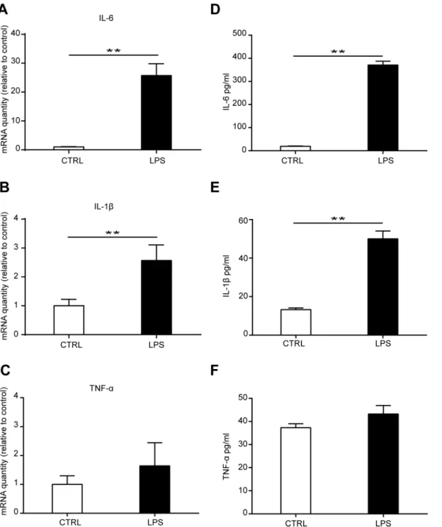
expression as detected using ELISA 3 days after LPS treatment (Fig 4D and 4E). No significant change of TNF-αmRNA and protein level was found after TLR4 ligand LPS treatment (Fig 4C
and 4F). These data suggested that TLR4 activation upregulated cytokines IL-6 and IL-1β
pro-duction in bone marrow MSCs.
TLR4 deletion eliminates the effects of LPS on MSC cytokine
production, proliferation and osteogenic differentiation
Previous studied suggested that LPS treatment could modify TLR4 expression [10,37]. Thus, we detected the mRNA and protein expression of TLR4 in MSCs after LPS treatment. We Fig 2. Effects of LPS treatment on the proliferation of MSCs. (A)MSCs were treated with 1000 ng/ml LPS for 1, 2, 3, or 6 days. Cell proliferation was detected with CCK-8 kit assay.(B)MSCs were treated with various concentrations of LPS. Cell proliferation was examined on the sixth day of treatment with CCK-8 kit assay.(C, D)MSCs were treated with 1000 ng/ml LPS for 6 days, and the proliferation of cells was analysis with EdU incorporation.(C)The representative images (Scale bar: 100μm), and(D)The statistical result. Data are presented as mean±SEM from four independent experiments.*P<0.05,
**P<0.01.
found that TLR4 mRNA and protein level both increased significantly after LPS treatment (S2 Fig). To further reveal the effects of TLR4 on MSC proliferation and osteogenic differentiation, we cultured MSCs from the bone marrow of TLR4-/-mice (S3 Fig). The effects of LPS on MSC cytokine production, proliferation and differentiation were then studied using TLR4-/-MSCs. We found that the effect of LPS on IL-6 and IL-1βproduction was abolished by TLR4 deletion
(S4 Fig). No difference in the cell proliferation of TLR4-/-MSCs was found after LPS treatment
Fig 3. Effects of LPS treatment on the osteogenic differentiation of MSCs. (A, B)MSCs were induced for osteogenic differentiation in the presence of 1000 ng/ml LPS as indicated for 7, 15, 20, or 25 days. Alizarin red staining and quantification was used to detect the osteogenic differentiation.(A)Representative images.
(B)Quantification of alizarin red.(C)ALP activity after MSCs were induced for osteogenic differentiation in the presence of 1000 ng/ml LPS for 3, 5, or 7 days. Data shown are representative of four independent experiments and presented as mean±SEM.*P<0.05,**P<0.01.
doi:10.1371/journal.pone.0149876.g003
for 6 days as detected by CCK-8 kit assay and EdU incorporation (Fig 5A–5C). Furthermore, results from alizarin red staining showed that no change in calcium deposit in the differentiated TLR4-/-cells after LPS treatment was found as detected 7, 15, and 25 days after differentiation. In addition, the ALP activity was not changed after LPS treatment for 3, 5, and 7 days. Together, these results suggested that TLR4 is essential for LPS-induced MSC proliferation, Fig 4. Effects of LPS treatment on cytokine production of MSCs.MSCs were treated with 1000 ng/ml LPS for 3 days. The mRNA production of IL-6(A), IL-1β(B), and TNF-α(C)was detected by Real-time PCR. Data are from four independent experiments and presented as mean±SEM.**P<0.01. MSCs were cultured in a 96 well plate and treated with 1000 ng/ml LPS for 3 days. The concentration of IL-6(D), IL-1β(E), and TNF-α(F)in the culture medium was detected by ELISA. Data are from three independent experiments and presented as mean±SEM.**P<0.01.
Fig 5. TLR4 knock out eliminate the effects of LPS on MSC proliferation and osteogenic
differentiation. (A)Wild type (WT) cells and TLR4 knock out (TLR4-/-) cells were treated with 1000 ng/ml LPS for 6 days. Cell proliferation was examined by CCK-8 kit assay.(B, C)TLR4-/-cells were treated with
osteogenic differentiation and cytokines production. The results further confirmed the role of TLR4 activation in inducing MSC proliferation and osteogenic differentiation.
LPS treatment upregulates the expression of Wnt3a and Wnt5a through
TLR4
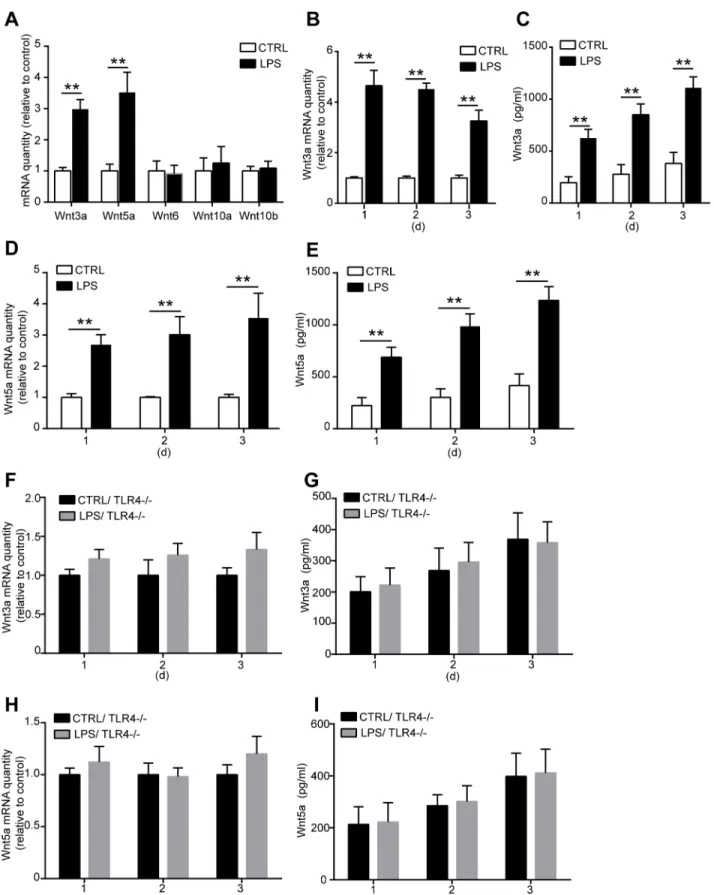
Wnt signaling is critical in fate decision of stem cell development during the embryonic phase. Recent researches indicate that Wnt signaling also plays an important role in regulating MSC proliferation and differentiation in adult [20,21,24]. To explore the underlying mechanisms of how TLR4 regulates MSC proliferation and osteogenic differentiation, we examined the involvement of Wnt family member. We detected the mRNA levels of Wnt3a, Wnt5a, Wnt6, Wnt10a, and Wnt10b in MSCs after LPS treated for 3 days. Only the mRNA expression of Wnt3a and Wnt5a was significantly increased after TLR4 activation by LPS (Fig 6A). We then analyzed the effects of LPS on the mRNA and protein expression of Wnt3a and Wnt5a. MSCs were treated with 1000 ng/ml LPS for 1, 2, and 3 days. Wnt3a and Wnt5a mRNA level in MSCs and the protein concentration in the culture supernatants were examined. We found that both Wnt3a and Wnt5a mRNA level were upregulated as detected in all the 3 days after LPS treat-ment (Fig 6B and 6C). Furthermore, the protein concentration of Wnt3a and Wnt5a in the cul-ture medium was also increased in a time-dependent manner as detected by ELISA (Fig 6D
and 6E). To further confirm the relationship among Wnt3a, Wnt5a, and TLR4, the TLR4
-/-MSCs was used in the experiments. No change of Wnt3a and Wnt5a mRNA expressions was found after LPS treatment in TLR4-/-MSCs (Fig 6F and 6H). In addition, no increase of the protein concentrations of Wnt3a and Wnt5a was found in the culture supernatants of TLR4 -/-MSCs (Fig 6G and 6I). These results strongly indicated that TLR4 is essential for LPS-induced Wnt3a and Wnt5a upregulation.
Wnt3a plays a critical role for TLR4-induced MSC proliferation, while
Wnt5a is involved in TLR4-induced MSC osteogenic differentiation
To explore the role of Wnt3a and Wnt5a in TLR4-induced MSC proliferation and osteogenic differentiation, specific siRNAs were used to silence Wnt3a and Wnt5a expression before LPS treatment (S5A and S5B Fig). We found that Wnt3a and Wnt5a silence has little effect on LPS-induced IL-6 and IL-1βsecretion (S5C–S5F Fig), which indicated that Wnt3a and Wnt5a are not involved in TLR4-induced cytokine production. The results from CCK-8 kit assay sug-gested that Wnt3a silencing remarkably inhibited the effects of LPS on MSC proliferation (Fig 7A). However, Wnt5a siRNA treatment has little effect on eliminating the effects of LPS on MSC proliferation (Fig 7B). The result was further confirmed by EdU incorporation assay (Fig7C and 7DandS6A and S6B Fig). These results indicated that Wnt3a is critical for
TLR4-in-duced MSC proliferation, while Wnt5a plays little effect in this process. Followed, the effects of LPS on the ALP activity in differentiated cells was detected after Wnt3a and Wnt5a specific siRNAs transfection. We found that Wnt3a siRNA treatment has little influence on changing the effects of LPS on ALP activity (Fig 7E). Wnt5a silence significantly antagonized the effects of LPS on increasing ALP activity (Fig 7F). Furthermore, the same role of Wnt3a and Wnt5a 1000 ng/ml LPS for 6 days. Cell proliferation was detected by EdU incorporation assay.(B)The statistical results.(C)Representative images of EdU staining (Scale bar: 50μm).(D, E)MSCs were differentiated with 1000 ng/ml LPS for 7, 15, or 25 days. The osteogenic differentiation was detected by alizarin red staining and quantification. Graph(D)shows the representative images, and(E)is the quantification results.(F)ALP activity of TLR4-/-MSC-differentiated cells in the presence of 1000 ng/ml LPS. Data are presented as mean±SEM from four independent experiments.**P<0.01.
Fig 6. LPS treatment upregulates Wnt3a and Wnt 5a expression through TLR4. (A)WT MSCs were treated with 1000 ng/ml LPS for 3 days, the mRNA expression of Wnt family members was detected by real-time PCR.(B-E)WT MSCs were treated with 1000 ng/ml LPS for 1, 2, or 3 days, then the mRNA and protein expression of Wnt3a and Wnt5a were detected by real-time PCR and ELISA respectively.(B)mRNA expression of Wnt3a in WT MSCs after LPS
siRNAs was found in influencing the calcium deposit in the differentiated cells. We found that Wnt5a siRNA treatment significantly antagonized the effects of LPS on calcium deposit, while Wnt3a siRNA has little effect on this process (Fig 7G and 7HandS6C and S6D Fig). These results suggested that Wnt5a is involved in TLR4-induced MSC osteogenic differentiation, while Wnt3a plays little effect in this process.
Discussion
TLR4 plays an important role in regulating MSC function. However, the exact functional roles and mechanisms of TLR4 on bone marrow MSC proliferation and differentiation need further study. Here, we found that activation of TLR4 enhances MSC proliferation and osteogenic dif-ferentiation. In addition, we also revealed that Wnt3a mediated the effects of TLR4-induced MSC proliferation, while Wnt5a is essential for the promotive effects of TLR4 on MSC osteo-genic differentiation.
MSCs from bone marrow give rise to bone tissue. It is the basic cellular unit of embryonic bone formation. MSCs also play a key role in fracture repair by differentiating into bone-form-ing osteoblasts. Many excitbone-form-ing findbone-form-ings have been obtained in resent MSC transplantation experiments in animal models. The enhancement of bone regeneration with MSCs is becoming a clinical reality [7]. Among all types of the adult stem cells, MSCs are the easiest to isolate, and are multipotent. In addition, MSCs can been easily expandedin vitro[38]. Thus, MSCs serve as the best candidate cells for tissue engineering. To promote the application of MSCs in bone regeneration and tissue engineering, an understanding of MSC biology is necessary.
MSC osteogenic differentiation is controlling by the coordinate activities of different signal-ing pathways that regulate the expression of various osteoblast-specific genes. Recently, TLR4 signaling is found expressed in bone marrow derived MSCs and play a critical role in regulating the function of MSCs [11]. Under pathological conditions, such as ischemia, traumatic injury, inflammatory process, endogenous and exogenous bacteria could release TLR4 ligand LPS which will activate TLR4 signaling in many different type of cells [39,40]. How MSCs maintain their multipotency and capacity to self-renewal and differentiate in these pathological condi-tions is not well understood. This is an important question for the further application of MSCs in the clinic.
Our results shown that, TLR4 activation by LPS promoted MSC proliferation and osteo-genic differentiationin vitro. Previous study has revealed that LPS is able to induce prolifera-tion and osteogenic differentiaprolifera-tion but reduce adipogenic differentiaprolifera-tion of human adipose tissue-derived MSCsin vitro[15]. However, the tissue origin of MSCs influences their TLR profile as well as their functional properties such as cytokine secretion, proliferation and osteo-genic potential [14,16]. The reason may due to the divergent levels of several important signal-ing pathways which mediate TLR4 functions, such as LBP and TGFβ1 signaling [41]. Our results provide evidence that TLR4 signaling plays an important role in regulating bone mar-row derived MSC function. Understanding the role and mechanisms of TLR4 on MSC prolifer-ation and differentiprolifer-ation may shed light on the bone remodeling. More importantly, it is also critical for development of new therapy using MSCs under pathological condition by modula-tion of TLR4 signaling activity.
treatment.(C)Protein level of Wnt3a in the culture supernatants after LPS treatment.(D)Wnt5a mRNA level in WT MSCs.(E)Wnt5a protein concentration in the culture supernatants.(F-I)TLR4-/-MSCs were treated with 1000 ng/ml LPS for 1, 2, or 3 days, then the mRNA and protein expression of Wnt3a and Wnt5a were detected as previously described.(F, G)Wnt3a mRNA expression in the TLR4-/-MSCs and protein concentration in the culture supernatants.(H,
I)Wnt5a mRNA level in the TLR4-/-MSCs and protein concentration in the culture supernatants. Data are from four independent experiments and presented as mean±SEM.**P<0.01.
It has become clear that MSCs also possess immunoregulatory properties during the past few years [5,6]. We have detected the cytokine production in MSCs after TLR4 activation. We found that IL-1βand IL-6 production in MSCs is upregulated after treated by TLR4 ligand LPS. However, we did not find any changes in cell survival, indicating the production of these cytokines does not kill the cells. Cytokines, IL-1βand IL-6 etc., induced by LPS may have the function of enhance TLR4 expression in MSCs [10,37]. Our result confirmed that as proved by the upregulation of TLR4 expression after LPS treatment. These results suggested that upregu-lating TLR4 expressing may be another mechanism by which LPS moduupregu-lating TLR4 signaling activity in MSCs. IL-1βand IL-6 can also induce MSCs migration by inducing the expression of chemokines [36]. Thus, the effects of LPS on cytokine secretion probably will enhance the function of TLR4 on bone tissue generation from MSCs. It is found that prolonged exposure to bacterial toxins downregulated expression of TLR4 in MSC-derived osteoprogenitors [42]. High concentration of LPS was found to reduce MSC proliferation, osteogenic differentiation, and increase cell death [43]. These results suggested that the time phase and dose of TLR4 ligands treatment are also key factors in regulating MSC functions.
Exploring the molecular mechanisms of TLR4 on MSC functions is critical for developing new therapy under pathological condition by modulating TLR4 activity. Till now, how TLR4 activation regulates MSC proliferation and differentiation is unclear. Published data suggest that Wnt signaling is critical in determining both embryonic and adult MSC fate. Wnt6, Wnt10a and Wnt10b are found to inhibit adipogenesis and stimulate osteoblastogenesis through aβ-catenin-dependent mechanism [25]. Wnt3a signaling promotes the proliferation, myogenic differentiation, and migration of bone marrow derived MSCs [20,21]. Wnt5a is nec-essary to maintain osteogenic potential of human MSCs, and it is also found to mediate the effects of the LIM-only protein FHL2 on osteogenic differentiation of MSCs [23,24]. However, the interactions of TLR4 and Wnt signaling and their functions in regulating the proliferation and differentiation of MSCs are unknown. We screened the mRNA expressions of Wnt family members Wnt3a, Wnt5a, Wnt6, Wnt10a, and Wnt10b in response to TLR4 ligand LPS treat-ment. We found that Wnt3a and Wnt5a mRNA and protein levels are significantly upregulated after TLR4 activation by LPS. Further studies using Wnt3a and Wnt5a specific siRNA revealed that Wnt3a is critical for TLR4-induced MSC proliferation. Wnt5a is essential for mediating the effects of TLR4 on MSC osteogenic differentiation. Our data revealed the interactions between TLR4 and Wnt signaling member Wnt3a and Wnt5a, and the functional role of this interaction in regulating MSC proliferation and osteogenic differentiation. It is found that LPS enhances Wnt5a expression through TLR4, myeloid differentiating factor 88, phosphatidylino-sitol 3-OH kinase/AKT and nuclear factor Kappa B pathways in human dental pulp stem cells [22]. TLR4 was also reported to downregulate the Wnt pathway in enterocytes in the ileum of newborn mice [44]. These results raise the possibility that TLR4 may play diverse role in regu-lating Wnt signaling in different tissues. Our data revealed that both Wnt3a and Wnt5a play Fig 7. Proliferation and osteogenic differentiation of MSCs in the presence of LPS after Wnt3a and Wnt5a silence. (A-D)MSCs were treated by Wnt3a and Wnt5a siRNA. The effect of LPS on MSC
proliferation was detected using CCK-8 kit assay and EdU incorporation.(A, B)MSCs were transfected with Wnt3a and Wnt5a siRNA, then the cells were treated with 1000 ng/ml LPS for 6 days. Cell proliferation was detected by CCK-8 kit assay.(C, D)Statistical results of cell proliferation from EdU incorporation as detected 6 days after LPS treatment. Representative images seeS6 Fig.(E-H)Wnt3a and Wnt5a expression were silenced by specific siRNA, then the cells were differentiated in the presence of 1000 ng/ml LPS. ALP activity and alizarin red staining was used to analyzed the effects of LPS on osteogenic differentiation of MSCs.(E, F)ALP activity in MSC-differentiated cells 7 days after LPS treatment.(G, H)Quantification of alizarin red staining in MSC-differentiated cells 15 days after LPS treatment. Representative images of alizarin red staining seeS6 Fig. Data are from three independent experiments and presented as mean±SEM.*P<0.05,
**P<0.01.
critical, but different, roles in TLR4-induced MSC proliferation and osteogenic differentiation. However, Wnt3a and Wnt5a silence has little effect on the LPS-induced cytokines production. Probably other mechanisms exist in mediating the effects of TLR4-induced cytokine secretion in MSCs. We did not analysis the dynamic changes and functions of all the Wnt family mem-bers during the whole process of MSC migration, proliferation, osteogenic differentiation, and maturation. In addition, we could not exclude that other Wnt family members is involved in MSC proliferation and osteogenic differentiation only depending on the results of their expres-sion in this process. Further researches are still needed to further exploring the dynamic role of different Wnt family members in controlling the MSC fate and their interaction with TLR4. To do this, the specific Wnt-CreER recombinase transgenic mice are probably needed to condi-tionally delete the Wnt expression at different stages to determine the role of Wnt signalingin vivo.
In conclusion, we have revealed the regulation of TLR4 on Wnt signaling in bone marrow derived MSCs and their function in determining the proliferation and differentiation of MSCs. These finding provide new data to understand the role of TLR4 in regulating MSC fates and the underlying molecular mechanisms. These data also provide new insight in developing new therapy using MSCs by modulating TLR4 signaling activity.
Supporting Information
S1 Fig. Identification of bone marrow derived MSCs from wild type mice.The flow cyto-metric analysis shows that the cultured cells are CD29(B)and CD44(C)positive. The cells are also CD31(D)and CD45(E)negative.
(TIF)
S2 Fig. LPS treatment increases TLR4 expression in MSCs. (A)The mRNA expression of TLR4 in MSCs was upregulated after 1000 ng/ml LPS treated for 3 days as detected by real-time PCR analysis.(B, C)The protein level of TLR4 is increased by LPS treatment as detected by western blotting analysis. Data are from three independent experiments and presented as mean ± SEM.P<0.05,P<0.01.
(TIF)
S3 Fig. (A) PCR identification of TLR4 KO mice. Genomic DNAs were extracted from tails of the mouse and were analyzed by PCR using the following primers: wild-type TLR4 primer (5'ATATGCATGATCAACACCACAG 3' and 5' TTTCCATTGCTGCCCTATAG 3'), mutant TLR4 primer (5'GCAAGTTTCTATATGCATTCTC 3' and 5' CCTCCATTTCCAATAGG-TAG 3'). The 140 bp band is the mutant TLR4 (lane 2 to 4), while the 390 bp band represented the wild-type genotype of TLR4 (lane 5 to 7). (B-E) Characteristic of bone marrow derived MSCs from TLR4 KO mice. The cultured cells are CD29+(B), CD44+(C), CD31-(D), and CD45-(E).
(TIF)
S4 Fig. TLR4 deletion eliminates the effect of LPS on MSC cytokine production. TLR4-/-MSCs were treated by 1000 ng/ml LPS for 3 days, the mRNA ecpression of IL-1β(A)and IL-6 (B)were detected by real-time PCR. No change was found in both of IL-1βand IL-6 mRNA expression. Data are from three independent experiments and presented as mean ± SEM. (TIF)
S5 Fig. (A, B)Wnt3a and Wnt5a siRNA transfection effectively silence Wnt3a and Wnt5a expression. MSCs were transfected with Wnt3a and Wnt5a siRNA respectively. The mRNA expression of Wnt3a(A)and Wnt5a(B)was detected by real-time PCR 2 and 4 days after
transfection. Data are from three independent experiments and presented as mean ± SEM.
P<0.01. (C-F) Effects of Wnt3a and Wnt5a silence on LPS-induced cytokine production in
MSCs. Wild type MSCs were transfected with Wnt3a (C, D) or Wnt5a (E, F) siRNA respec-tively, the mRNA expression of IL-1βand IL-6 was then detected by real-time PCR. Data are from three independent experiments and presented as mean ± SEM.P<0.01.
(TIF)
S6 Fig. (A, B)Representative images of EdU incorporation as detected 6 days after LPS treat-ment in Wnt3a and Wnt5a silence cells. Scale bar: 50μm.(C, D)Representative images of aliz-arin red staining as detected 15 days after LPS treatment. Scale bar: 100μm.
(TIF)
Acknowledgments
We thank Liting Wang and Wei Sun from Biomedical Analysis Center of Third Military Medi-cal University for their excellent techniMedi-cal assistance. This work was supported by the National Natural Sciences Foundation of China (Grant No. 81472096).
Author Contributions
Conceived and designed the experiments: XH YX. Performed the experiments: XH HW TJ. Analyzed the data: XH YX JY. Contributed reagents/materials/analysis tools: XH HW TJ. Wrote the paper: XH YX LM JY.
References
1. Pittenger MF, Mackay AM, Beck SC, Jaiswal RK, Douglas R, Mosca JD, et al. Multilineage potential of adult human mesenchymal stem cells. Science. 1999; 284(5411):143–7. Epub 1999/04/02. PMID:
10102814.
2. Gronthos S, Zannettino AC, Hay SJ, Shi S, Graves SE, Kortesidis A, et al. Molecular and cellular char-acterisation of highly purified stromal stem cells derived from human bone marrow. J Cell Sci. 2003; 116(Pt 9):1827–35. Epub 2003/04/01. PMID:12665563.
3. Bruder SP, Kurth AA, Shea M, Hayes WC, Jaiswal N, Kadiyala S. Bone regeneration by implantation of purified, culture-expanded human mesenchymal stem cells. J Orthop Res. 1998; 16(2):155–62. Epub
1998/06/11. doi:10.1002/jor.1100160202PMID:9621889.
4. Livingston TL, Gordon S, Archambault M, Kadiyala S, McIntosh K, Smith A, et al. Mesenchymal stem cells combined with biphasic calcium phosphate ceramics promote bone regeneration. J Mater Sci Mater Med. 2003; 14(3):211–8. Epub 2004/09/07. 5119192 [pii]. PMID:15348466.
5. Glenn JD, Whartenby KA. Mesenchymal stem cells: Emerging mechanisms of immunomodulation and therapy. World J Stem Cells. 2014; 6(5):526–39. Epub 2014/11/27. doi:10.4252/wjsc.v6.i5.526PMID:
25426250; PubMed Central PMCID: PMC4178253.
6. Glennie S, Soeiro I, Dyson PJ, Lam EW, Dazzi F. Bone marrow mesenchymal stem cells induce divi-sion arrest anergy of activated T cells. Blood. 2005; 105(7):2821–7. Epub 2004/12/14. 2004-09-3696
[pii]. doi:10.1182/blood-2004-09-3696PMID:15591115.
7. Jones E, Yang X. Mesenchymal stem cells and bone regeneration: current status. Injury. 2011; 42 (6):562–8. Epub 2011/04/15. doi:10.1016/j.injury.2011.03.030S0020-1383(11)00124-0 [pii]. PMID:
21489533.
8. Akira S, Takeda K, Kaisho T. Toll-like receptors: critical proteins linking innate and acquired immunity. Nat Immunol. 2001; 2(8):675–80. Epub 2001/07/31. doi:10.1038/90609. 90609[pii]. PMID:11477402. 9. Medzhitov R. Toll-like receptors and innate immunity. Nat Rev Immunol. 2001; 1(2):135–45. Epub
2002/03/22. doi:10.1038/35100529PMID:11905821.
10. Raicevic G, Rouas R, Najar M, Stordeur P, Boufker HI, Bron D, et al. Inflammation modifies the pattern and the function of Toll-like receptors expressed by human mesenchymal stromal cells. Hum Immunol. 2010; 71(3):235–44. Epub 2009/12/26. doi:10.1016/j.humimm.2009.12.005S0198-8859(09)00659-4
11. Liotta F, Angeli R, Cosmi L, Fili L, Manuelli C, Frosali F, et al. Toll-like receptors 3 and 4 are expressed by human bone marrow-derived mesenchymal stem cells and can inhibit their T-cell modulatory activity by impairing Notch signaling. Stem Cells. 2008; 26(1):279–89. Epub 2007/10/27. 2007–0454 [pii]. doi:
10.1634/stemcells.2007-0454PMID:17962701.
12. Wang ZJ, Zhang FM, Wang LS, Yao YW, Zhao Q, Gao X. Lipopolysaccharides can protect mesenchy-mal stem cells (MSCs) from oxidative stress-induced apoptosis and enhance proliferation of MSCs via Toll-like receptor(TLR)-4 and PI3K/Akt. Cell Biol Int. 2009; 33(6):665–74. Epub 2009/04/21. doi:10.
1016/j.cellbi.2009.03.006S1065-6995(09)00075-4 [pii]. PMID:19376254.
13. Brewster BD, Rouch JD, Wang M, Meldrum DR. Toll-like receptor 4 ablation improves stem cell survival after hypoxic injury. J Surg Res. 2012; 177(2):330–3. Epub 2012/06/19. doi:10.1016/j.jss.2012.04.042
S0022-4804(12)00402-7 [pii]. PMID:22703984.
14. Raicevic G, Najar M, Pieters K, De Bruyn C, Meuleman N, Bron D, et al. Inflammation and Toll-like receptor ligation differentially affect the osteogenic potential of human mesenchymal stromal cells depending on their tissue origin. Tissue Eng Part A. 2012; 18(13–14):1410–8. Epub 2012/03/21. doi:
10.1089/ten.TEA.2011.0434PMID:22429150.
15. Fiedler T, Salamon A, Adam S, Herzmann N, Taubenheim J, Peters K. Impact of bacteria and bacterial components on osteogenic and adipogenic differentiation of adipose-derived mesenchymal stem cells. Exp Cell Res. 2013; 319(18):2883–92. Epub 2013/08/31. doi:10.1016/j.yexcr.2013.08.020
S0014-4827(13)00363-7 [pii]. PMID:23988607.
16. Raicevic G, Najar M, Stamatopoulos B, De Bruyn C, Meuleman N, Bron D, et al. The source of human mesenchymal stromal cells influences their TLR profile as well as their functional properties. Cell Immu-nol. 2011; 270(2):207–16. Epub 2011/06/28. doi:10.1016/j.cellimm.2011.05.010S0008-8749(11)
00120-1 [pii]. PMID:21700275.
17. Moon RT, Kohn AD, De Ferrari GV, Kaykas A. WNT and beta-catenin signalling: diseases and thera-pies. Nat Rev Genet. 2004; 5(9):691–701. Epub 2004/09/17. doi:10.1038/nrg1427nrg1427 [pii]. PMID:
15372092.
18. Yu JM, Kim JH, Song GS, Jung JS. Increase in proliferation and differentiation of neural progenitor cells isolated from postnatal and adult mice brain by Wnt-3a and Wnt-5a. Mol Cell Biochem. 2006; 288(1–
2):17–28. Epub 2006/04/04. doi:10.1007/s11010-005-9113-3PMID:16583142.
19. Reya T, Clevers H. Wnt signalling in stem cells and cancer. Nature. 2005; 434(7035):843–50. Epub
2005/04/15. nature03319 [pii]. doi:10.1038/nature03319PMID:15829953.
20. Boland GM, Perkins G, Hall DJ, Tuan RS. Wnt 3a promotes proliferation and suppresses osteogenic differentiation of adult human mesenchymal stem cells. J Cell Biochem. 2004; 93(6):1210–30. Epub
2004/10/16. doi:10.1002/jcb.20284PMID:15486964.
21. Shang YC, Wang SH, Xiong F, Zhao CP, Peng FN, Feng SW, et al. Wnt3a signaling promotes prolifera-tion, myogenic differentiaprolifera-tion, and migration of rat bone marrow mesenchymal stem cells. Acta Phar-macol Sin. 2007; 28(11):1761–74. Epub 2007/10/26. doi:10.1111/j.1745-7254.2007.00671.xPMID:
17959027.
22. He W, Wang Z, Zhou Z, Zhang Y, Zhu Q, Wei K, et al. Lipopolysaccharide enhances Wnt5a expression through toll-like receptor 4, myeloid differentiating factor 88, phosphatidylinositol 3-OH kinase/AKT and nuclear factor kappa B pathways in human dental pulp stem cells. J Endod. 2014; 40(1):69–75. Epub
2013/12/18. doi:10.1016/j.joen.2013.09.011S0099-2399(13)00818-2 [pii]. PMID:24331994.
23. Bilkovski R, Schulte DM, Oberhauser F, Gomolka M, Udelhoven M, Hettich MM, et al. Role of WNT-5a in the determination of human mesenchymal stem cells into preadipocytes. J Biol Chem. 2010; 285 (9):6170–8. Epub 2009/12/25. doi:10.1074/jbc.M109.054338M109.054338 [pii]. PMID:20032469;
PubMed Central PMCID: PMC2825412.
24. Brun J, Fromigue O, Dieudonne FX, Marty C, Chen J, Dahan J, et al. The LIM-only protein FHL2 con-trols mesenchymal cell osteogenic differentiation and bone formation through Wnt5a and Wnt10b. Bone. 2013; 53(1):6–12. Epub 2012/12/04. doi:10.1016/j.bone.2012.11.020S8756-3282(12)01382-8
[pii]. PMID:23201222.
25. Cawthorn WP, Bree AJ, Yao Y, Du B, Hemati N, Martinez-Santibanez G, et al. Wnt6, Wnt10a and Wnt10b inhibit adipogenesis and stimulate osteoblastogenesis through a beta-catenin-dependent mechanism. Bone. 2012; 50(2):477–89. Epub 2011/08/30. doi:10.1016/j.bone.2011.08.010
S8756-3282(11)01153-7 [pii]. PMID:21872687; PubMed Central PMCID: PMC3261372.
26. Chen Q, Shou P, Zhang L, Xu C, Zheng C, Han Y, et al. An osteopontin-integrin interaction plays a criti-cal role in directing adipogenesis and osteogenesis by mesenchymal stem cells. Stem Cells. 2014; 32 (2):327–37. Epub 2013/10/15. doi:10.1002/stem.1567PMID:24123709; PubMed Central PMCID:
PMC3961005.
27. Xu N, Liu H, Qu F, Fan J, Mao K, Yin Y, et al. Hypoxia inhibits the differentiation of mesenchymal stem cells into osteoblasts by activation of Notch signaling. Exp Mol Pathol. 2013; 94(1):33–9. Epub 2012/
09/12. doi:10.1016/j.yexmp.2012.08.003S0014-4800(12)00117-7 [pii]. PMID:22964414.
28. Chen C, Ma Q, Chen X, Zhong M, Deng P, Zhu G, et al. Thyroid Hormone-Otx2 Signaling Is Required for Embryonic Ventral Midbrain Neural Stem Cells Differentiated into Dopamine Neurons. Stem Cells Dev. 2015; 24(15):1751–65. Epub 2015/04/14. doi:10.1089/scd.2014.0489PMID:25867707; PubMed
Central PMCID: PMC4507356.
29. Fu YC, Lin CC, Chang JK, Chen CH, Tai IC, Wang GJ, et al. A novel single pulsed electromagnetic field stimulates osteogenesis of bone marrow mesenchymal stem cells and bone repair. PLoS One. 2014; 9 (3):e91581. Epub 2014/03/19. doi:10.1371/journal.pone.0091581PONE-D-13-39187 [pii]. PMID: 24632682; PubMed Central PMCID: PMC3954729.
30. Chen C, Zhou Z, Zhong M, Li M, Yang X, Zhang Y, et al. Excess thyroid hormone inhibits embryonic neural stem/progenitor cells proliferation and maintenance through STAT3 signalling pathway. Neuro-tox Res. 2011; 20(1):15–25. Epub 2010/08/17. doi:10.1007/s12640-010-9214-yPMID:20711698. 31. Gao S, Mao F, Zhang B, Zhang L, Zhang X, Wang M, et al. Mouse bone marrow-derived mesenchymal
stem cells induce macrophage M2 polarization through the nuclear factor-kappaB and signal trans-ducer and activator of transcription 3 pathways. Exp Biol Med (Maywood). 2014; 239(3):366–75. Epub
2014/02/07. doi:10.1177/15353702135181691535370213518169 [pii]. PMID:24500984.
32. Rahmat Z, Jose S, Ramasamy R, Vidyadaran S. Reciprocal interactions of mouse bone marrow-derived mesenchymal stem cells and BV2 microglia after lipopolysaccharide stimulation. Stem Cell Res Ther. 2013; 4(1):12. Epub 2013/01/30. doi:10.1186/scrt160scrt160 [pii]. PMID:23356521; PubMed Central PMCID: PMC3706938.
33. Ebert R, Benisch P, Krug M, Zeck S, Meissner-Weigl J, Steinert A, et al. Acute phase serum amyloid A induces proinflammatory cytokines and mineralization via toll-like receptor 4 in mesenchymal stem cells. Stem Cell Res. 2015; 15(1):231–9. Epub 2015/07/03. doi:10.1016/j.scr.2015.06.008
S1873-5061(15)00081-1 [pii]. PMID:26135899.
34. Hwang SH, Cho HK, Park SH, Lee W, Lee HJ, Lee DC, et al. Toll like receptor 3 & 4 responses of human turbinate derived mesenchymal stem cells: stimulation by double stranded RNA and lipopoly-saccharide. PLoS One. 2014; 9(7):e101558. Epub 2014/07/09. doi:10.1371/journal.pone.0101558 PONE-D-14-08939 [pii]. PMID:25004159; PubMed Central PMCID: PMC4086816.
35. Kondo M, Yamaoka K, Sakata K, Sonomoto K, Lin L, Nakano K, et al. Contribution of the Interleukin-6/ STAT-3 Signaling Pathway to Chondrogenic Differentiation of Human Mesenchymal Stem Cells. Arthri-tis Rheumatol. 2015; 67(5):1250–60. Epub 2015/01/22. doi:10.1002/art.39036PMID:25604648. 36. Carrero R, Cerrada I, Lledo E, Dopazo J, Garcia-Garcia F, Rubio MP, et al. IL1beta induces
mesenchy-mal stem cells migration and leucocyte chemotaxis through NF-kappaB. Stem Cell Rev. 2012; 8 (3):905–16. Epub 2012/04/03. doi:10.1007/s12015-012-9364-9PMID:22467443; PubMed Central
PMCID: PMC3412085.
37. Romieu-Mourez R, Francois M, Boivin MN, Bouchentouf M, Spaner DE, Galipeau J. Cytokine modula-tion of TLR expression and activamodula-tion in mesenchymal stromal cells leads to a proinflammatory pheno-type. J Immunol. 2009; 182(12):7963–73. Epub 2009/06/06. doi:10.4049/jimmunol.0803864182/12/
7963 [pii]. PMID:19494321.
38. Satija NK, Gurudutta GU, Sharma S, Afrin F, Gupta P, Verma YK, et al. Mesenchymal stem cells: molecular targets for tissue engineering. Stem Cells Dev. 2007; 16(1):7–23. Epub 2007/03/14. doi:10.
1089/scd.2006.9998PMID:17348802.
39. Sharifnia T, Antoun J, Verriere TG, Suarez G, Wattacheril J, Wilson KT, et al. Hepatic TLR4 signaling in obese NAFLD. Am J Physiol Gastrointest Liver Physiol. 2015; 309(4):G270–8. Epub 2015/06/27. doi:
10.1152/ajpgi.00304.2014ajpgi.00304.2014 [pii]. PMID:26113297; PubMed Central PMCID: PMC4537925.
40. He S, Liang Y, Shao F, Wang X. Toll-like receptors activate programmed necrosis in macrophages through a receptor-interacting kinase-3-mediated pathway. Proc Natl Acad Sci U S A. 2011; 108 (50):20054–9. Epub 2011/11/30. doi:10.1073/pnas.11163021081116302108 [pii]. PMID:22123964;
PubMed Central PMCID: PMC3250173.
41. Levin S, Pevsner-Fischer M, Kagan S, Lifshitz H, Weinstock A, Gataulin D, et al. Divergent levels of LBP and TGFbeta1 in murine MSCs lead to heterogenic response to TLR and proinflammatory cytokine activation. Stem Cell Rev. 2014; 10(3):376–88. Epub 2014/03/26. doi:10.1007/s12015-014-9498-z
PMID:24664302.
43. Tang J, Wu T, Xiong J, Su Y, Zhang C, Wang S, et al. Porphyromonas gingivalis lipopolysaccharides regulate functions of bone marrow mesenchymal stem cells. Cell Prolif. 2015; 48(2):239–48. Epub
2015/02/14. doi:10.1111/cpr.12173PMID:25676907.
44. Sodhi CP, Shi XH, Richardson WM, Grant ZS, Shapiro RA, Prindle T Jr., et al. Toll-like receptor-4 inhib-its enterocyte proliferation via impaired beta-catenin signaling in necrotizing enterocolitis. Gastroenter-ology. 2010; 138(1):185–96. Epub 2009/09/30. doi:10.1053/j.gastro.2009.09.045S0016-5085(09)
01695-3 [pii]. PMID:19786028; PubMed Central PMCID: PMC2813409.