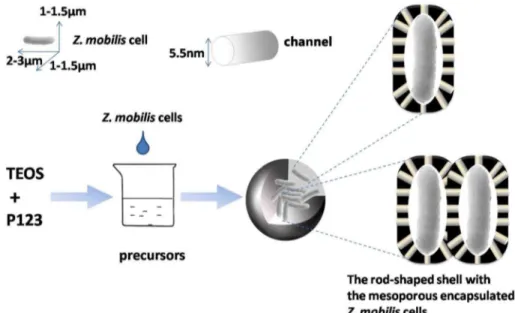
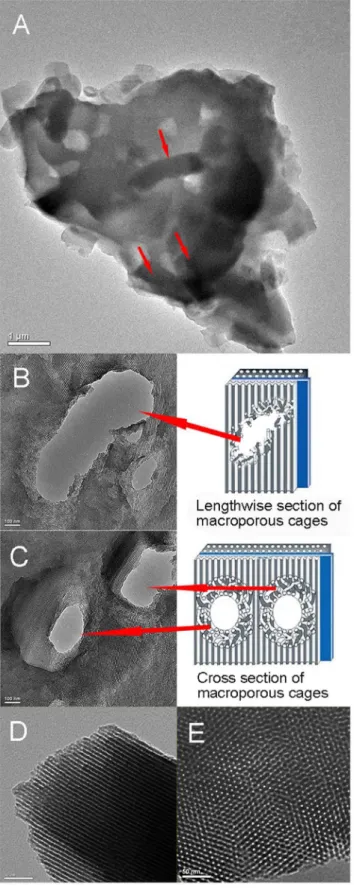
"Fish-in-net", a novel method for cell immobilization of Zymomonas mobilis.
Texto
Imagem
Documentos relacionados
Por sua vez, os granulitos básicos são constituídos, no geral, pela mesma mineralogia dos metatonalitos, exceto pelo empobrecimento em quartzo e o enriquecimento em piroxênios,
Vale salientar que há pessoas que são apenas figurantes, em nada contribuindo para o trabalho das comissões; Em relação às negociações para captação de recursos financeiros, a
“Es cierto que, en principio de su estancia en América, Las Casas recibió indígenas como esclavos: cierto que llegó a sugerir que se importaran negros para aliviar la miserable
Atendendo ao objetivo geral da pesquisa, ou seja, analisar as principais características da liderança transformacional no filme Coach Carter – Treino para a
Palavras-chave: Fluxo de potência sem solução, matriz Jacobiana singular, método de Newton-Raphson amortecido, método de otimização de Newton, método de otimização
Sobre a prática de festivais de cinema como política pública para a promoção cinematográfica, foi analisado que embora a literatura entenda que os festivais são uma prática
The production of sorbitol by permeabilized and immobilized cells of Zymomonas mobilis in Luffa cylindrica was investigated in sucrose medium.. The cell permeabilization showed
Sugar Cane Juice Fermentation by Zymomonas mobilis CP4 Subjected to Inhibition by Ethanol and High Initial Concentration of Substrate..
