Strategy for RNA-Seq Analysis of the
Francisella
Tularensis
LVS Transcriptome during Infection of Murine
Macrophages
Zachary W. Bent
*, David M. Brazel, Mary B. Tran-Gyamfi, Rachelle Y. Hamblin, Victoria A. VanderNoot,
Steven S. Branda
Sandia National Laboratories, Livermore, California, United States of America
Abstract
Francisella tularensis is a zoonotic intracellular pathogen that is capable of causing potentially fatal human infections. Like all successful bacterial pathogens, F. tularensis rapidly responds to changes in its environment during infection of host cells, and upon encountering different microenvironments within those cells. This ability to appropriately respond to the challenges of infection requires rapid and global shifts in gene expression patterns. In this study, we use a novel pathogen transcript enrichment strategy and whole transcriptome sequencing (RNA-Seq) to perform a detailed characterization of the rapid and global shifts in F. tularensis LVS gene expression during infection of murine macrophages. We performed differential gene expression analysis on all bacterial genes at two key stages of infection: phagosomal escape, and cytosolic replication. By comparing the F. tularensis transcriptome at these two stages of infection to that of the bacteria grown in culture, we were able to identify sets of genes that are differentially expressed over the course of infection. This analysis revealed the temporally dynamic expression of a number of known and putative transcriptional regulators and virulence factors, providing insight into their role during infection. In addition, we identified several F. tularensis genes that are significantly up-regulated during infection but had not been previously identified as virulence factors. These unknown genes may make attractive therapeutic or vaccine targets.
Citation: Bent ZW, Brazel DM, Tran-Gyamfi MB, Hamblin RY, VanderNoot VA, et al. (2013) Use of a Capture-Based Pathogen Transcript Enrichment Strategy for RNA-Seq Analysis of the Francisella Tularensis LVS Transcriptome during Infection of Murine Macrophages. PLoS ONE 8(10): e77834. doi: 10.1371/journal.pone.0077834
Editor: Yung-Fu Chang, Cornell University, United States of America
Received July 3, 2013; Accepted September 9, 2013; Published October 14, 2013
This is an open-access article, free of all copyright, and may be freely reproduced, distributed, transmitted, modified, built upon, or otherwise used by anyone for any lawful purpose. The work is made available under the Creative Commons CC0 public domain dedication.
Funding: Sandia National Laboratories is a multi-program laboratory managed and operated by Sandia Corporation, a wholly owned subsidiary of Lockheed Martin Corporation, for the U.S. Department of Energy's National Nuclear Security Administration under contract DE-AC04-94AL85000. The funders had no role in study design, data collection and analysis, decision to publish, or preparation of the manuscript.
Competing interests: The authors have declared that no competing interests exist. * E-mail: [email protected]
Introduction
Francisella tularensis, the causative agent of tularemia, is a Gram-negative facultative intracellular pathogen that is capable of infecting a wide variety of hosts, including mammals, birds, amphibians, fish, and insects [1]. Originally isolated in 1911 in Tulare County, California during a plague-like outbreak in the rodent population, F. tularensis was subsequently found to be endemic in most of the northern hemisphere [2]. Human infections most commonly occur upon contact with infected animals or from the bite of an infected tick, leading to cutaneous ulceroglandular tularemia [3]. A pneumonic infection can result from inhalation of as few as 10 bacteria, leading to severe and often fatal disease [4]. Because of the seriousness of its disease, ability to be aerosolized, and extremely low
As an intracellular pathogen, F. tularensis must adapt to multiple environments throughout the course of an infection. The bacteria enter host cells via phagocytosis, escape the phagosome, replicate within the host cell cytosol, and at later stages of infection are found within a double membranous compartment that resembles an autophagosome [10]. The bacteria infect a variety of cell types in a variety of locations throughout the body, each presenting different stresses and challenges to bacterial survival [7,11]. The bacteria must appropriately respond to each of these microenvironments for the infection to proceed. To accomplish this, F. tularensis must rapidly alter transcription of numerous genes in a coordinated manner as it moves from site to site within the host, as well as within the compartments of individual host cells. Typically, pathogens respond to infection-associated stresses through up-regulation of virulence factors - genes that have been demonstrated by mutational or genetic analysis to play a critical role in the bacteria’s ability to cause disease. These genes encode a wide range of products, including secretion systems, adhesins, invasins, iron uptake pathways, and toxins. During infection pathogens also down-regulate expression of transcripts that are no longer necessary, or are potentially detrimental, in a given microenvironment. These changes in gene expression are ultimately responsible for the success of the pathogen in evading or subverting the immune response and surviving within its host.
Previous work to characterize F. tularensis transcriptome dynamics during infection has focused on the type A strain SCHU S4. Wehrly and colleagues used microarray analysis to track the transcriptional profiles of the bacteria during infection of murine bone marrow-derived macrophages (BMDM) [12]. Walters and colleagues used RNA-Seq to investigate the transcriptome of the bacteria at late time points in the lungs of infected mice [13]. Both studies revealed up-regulation of known and previously unknown virulence factors, demonstrating that distinct stages of F. tularensis infection are accompanied by global changes in transcriptional profile.
Given the importance of these global transcriptional shifts to the virulence and persistence of the bacteria within the host, it is critical to understand when and how these shifts occur. Using RNA-Seq to address this issue presents a technical challenge, because the vast majority of transcripts present in infected cells are derived from the host, rather than from the bacteria of interest. In this study, we use a newly developed capture-based bacterial transcript enrichment strategy [14] to obtain enough pathogen reads , despite the host-dominated background, to analyze the complete transcriptome of F. tularensis LVS during phagosomal escape and cytosolic replication within murine macrophages. Comparison of the transcriptional profiles of the bacteria at these two distinct time points, relative to that of the bacteria in culture, revealed up-regulation of numerous known virulence factors as well as many genes with unknown function that play, as yet, undetermined roles in F. tularensis virulence. Further, by analyzing the expression of the known and putative transcriptional regulators encoded by the F. tularensis genome, we were able to identify pathways and products that are important at each stage of infection.
Results
F. tularensis transcriptional changes during infection F. tularensis bacteria grown to exponential phase in a rich medium, such as the one used in this study, are highly invested in replication, and therefore would be expected to express genes associated with metabolic functions and cell division. Switching from culture in a rich medium to an active infection of host cells presents a specific series of challenges to the bacteria that are addressed through global transcriptional changes. The first phase of F. tularensis infection requires that the bacteria be taken up into the host cell by phagocytosis. This is followed by escape from the phagosome, and establishment of a replicating population within the cytosol of the host cell. The exact timing of these events varies in different F. tularensis strains, host cell types, and infection protocols. However, it is well documented that in the murine macrophage cell line J774A.1, as well as in murine bone marrow derived macrophages (BMDM), after 4 hours of infection the bacteria are in a transition state in which some are still within phagosomes while others have managed to escape into the cytosol [10,15-18]. After 8 hours the vast majority of bacteria are located within the cytosol [10,12,15,18-20]. Work by Mack and colleagues [21] as well as Edwards and colleagues [22] has demonstrated, through direct comparisons, that F. tularensis infections show an essentially identical disease progression in J774A.1 cells as P388D1 cells, the murine macrophage line used in this study.
To characterize the F. tularensis transcriptome dynamics associated with transition from growth in culture to infection of host cells, as well as the transition from phagosome to the cytosol, we performed a differential gene expression analysis of F. tularensis LVS before infection and after 4 or 8 hours of infection. Table S1 presents the biological duplicate FPKM (fragments per kilobase of transcript per million mapped reads) values and differential expression results for all F. tularensis
and 60 genes after 8 hours (Tables S2 and S3, respectively). The highly up-regulated unknown genes at each time-point are especially interesting because they are likely to be important in specific stages of pathogenesis and yet have not been previously identified as virulence factors.
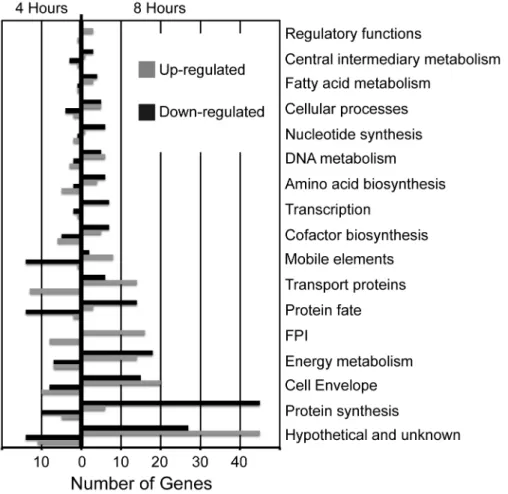
Table 1 lists the F. tularensis genes showing the largest changes in expression during transition from culture to infection. Strikingly, 30-40% of the most strongly up-regulated genes are of unknown function. Among the genes of known function, those that were most strongly up-regulated are involved in purine and amino acid biosynthesis, peptide transport, and competence. The genes most strongly down-regulated are involved in protein synthesis and central metabolism - functions predicted to play diminished roles during infection, as compared to exponential growth in culture. Interestingly, although the genes differentially expressed after 4 hours versus 8 hours of infection are closely related with respect to their annotated functions, only 39 (20.1%) of the up-regulated genes, and 46 (21.8%) of the down-up-regulated genes, were differentially expressed at both time points (Figure 2). The consistency with which these genes were differentially expressed suggests that they represent a core set of genes
whose regulation is sensitive to the environmental changes associated with transition from culture to infection. Of the genes consistently up-regulated during infection (Table S4), ~20% are located within the FPI. Additionally, ~18% of these genes were categorized as transport and binding proteins, a group of proteins that include several genes implicated in virulence such as siderophore synthesis, and transmembrane peptide transport. Of the genes consistently down-regulated during infection (Table S5), ~49% are involved in protein synthesis and fate, and several others in biosynthesis of enzyme cofactors such as riboflavin, cyanophycin, and anthranilate.
Genomic localization of genes differentially expressed during infection
To determine whether the genes differentially expressed during infection are located in particular regions of the F. tularensis genome, we identified the genes showing the largest changes in expression (> 4 fold) after 4 hours and 8 hours of infection, and mapped their locations within the genome. This analysis revealed that most of the differentially expressed genes are broadly distributed throughout the F. tularensis Figure 1. Number of genes up- and down-regulated, by functional category. All differentially expressed F. tularensis LVS genes were categorized by function, and the number of genes in each category were plotted according to whether their expression increased or decreased at 4 hours (left) and 8 hours (right) after infection.
genome (Figure 3). However, notable exceptions include up-regulated genes mapping to the FPI (two copies in F. tularensis
LVS); and two clusters of down-regulated genes, encoding ribosomal proteins and ATP synthase subunits, respectively. These results indicate that the F. tularensis genes most strongly induced or repressed during infection generally are not found within coordinately regulated gene clusters or chromosomal regions, with a few notable exceptions.
Differential expression of transcriptional regulators
To gain further insight into the transcriptional shifts that occur during infection, we analyzed the expression of all known and putative F. tularensis LVS transcriptional regulators [24-31] (Table 2). After 4 hours of infection, the only transcriptional regulators up-regulated by at least 2-fold were fevR and FTL_1216. FevR has been shown to positively regulate expression of genes located within the FPI, as well as several other genes encoding virulence factors [32,33]. FTL_1216 is a putative transcriptional regulator that is conserved across many Gram-negative bacteria, but it has no known function or regulon. Given that FTL_1216 is the transcriptional regulator most strongly up-regulated after 4 hours of infection, but is not significantly up-regulated after 8 hours of infection, it seems likely that it plays an important role in phagosomal escape, but not in replication within the cytosol. The only transcriptional regulators significantly down-regulated after 4 hours of infection were rpoD and migR. Down-regulation of rpoD, which encodes the primary sigma factor, is consistent with the decrease in expression of numerous genes involved in aspects of cell growth (see Figure 1). On the other hand, down-regulation of
migR would appear to contradict previous studies that demonstrated that MigR positively regulates fevR through the stress alarmone ppGpp [33,34]. This apparent contradiction may be explained by the high FPKM values of mglA, sspA, and
pmrA, which also positively regulate fevR [32,34,35] and therefore may compensate for reduced expression of migR.
We also observed up-regulation of oxyR, fur, and a diguanylate cyclase (DGC) (FTL_1218) after 8 hours of infection (Table 2). The oxyR and fur transcriptional regulators have been shown to promote formation of Fe-S clusters and mitigate oxidative damage in E. coli and many other bacteria [36-38] The gene product of oxyR was shown to be important in the response to oxidative stress in F. novicida during infection of Drosophila cells [39]. The ferric uptake regulator, or Fur, coordinates bacterial response to the iron-limited environment of the host, by promoting expression of the fig-fsl
operon (siderophore production) [25,40,41] and other genes encoding virulence factors, including several located within the FPI [33]. Taken together, up-regulation of these three transcriptional regulators indicates that avoiding oxidative stress and scavenging iron and other limiting nutrients, is important for F. tularensis replication within the host cytoplasm. Diguanylate cyclase is responsible for the synthesis of cyclic di-GMP (cddi-GMP), a secondary messenger that promotes biofilm formation and inhibits virulence in F. novicida [42]. Expression of this gene during an infection might partially explain the decreased virulence of this Type B strain compared to more virulent Type A strains. Interestingly, all of the transcriptional regulators that were down-regulated after 8 hours of infection were also down-regulated at 4 hours. This suggests that despite their localization to different intracellular compartments
Table 1. Genes showing the largest changes in expression during infection.
Gene ID Name/Function 4hr Fold Change Adj P-Value Gene ID Name/Function 8hr Fold Change Adj P-Value
FTL_0721 DedA family protein* 9.69 0.016 FTL_0815 PRC-barrel protein* 28.51 <0.001
FTL_1213 Unknown* 8.39 <0.001 FTL_1402 ISFtu1 transposase 13.91 <0.001
FTL_1216 Unknown* 8.16 <0.001 FTL_0953 methyltransferase 12.72 <0.001
FTL_1876 Outer membrane protein* 8.14 0.001 FTL_0814 PRC-barrel protein* 12.03 0.003
FTL_1509 Carboxypeptidase 7.82 0.007 FTL_0924 Oligopeptide transporter 10.90 0.037
FTL_0765 vacJ/lipoprotein 7.70 <0.001 FTL_1219 Aminotransferase 8.76 <0.001
FTL_0731 YhhQ family/purine regulon 6.69 0.011 FTL_1957 Heat shock 8.43 <0.001
FTL_0700 comL/competence 5.80 0.001 FTL_0816 Unknown* 8.18 .05
FTL_1219 Aminotransferase 5.42 <0.001 FTL_0123 Short chain dehydrogenase 8.13 <0.001
FTL_0691 Oligopeptide transport 5.24 <0.001 FTL_0473 Peptide deformylase 7.80 <0.001
FTL_1127 ISFtu1 transposase -4.10 <0.001 FTL_0916 Ketol-acid reductoisomerase -6.76 <0.001
FTL_0128 ISFtu1 transposase -4.10 0.001 FTL_0243 rplP/ribosomal protein -6.99 <0.001
FTL_0579 Nicotinate metabolism -4.12 0.028 FTL_0204 Unknown* -7.14 0.002
FTL_0732 Lactoylglutathione lyase -4.17 0.001 FTL_0239 rplB/ribosomal protein -7.25 <0.001
FTL_0227 Ribosome recycling factor -4.31 0.002 FTL_0075 Riboflavin synthase -7.35 <0.001
FTL_0266 ISFtu1 transposase -4.81 0.028 FTL_1139 3-oxoacyl reductase -7.52 <0.001
FTL_0799 Type IV pili lipoprotein* -4.88 0.035 FTL_0241 rplV/ribosomal protein -8.70 <0.001
FTL_0964 hslU/heat shock -5.03 <0.001 FTL_1796 ATP synthase γ-subunit -9.71 <0.001
FTL_0965 hslV/heat shock -7.19 0.005 FTL_0244 L29 ribosomal protein -11.24 <0.001
FTL_1128 Unknown* -10.80 <0.001 FTL_0238 rplW/ribosomal protein -11.49 <0.001
at these two time points, the bacteria rely upon similar mechanisms to survive both stages of infection.
Up-regulation of genes within the FPI
The FPI is an ~30 kb region encompassing 18 genes, that primarily encode an atypical type VI secretion system (T6SS) [43,44]. Most of the genes within the FPI have been implicated in virulence during at least one stage of infection, and were among the first recognized Francisella virulence factors [23,45]. We compared their expression levels during growth in culture
versus after 4 hours or 8 hours of infection (Table 3). Consistent with our observation that fevR, a key positive regulator of FPI gene expression [32,33,46], is strongly up-regulated during infection, we found that the FPI genes were up-regulated after 4 hours of infection. Roughly half of the FPI genes show significant increases in expression at this stage of infection, and all except pdpC show higher levels of expression than measured in the bacteria grown in culture. After 8 hours of infection, all FPI genes were significantly up-regulated except for anmK and pdpD, both of which showed extremely low expression levels throughout the entire course of the
Figure 2. Comparison of the genes up- and down-regulated at each time point. The Venn diagrams depict the number of genes with significant changes in expression at both the 4 and 8-hour post-infection time points, with the number in the middle representing genes up- or down-regulated at both time points. A) Up-regulated genes. B) Down-regulated genes.
doi: 10.1371/journal.pone.0077834.g002
experiment. Interestingly, pdpE, the only gene in the FPI that has not been shown to be necessary for full virulence [23], was significantly up-regulated after 8 hours of infection, indicating that while it may not be required for virulence, pdpE likely plays a role in F. tularensis replication in the cytoplasm. Also consistent with the FPI being coordinately regulated is the fact that the recognized virulence factors within the FPI all clustered with regard to their transcriptional shifts at 4 and 8 hours post-infection, showing the general trend of slight up-regulation at 4 hours followed by strong up-regulation at 8 hours (Figure 4). Overall, these results are consistent with those from previous studies that implicate the FPI and its encoded T6SS in F. tularensis virulence, and reinforce the idea that FevR regulates these genes.
Expression of non-FPI genes encoding virulence factors
While the FPI genes encode the best characterized of F. tularensis virulence factors, numerous non-FPI genes have been shown to be required for full virulence in vitro and in vivo, through use of mutational analysis screens [47-55]. However, mutational analysis has provided limited insight into the role of each virulence factor in the different stages of infection. For example, mutant bacteria that fail to enter the host cell or escape the phagosome are attenuated for overall virulence, yet they might be capable of intracellular growth and replication. To further elucidate the roles of non-FPI genes previously shown to be required for full virulence, we analyzed their expression during phagosomal escape and intracellular replication and compared it to their expression during growth in culture (Table 4). We found that five of the non-FPI genes encoding virulence factors were significantly up-regulated after 4 hours of infection, but only one of these was also significantly up-regulated after 8 hours of infection. Figure 4 shows two gene clusters (Cluster 2 and 3) that differ in expression from the pattern seen in the FPI. Cluster 2 contains genes that are up-regulated after four hours of infection, and show reduced expression after 8 hours of infection. Cluster 3 is comprised of the few virulence factors that are down-regulated at both time points relative to the bacteria in culture, suggesting that they may not be important for infection under the particular conditions of our model system, or that they play a role at earlier or later stages of infection not analyzed in this study [10].
required for full virulence in cultured macrophages as well as in mice [46]; however, Wehrly and colleagues found no such requirement when infecting cultured macrophages [12]. The fact that we observed high levels of FTL_1213 expression after 4 hours of infection, and reduced levels of expression after 8 hours of infection, suggests that its gene product may be more important for phagosomal escape than for subsequent stages of infection. FTL_1306 (FTT_0369c), the only non-FPI virulence-associated gene that was significantly up-regulated at both time points, has been shown to be required for F. tularensis replication in the cytosol [12]; however, its specific function has yet to be determined.
It should be noted that two non-FPI genes encoding virulence factors (htpG and purCD) were significantly down-regulated after 4 hours and 8 hours of infection. HtpG is a heat shock protein that was shown to be required for virulence in macrophages and mice [54,55], whereas purCD was shown to be required for purine biosynthesis during infection [57,58]. It remains to be determined whether down-regulation of htpG and
purCD during infection is observed in other model systems, or is a unique feature of F. tularensis infection of P388D1 macrophages.
In summary, the results of these analyses, as well as those presented in Figure 4, fit well with those from previous studies, and show that expression of the non-FPI virulence-associated genes is less coordinated than that of the FPI genes. Further research will be necessary to elucidate the specific functions and roles in pathogenesis of the uncharacterized but up-regulated virulence factors FTL_0073, FTL_1213, and FTL_1306.
Discussion
F. tularensis has the ability to infect multiple cell types and exist within multiple intracellular compartments during infection of the host. The requirements for survival and proliferation during infection have been studied both in vitro and in vivo, primarily through mutational analysis to identify the genes that are critical for virulence [47-55]. While this approach has been highly successful in discovering genes that are required for full virulence of the bacteria in a given model system of infection, it is not without its disadvantages. One significant drawback to these types of studies is that they often fail to determine the stage(s) of infection for which the genes are required. For example, a gene that is required for the initial entry into a host cell will be identified as critical for virulence, however it is typically not possible to determine whether this gene is also involved in phagosomal escape or replication within the host cell cytosol, as a mutant for that gene will not proceed to those stages of infection. To understand when and where genes are expressed throughout the course of an infection, transcriptional analyses are required. Global analysis of the transcriptome can be performed using either microarrays designed specifically for the pathogen of interest or, more recently, by sequencing total RNA (RNA-Seq) from an infected sample. While RNA-Seq is a relatively new technology, its sensitivity, dynamic range, low cost, and ability to detect non-protein-coding transcripts is unmatched by microarray-based approaches.
A major consideration for either transcriptomics approach is that the RNA recovered from virtually any infection is primarily host-derived, with the pathogen RNA outnumbered by well over 100-fold [14,59]. Using RNA-Seq to analyze the pathogen transcriptome under these circumstances becomes expensive, as deeper sequencing is required to get enough reads for a
Figure 3. Differentially expressed genes plotted across the F. tularensis genome. All genes that had at least a 4-fold change in expression at either 4 hours (red) or 8 hours (blue) were plotted according to their gene ID number across the genome. The two copies of the FPI are highlighted in the up-regulated portion of the figure, and the ribosomal proteins and ATP synthase subunits are highlighted in the down-regulated portion of the figure.
gene-level analysis throughout the course of an infection. Prior work in our lab has demonstrated the effectiveness of a capture-based approach to enrich for pathogen transcripts from infected cells [14]. This technique relies on the use of biotinylated probe sequences randomly generated from the entire bacterial genome, ensuring that all possible transcripts can be captured. Double-stranded and tagged cDNA generated from the infected sample [60] are mixed with a large excess of capture probe. The mixture is denatured, and stringent hybridization conditions are established to allow the pathogen-derived cDNA to anneal to complementary capture probe sequences. The hybridization mixture is then adsorbed to a monomeric avidin column, washed repeatedly to remove cDNA non-specifically bound to the probe, and the remaining cDNA released from the column to generate a pool enriched for pathogen-derived sequences. The short tags at the ends of the cDNA allow PCR-mediated addition of full-length sequencing adaptors, whereas the probes lack these tags preventing inadvertent sequencing of the probe. Using a F. tularensis LVS
infection model, we previously demonstrated unbiased enrichment of bacterial transcripts by upwards of 50-fold [14]. This enrichment of pathogen transcripts allows for much more efficient sequencing of the bacterial transcriptome at any stage of infection, as compared to brute-force RNA-Seq without enrichment.
Given the proven ability of our pathogen capture approach to enrich for F. tularensis transcripts present in infected samples, we employed the technique to perform a differential gene expression analysis comparing the transcriptomes of the bacteria before and after infection. By observing the transcriptional profiles of the bacteria at two distinct time points after entry into the host cell, we hypothesized that it would be possible to determine sets of genes that were important in two critical stages of the infection: phagosomal escape, and cytosolic growth. At each time point we analyzed the global transcriptional shifts with respect to changes in expression of functional categories of genes as well as sets of known and putative transcriptional regulators and virulence factors.
Table 2. Expression of known and putative transcriptional regulators.
Gene ID Name/Family
Control Expression (FPKM)
4hr Expression (FPKM)
DESeq Fold
Change Adj P-Value
8hr Expression (FPKM)
DESeq Fold
Change Adj P-Value
FTL_0040 LysR family 2.19 1.82 1.06 1 0.21 0.17 0.793
FTL_0062 LysR famiy 1.56 1.19 1.89 1 0.40 1.38 1
FTL_0449 fevR 53.85 155.02 3.19* <0.001 201.04 6.80* <0.001
FTL_0552 pmrA 67.28 31.23 0.54 0.209 26.04 0.59 0.625
FTL_0662 lexA 21.49 9.16 0.62 0.763 11.84 1.06 1
FTL_0671 panK1 17.95 13.55 1.25 0.945 9.79 1.36 0.624
FTL_0689 AraC family 1.94 2.50 1.42 1 1.31 0.87 1
FTL_0742 LysR famliy 2.44 0.84 1.20 1 0.90 2.28 0.793
FTL_0780 Csp family 1.10 1.88 2.24 1 0.50 0.88 1
FTL_0844 LysR family 3.64 1.94 0.90 1 1.68 2.60 0.477
FTL_0851 rpoH 44.85 11.39 0.54 0.09 9.72 0.68 0.229
FTL_1014 oxyR 4.82 2.79 2.40 0.306 1.71 3.59* 0.004
FTL_1050 rpoD 37.00 10.44 0.38* <0.001 8.08 0.44* <0.001
FTL_1125 hipA 1.79 2.03 1.58 1 0.36 0.54 0.939
FTL_1126 XRE family 20.16 10.96 0.89 1 3.10 0.21 0.781
FTL_1176 LysR family 0 0 NA 1 0 NA 1
FTL_1185 mglA 45.19 53.93 1.86 0.062 10.63 0.42 0.088
FTL_1193 LysR family 1.00 0.49 0.82 0.823 0.64 0.86 1
FTL_1216 Unknown 3.50 18.8 8.11* <0.001 2.01 2.38 0.391
FTL_1218 DGC 25.35 24.91 1.38 0.568 34.00 2.66* <0.001
FTL_1231 iscR 12.83 14.33 2.08 0.458 18.78 3.21 0.088
FTL_1277 ROK famliy 7.97 4.48 0.80 1 2.12 0.71 0.838
FTL_1364 IclR family 66.38 25.62 0.55 0.085 24.03 0.67 0.21
FTL_1542 migR 26.86 5.49 0.29* <0.001 3.77 0.32* <0.001
FTL_1568 LysR family 0.76 0.40 1.22 1 0.16 1.26 1
FTL_1606 sspA 60.64 24.38 0.61 0.384 35.58 1.20 0.694
FTL_1634 LysR family 5.62 4.69 1.16 1 4.15 1.38 0.83
FTL_1665 panK2 0.70 1.18 2.34 0.713 1.00 2.65 0.371
FTL_1763 qseC 2.53 1.05 0.90 1 2.02 1.57 0.84
FTL_1831 fur 32.00 34.90 1.49 0.751 38.49 2.26* 0.037
FTL_1878 kdpD 5.27 4.05 1.1 1 3.37 1.68 0.296
The challenge faced by bacteria as they shift from culture to infection of host cells can be summed up as a change from replication in a protected environment to survival in a threatening environment. Consistent with this idea, we found
that transition from culture to infection was generally associated with up-regulation of genes involved in virulence and stress response, and down-regulation of genes involved in replication. These results were consistent with expectations,
Table 3. Expression of Francisella pathogenicity island (FPI) genes
Gene ID Name
Control Expression (FPKM)
4hr Expression
(FPKM) DESeq Fold Change Adj P-Value
8hr Expression
(FPKM) DESeq Fold Change Adj P-Value
FTL_0109 anmK 2.30 1.08 1.52 1 1.21 1.63 1
FTL_0110 pdpD 0 0 NA 1 0 NA 1
FTL_0111 iglA 80.43 114.70 2.52* <0.001 162.89 4.28* <0.001
FTL_0112 iglB 87.71 127.90 2.14* <0.001 138.17 3.74* <0.001
FTL_0113 iglC 369.21 452.55 1.97 <0.001 908.81 5.43* <0.001
FTL_0114 iglD 39.91 37.32 1.69 0.168 47.34 3.95* <0.001
FTL_0115 pdpE 14.45 8.98 1.53 0.803 13.14 2.43* 0.025
FTL_0116 pdpC 11.10 7.34 0.94 1 11.27 2.21* <0.001
FTL_0117 iglJ 14.78 15.49 1.73 0.347 13.44 3.01* <0.001
FTL_0118 iglI 16.90 24.46 2.03* 0.047 30.40 2.96* <0.001
FTL_0119 dotU 23.43 36.80 2.24 0.093 32.17 2.88* 0.002
FTL_0120 iglH 10.12 18.47 2.53* 0.004 22.44 4.40* <0.001
FTL_0121 iglG 31.14 61.00 2.99* 0.033 88.83 6.12* <0.001
FTL_0122 iglF 3.47 5.13 2.37 0.489 10.13 6.29* <0.001
FTL_0123 vrgG 5.29 16.76 4.27* 0.047 15.51 8.13* <0.001
FTL_0124 iglE 8.79 8.95 2.07 0.705 16.09 6.23* <0.001
FTL_0125 pdpB 7.87 10.48 1.91 0.016 19.24 4.93* <0.001
FTL_0126 pdpA 10.06 22.51 3.17* <0.001 34.34 6.85* <0.001
*. Differential expression (≥2 fold change in expression, p≤0.05) doi: 10.1371/journal.pone.0077834.t003
Table 4. Expression of non-FPI genes encoding virulence factors.
Gene ID Name
Control Expression (FPKM)
4hr Expression (FPKM)
DESeq Fold
Change Adj P-Value
8hr Expression (FPKM)
DESeq Fold
Change Adj P-Value
FTL_0028 pyrB 1.81 2.89 2.01 0.879 1.24 1.00 1
FTL_0029 carB 4.97 6.32 1.86 0.118 1.28 0.64 0.461
FTL_0030 carA 4.86 12.12 3.71* 0.007 2.92 1.67 0.576
FTL_0031 Hap 3.92 2.85 0.98 1 0.63 0.30 0.415
FTL_0073 Lipoprotein 23.98 40.22 2.44* <0.001 19.02 1.57 0.161
FTL_0158 acpA 7.98 5.50 1.36 0.818 6.07 1.83 0.183
FTL_0267 htpG 135.54 30.60 0.32* <0.001 16.20 0.23* <0.001
FTL_0395 purM 31.28 18.23 0.81 0.922 10.96 0.73 0.489
FTL_0396 purCD 29.41 13.04 0.63 0.066 6.11 0.40* <0.001
FTL_0552 pmrA 67.28 31.23 0.55 0.209 26.04 0.83 0.625
FTL_0766 ggt 4.38 8.06 2.59* 0.04 4.00 1.93 0.182
FTL_0889 acpC 0 0 NA 1 0 NA 1
FTL_1096 Thioredoxin 26.23 27.47 1.49 0.289 20.31 1.54 0.147
FTL_1184 mglB 31.23 24.99 1.60 0.971 9.16 0.75 0.98
FTL_1213 Unknown 2.67 16.30 8.39* <0.001 1.09 0.99 1
FTL_1306 Unknown 13.50 29.18 2.99* <0.001 14.13 2.10* 0.029
FTL_1670 dsbB 9.39 14.51 2.25 0.522 4.12 0.95 1
FTL_1732 acpB 11.70 5.21 0.59 0.868 0.98 0.46 0.437
FTL_1914 ripA 167.89 138.03 1.20 0.608 126.99 1.42 0.095
and indicated that our techniques are effective at detecting the previously-identified transcriptional shifts that occur during infection. However, the switch from growth in culture to infection of host cells is both spatially and temporally dynamic, with different cell types and intracellular compartments presenting different challenges to bacterial survival. This is why, when the two stages of infection were analyzed in greater detail, we observed analogous but distinctive gene expression patterns associated with phagosomal escape and cytosolic replication.
Perhaps the greatest insight into the global transcriptional shifts that occur during different stages of infection can be obtained through analysis of transcriptional regulators. Interestingly, very few regulatory proteins have been identified in F. tularensis; indeed, its genome shows a complete lack of classically arranged two-component regulatory systems and only one alternative sigma factor [24,35,61,62]. Previous work has identified only 31 potential transcriptional regulators in F. tularensis LVS [24-30], a number that is considerably lower
Figure 4. Heat map of virulence factor genes up- and down-regulated at each time point. Change in expression was determined for previously identified F. tularensis virulence factor genes at both post-infection time points, and then clustered to identify genes that are coordinately regulated. The cluster analysis segregated the genes into three groups. Cluster 1, in which the genes are up-regulated at both post-infection time points, is comprised entirely of genes in the FPI.
doi: 10.1371/journal.pone.0077834.g004
than in related bacteria such as E. coli, which has over 250 putative transcription factors [63]. Despite the substantial number of genes that showed significant changes in expression during the course of the infection (405 in total), we found that only 7 of the previously identified regulators showed a significant change in expression relative to the bacteria grown in culture. Combined with the fact that there are so few transcription factors identified to date, this may indicate that F. tularensis regulates gene expression using different mechanisms than those examined in this study. These could include use of unusual transcription factors, anti-sense RNAs, or post-transcriptional modifications that lead to the transcriptome shifts that we observed. One of the more interesting observations in this work is the up-regulation of the putative transcription factor FTL_1216 after 4 hours of infection. Though its function is not yet known, the gene is conserved in many bacteria and, based on our results, likely plays a role in regulating the expression of genes involved in phagosomal escape. More work will be necessary to determine the exact nature of its role in virulence and to discover the genes in its regulon.
Like all pathogens, F. tularensis expresses a suite of virulence factors required for completion of its pathogenic life cycle. The best characterized of these are the genes comprising the FPI and its encoded T6SS. While there is still much work to be done to understand the precise functions of these genes, it is clear from our study, and from those of others, that FPI genes play important roles in phagosomal escape and cytosolic replication. Indeed, the FPI genes were the only virulence-associated genes (aside from FTL_1306) that showed up-regulation after both 4 hours and 8 hours of infection. A number of non-FPI genes are associated with virulence, but strikingly few were differentially expressed during infection in our model system. One intriguing exception is FTL_1213, which showed up-regulation after 4 hours of infection, suggesting that this virulence-associated gene may play an important role in phagosomal escape. Its chromosomal proximity and similar expression profile to FTL_1216, a putative transcriptional regulator, suggests that FTL_1213 may belong to the regulon controlled by FTL_1216, perhaps acting in concert with other similarly regulated virulence factors to promote escape from the phagosome.
there is still a significant amount to learn about the molecular mechanisms of F. tularensis pathogenesis.
Materials and Methods
Infection of Murine Macrophages and RNA Extraction
Infections and RNA extraction were performed in biological duplicates as previously described [14]. Briefly, P388D1 cells obtained from ATCC (ATCC® CCL-46™) were grown in six-well
microtiter plates overnight in RPMI media (Life Technologies) supplemented to a final concentration of 10% fetal bovine serum. Francisella tularensis spp. holarctica LVS obtained from BEI (NR-646) was grown in Francisella broth (BHI supplemented with 17.5g/L casamino acids and 2% isovitalex) overnight at 37°C in a shaking incubator. Bacterial concentrations were determined by OD600 with comparison to a
previously established standard curve. Approximately 1.45x10⁶
P388D1 cells were infected with the overnight culture of F. tularensis LVS to produce a multiplicity of infection (MOI) of 10. Biological duplicate samples were taken at 4 hours and 8 hours post infection, washed twice with PBS to remove non-adherent or non-internalized bacteria, and 1mL of RNAzol (Molecular Research Center, Inc.) was immediately added to each well. RNA was extracted in combination with the Direct-zol kit (Zymo Research) according to the manufacturer’s instructions. Total RNA was also extracted in biological duplicates from 1mL of the overnight F. tularensis culture used as the inoculum, using the technique described above. In all cases, the RNA was quantitated using a Qubit (Life Technologies) and run on a BioAnalyzer (Agilent) to determine its integrity.
RNA-Seq library preparation and sequencing
Double-stranded, tagged cDNA was generated from total RNA as previously described [14,60]. For the infection samples, 20ng of cDNA was mixed with 2μg of biotinylated probes generated against the entire F. tularensis genome using the BioPrime DNA Labeling System (Life Technologies), denatured, and allowed to hybridize overnight. Following hybridization, probe-bound F. tularensis transcripts were selectively removed from the pool using monomeric avidin agarose (Pierce/Thermo) as previously described [14]. The optimal cycle number for indexing PCR was determined by qPCR, and samples were barcoded using custom indexing primers [60]. Libraries were combined in equal molar amounts and visualized using the Bionalyzer (Agilent). The Vincent J. Coates Genomics Sequencing Laboratory (University of California, Berkeley) performed 100-base, single-end sequencing using an Illumina HiSeq 2000. All quality filtered reads have been deposited in the NCBI Sequence Read Archive (SRA) with the accession number PRJNA213748
RNA-Seq data analysis and statistical determination of differentially expressed genes
Raw reads were processed using a previously described quality filter designed to remove low quality reads or sections of reads as well as any sequences derived from the sequencing adaptors or primers [64]. The quality filtered FASTQ files were
mapped to the Francisella tularensis LVS genome (NC_007880) with Bowtie 2 in local alignment mode [65]. The alignments were converted and sorted with the SAMtools package [66]. For the differential expression analysis, read counts were generated for each CDS in the NCBI RefSeq annotation of the LVS genome with the BEDTools multicov tool [67]. Differentially expressed genes were identified at each time point with the R package DESeq [68], by comparing the read counts of each CDS at four and eight hours to those in the culture-grown control. This package tests for differential expression through the application of the negative binomial distribution and a shrinkage estimator for the distribution’s variance. Normalized expression levels among the various samples were obtained by estimating the total sequencing depths for each sample as the median of the ratios of the sample’s counts to geometric mean across all samples. Further details of the statistical analyses can be found in the DESeq vignette (http://www.bioconductor.org/ packages/2.12/bioc/ vignettes/DESeq/inst/doc/DESeq.pdf). Genes were identified as differentially expressed when the DESeq calculated adjusted p-value was less than 0.05 and the change in expression was at least two-fold up or down. FPKM values for each annotated CDS were calculated from the alignments by providing Cufflinks with a reference annotation [69]. Each gene’s functional category was determined by the J. Craig Venter Institute’s Comprehensive Microbial Resource (cmr.jcvi.org)
Supporting Information
Table S1. FPKM values and differential expression results for all F. tularensis LVS genes in duplicate at each time point compared to the control.
(XLSX)
Table S2. Genes of unknown function that are differential expressed after 4 hours of infection.
(DOC)
Table S3. Genes of unknown function that are differential expressed after 8 hours of infection.
(DOC)
Table S4. Genes up-regulated at both the 4 and 8-hour time points.
(DOC)
Table S5. Genes down-regulated at both the 4 and 8-hour time points.
(DOC)
Acknowledgements
Repository, NIAD, NIH: Francisellatularensis subsp. holarctica
LVS (FSC155), NR-646.
Author Contributions
Conceived and designed the experiments: ZWB DMB VAV SSB. Performed the experiments: ZWB DMB MBT RYH.
Analyzed the data: ZWB DMB. Wrote the manuscript: ZWB DMB SSB.
References
1. Mörner T (1992) The ecology of tularaemia. Rev Sci Tech 11: 1123-1130. PubMed: 1305858.
2. Ellis J, Oyston PC, Green M, Titball RW (2002) Tularemia. Clin Microbiol Rev 15: 631-646. doi:10.1128/CMR.15.4.631-646.2002. PubMed: 12364373.
3. McLendon MK, Apicella MA, Allen LA (2006) Francisellatularensis: taxonomy, genetics, and Immunopathogenesis of a potential agent of biowarfare. Annu Rev Microbiol 60: 167-185. doi:10.1146/ annurev.micro.60.080805.142126. PubMed: 16704343.
4. Oyston PC, Sjostedt A, Titball RW (2004) Tularaemia: bioterrorism defence renews interest in Francisellatularensis. Nat Rev Microbiol 2: 967-978. doi:10.1038/nrmicro1045. PubMed: 15550942.
5. Dennis DT, Inglesby TV, Henderson DA, Bartlett JG, Ascher MS et al. (2001) Tularemia as a biological weapon: medical and public health management. JAMA 285: 2763-2773. doi:10.1001/jama.285.21.2763. PubMed: 11386933.
6. Rotz LD, Khan AS, Lillibridge SR, Ostroff SM, Hughes JM (2002) Public health assessment of potential biological terrorism agents. Emerg Infect Dis 8: 225-230. doi:10.3201/eid0802.010164. PubMed: 11897082. 7. Oyston PC (2008) Francisellatularensis: unravelling the secrets of an
intracellular pathogen. J Med Microbiol 57: 921-930. doi:10.1099/jmm. 0.2008/000653-0. PubMed: 18628490.
8. Crane DD, Scott DP, Bosio CM (2012) Generation of a convalescent model of virulent Francisellatularensis infection for assessment of host requirements for survival of tularemia. PLOS ONE 7: e33349. doi: 10.1371/journal.pone.0033349. PubMed: 22428026.
9. Fortier AH, Slayter MV, Ziemba R, Meltzer MS, Nacy CA (1991) Live vaccine strain of Francisella tularensis: infection and immunity in mice. Infect Immun 59: 2922-2928. PubMed: 1879918.
10. Checroun C, Wehrly TD, Fischer ER, Hayes SF, Celli J (2006) Autophagy-mediated reentry of Francisellatularensis into the endocytic compartment after cytoplasmic replication. Proc Natl Acad Sci U S A 103: 14578-14583. doi:10.1073/pnas.0601838103. PubMed: 16983090.
11. Ray K, Marteyn B, Sansonetti PJ, Tang CM (2009) Life on the inside: the intracellular lifestyle of cytosolic bacteria. Nat Rev Microbiol 7: 333-340. doi:10.1038/nrmicro2112. PubMed: 19369949.
12. Wehrly TD, Chong A, Virtaneva K, Sturdevant DE, Child R et al. (2009) Intracellular biology and virulence determinants of Francisellatularensis
revealed by transcriptional profiling inside macrophages. Cell Microbiol 11: 1128-1150. doi:10.1111/j.1462-5822.2009.01316.x. PubMed: 19388904.
13. Walters KA, Olsufka R, Kuestner RE, Cho JH, Li H et al. (2013)
Francisellatularensis subsp. tularensis Induces a Unique Pulmonary Inflammatory Response: Role of Bacterial Gene Expression in Temporal Regulation of Host Defense Responses. PLOS ONE 8: e62412. doi:10.1371/journal.pone.0062412. PubMed: 23690939. 14. Bent ZW, Tran-Gyamfi MB, Langevin SA, Brazel DM, Hamblin RY et al.
(2013) Enriching pathogen transcripts from infected samples: A capture-based approach to enhanced host-pathogen RNA sequencing. Anal Biochem 438: 90-96. doi:10.1016/j.ab.2013.03.008. PubMed: 23535274.
15. Clemens DL, Lee BY, Horwitz MA (2004) Virulent and avirulent strains of Francisellatularensis prevent acidification and maturation of their phagosomes and escape into the cytoplasm in human macrophages. Infect Immun 72: 3204-3217. doi:10.1128/IAI.72.6.3204-3217.2004. PubMed: 15155622.
16. Golovliov I, Baranov V, Krocova Z, Kovarova H, Sjöstedt A (2003) An attenuated strain of the facultative intracellular bacterium Francisella tularensis can escape the phagosome of monocytic cells. Infect Immun 71: 5940-5950. doi:10.1128/IAI.71.10.5940-5950.2003. PubMed: 14500514.
17. McCaffrey RL, Allen LA (2006) Francisellatularensis LVS evades killing by human neutrophils via inhibition of the respiratory burst and phagosome escape. J Leukoc Biol 80: 1224-1230. doi:10.1189/jlb. 0406287. PubMed: 16908516.
18. Santic M, Molmeret M, Abu Kwaik Y (2005) Modulation of biogenesis of the Francisella tularensis subsp. novicida-containing phagosome in quiescent human macrophages and its maturation into a phagolysosome upon activation by IFN-gamma. Cell Microbiol 7: 957-967. doi:10.1111/j.1462-5822.2005.00529.x. PubMed: 15953028. 19. Chong A, Wehrly TD, Nair V, Fischer ER, Barker JR et al. (2008) The
early phagosomal stage of Francisellatularensis determines optimal phagosomal escape and Francisella pathogenicity island protein expression. Infect Immun 76: 5488-5499. doi:10.1128/IAI.00682-08. PubMed: 18852245.
20. Lindgren H, Golovliov I, Baranov V, Ernst RK, Telepnev M et al. (2004) Factors affecting the escape of Francisella tularensis from the phagolysosome. J Med Microbiol 53: 953-958. doi:10.1099/jmm. 0.45685-0. PubMed: 15358816.
21. Mack K, Fulop M, Manchee RJ, Stirling CJ (1994) A new cell assay to determine the virulence of Francisellatularensis. Lett Appl Microbiol 19: 158-160. doi:10.1111/j.1472-765X.1994.tb00931.x.
22. Edwards JA, Zemska O, Rappleye CA (2011) Discovery of a role for Hsp82 in Histoplasma virulence through a quantitative screen for macrophage lethality. Infect Immun 79: 3348-3357. doi:10.1128/IAI. 05124-11. PubMed: 21606189.
23. Broms JE, Sjostedt A, Lavander M (2010) The Role of the Francisella tularensis Pathogenicity Island in Type VI Secretion, Intracellular Survival, and Modulation of Host Cell Signaling. Front Microbiol 1: 136. PubMed: 21687753.
24. Dai S, Mohapatra NP, Schlesinger LS, Gunn JS (2010) Regulation of
Francisella tularensis virulence. Front Microbiol 1: 144. PubMed: 21687801.
25. Deng K, Blick RJ, Liu W, Hansen EJ (2006) Identification of Francisella tularensis genes affected by iron limitation. Infect Immun 74: 4224-4236. doi:10.1128/IAI.01975-05. PubMed: 16790797.
26. Charity JC, Blalock LT, Costante-Hamm MM, Kasper DL, Dove SL (2009) Small molecule control of virulence gene expression in
Francisellatularensis. PLOS Pathog 5: e1000641. PubMed: 19876386. 27. Charity JC, Costante-Hamm MM, Balon EL, Boyd DH, Rubin EJ et al.
(2007) Twin RNA polymerase-associated proteins control virulence gene expression in Francisellatularensis. PLOS Pathog 3: e84. doi: 10.1371/journal.ppat.0030084. PubMed: 17571921.
28. Mortensen BL, Fuller JR, Taft-Benz S, Kijek TM, Miller CN et al. (2010) Effects of the putative transcriptional regulator IclR on Francisella tularensis pathogenesis. Infect Immun 78: 5022-5032. doi:10.1128/IAI. 00544-10. PubMed: 20921148.
29. Grall N, Livny J, Waldor M, Barel M, Charbit A et al. (2009) Pivotal role of the Francisella tularensis heat-shock sigma factor RpoH. Microbiology 155: 2560-2572. doi:10.1099/mic.0.029058-0. PubMed: 19443547.
30. Bönquist L, Lindgren H, Golovliov I, Guina T, Sjöstedt A (2008) MglA and Igl proteins contribute to the modulation of Francisellatularensis
live vaccine strain-containing phagosomes in murine macrophages. Infect Immun 76: 3502-3510. doi:10.1128/IAI.00226-08. PubMed: 18474647.
31. Sammons-Jackson WL, McClelland K, Manch-Citron JN, Metzger DW, Bakshi CS et al. (2008) Generation and characterization of an attenuated mutant in a response regulator gene of Francisella tularensis live vaccine strain (LVS). DNA Cell Biol 27: 387-403. doi: 10.1089/dna.2007.0687. PubMed: 18613792.
32. Brotcke A, Monack DM (2008) Identification of fevR, a novel regulator of virulence gene expression in Francisellanovicida. Infect Immun 76: 3473-3480. doi:10.1128/IAI.00430-08. PubMed: 18559431.
33. Buchan BW, McCaffrey RL, Lindemann SR, Allen LA, Jones BD (2009) Identification of migR, a regulatory element of the Francisellatularensis
live vaccine strain iglABCD virulence operon required for normal replication and trafficking in macrophages. Infect Immun 77: 2517-2529. doi:10.1128/IAI.00229-09. PubMed: 19349423.
FPI gene regulation and intracellular growth by modulation of the stress alarmone ppGpp. Infect Immun.
35. Mohapatra NP, Soni S, Bell BL, Warren R, Ernst RK et al. (2007) Identification of an orphan response regulator required for the virulence of Francisella spp. and transcription of pathogenicity island genes. Infect Immun 75: 3305-3314. doi:10.1128/IAI.00351-07. PubMed: 17452468.
36. Yeo WS, Lee JH, Lee KC, Roe JH (2006) IscR acts as an activator in response to oxidative stress for the suf operon encoding Fe-S assembly proteins. Mol Microbiol 61: 206-218. doi:10.1111/j. 1365-2958.2006.05220.x. PubMed: 16824106.
37. Lee KC, Yeo WS, Roe JH (2008) Oxidant-responsive induction of the suf operon, encoding a Fe-S assembly system, through Fur and IscR in
Escherichiacoli. J Bacteriol 190: 8244-8247. doi:10.1128/JB.01161-08. PubMed: 18849427.
38. Chiang SM, Schellhorn HE (2012) Regulators of oxidative stress response genes in Escherichiacoli and their functional conservation in bacteria. Arch Biochem Biophys 525: 161-169. doi:10.1016/j.abb. 2012.02.007. PubMed: 22381957.
39. Moule MG, Monack DM, Schneider DS (2010) Reciprocal analysis of
Francisella novicida infections of a Drosophila melanogaster model reveal host-pathogen conflicts mediated by reactive oxygen and imd-regulated innate immune response. PLOS Pathog 6: e1001065. PubMed: 20865166.
40. Sullivan JT, Jeffery EF, Shannon JD, Ramakrishnan G (2006) Characterization of the siderophore of Francisellatularensis and role of
fslA in siderophore production. J Bacteriol 188: 3785-3795. doi: 10.1128/JB.00027-06. PubMed: 16707671.
41. Kiss K, Liu W, Huntley JF, Norgard MV, Hansen EJ (2008) Characterization of fig operon mutants of Francisellanovicida U112. FEMS Microbiol Lett 285: 270-277. doi:10.1111/j. 1574-6968.2008.01237.x. PubMed: 18564336.
42. Zogaj X, Wyatt GC, Klose KE (2012) Cyclic di-GMP stimulates biofilm formation and inhibits virulence of Francisellanovicida. Infect Immun 80: 4239-4247. doi:10.1128/IAI.00702-12. PubMed: 22988021. 43. de Bruin OM, Ludu JS, Nano FE (2007) The Francisella pathogenicity
island protein IglA localizes to the bacterial cytoplasm and is needed for intracellular growth. BMC Microbiol 7: 1. doi:10.1186/1471-2180-7-1. PubMed: 17233889.
44. Nano FE, Schmerk C (2007) The Francisella pathogenicity island. Ann N Y Acad Sci 1105: 122-137. doi:10.1196/annals.1409.000. PubMed: 17395722.
45. Nano FE, Zhang N, Cowley SC, Klose KE, Cheung KK et al. (2004) A
Francisellatularensis pathogenicity island required for intramacrophage growth. J Bacteriol 186: 6430-6436. doi:10.1128/JB. 186.19.6430-6436.2004. PubMed: 15375123.
46. Brotcke A, Weiss DS, Kim CC, Chain P, Malfatti S et al. (2006) Identification of MglA-regulated genes reveals novel virulence factors in
Francisella tularensis. Infect Immun 74: 6642-6655. doi:10.1128/IAI. 01250-06. PubMed: 17000729.
47. Alkhuder K, Meibom KL, Dubail I, Dupuis M, Charbit A (2009) Glutathione provides a source of cysteine essential for intracellular multiplication of Francisella tularensis. PLOS Pathog 5: e1000284. PubMed: 19158962.
48. Gray CG, Cowley SC, Cheung KK, Nano FE (2002) The identification of five genetic loci of Francisellanovicida associated with intracellular growth. FEMS Microbiol Lett 215: 53-56. doi:10.1111/j. 1574-6968.2002.tb11369.x. PubMed: 12393200.
49. Kraemer PS, Mitchell A, Pelletier MR, Gallagher LA, Wasnick M et al. (2009) Genome-wide screen in Francisellanovicida for genes required for pulmonary and systemic infection in mice. Infect Immun 77: 232-244. doi:10.1128/IAI.00978-08. PubMed: 18955478.
50. Maier TM, Casey MS, Becker RH, Dorsey CW, Glass EM et al. (2007) Identification of Francisella tularensis Himar1-based transposon mutants defective for replication in macrophages. Infect Immun 75: 5376-5389. doi:10.1128/IAI.00238-07. PubMed: 17682043.
51. Qin A, Mann BJ (2006) Identification of transposon insertion mutants of
Francisellatularensistularensis strain Schu S4 deficient in intracellular replication in the hepatic cell line HepG2. BMC Microbiol 6: 69. doi: 10.1186/1471-2180-6-69. PubMed: 16879747.
52. Schulert GS, McCaffrey RL, Buchan BW, Lindemann SR, Hollenback C et al. (2009) Francisellatularensis genes required for inhibition of the
neutrophil respiratory burst and intramacrophage growth identified by random transposon mutagenesis of strain LVS. Infect Immun 77: 1324-1336. doi:10.1128/IAI.01318-08. PubMed: 19204089.
53. Su J, Yang J, Zhao D, Kawula TH, Banas JA et al. (2007) Genome-wide identification of Francisella tularensis virulence determinants. Infect Immun 75: 3089-3101. doi:10.1128/IAI.01865-06. PubMed: 17420240.
54. Tempel R, Lai XH, Crosa L, Kozlowicz B, Heffron F (2006) Attenuated
Francisellanovicida transposon mutants protect mice against wild-type challenge. Infect Immun 74: 5095-5105. doi:10.1128/IAI.00598-06. PubMed: 16926401.
55. Weiss DS, Brotcke A, Henry T, Margolis JJ, Chan K et al. (2007) Invivo
negative selection screen identifies genes required for Francisella virulence. Proc Natl Acad Sci U S A 104: 6037-6042. doi:10.1073/pnas. 0609675104. PubMed: 17389372.
56. Ireland PM, LeButt H, Thomas RM, Oyston PC (2011) A Francisella tularensis SCHU S4 mutant deficient in gamma-glutamyltransferase activity induces protective immunity: characterization of an attenuated vaccine candidate. Microbiology 157: 3172-3179. doi:10.1099/mic. 0.052902-0. PubMed: 21852349.
57. Pechous RD, McCarthy TR, Mohapatra NP, Soni S, Penoske RM et al. (2008) A Francisellatularensis Schu S4 purine auxotroph is highly attenuated in mice but offers limited protection against homologous intranasal challenge. PLOS ONE 3: e2487. doi:10.1371/journal.pone. 0002487. PubMed: 18575611.
58. Pechous R, Celli J, Penoske R, Hayes SF, Frank DW et al. (2006) Construction and characterization of an attenuated purine auxotroph in a Francisella tularensis live vaccine strain. Infect Immun 74: 4452-4461. doi:10.1128/IAI.00666-06. PubMed: 16861631.
59. Westermann AJ, Gorski SA, Vogel J (2012) Dual RNA-seq of pathogen and host. Nat Rev Microbiol 10: 618-630. doi:10.1038/nrmicro2852. PubMed: 22890146.
60. Langevin SA, Bent ZW, Solberg OD, Curtis DJ, Lane PD et al. (2013) Peregrine: A rapid and unbiased method to produce strand-specific RNA-Seq libraries from small quantities of starting material. RNA Biol 10: 502–15. PubMed: 23558773.
61. Bell BL, Mohapatra NP, Gunn JS (2010) Regulation of virulence gene transcripts by the Francisella novicida orphan response regulator PmrA: role of phosphorylation and evidence of MglA/SspA interaction. Infect Immun 78: 2189-2198. doi:10.1128/IAI.00021-10. PubMed: 20231408.
62. Fuller JR, Kijek TM, Taft-Benz S, Kawula TH (2009) Environmental and intracellular regulation of FrancisellatularensisripA. BMC Microbiol 9: 216. doi:10.1186/1471-2180-9-216. PubMed: 19821974.
63. Grainger DC, Aiba H, Hurd D, Browning DF, Busby SJ (2007) Transcription factor distribution in Escherichiacoli: studies with FNR protein. Nucleic Acids Res 35: 269-278. doi:10.1093/nar/gkl891. PubMed: 17164287.
64. Vandernoot VA, Langevin SA, Solberg OD, Lane PD, Curtis DJ et al. (2012) cDNA normalization by hydroxyapatite chromatography to enrich transcriptome diversity in RNA-seq applications. BioTechniques 53: 373-380. PubMed: 23227988.
65. Langmead B, Salzberg SL (2012) Fast gapped-read alignment with Bowtie 2. Nat Methods 9: 357-359. doi:10.1038/nmeth.1923. PubMed: 22388286.
66. Li H, Handsaker B, Wysoker A, Fennell T, Ruan J et al. (2009) The Sequence Alignment/Map format and SAMtools. Bioinformatics 25: 2078-2079. doi:10.1093/bioinformatics/btp352. PubMed: 19505943. 67. Quinlan AR, Hall IM (2010) BEDTools: a flexible suite of utilities for
comparing genomic features. Bioinformatics 26: 841-842. doi:10.1093/ bioinformatics/btq033. PubMed: 20110278.
68. Anders S, Huber W (2010) Differential expression analysis for sequence count data. Genome Biol 11: R106. doi:10.1186/ gb-2010-11-10-r106. PubMed: 20979621.