CD8
a
Dendritic Cells Drive Establishment of HSV-1
Latency
Kevin R. Mott1, Sariah J. Allen1, Mandana Zandian1, Bindu Konda2, Behrooz G. Sharifi3, Clinton Jones4, Steven L. Wechsler5, Terrence Town6, Homayon Ghiasi1*
1Center for Neurobiology and Vaccine Development, Ophthalmology Research, Department of Surgery, Cedars-Sinai Burns & Allen Research Institute, Los Angeles, California, United States of America,2Departments of Neurosurgery and Biomedical Sciences, Maxine Dunitz Neurosurgical Institute, Cedars-Sinai Medical Center, Los Angeles, California, United States of America,3Division of Cardiology, Department of Medicine, Cedars-Sinai Medical Center, Los Angeles, California, United States of America,4School of Veterinary Medicine and Biomedical Sciences, Nebraska Center for Virology, University of Nebraska, Lincoln, Nebraska, United States of America,
5Gavin Herbert Eye Institute, The Department of Ophthalmology, The Department of Microbiology and Molecular Genetics, and the Center for Virus Research, University of California Irvine, School of Medicine, Irvine, California, United States of America,6Zilkha Neurogenetic Institute, Department of Physiology and Biophysics, Keck School of Medicine of the University of Southern California, Los Angeles, California, United States of America
Abstract
It is generally accepted that CD8 T cells play the key role to maintain HSV-1 latency in trigeminal ganglia of ocularly infected mice. Yet, comparably little is known about the role of innate immunity in establishment of viral latency. In the current study, we investigated whether CD8aDCs impact HSV-1 latency by examining latency in the trigeminal ganglia (TG) of wild-type (WT) C57BL/6 versus CD8a2/2(lack functional CD8 T cells and CD8a+DCs), CD8b2/2(have functional CD8a+T cells and CD8a+DCs), andb2m2/2(lack functional CD8 T cells but have CD8a+DCs) mice as well as BXH2 (have functional CD8 T cells but lack CD8a+DCs) versus WT C3H (have functional CD8aT cells and CD8a+DCs) mice. We also determined whether the phenotype of CD8a2/2and BXH2 mice could be restored to that of WT mice by adoptive transfer of WT CD8+T cells or bone marrow (BM) derived CD8a+ DCs. Our results clearly demonstrate that CD8a DCs, rather than CD8 T cells, are responsible for enhanced viral latency and recurrences.
Citation:Mott KR, Allen SJ, Zandian M, Konda B, Sharifi BG, et al. (2014) CD8aDendritic Cells Drive Establishment of HSV-1 Latency. PLoS ONE 9(4): e93444. doi:10. 1371/journal.pone.0093444
Editor:Ashok Kumar, Wayne State University School of Medicine, United States of America
ReceivedFebruary 3, 2014;AcceptedMarch 3, 2014;PublishedApril 2, 2014
Copyright:ß2014 Mott et al. This is an open-access article distributed under the terms of the Creative Commons Attribution License, which permits unrestricted use, distribution, and reproduction in any medium, provided the original author and source are credited.
Funding:This work was supported by Public Health Service grants AI093941, EY13615, and T32 AI89553. The funders had no role in study design, data collection and analysis, decision to publish, or preparation of the manuscript.
Competing Interests:The authors have declared that no competing interests exist.
* E-mail: [email protected]
Introduction
HSV-1 infections are among the most frequent infections in the U.S. In addition to eye disease, HSV-1 can cause recurring orolabial lesions, pharyngitis, genital lesions, and less commonly, encephalitis [1–3]. It is estimated that 70–90% of the adult population in the U.S. has antibodies to HSV-1 and/or HSV-2, with about 25% showing clinical symptoms [4–6]. At various times throughout the life of the latently-infected individual, the virus may reactivate, travel back to the original site of infection and cause recurrent disease [2,3]. Episodic recurrences2not primary infection2are the causative mechanism of corneal scarring (CS), which is also broadly referred to as herpes stromal keratitis (HSK) [7–10]. Despite the seriousness of recurrent ocular herpes, no drug has been FDA approved that prevents ocular recurrences. There is a critical need to understand the mechanism(s) behind HSV-1 latency so that effective methods for prevention and control of serious HSV-1-induced ocular syndromes can be devised.
Previously, it was shown that CD8 T cells infiltrate trigeminal ganglia (TG) at the time of HSV-1 latency establishment, where they have been hypothesized to inhibit reactivation from latency [11]. During latency, a subset of CD8 T cells remain in direct contact with infected neurons [12]. These cells can block HSV-1 reactivation from latency inex vivocultures of TG from
latently-infected mice [11,12]. We previously demonstrated that mice latently infected with wild-type (WT) HSV-1 have increased Latency-Associated Transcript (LAT) mRNA, and both increased CD8 and greater abundance of PD-1 mRNAs in their TGs, relative to mice latently infected with LAT deficient virus [13,14]. More recently, our group found significantly decreased HSV-1 latency in PD-12/2and PD-L12/2mice compared to WT or PD-L22/2mice [13]. Further, we reported that mice depleted of their DCs by diphtheria toxin had lower levels of latency than mock-depleted control counterparts, and that myeloid DCs regulated this process [15]. Collectively, these studies suggest that DCs have a previously unappreciated function to modulate HSV-1 latency. While the potential role of LAT in this process is unclear, greater LAT production may result in increased DC infectivity, less anti-viral responses, and thus greater propensity for latency.
In this report, we examined latency in TG of WT versus CD8a2/2and BXH2 mice, both of which do not have functional
CD8a+DCs [16,17]; while CD8a2/2mice lack and BXH2 mice
have CD8 T cells. Additionally,b2m2/2(lack functional CD8 T cells) and CD8b2/2(have functional CD8 T cells) mice that have
CD8a+DCs were utilized. We determined if DCs from these mice
behave differently following infection with LAT(+) versus LAT(2) viruses, and whether the phenotype of CD8a2/2 mice could be
Figure 1. Quantitation of gB DNA and LAT RNA in trigeminal ganglia of HSV-1 latently-infected mice.Wild-type (WT) C57BL/6,b2m2/2, or CD8a2/2mice were ocularly-infected with HSV-1 strain McKrae (LAT(
cells or bone marrow (BM) derived CD8a DCs. These studies point to a key role for CD8a+ DCs in establishment and
maintenance of HSV-1 latency.
Materials and Methods
Ethics Statement
All animal procedures were performed in strict accordance with the Association for Research in Vision and Ophthalmology Statement for the Use of Animals in Ophthalmic and Vision Research and the NIHGuide for the Care and Use of Laboratory Animals
(ISBN 0-309-05377-3). Animal research protocol was approved by the Institutional Animal Care and Use Committee of Cedars-Sinai Medical Center.
Virus and Mice
Plaque-purified virulent HSV-1 strains McKrae and avirulent KOS were grown in rabbit skin (RS) cell monolayers in minimal essential medium (MEM) containing 5% fetal calf serum (FCS), as described previously [18]. RS (rabbit skin) cells (from Steven L Wechsler) was described previously [19].
WT C57BL/6, C57BL/6-CD8a2/2, C57BL/6-b 2m2/2, C57BL/6-DTR, C57BL/6-GFP, BXH2/TyJ and C3H/HEJ mice were purchased from Jackson Laboratories. C57BL/6-CD8b2/2 mice have been reported previously [20] and were
bred in-house.
Ocular Infection
Mice on the C57BL/6 background were infected ocularly with 26105PFU per eye of HSV-1 strain McKrae, while BXH2/TyJ and C3H/HEJ mice were ocularly infected with 26103 of McKrae or 26105 PFU/eye of avirulent HSV-1 strain KOS due to their susceptibility to infection with higher PFU of HSV-1 strain McKrae. Each virus was suspended in 2ml of tissue culture medium and administered as an eye drop without prior corneal scarification. Survival data in different mouse strains following infection with various PFU of HSV-1 strain McKrae are shown in Supplementary Table 1.
DC Culture
Six week-old mice were used as a source of bone marrow (BM) for the generation of mouse DCs in culture. BM cells were isolated by flushing femurs with PBS. Pelleted cells were briefly resuspended in red blood lysing buffer (Sigma) to lyse red blood cells and stabilized by adding complete medium (RPMI 1640, 10% fetal bovine serum, 100 U/ml penicillin, 100mg/ml strep-tomycin, 2 mM L-glutamine). The cells were centrifuged and resuspended in complete medium supplemented with GM-CSF (100 ng/ml; Peprotech, NJ) to enhance replication of DCs [21,22]. Afterwards, cells were plated in non-tissue culture plastic Petri dishes (1 bone per 10 cm dish) for 5 days at 37uC with CO2.
Infection of DCs
Monolayers of DCs from WT, b2m2/2, CD8a2/2, or CD8b2/2 were infected with 1 PFU/cell of HSV-1 strains
McKrae. One hr after infection at 37uC, virus was removed and the infected cells were washed three times with fresh medium and growth medium was replaced. At 24 hr post infection, infected
GAPDH expression was used to normalize the relative expression of viral (gB) DNA and LAT RNA in the TG. Each data point represents the mean6
SEM from 20 TGs for WT and 18 TGs forb2m2/2or CD8a2/2mice from two separate experiments. Panels: A) gB DNA; and B) LAT RNA (the Y-scale for LAT in CD8a2/2mice TG is different than the Y-scale for WT andb
2m2/2mice). doi:10.1371/journal.pone.0093444.g001
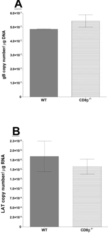
Figure 2. Quantitation of HSV-1 latency in CD8b2/2infected mice. Wild-type (WT) C57BL/6 and CD8b2/2 mice were infected as
described in Materials and Methods. Twenty-eight days PI, TGs from infected mice were harvested and quantitative PCR and RT-PCR were performed on each individual mouse TG. Each data point for gB DNA represents the mean6SEM from 24 TGs. For LAT RNA, each data point represents 22 TGs for CD8b2/2 and 18 TGs for WT mice from two
DCs monolayers were harvested and the presence of gB transcript was determined by qRT-PCR.
Adoptive Transfer of BM or CD8aDCs to Recipient Mice
BM from C57BL/6-GFP mice was isolated and each recipient CD8a2/2 mouse received BM from one donor mouse. We compared Intravenous (IV) and Intraperitoneal injection (IP) routes of BM transfer and found that IV transfer was more efficient than IP; therefore, all the experiments described here are based on IV transfer of BM. Two weeks post-transfer, the CD8a2/ 2 recipient mice were ocularly infected with HSV-1 strain
McKrae. TG, BM, spleen, and thymus from recipient mice were isolated on the same day as ocular infection, and 14 and 28 days post ocular infection. Tissues were used for flow cytometry and IHC. TG from infected mice were also isolated for detection of LAT RNA and gB DNA on day 28 PI. For transfer to BXH2 mice, cultured DCs from C3H/HEJ mice were grown as above,
harvested on day 5, and CD8aDCs were isolated by two cycles of positive selection per the manufacturer’s protocol (Miltenyi Biotec, Auburn CA). The purity of isolated CD8a DCs was more than 97% as confirmed by FACS analysis using anti-CD11c/anti-CD8a antibodies. Each recipient BXH2 mouse received CD8a
DCs from one donor mouse and two weeks post-transfer, the recipient mice were ocularly infected with HSV-1 strain KOS. TG from infected mice were also isolated for detection of LAT RNA on day 28 PI.
Adoptive Transfer of CD8 T Cells
Donor WT C57BL/6-GFP mouse spleens were pooled, and single-cell suspensions were prepared as described previously [23]. CD8 T cells were isolated using magnetic beads as described by the manufacturer (Miltenyi Biotec). After two cycles of purifica-tion, the purity of isolated CD8 T cells was 97% as confirmed by FACS analysis using anti-CD3/anti-CD8a antibodies. Each
Figure 3. IHC of DCs isolated from different knockout of mice.BM-derived DCs from WT,b2m2/2, CD8a2/2, and CD8b2/2mice were isolated and grown on Lab-Tex chamber slides. At 24 hr post culture, DCs were fixed, stained with anti-CD11c/anti-CD4, CD11c/anti-CD8a, or CD11c/anti-CD8bantibodies followed by incubation with relevant secondary antibody to each primary antibody as described in Materials and Methods. DAPI is shown as a nuclear counter-stain. A (WT DCs). Upper panels from left to right DAPI, CD11c (FITC), CD4 (TRITC), Merge; middle panels: from left to right DAPI, CD11c (FITC), CD8a(TRITC), Merge and bottom panels: from left to right DAPI, CD11c (FITC), CD8b(TRITC), Merge; B (b2m2/2DCs). Upper panels from left to right DAPI, CD11c (FITC), CD4 (TRITC), Merge; middle panels: from left to right DAPI, CD11c (FITC), CD8a(TRITC), Merge, and bottom panels: from left to right DAPI, CD11c (FITC), CD8b(TRITC), Merge; C (CD8a2/2DCs). Upper panels from left to right DAPI, CD11c (FITC), CD4
(TRITC), Merge; middle panels: from left to right DAPI, CD11c (FITC), CD8a(TRITC), Merge, and bottom panels: from left to right DAPI, CD11c (FITC), CD8b(TRITC), Merge; and D (CD8b2/2DCs). Upper panels from left to right DAPI, CD11c (FITC), CD4 (TRITC), Merge; middle panels: from left to right
recipient mouse was injected once with CD8+T cells from one
donor mouse in 300ml of MEM intraperitoneally. The control mice received 300ml MEM alone. We compared IV and IP routes of CD8+T cells transfer and detected no differences; therefore, all
the experiments described here are based on IP transfer of CD8 T cells. The recipient and control mice were infected ocularly with strain McKrae 2 weeks after transfer of the CD8+T cells. Presence
of CD8 T cells in the spleen, thymus, BM, and TG of recipient mice was determined by IHC and flow cytometry before ocular infection, and at weeks 2 and 4 PI. TG from infected mice were also isolated for detection of LAT RNA and gB DNA on day 28 PI.
Confocal Microscopy and Image Analysis
DCs isolated from different strains of mice were grown on Lab-Tex chamber slides (Sigma-Alderich) as we described previously [24]. Briefly, cells were fixed by incubating slides in methanol for 10 min followed by acetone for 5 min at 220uC. Afterwards, slides were rinsed three times for 5 min each at ambient temperature in PBS containing 0.05% v/v Tween-20 (PBS-T). Slides were then blocked for 30 min at ambient temperature in PBS-T containing 1% w/v BSA. Rat anti-CD8a (clone 53–6.7, eBioscience, San Diego, CA), rat anti-CD4 (clone Gk1.5, eBioscience), rat anti-CD8b(clone YTS156.7.7, BioLegend), and hamster anti-CD11c (clone HL3, BD Biosciences) were used for IHC. Immunostaining was done using CD11c/CD4, CD11c/ CD8a, or CD11c/CD8bantibody combinations and staining for 1 h at 25uC. After three rinses for 5 min each in PBS, slides were incubated for 1 h at 25uC with secondary antibodies labeled with anti-hamster FITC or anti-Rat TRITC (Invitrogen). Slides were washed three times with PBS, air-dried and mounted with Prolong Gold DAPI mounting medium (Invitrogen). Images were captured at 102461024 pixels (original magnification = 20X) in indepen-dent fluorescence channels using a Nikon C1 eclipse inverted confocal microscope.
DNA Extraction and PCR Analysis for HSV-1 Genomic DNA
DNA was isolated from homogenized individual TG using the commercially available DNeasy Blood &Tissue Kit (Qiagen, Stanford, CA) according to the manufacturer’s instructions. PCR analyses were done using gB specific primers (Forward - 59 -AACGCGACGCACATCAAG-39; Reverse - 59 -CTGGTACGC-GATCAGAAAGC-39; and Probe - 59 -FAM-CAGCCGCAG-TACTACC-39). The amplicon length for this primer set was 72 bp. Relative copy numbers for gB DNA were calculated using standard curves generated from the plasmid pAc-gB1. In all experiments, GAPDH was used for normalization of transcripts.
RNA Extraction, cDNA Synthesis, and TaqMan RT-PCR
RNA was extracted from latent TG or infected DCs as we have described previously [24–26]. Following RNA extraction, 1mg of total RNA was reverse-transcribed using random hexamer primers and Murine Leukemia Virus (MuLV) Reverse Transcriptase from the High Capacity cDNA Reverse Transcription Kit (Applied Biosystems, Foster City, CA), in accordance with the manufac-turer’s recommendations. The levels of various transcripts were evaluated using commercially available TaqMan Gene Expression Assays (Applied Biosystems) with optimized primer and probe concentrations. Primer-probe sets consisted of two unlabeled PCR primers and the FAMTM dye-labeled TaqMan MGB probe formulated into a single mixture. Additionally, all cellular amplicons included an intron-exon junction to eliminate signal
Figure 4. LAT and gB levels in latently-infected CD8a2/2mice
following adoptive transfer of bone marrow or CD3+CD8+ T
cells. Each recipient CD8a2/2 mice received bone marrow (BM) or
CD3+CD8+T cells as described in Materials and Methods. Control mice did not receive adoptive transfer. Two weeks post-transfer, mice were ocularly infected as described in Materials and Methods. Quantitative RT-PCR and PCR was performed on RNA and DNA isolated from the TG of surviving mice to determine copy number of the LAT or gB transcripts, respectively. Each data point represents the mean6SEM from 14 TGs. Panels: A) LAT RNA; and B) gB DNA.
from genomic DNA contamination. The HSV-1 gB and LAT primers and probe used were as follows: 1) gB: forward primer, 59 -AACGCGACGCACATCAAG-39, reverse primer, 59 -CTGGTACGCGATCAGAAAGC-39; and probe, 59 -FAM-CAGCCGCAGTACTACC-39 – Amplicon length = 72 bp; and 2)LAT: forward primer 59-GGGTGGGCTCGTGTTACAG-39; reverse primer, 59 -GGACGGGTAAGTAACAGAGTCTCTA-39; and probe, 59- FAM-ACACCAGCCCGTTCTTT-39– Am-plicon Length = 81 bp, corresponding to LAT nts 119553– 119634. As an internal control, a set of GAPDH primers from Applied Biosystems (ASSAY I.D. m999999.15_G1 - Amplicon Length = 107 bp) was used.
Quantitative real-time PCR (qRT-PCR) was performed using an ABI PRISM 7900HT Sequence Detection System (Applied Biosystems, Foster City, CA) in 384-well plates as we described previously [25,26]. Real-time PCR was performed in triplicate for each tissue sample. The threshold cycle (Ct) values, which represents the PCR cycle at which there is a noticeable increase in the reporter fluorescence above baseline, were determined using SDS 2.2 Software. GAPDH transcript was used for normalization purposes.
Statistical Analysis
Student’s t test and chi-squared tests were performed using the computer program Instat (GraphPad, San Diego). Results were considered statistically significant when thePvalue was,0.05. Results
Effect of CD8 T Cells on the Establishment of Latency in Latently Infected Mice
It was now generally accepted that CD8 T cells play a dominant role to maintain HSV-1 latency [11]. We ocularly infected WT C57BL/6, C57BL/6-b2m2/2, and C57BL/6-CD8a2/2 mice with 26105PFU/eye of WT HSV-1 strain McKrae. C57BL/6-CD8a2/2mice are devoid of CD8+T cells, and C57BL/6-b
2m2/ 2mice lack functional CD8+T cells due to the absence of major
histocompatibility complex I (MHC-I). Individual TG from surviving mice were isolated on day 28 post-infection (PI), and total DNA was prepared. TaqMan PCR was performed as described in Materials and Methods to quantitate viral gB DNA as a measure of HSV-1 genome copies and hence, latency. The combined data from two separate experiments are shown in Figure 1.
Surprisingly, the amount of viral DNA during latency in WT mice was similar to that of b2m2/2 mice (Figure 1A; WT vs.
b2m2/2; p = 0.9). This suggests that lack of MHC-I and functional classical CD8 T cells does not impact viral latency but does not rule out the involvement of non-classical T cells in latency as was reported for gamma-herpesvirus 68 [27]. By contrast, gB copy number in TG of CD8a2/2 mice was reduced at least 6-fold
compared to WT and b2m2/2 mice (Figure 1A, P,0.0001 for each comparison). These data demonstrate that the phenotype of CD8a2/2 mice significantly differs from b2m2/2 or WT mice with regards to latency.
During HSV-1 neuronal latency, only the LAT transcript is consistently expressed at high levels [28,29]. We next sought to confirm our gB DNA latency results by quantitating LAT
expression levels. On day 28, TG from surviving mice were harvested and LAT transcript was determined by TaqMan RT-PCR from total RNA isolated from individual TG. The amount of LAT RNA detected in the TG ofb2m2/2mice was significantly higher compared with WT mice (Figure 1B, p,.001). In agreement with the gB DNA results (Figure 1A), TG from CD8a2/2mice had LAT copy numbers that were over 4 orders of
magnitude lower than WT orb2m2/2mice (Figure 1B, P,0.0001 for each comparison). These striking results demonstrate that absence of CD8a, but notb2m, reduces HSV-1 latency.
CD8 consists of eitheraahomodimers orabheterodimers [30]. To follow-up from our CD8a2/2mouse results, we next evaluated whether CD8b impacted viral latency using CD8b2/2 mice.
CD8b2/2 and WT mice were ocularly infected as above, TG
were harvested on day 28 PI, and TaqMan PCR and RT-PCR were performed as detailed above. Combined data from two separate experiments are shown in Figure 2. The amount of latency, as determined by viral DNA and LAT RNA, was similar between CD8b2/2 and WT mice (Figure 2A and B; p.0.05). Interestingly, absence of the CD8b chain did not affect latency. CD8b2/2 mice have attenuated T cell function but normal
numbers of functional CD8a DCs [20]. These results raise the possibility that CD8a DCs2rather than CD8 T cells2play the primary role in HSV-1 latency establishment.
Nonetheless, lower latency in CD8a2/2 compared with WT,
b2m2/2, or CD8b2/2mice could be owed to absence of CD8aT cells, CD8aDCs, or both. Mouse plasmacytoid DCs (pDCs) were previously thought to express CD8a, but not CD8b. However, it was recently shown that at least one DC subclass does express CD8b [31]. To evaluate expression profiles of CD8aand CD8b
on DCs, BM-derived DCs from naive WT,b2m2/2, CD8a2/2, and CD8b2/2mice were grown to confluency on chamber slides
as described in Materials and Methods. Cells were stained with various combinations of CD11c, CD8aor CD8bantibodies, and CD11c/CD4 immunostaining was used as a positive control for each mouse strain. Immunohistochemistry (IHC) results are shown in Figure 3. CD4, CD8a, and CD8b were all detected on DCs isolated from WT andb2m2/2 mice (Figure 3A, WT DCs; 3B,
b2m2/2 DCs). By contrast, CD8a or CD8b DCs were not detected on DCs isolated from CD8a2/2mice, while CD4 DCs
were present (Figure 3C, CD8a2/2 DCs). These results are
consistent with a previous study showing that mouse CD8bcannot be expressed as abbhomodimer in the absence of CD8a[32,33]. In contrast, CD8b2/2mice lacked CD8bDCs, while CD8aDCs
were not affected (Figure 3D, CD8b2/2DCs). Thus, these results
allow us to group pDCs into CD8a+b+, CD8a+b
-, and CD8a-b-. These data dovetail with a previous report showing distinct populations of pDCs according to their surface expression of CD8a or CD8b [31]. Taken together, these results suggest that lack of CD8a DCs alone or in combination with CD8 T cells reduces latency in CD8a2/2mice compared with WT,b
2m2/2, or CD8b2/2mice.
Adoptive Transfer of CD8aDCs, but not CD8 T Cells, Increases Latency in HSV-1-Infected CD8a2/2Mice
In the above experiments using CD8a2/2 mice, we found significantly reduced viral latency compared with WT,b2m2/2, or CD8b2/2 mice. Therefore, it was of interest to determine if isolated and grown on Lab-Tex chamber slides. At 24 hr post culture, DCs were fixed, stained with anti-CD11c/anti-CD8aor anti-CD11c/anti-CD8b
antibodies followed by incubation with relevant secondary antibody to each primary antibody as described in Materials and Methods. DAPI is shown as a nuclear counter-stain. Upper panels from left to right DAPI, CD11c (FITC), CD8a(TRITC), and Merge; and bottom panels: from left to right DAPI, CD11c (FITC), CD8b(TRITC), and Merge.
decreased latency was due to attenuated CD8a DCs or CD8 T cells. CD8a2/2 mice adoptively received either BM-derived
CD8a DCs or T cells from WT GFP mice as described in Materials and Methods. Abundance of LAT RNA in mice that received BM-derived CD8aDCs was at least 6-fold higher than in mice that received CD8 T cells or no adoptive transfer (Figure 4A; BM DC transfer vs. no transfer or CD8 T cell transfer; p,0.0001). There was no difference in latency between mice that received CD8 T cells and no transfer control mice (Figure 4A, p.0.05). A similar pattern of results was observed when latency levels were judged by gB DNA (genomic DNA) levels (Figure 4B). Thus, adoptive transfer of BM-derived CD8aDCs significantly increased the levels of LAT and gB DNA in CD8a2/2mice, while transfer
of CD8 T cells had no effect. Figure S1A and S1B shows detection of GFP+CD8a+T cells or GFP+CD8a+DCs in the TG, BM, and
spleen of recipient mice both before infection and at 14 and 28 days PI. Figure S1C shows the presence of GFP+CD8a+T cells in
the spleen, thymus and TG of ocularly infected mice on day 28 PI via immunohistochemistry. In summary, these results indicate that CD8a DCs, and not CD8 T cells, play an indispensable role in increasing HSV-1 latency.
BXH2 Mice have Similar Viral Latency as CD8a2/2 Mice
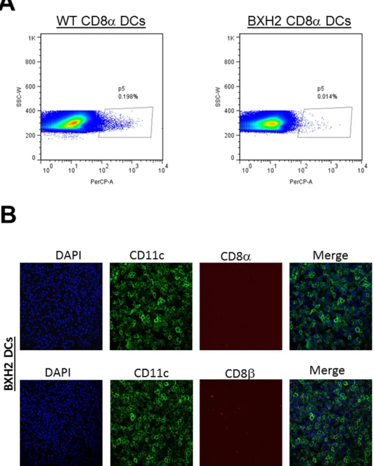
Previously, it was shown that interferon regulatory factor 8 (IRF-8) is essential for the development of CD8amyeloid DCs and pDCs [34]. More recently, a spontaneous point mutation was identified in the IRF-8 gene in the BXHBXH2 mouse [35]. This mutant has a similar phenotype as the IRF-8 knockout mouse, but without impaired pDC development and these mice are compe-tent to produce type 1 IFNs [16]. As shown in Figure 5A, DCs isolated from BXH2 mice had no significant numbers of CD8a
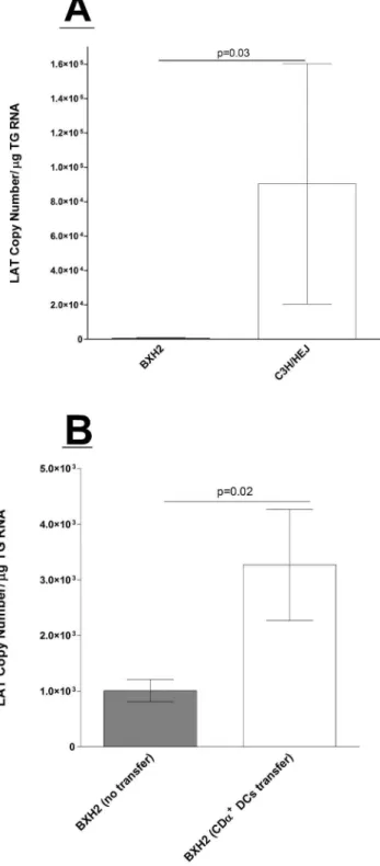
DCs compared with C3H/HEJ control mice. IHC results also confirmed the absence of CD8aas well as CD8b on these cells (Figure 5B). These results are similar to a previous report by Tailor et al showing that BXH2 mice do not have CD8aDCs but have intact CD8 T cell responses [16]. Thus, similar to CD8a2/2mice, BXH2 mice lack CD8aDCs, but in contrast to CD8a2/2mice, they have normal CD8 T cells. To determine if the absence of CD8a DCs, as opposed to CD8 T cells, contributed to higher latency in the CD8a2/2mouse, we ocularly infected BXH2 mice with 26103 PFU/eye of HSV-1 strain McKrae. Control C3H/ HEJ mice were similarly infected. The amount of LAT RNA detected in TG of BXH2 mice was significantly lower than control C3H/HEJ mice (Figure 6A,p,0.001).
To further confirm the role of CD8aDCs in establishment of viral latency, BXH2 mice adoptively received BM-derived CD8a
DCs from WT C3H/HEJ mice as described in Materials and Methods. BXH2 untreated mice, BXH2 mice that received CD8a+DCs, and control C3H/HEJ mice were ocularly infected
with 26105PFU/eye of avirulent HSV-1 strain KOS to reduce death of infected animals. Abundance of LAT RNA in mice that received BM-derived CD8aDCs was similar to control C3H/HEJ mice, and both were significantly higher when compared to untreated BXH2 mice (Figure 6B). Thus, adoptive transfer of CD8a DCs recovers the phenotype of the BXH2 mice. These results further bolster the notion that CD8aDCs2and not CD8 T cells2function to increase viral latency.
Figure 6. Quantitation of latency in BXH2 mice infected with HSV-1.A) Infection of mice with virulent HSV-1 strain McKrae. BXH2 or wild-type (WT) C3H/HEJ mice were infected with 26103 PFU/eye of
virulent HSV-1 strain McKrae. Twenty eight days PI, TGs from infected mice were harvested and quantitative RT-PCR was performed on each individual mouse TG as described in Materials and Methods. Each data point represents the mean6SEM from 18 or 12 TGs from BXH2 or WT mice from two separate experiments. B) Latency following adoptive transfer of CD8aDCs.Each recipient BXH2 mouse received BM-derived CD8aDCs from WT mice as described in Materials and Methods. A subset of BXH2 mice did not receive adoptive transfer, as a negative control. BM-derived recipient BXH2, untreated BXH2, and WT mice were ocularly infected with 26105PFU/eye of avirulent HSV-1 strain KOS.
Twenty-eight days PI, TG from infected mice were harvested and
quantitative RT-PCR were performed on each individual mouse TG. Each data point represents the mean6SEM from 18 TG from BM-derived recipient BXH2 or 16 TG from untreated BXH2 mice.
HSV-1 Infectivity is Reduced in DCs Isolated from CD8a2/
2Mice
The above experiments suggested that absence of CD8aDCs affected the level of latencyin vivo. To determine if this effect also operatedin vitro, DCs were isolated and cultured from naive WT,
b2m2/2, CD8b2/2, or CD8a2/2mice. DCs were then infected with 1 PFU/cell of HSV-1 strain McKrae, as described in Materials and Methods. At 24 h PI, RNA was isolated and gB transcript abundance was measured by qRT-PCR (Figure 7). Similar to our results with knockout mice, infected CD8a2/2DCs
had at least 10-fold less gB RNA than DCs from WT,b2m2 /2, or
CD8b2/2mice (Figure 7, p,0.0001).
Discussion
One of the hallmarks of HSV infection is the ability of the virus to establish latency in neurons of an infected host [36–38]. Once acquired, latent infections demonstrate a lifelong pattern of episodic recurrence, such that infected individuals serve as permanent carriers who are intermittently infectious [7–9]. The most efficient way to decrease latency and subsequent recurrent infections is to reduce establishment of latency, thus decreasing the probability of subsequent reactivations and recurrent eye disease. Previously, we showed that lymphoid-related DCs enhance HSV-1 latency in TG of infected mice and that immunization of mice with Flt3L, which increases lymphoid-related DCs, increases latent virus in the TG [15,39].
DCs have been classified into several subsets based on their immunophenotype, resident location, and functional differences
[40,41]. CD8a DCs are specialized cells known for cross-presentation of antigens by MHC-I molecules and for their major role in priming cytotoxic CD8 T cell responsesin vivo[42–45]. In addition, they have a high capacity to engulf apoptotic cells, which are used as a source of antigens for cross-priming [46]. Human blood DC antigen 3 (CD141, BDCA3) DCs are reported to be the homologue of mouse CD8a DCs [47]. To elucidate whether a relationship existed between CD8aDCs and HSV-1 latency, we utilized a multitude of gene deficient murine models. In this report, we show that increased viral latency is determined by CD8a DCs, but not CD8 T cells. Interestingly, we previously reported increased latency in CD42/2 mice, indicating the importance of CD4 T cells in orchestrating immune responses against HSV-1 latency [39].
In contrast to CD8a+ DCs, CD8a- DCs preferentially drive activation of CD4 T cells [48], can produce IFNs and IL-12 [49], acquire the ability to cross-present antigen [50], and induce CD4 cytotoxic regulatory T cells [51–54]. In addition, CD8a
-DCs are more effective than CD8a+ DCs at activating antigen-specific
CD4 T cells [43,55]. On the other hand, cross-presenting self-Ags is unique to CD8a+DCs, and is either completely absent or less
effective in other DC subsets [56]. However, the cross-presentation capacity of CD8a+ DCs is a double-edged sword, as it also
increases their susceptibility to infection [57,58]. Consequently, infected CD8a+ DCs may lose their function and/or block the
APC function of other DC subtypes. This may lead to greater viral latency and loss of CD8 T cell function. Thus, lower latency in the absence of CD8a+DCs, as found in this report, may be due to
APC subtypes other than CD8a+ DCs being able to process
Figure 7. Replication of HSV-1 in bone marrow-derived DCs.Sub-confluent monolayers of DCs cultured from WT,b2m2/2, CD8b2/2, or CD8a2/2mice were infected with 1 PFU/cell of HSV-1 strain McKrae for 24 h as described in Materials and Methods. RNA was isolated and the
antigen and induce a more effective antiviral response. Consistent with this notion, absence of CD8a+ DCs in mice enhances
resistance to the intracellular bacteriumListeria monocytogenes[58]. These results are also consistent with reports on the negative role of certain DC populations in control of Vaccinia virus [59], HIV-1 [60,61], and dengue virus [62]. In addition, the potential for autoimmunity induced by DCs, particularly in response to persistent viral infection, has been suggested by others [63].
Because of the critical role that DCs play in orchestrating immune responses, there is increasing interest in using signals that are known to activate DCs to stimulate and improve vaccine efficacy. However, the negative aspects of DCs have received far less attention than their positive roles, likely due to enthusiasm for their immunotherapeutic potential (reviewed in [64]). It was shown that Langerhans cells are negative regulators of the
anti-Leishmania response in mice [65], similar to our results reported here for CD8a DCs and HSV-1. Additionally, Vaccinia virus abortively infects both mature and immature DCs and blocks their maturation; hence, T cell activation is impaired [59]. By inhibiting maturation pathways in DCs and inducing their death, Vaccinia virus can subvert the development of efficient antiviral T cell immunity. Similarly, we found here that DCs isolated from CD8a2/2 mice harbor fewer viruses than other populations of
DCs. Consequently, HSV-1 infected DCs may directly, or via a negative feedback control mechanism, interfere with DC function, thus inhibiting induction of antiviral responses leading to even higher latency. Previously, it was shown that depletion of DCs during primary ocular infection reduces NK cells function and results in impaired clearance of HSV-1 from the eye of infected mice [66]. Maturation of DCs is required to induce potent immune responses, and HSV-1 infection elicits immune responses incapable of preventing or eradicating infection. Furthermore, we have previously shown that mature murine DCs can be infected by HSV-1, but the virus does not replicate in infected DCs [24]. Similarly, it was shown that following infection with HSV-1, immature DCs generate infectious viral particles, while mature DCs do not support virus production [67]. Finally, HSV-1 infection of human DCs results in functional impairment of the infected cells [68–70].
It has been shown that TG-resident CD8 T cells block HSV-1 reactivation from latency, and that these cells are necessary for maintaining viral latency [11,12]. Additionally, CD8 T cells can actively suppress viral reactivation through release of IFN-c[71]. It was reported that adding anti-CD8 antibody to cells isolated from TG of latently infected mice increases reactivation of latent virus [11]. Interestingly however, these studies all relied on CD8a
antibody, and so their results might be reinterpreted as implicating CD8aDCs as opposed to CD8 T cells.
A key finding in our study is that increased latency is due to CD8a+DCs. These results might also translate into
neuroprotec-tion, since increased latency usually means more neurons survive the primary infection. Along these lines, our published studies have shown that transfer of CD11c+CD8a+cells significantly enhances
latency in the TG of WT infected mice, whereas transfer of CD11c+CD8a
-cells reduces latency [39].
Overall, our results suggest that CD8a+DCs, rather than CD8+
T cells, play a non-redundant role in increasing HSV-1 latency. Thus, we propose that infection of CD8a+DCs by HSV-1 reduces
the antiviral response, resulting in greater latency, which in turn leads to increased recruitment of CD8 T cells at the site of latent infection. Continuous exposure of these CD8 T cells to viral antigens results in increased PD-1/PD-L1 and T cell exhaustion as
we reported recently [13]. Our results reveal a previously unappreciated role of innate immunity in maintaining HSV-1 latency.
Conclusion
Previously, it was reported that CD8+ T cells infiltrate
trigeminal ganglia (TG) at the time of latency establishment and contributing to increase of latency as well as inhibiting HSV-1 reactivation from latency. This has become the standard mechanism of HSV-1 latency-reactivation among herpes research-ers. To determine factors that are contributing to increase of latency, we examined latency in TG of WT mice versus CD8a2/2
and BXH2 mice, both of which lack CD8a DCs but CD8a2/2
mice lack CD8+T cells, while BXH2 mice have CD8+ T cells.
Additionally,b2m2/2and CD8b2/2mice that have CD8aDCs
were utilized. Overall, we report here that, in the absence of CD8a+DCs, HSV-1 latency was significantly decreased in vivo.
Thus, these studies point to a key role for CD8a+DCs rather than
CD8+T cells in establishment and maintenance of HSV-1 latency
in TG of infected mice.
Supporting Information
Figure S1 Efficiency of adoptive CD8a+T cells or BM to
recipient CD8a2/2 mice.Naive CD8a+T cells or BM were
isolated from naive C57BL/6-GFP+mice as described in Materials
and Methods. Isolated CD8a+T cells or BM were transferred IP
or IV into recipient CD8a2/2mice, respectively. Two weeks post
transfer some of the recipient mice were infected ocularly with 26105PFU/eye of WT HSV-1 strain McKrae. On day 14 before ocular infection and on days 14 and 28 PI some of the mice were euthanized and the presence of GFP+cells in TG, BM, spleen and
thymus were determined by FACS and IHC. A) Transfer of CD8a+GFP+T cells to recipient mice. Presence of total GFP+cells
in TG, BM, and spleen of donor WT-GFP+mice are shown as
control in the left side under the WT-GFP column. Marked area inside each quadrant show presence of CD8a+GFP+T cells in TG,
BM, and spleen of recipient mice before and after infection; B) Transfer of BM-GFP+cells to recipient mice. Marked area inside
each quadrant show presence of GFP+in TG, BM, and spleen of
recipient mice before and after infection; and C) Detection of CD8a+GFP+T cells in recipient mice. Presence of CD8a+GFP+T
cells in TG, thymus, and spleen of recipient mice were determined by IHC using anti-GFP-488 antibody to enhance the signal. DAPI is shown as a nuclear counter-stain. Spleen (upper panels): from left to right DAPI, anti-GFP-488, and Merge; Thymus (middle panels): from left to right DAPI, anti-GFP-488, and Merge; and TG (Bottom): from left to right DAPI, anti-GFP-488, and Merge. (PPT)
Table S1 Survival of different mouse strains following ocular infection.
(DOCX)
Acknowledgments
We thank Dr. Dan Littman (New York University) for gifting the CD8b
knockout mice.
Author Contributions
References
1. Dawson CR (1984) Ocular herpes simplex virus infections. Clin Dermatol 2: 56– 66.
2. Barron BA, Gee L, Hauck WW, Kurinij N, Dawson CR, et al. (1994) Herpetic Eye Disease Study. A controlled trial of oral acyclovir for herpes simplex stromal keratitis. Ophthalmology 101: 1871–1882.
3. Wilhelmus KR, Dawson CR, Barron BA, Bacchetti P, Gee L, et al. (1996) Risk factors for herpes simplex virus epithelial keratitis recurring during treatment of stromal keratitis or iridocyclitis. Herpetic Eye Disease Study Group. Br J Ophthalmol 80: 969–972.
4. Oh JO, Kimura SJ, Ostler HB, Dawson CR, Smolin G (1976) Oculogenital transmission of type 2 herpes simplex virus in adults. Surv Ophthalmol 21: 106– 109.
5. Pavan-Langston D (1983) Ocular viral infections. Med Clin North Am 67: 973– 990.
6. Corey L (1994) The current trend in genital herpes. Progress in prevention. Sex Transm Dis 21: S38–S44.
7. Gordon YJ (1990) Pathogenesis and latency of herpes simplex virus type 1 (HSV-1): an ophthalmologist’s view of the eye as a model for the study of the virus- host relationship. Adv Exp Med Biol 278: 205–209.
8. Kaufman HE, Azcuy AM, Varnell ED, Sloop GD, Thompson HW, et al. (2005) HSV-1 DNA in tears and saliva of normal adults. Invest Ophthalmol Vis Sci 46: 241–247.
9. Steiner I (1996) Human herpes viruses latent infection in the nervous system. Immunol Rev 152: 157–173.
10. Doymaz MZ, Rouse BT (1992) Herpetic stromal keratitis: an immunopathologic disease mediated by CD4+T lymphocytes. Invest Ophthalmol Vis Sci 33: 2165– 2173.
11. Liu T, Khanna KM, Chen X, Fink DJ, Hendricks RL (2000) CD8(+) T cells can block herpes simplex virus type 1 (HSV-1) reactivation from latency in sensory neurons. J Exp Med 191: 1459–1466.
12. Khanna KM, Bonneau RH, Kinchington PR, Hendricks RL (2003) Herpes simplex virus-specific memory CD8+ T cells are selectively activated and retained in latently infected sensory ganglia. Immunity 18: 593–603. 13. Allen SJ, Hamrah P, Gate DM, Mott KR, Mantopoulos D, et al. (2011) The role
of LAT in increased CD8+T cell exhaustion in trigeminal ganglia of mice latently infected with herpes simplex virus type 1. J Virol 85: 4184–4197. 14. Mott KR, Bresee CJ, Allen SJ, BenMohamed L, Wechsler SL, et al. (2009) Level
of herpes simplex virus type 1 latency correlates with severity of corneal scarring and exhaustion of CD8+T cells in trigeminal ganglia of latently infected mice. J Virol 83: 2246–2254.
15. Mott KR, Ghiasi H (2008) Role of dendritic cells in enhancement of herpes simplex virus type 1 latency and reactivation in vaccinated mice. Clin Vaccine Immunol 15: 1859–1867.
16. Tailor P, Tamura T, Morse HC, 3rd, Ozato K (2008) The BXH2 mutation in IRF8 differentially impairs dendritic cell subset development in the mouse. Blood 111: 1942–1945.
17. Fung-Leung WP, Schilham MW, Rahemtulla A, Kundig TM, Vollenweider M, et al. (1991) CD8 is needed for development of cytotoxic T cells but not helper T cells. Cell 65: 443–449.
18. Osorio Y, Ghiasi H (2003) Comparison of adjuvant efficacy of herpes simplex virus type 1 recombinant viruses expressing TH1 and TH2 cytokine genes. J Virol 77: 5774–5783.
19. Perng GC, Dunkel EC, Geary PA, Slanina SM, Ghiasi H, et al. (1994) The latency-associated transcript gene of herpes simplex virus type 1 (HSV-1) is required for efficient in vivo spontaneous reactivation of HSV-1 from latency. J Virol 68: 8045–8055.
20. Crooks ME, Littman DR (1994) Disruption of T lymphocyte positive and negative selection in mice lacking the CD8 beta chain. Immunity 1: 277–285. 21. Gilliet M, Boonstra A, Paturel C, Antonenko S, Xu XL, et al. (2002) The
development of murine plasmacytoid dendritic cell precursors is differentially regulated by FLT3-ligand and granulocyte/macrophage colony-stimulating factor. J Exp Med 195: 953–958.
22. Balkhi MY, Latchumanan VK, Singh B, Sharma P, Natarajan K (2004) Cross-regulation of CD86 by CD80 differentially regulates T helper responses from Mycobacterium tuberculosis secretory antigen-activated dendritic cell subsets. J Leukoc Biol 75: 874–883.
23. Ahmed R, King CC, Oldstone MB (1987) Virus-lymphocyte interaction: T cells of the helper subset are infected with lymphocytic choriomeningitis virus during persistent infection in vivo. J Virol 61: 1571–1576.
24. Mott KR, Underhill D, Wechsler SL, Town T, Ghiasi H (2009) A role for the JAK-STAT1 pathway in blocking replication of HSV-1 in dendritic cells and macrophages. Virol J 6: 56.
25. Mott KR, Perng GC, Osorio Y, Kousoulas KG, Ghiasi H (2007) A Recombinant Herpes Simplex Virus Type 1 Expressing Two Additional Copies of gK Is More Pathogenic than Wild-Type Virus in Two Different Strains of Mice. J Virol 81: 12962–12972.
26. Mott KR, Osorio Y, Brown DJ, Morishige N, Wahlert A, et al. (2007) The corneas of naive mice contain both CD4+and CD8+T cells. Mol Vis 13: 1802– 1812.
27. Braaten DC, McClellan JS, Messaoudi I, Tibbetts SA, McClellan KB, et al. (2006) Effective control of chronic gamma-herpesvirus infection by unconven-tional MHC Class Ia-independent CD8 T cells. PLoS Pathog 2: e37. 28. Rock DL, Nesburn AB, Ghiasi H, Ong J, Lewis TL, et al. (1987) Detection of
latency-related viral RNAs in trigeminal ganglia of rabbits latently infected with herpes simplex virus type 1. J Virol 61: 3820–3826.
29. Dobson AT, Margolis TP, Sedarati F, Stevens JG, Feldman LT (1990) A latent, nonpathogenic HSV-1-derived vector stably expresses beta- galactosidase in mouse neurons. Neuron 5: 353–360.
30. Zamoyska R (1994) The CD8 coreceptor revisited: one chain good, two chains better. Immunity 1: 243–246.
31. Lombardi V, Speak AO, Kerzerho J, Szely N, Akbari O (2012) CD8alpha(+ )beta(2) and CD8alpha(+)beta(+) plasmacytoid dendritic cells induce Foxp3(+) regulatory T cells and prevent the induction of airway hyper-reactivity. Mucosal Immunol 5: 432–443.
32. Devine L, Kieffer LJ, Aitken V, Kavathas PB (2000) Human CD8 beta, but not mouse CD8 beta, can be expressed in the absence of CD8 alpha as a beta beta homodimer. J Immunol 164: 833–838.
33. Gorman SD, Sun YH, Zamoyska R, Parnes JR (1988) Molecular linkage of the Ly-3 and Ly-2 genes. Requirement of Ly-2 for Ly-3 surface expression. J Immunol 140: 3646–3653.
34. Aliberti J, Schulz O, Pennington DJ, Tsujimura H, Reis e Sousa C, et al. (2003) Essential role for ICSBP in the in vivo development of murine CD8alpha+ dendritic cells. Blood 101: 305–310.
35. Turcotte K, Gauthier S, Mitsos LM, Shustik C, Copeland NG, et al. (2004) Genetic control of myeloproliferation in BXH-2 mice. Blood 103: 2343–2350. 36. Stevens JG (1989) Human herpesviruses: a consideration of the latent state.
Microbiol Rev 53: 318–332.
37. Wechsler SL, Nesburn AB, Watson R, Slanina S, Ghiasi H (1988) Fine mapping of the major latency-related RNA of herpes simplex virus type 1 in humans. J Gen Virol 69: 3101–3106.
38. Fraser NW, Valyi-Nagy T (1993) Viral, neuronal and immune factors which may influence herpes simplex virus (HSV) latency and reactivation. Microb Pathog 15: 83–91.
39. Mott KR, UnderHill D, Wechsler SL, Ghiasi H (2008) Lymphoid-related CD11c+CD8a+dendritic cells are involved in enhancing HSV-1 latency. J Virol 82: 9870–9879.
40. Shortman K, Naik SH (2007) Steady-state and inflammatory dendritic-cell development. Nat Rev Immunol 7: 19–30.
41. Belz GT, Nutt SL (2012) Transcriptional programming of the dendritic cell network. Nat Rev Immunol 12: 101–113.
42. den Haan JM, Lehar SM, Bevan MJ (2000) CD8(+) but not CD8(2) dendritic cells cross-prime cytotoxic T cells in vivo. J Exp Med 192: 1685–1696. 43. Dudziak D, Kamphorst AO, Heidkamp GF, Buchholz VR, Trumpfheller C, et
al. (2007) Differential antigen processing by dendritic cell subsets in vivo. Science 315: 107–111.
44. Belz GT, Smith CM, Eichner D, Shortman K, Karupiah G, et al. (2004) Cutting edge: conventional CD8alpha(+) dendritic cells are generally involved in priming CTL immunity to viruses. J Immunol 172: 1996–2000.
45. Belz GT, Behrens GM, Smith CM, Miller JF, Jones C, et al. (2002) The CD8alpha(+) dendritic cell is responsible for inducing peripheral self-tolerance to tissue-associated antigens. J Exp Med 196: 1099–1104.
46. Iyoda T, Shimoyama S, Liu K, Omatsu Y, Akiyama Y, et al. (2002) The CD8+ dendritic cell subset selectively endocytoses dying cells in culture and in vivo. J Exp Med 195: 1289–1302.
47. Jongbloed SL, Kassianos AJ, McDonald KJ, Clark GJ, Ju X, et al. (2007) Human CD141+(BDCA-3)+dendritic cells (DCs) represent a unique myeloid DC subset that cross-presents necrotic cell antigens. J Exp Med 207: 1247–1260. 48. Allenspach EJ, Lemos MP, Porrett PM, Turka LA, Laufer TM (2008) Migratory and lymphoid-resident dendritic cells cooperate to efficiently prime naive CD4 T cells. Immunity 29: 795–806.
49. Maldonado-Lopez R, Moser M (2001) Dendritic cell subsets and the regulation of Th1/Th2 responses. Semin Immunol 13: 275–282.
50. den Haan JM, Bevan MJ (2002) Constitutive versus activation-dependent cross-presentation of immune complexes by CD8(+) and CD8(2) dendritic cells in vivo. J Exp Med 196: 817–827.
51. Kawamura K, Kadowaki N, Kitawaki T, Uchiyama T (2006) Virus-stimulated plasmacytoid dendritic cells induce CD4+cytotoxic regulatory T cells. Blood 107: 1031–1038.
52. Brown DM, Lee S, Garcia-Hernandez Mde L, Swain SL (2012) Multifunctional CD4 cells expressing gamma interferon and perforin mediate protection against lethal influenza virus infection. J Virol 86: 6792–6803.
53. Zhou Y, Callendret B, Xu D, Brasky KM, Feng Z, et al. (2012) Dominance of the CD4(+) T helper cell response during acute resolving hepatitis A virus infection. J Exp Med 209: 1481–1492.
54. Lee HK, Zamora M, Linehan MM, Iijima N, Gonzalez D, et al. (2009) Differential roles of migratory and resident DCs in T cell priming after mucosal or skin HSV-1 infection. J Exp Med 206: 359–370.
56. Schnorrer P, Behrens GM, Wilson NS, Pooley JL, Smith CM, et al. (2006) The dominant role of CD8+dendritic cells in cross-presentation is not dictated by antigen capture. Proc Natl Acad Sci U S A 103: 10729–10734.
57. Alaniz RC, Sandall S, Thomas EK, Wilson CB (2004) Increased dendritic cell numbers impair protective immunity to intracellular bacteria despite augment-ing antigen-specific CD8(+) T lymphocyte responses. J Immunol 172: 3725– 3735.
58. Edelson BT, Bradstreet TR, Hildner K, Carrero JA, Frederick KE, et al. (2011) CD8alpha(+) dendritic cells are an obligate cellular entry point for productive infection by Listeria monocytogenes. Immunity 35: 236–248.
59. Engelmayer J, Larsson M, Subklewe M, Chahroudi A, Cox WI, et al. (1999) Vaccinia virus inhibits the maturation of human dendritic cells: a novel mechanism of immune evasion. J Immunol 163: 6762–6768.
60. Granelli-Piperno A, Delgado E, Finkel V, Paxton W, Steinman RM (1998) Immature dendritic cells selectively replicate macrophagetropic (M-tropic) human immunodeficiency virus type 1, while mature cells efficiently transmit both M- and T-tropic virus to T cells. J Virol 72: 2733–2737.
61. Geijtenbeek TB, Kwon DS, Torensma R, van Vliet SJ, van Duijnhoven GC, et al. (2000) DC-SIGN, a dendritic cell-specific HIV-1-binding protein that enhances trans-infection of T cells. Cell 100: 587–597.
62. Wu SJ, Grouard-Vogel G, Sun W, Mascola JR, Brachtel E, et al. (2000) Human skin Langerhans cells are targets of dengue virus infection. Nature Medicine 6: 816–820.
63. Paroli M, Schiaffella E, Di Rosa F, Barnaba V (2000) Persisting viruses and autoimmunity. J Neuroimmunol 107: 201–204.
64. Ganguly D, Haak S, Sisirak V, Reizis B (2013) The role of dendritic cells in autoimmunity. Nat Rev Immunol 13: 566–577.
65. Kautz-Neu K, Noordegraaf M, Dinges S, Bennett CL, John D, et al. (2011) Langerhans cells are negative regulators of the anti-Leishmania response. J Exp Med 208: 885–891.
66. Frank GM, Buela KA, Maker DM, Harvey SA, Hendricks RL (2012) Early responding dendritic cells direct the local NK response to control herpes simplex virus 1 infection within the cornea. J Immunol 188: 1350–1359.
67. Kobelt D, Lechmann M, Steinkasserer A (2003) The interaction between dendritic cells and herpes simplex virus-1. Curr Top Microbiol Immunol 276: 145–161.
68. Salio M, Cella M, Suter M, Lanzavecchia A (1999) Inhibition of dendritic cell maturation by herpes simplex virus. Eur J Immunol 29: 3245–3253. 69. Kruse M, Rosorius O, Kratzer F, Stelz G, Kuhnt C, et al. (2000) Mature
dendritic cells infected with herpes simplex virus type 1 exhibit inhibited T-cell stimulatory capacity. J Virol 74: 7127–7136.
70. Stefanidou M, Ramos I, Mas Casullo V, Trepanier JB, Rosenbaum S, et al. (2013) HSV-2 Prevents Dendritic Cell Maturation, Induces Apoptosis and Triggers Release of Pro-inflammatory Cytokines: Potential Links to HSV-HIV Synergy. J Virol 87: 1443–1453.