Chronic Administration of Thymosin
b
-4 in the
Dystrophin Deficient Mouse
Christopher F. Spurney1, Hee-Jae Cha2, Arpana Sali3, Gouri S. Pandey3, Emidio Pistilli4, Alfredo D. Guerron3, Heather Gordish-Dressman3, Eric P. Hoffman3, Kanneboyina Nagaraju3*
1Division of Cardiology, Children’s National Medical Center, Washington, District of Columbia, United States of America,2Department of Parasitology and Genetics, Kosin University College of Medicine, Amnam-dong, Seo-gu, Busan, South Korea,3Center for Genetic Medicine Research, Children’s National Medical Center, Washington, District of Columbia, United States of America,4Pennsylvania Muscle Institute, School of Medicine, University of Pennsylvania, Philadelphia, Pennsylvania, United States of America
Abstract
Thymosin beta-4 (Tb4) is a ubiquitous protein with many properties relating to cell proliferation and differentiation that promotes wound healing and modulates inflammatory mediators. We studied the effects of chronic administration of Tb4 on the skeletal and cardiac muscle of dystrophin deficientmdxmice, the mouse model of Duchenne muscular dystrophy. Female wild type (C57BL10/ScSnJ) andmdxmice, 8–10 weeks old, were treated with 150mg of Tb4 twice a week for 6
months. To promote muscle pathology, mice were exercised for 30 minutes twice a week. Skeletal and cardiac muscle function were assessed via grip strength and high frequency echocardiography. Localization of Tb4 and amount of fibrosis were quantified using immunohistochemistry and Gomori’s tri-chrome staining, respectively.Mdxmice treated with Tb4 showed a significant increase in skeletal muscle regenerating fibers compared to untreated mdx mice. Tb4 stained exclusively in the regenerating fibers ofmdxmice. Although untreatedmdxmice had significantly decreased skeletal muscle strength compared to untreated wild type, there were no significant improvements inmdxmice after treatment. Systolic cardiac function, measured as percent shortening fraction, was decreased in untreatedmdxmice compared to untreated wild type and there was no significant difference after treatment inmdxmice. Skeletal and cardiac muscle fibrosis were also significantly increased in untreatedmdxmice compared to wild type, but there was no significant improvement in treated mdxmice. In exercised dystrophin deficient mice, chronic administration of Tb4 increased the number of regenerating fibers in skeletal muscle and could have a potential role in treatment of skeletal muscle disease in Duchenne muscular dystrophy.
Citation:Spurney CF, Cha H-J, Sali A, Pandey GS, Pistilli E, et al. (2010) Evaluation of Skeletal and Cardiac Muscle Function after Chronic Administration of Thymosinb-4 in the Dystrophin Deficient Mouse. PLoS ONE 5(1): e8976. doi:10.1371/journal.pone.0008976
Editor:Antoni L. Andreu, Hospital Vall d’Hebron, Spain
ReceivedSeptember 4, 2009;AcceptedDecember 29, 2009;PublishedJanuary 29, 2010
Copyright:ß2010 Spurney et al. This is an open-access article distributed under the terms of the Creative Commons Attribution License, which permits unrestricted use, distribution, and reproduction in any medium, provided the original author and source are credited.
Funding:This work was supported by funding provided by the following: National Institutes of Health (NIH)/National Institute of Child Health and Human Development/Child Health Research Career Development Award K12HD001399-04 (PI: Spurney); Department of Defense United States Army Medical Research Acquisition Activity grant W81XWH-05-1-0616 (Mouse Drug Screening Core to K. Nagaraju and E. Hoffman), Foundation to Eradicate Duchenne, Inc. (www. duchennemd.org), Muscular Dystrophy Association (www.mda.org), Charley’s Fund (K. Nagaraju)(www.charleysfund.com), NIH grant 1U54HD053177-01A1 (Wellstone Muscular Dystrophy Center to E. Hoffman and K. Nagaraju), and NIH grant R01-AR050478 (K. Nagaraju). The funders had no role in study design, data collection and analysis, decision to publish, or preparation of the manuscript.
Competing Interests:The authors have declared that no competing interests exist.
* E-mail: [email protected]
Introduction
Duchenne muscular dystrophy (DMD) is an inherited X-linked disorder with an incidence of 1 in 3,500 male births that is due to the absence of dystrophin, a large protein linking the intracellular cytoskeleton to the extracellular matrix.[1] The animal model of DMD, the mdx mouse, is genetically similar to the human deletion.[1–3] Although the underlying gene defect is the same in human and themdxmouse, the clinical picture is quite different. The mdx skeletal muscle undergoes an early acute phase of degeneration at 3–4 weeks of age followed by a successful regeneration phase. The histopathology after this acute phase shows a relatively mild picture, although specific muscles (e.g. diaphragm) and older mice can show more severe pathology consistent with human DMD muscle at presentation (failed regeneration and fibrosis). Commensurate with the pathology,
the physical symptoms of themdxmouse tend to be relatively mild, with muscle weakness more obvious after exercise or lengthening contractions.[4–6] Mdx mice also develop decreased cardiac systolic function slowly over time. This decreased function can be measured at significant levels by non-invasive echocardiogra-phy around nine months of age.[7,8]
modulates inflammatory mediators.[26,27] Tb4 was recently shown to promote cardiomyocyte migration, survival and repair in a coronary ligation model.[28]. Based on these mechanisms, we studied the effects of chronic Tb4 administration on skeletal and cardiac muscle function in exercised dystrophin deficient mice. While we found no significant differences in muscle function, we did see significantly increased skeletal muscle regeneration in Tb4 treatedmdxmice and these regenerating fibers distinctly stained for Tb4.
Methods
Animal Care
All animals were handled in strict accordance with good animal practice as defined by the relevant national and/or local animal welfare bodies, and all animal work was approved by the Institutional Animal Care and Use Committee at the Children’s National Medical Center, Washington, DC and the Veterans Administration Medical Center, Washington, DC (Protocol #01079). Eight to ten week old female C57BL/10ScSn-Dmdmdx/J (mdx) and C57BL/10ScSnJ (wild type) mice weighing 20–30 grams were purchased from The Jackson Laboratory (Bar Harbor, Ma). All mice were housed in an individually vented cage system with a 12 hour light-dark cycle and received standard mouse chow and waterad libitum. All mice were rested at least 10– 14 days before starting acclimations and baseline recordings.
Treatment with Thymosin Beta-4
Tb4 (RegeneRx Biopharmaceuticals Inc., Bethesda, Md) was given via intraperitoneal injection twice weekly over a 6 month period tomdxand wild type mice at a dose of 150mg in 200ml PBS. Buffer was given at the same times to mdx and wild type control groups.
Treadmill Exercise
The treadmill exercise uses a common commercially available setup (Columbus Instruments, Columbus, Ohio) which employs a moving belt. We subjected all experimental mice to a 30-minute run on a horizontal treadmill at 12 m/min, twice a week. This test was performed during the morning hours twice weekly during the 6 months except in those days on which functional data was obtained.
Grip Strength Test
Grip Strength was assessed using a grip strength meter consisting of horizontal forelimb mesh and an angled hind limb mesh (Columbus Instruments, Columbus, OH). Five successful hindlimb and forelimb strength measurements within 2 minutes were recorded and normalized to body weight as previously described.[29]
Rotarod Test
Mice were trained on the Rotarod (Ugo Basile, VA, Italy) for two days before collecting data. Each acclimatization session consisted of four training sessions, 2 per day and each session lasting 120 seconds at a speed of 5 rpm. Each trial consisted of placing the mice on the rod at 10 rpm for 60 seconds (stabilizing period) followed by an acceleration from 10 rpm to 40 rpm within the first 25 seconds until the animal falls from the rod or until 180 seconds are reached. If the animals fell during the stabilizing period, they were placed back on the rod to complete the session. The total testing time is 240 seconds (60 sec stabilization time and 180 seconds test time). Each trial was done twice a day (a gap of 2 h interval between sessions) for 3 consecutive days. The latency
to fall (seconds) was recorded and all six scores per mouse were averaged and recorded as latency to fall (in seconds) for each mouse.
Echocardiography
Mice were anesthetized with 1–2% isoflurane in 100% oxygen and scanning was performed over 20 minutes using a high frequency ultrasound probe (RMZ 702a, Vevo 660, VisualSonics, Toronto, Canada) as previously described.[8] Qualitative and quantitative measurements were made offline using analytic software (VisualSonics, Toronto, Canada).
Histological Evaluations
At the end of the trial, all animals were euthanized and tissue samples were taken for further testing. Histological evaluations were done by two individuals in a blinded manner using coded H&E stained slides and their results were averaged. The number of tissues examined per group varied based on tissue availability. Quantitative stereology (Olympus C.A.S.T. Stereology System, Olympus America Inc., Center Valley, PA) was used to evaluate the slides. Assessment criteria included: assessment of total fibers present, total fibers with central nuclei, total peripheral nuclei, total central nuclei, regenerating fibers (highly basophilic fibers), degenerating fibers, and inflammation (an interstitial group of more than 10 smaller inflammatory cell dark blue nuclei in a high power field) in five high power (40x) non-overlapping fields in normal andmdxgastrocnemius muscle sections. Fibers intersecting the left and top borders of the field where not counted and nuclei further than one nuclear diameter from the fiber border were deemed ‘‘central’’.[29]
For immunohistochemistry, tissue slides of untreated mdx gastrocnemius skeletal muscle were deparaffinized and hydrated. For antigen retrieval, slides were immersed in citrate buffer (0.01 M, pH 6.0) and heated twice in a microwave (700 W or high) for 5 min. The slides were quenched with endogenous peroxidase by incubation with 3% hydrogen peroxide solution for 5 minutes and washed three times in PBS for 5 minutes. Slides were then immunostained with rabbit polyclonal antibody to thymosin b4 (1:2000 dilution; ALPCO Diagnostics, Windham, NH, USA) at 4uC overnight. After primary antibody incubation, slides were washed three times in PBS for 5 minutes and incubated with secondary antibody for 1 hour. Then, slides were washed four times in PBS for 5 minutes each and the color reaction was developed with DAB and slides were counterstained Meyer’s hematoxylin (DAKO, Carpinteria, CA, USA) for 20 seconds, dehydrated, and mounted with Permount (Fisher Scientific, Pittsburgh, PA, USA).
Quantification of Fibrosis
Creatine Kinase (CK) Determination
Blood was obtained by heart puncture immediately after euthanasia. 250mL of blood was collected into eppendorf tubes,
allowed to clot and kept at room temperature to allow clot-contraction prior to centrifugation and serum collection. CK determination was performed according to the manufacturer’s instructions using standard spectrophotometric method with enzyme-coupled assay reagent from Fisher Scientific (CK10).[30] Absorption at 340 nm was measured every min for 2 min at 37uC to calculate enzyme activity. Duplicate measurements were done on each serum sample and the data was expressed as U/L.
Statistical Analysis
Measurements between wild type andmdxmice were compared at each time point using an analysis of variance with Sidak adjustment for multiple comparisons (body weight, GSM, Rotarod, echocardiography, percent collagen). Normality of each quantitative measurement was confirmed prior to analysis and those not conforming to normality underwent data transforma-tions. Histology measurements (degeneration fibers, regenerating fibers, inflammation, calcification, and central and peripheral nuclei) were first compared between two investigators to determine their consistency. Comparisons were then made using Poisson regression or using negative binomial regression where the Poisson model did not fit the data due to over dispersion.
Results
Both wild type and mdx mice were treated with Tb4 for 6 months and the behavioral data was collected at baseline (3 months of age), mid trial (5–6 months of age), and end of the trial (9 months of age). All results are presented as mean6standard deviation except those in figures 1 and 2.
Body Weight
No significant differences were seen in body weights between treated and untreated mice within the same strain. When comparing mdx and wild type mice, there was a significant increase in the body weight ofmdxmice at baseline and 6 months but not at 9 months of age (Table S1).
Grip Strength
Untreated mdx mice had significantly decreased normalized forelimb grip strength (kilogram force per kilogram; KGF/kg) compared to untreated wild type mice at 3, 6 and 9 months of age. Comparing the normalized hindlimb grip strength of these same groups showed a significant decrease in mdx mice at 3 and 6 months but not at 9 months of age. There were no significant differences in normalized forelimb or hindlimb grip strength between treated and untreated mice within the same strain during the trial (Table S1).
Rotarod
There were no significant differences in latency time to fall on the Rotarod apparatus between wild type andmdxmice. There were also no differences between treated and untreated mice within the strain, but treatedmdxmice showed significantly lower performance in comparison with treated wild type mice at 9 months of age (Table S1).
Echocardiography
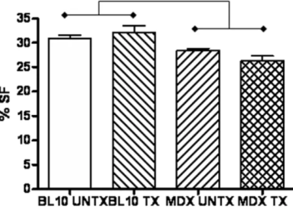
High frequency echocardiography found decreased cardiac function, measured as percent shortening fraction, in untreated mdx(27.961.86%) mice compared to untreated (30.662.6%) and treated (32.065.2; p = 0.045) wild type mice (Figure 1). Treated mdx(26.263.1%) mice also showed significantly decreased cardiac function compared to treated (p,0.01) and untreated (p,0.05) wild type mice. There were no significant differences between treated and untreated mice within the same strain. No significant differences were found in measurements of left ventricular chamber size or wall thickness betweenmdxand wild type mice, showing no dilation in the hearts of mdx mice with decreased
Figure 1. Significantly decreased cardiac function (mean 6
SEM) measured as percent shortening fraction (%SF) inmdx
mice is seen after 6 months of treatment with thymosin-beta 4 compared to wild type mice. There is no significant difference between treated and untreatedmdxmice.
doi:10.1371/journal.pone.0008976.g001
function. No significant differences were found in heart rates or Doppler measurements of aortic, pulmonary, tricuspid or mitral blood flow velocities between treated and untreated mdx mice (Table S2).
Skeletal Muscle Histology
Evaluation of the gastrocnemius skeletal muscle histology found significantly increased number of regenerating fibers in treatedmdx mice (11.6613.5) compared to untreated mdx mice (2.661.1; p = 0.03). Both treated and untreated mdx mice had increased central nuclei, central nuclei per fiber and central nucleated fibers compared to treated and untreated wild type. Untreatedmdxmice showed significantly increased total peripheral nuclei compared to treatedmdxmice (p = 0.014). There was also significantly increased inflammation (3+) betweenmdxand wild type mice that was not significantly altered in the treated groups (Table S3).
Quantification of Fibrosis
Using Gomori’s tri-chrome staining, an analysis of percent collagen showed significantly increased collagen found in the left ventricles of untreated (3.8360.9%) and treated (4.3961.2%)mdx mice compared to both untreated (1.661.1%) and treated (1.8260.9%) wild type controls (all p values,0.05) (Figures 2 and 3). The diaphragm also showed significantly increased percent collagen in the untreated (22.967.5%) and treated (25.066.6%) mdx mice compared to untreated (7.460.8%) and treated (7.961.8%) control mice (all p values,0.01). The gastrocnemius also showed significantly increased percent collagen in untreated mdx mice (6.2561.7%) compared to untreated control mice (3.061.1%; p,0.05) (Figure 2). There were no significant differences in percent collagen between treated and untreated groups within the same strain.
Serum Creatine Kinase
There was a significant increase in serum creatine kinase in both treated (520561785 U/L, n = 8) and untreated (578862494 U/ L, n = 10)mdxmice compared to treated (1416108 U/L, n = 14) and untreated (85675 U/L, n = 13) wild type controls (p,0.001). There was no significant difference between treated and untreated mdxmice.
Tb4 Localization Using Immunohistochemistry
Staining of untreated wild type and mdx skeletal muscle (gastrocnemius) with anti-Tb4 antibody shows localized staining in regenerating fibers. Sequential slides were stained for desmin, a marker for regenerating fibers, and this staining corresponded to Tb4 staining. There was no staining of fibers with either anti-Tb4 or anti-desmin antibodies in wild type tissue (Figure 4).
Discussion
We completed a six month trial using Tb4 in exercisedmdxand wild type mice. We found no significant improvement in treated mdxskeletal or cardiac muscle function compared to untreatedmdx mice. However, we did find significantly increased regenerating fibers in treatedmdxskeletal muscle and these fibers convincingly stained for Tb4. While Tb4 led to increased regeneration inmdx skeletal muscle, it did not improve fibrosis in the cardiac, diaphragmatic or skeletal muscle of treatedmdxmice. This study shows that chronic Tb4 administration is beneficial for skeletal muscle fiber regeneration in dystrophin deficient mice.
Previous gene profiling experiments showed increased Tb4 expression in skeletal muscle of mdx mice. Tseng et al. (2002) showed that in 16 week oldmdxmice, the gastrocnemius muscle showed a two-fold increase in Tb4 mRNA expression. Boer et al. (2002) showed that another member of the thymosin family with similar properties, thymosin beta-10, showed a 4-fold increased expression in 13–15 week oldmdxgastrocnemius muscle compared to wild type.[9] Nakayama et al. (2004) found up-regulation of Tb4 in 2 month oldmdxhindlimb skeletal muscle cell culture and showed that it was not altered after the addition of micro-dystrophin to the
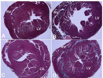
Figure 3. Gomori’s tri-chrome stained slides of cardiac tissue showing increased fibrosis inmdxmice. A) Untreated wild type cardiac tissue showing minimal collagen staining (light blue color) corresponding to a percent collagen of 1.8260.5%. B) Untreatedmdx cardiac tissue showing diffuse fibrosis in the LV and RV ventricular walls corresponding to a percent collagen of 3.8360.5%. C) Tb4 treated wild type mice showing few increased areas of collagen staining corre-sponding to a percent collagen of 1.660.5%. D) Tb4 treated mdx cardiac tissue showing large areas of collagen staining in the LV and RV walls corresponding to a percent collagen of 4.3960.7%. (LV – left ventricle, RV – right ventricle).
doi:10.1371/journal.pone.0008976.g003
Figure 4. Peroxidase staining of regenerating fibers using anti-TB4 antibodies in skeletal muscle (gastrocnemius). A) Mdx muscle treated with anti-TB4 antibody shows peroxidase staining of regenerating fibers (*); B) Mdx muscle treated with anti-desmin antibody shows staining in regenerating fibers (#), the same as in plate A; C) Wild type muscle treated with anti-TB4 antibodies shows no staining; D) Wild type muscle treated with anti-desmin antibodies shows no staining.
culture.[11] However, the authors did not find a similar increase in DMD patient derived cell lines. Turk et al. (2005) found significantly increased prothymosin beta-4 (Ptmb4), the precursor protein, at 8 of 9 time points ranging from 1 to 20 weeks in mdx hindlimb muscle.[17] Hara et al. (2005) found up-regulation of Tb4 expression in mdx skeletal muscle cell cultures and that Tb4 stimulated migration and chemotaxis of myoblasts.[18] All of these studies used mice from 1 to 20 weeks old, a time period of rapid degeneration and regeneration in the mdx skeletal muscle and demonstrate that Tb4 is important in skeletal muscle regenerative pathways. Our study supports these previous reports. We demonstrate the presence of Tb4 in regenerating fibers (Figure 4) of mdx gastrocnemius muscle. We also showed a significantly increased number of regenerating fibers in the gastrocnemius of Tb4 treatedmdxmice (Table S3). This parameter has significant variation because the gastrocnemius muscle develops patchy areas of regeneration and the majority of the muscle that is sampled shows no areas of regeneration at all. In another study, Tb4 stimulated the migration of stem cells in hair follicles leading to increased hair growth. [31] Tb4 may likewise stimulate satellite cell migration in skeletal muscle cells, leading to improved regeneration. Importantly, this is the first correlation of gene expression data with in vivo administration and histological localization and supports an integral role for Tb4 in muscle regeneration.
Another potential mechanism of Tb4 mediated regeneration is the inhibition of apoptosis. Tb4 was shown to decrease apoptosis in an ethanol-treated corneal epithelial model and inhibit activation of NF-kB during TNF-astimulation in human corneal epithelial cells. [32,33] In cardiac tissue, Bock-Marquette et al. (2004) showed that Tb4 decreased cardiac fibrosis secondary to ischemic damage in a coronary ligation model. This beneficial effect of Tb4 on myocyte cell survival was also related to decreased apoptosis and found to be mediated by PINCH, ILK and Akt. [28] Previous C2C12 muscle cell culture experiments from our lab also showed that Tb4 directly decreased NF-kB activation from TNF-a stimulation. [34] These studies support the direct action of Tb4 on muscle cells to inhibit NF-kB and consequently apoptosis and potentially improve muscle regenerative capacity.
Studies of Tb4 in multiple tissue models showed modulation of various inflammatory cytokines. [35–37] While, these changes may acutely promote wound healing, the effects of chronic Tb4 treatment on different cytokine levels are not known. Chronic treatment could induce a more prolonged cytokine response that may become more pro-inflammatory and pro-fibrotic, decreasing the beneficial effects seen with acute Tb4 administration. This might explain why this study found no significant changes in the amounts of collagen in skeletal and cardiac muscle in chronically Tb4 treatedmdxmice.
Also, previous studies showed that decreased levels of Ac-SDKP, the active tetrapeptide that is released from Tb4, led to
increased cardiac and renal perivascular fibrosis.[38] Pokharel et al. (2004) also showed that in rats over-expressing angiotensin-converting enzyme, which decreases levels of Ac-SDKP, there was increased cardiac collagen content.[39] Thus, the chronic exposure of cardiac and skeletal muscle to Tb4 may lead to a down-regulation of Tb4 expression or receptor activity and a decrease in Ac-SDKP.
Although we did not directly measure Tb4 levels in treated mice, a previous study showed significantly increased levels in the hearts and skeletal muscle of mice after treatment with 400 micrograms of Tb4 via intraperitoneal injection.[40] Chronic exposure of Tb4 could potentially lead to the development of anti-Tb4 antibodies. These antibodies could neutralize Tb4 and prevent any beneficial effects on cell survival and decreased fibrosis. The presence of any antibodies was not assessed in this study.
This study found a significant increase in Tb4 positive regenerating fibers in the skeletal muscle of exercisedmdxmice. There were no beneficial effects of chronic Tb4 treatment on muscle function or fibrosis. This study provides histological correlation for previous gene expression studies showing the importance of Tb4 in skeletal muscle regeneration.
Supporting Information
Table S1 Body weight, normalized grip strength, and Rotarod latency to fall measurements in treated and untreated wild type (BL10) and mdx mice after 6 months of treatment with thymosin beta-4.
Found at: doi:10.1371/journal.pone.0008976.s001 (0.05 MB DOC)
Table S2 Cardiac M-mode and spectral Doppler echocardiog-raphy measurements in treated and untreated wild type (BL10) and mdx mice after 6 months of treatment with thymosin beta-4. Found at: doi:10.1371/journal.pone.0008976.s002 (0.05 MB DOC)
Table S3 Gastrocnemius skeletal muscle histology
measure-ments in treated and untreated wild type (BL10) and mdx mice after 6 months of treatment with thymosin beta-4.
Found at: doi:10.1371/journal.pone.0008976.s003 (0.04 MB DOC)
Author Contributions
Conceived and designed the experiments: CFS EH KN. Performed the experiments: CFS HJC AS GP EP ADG EH KN. Analyzed the data: CFS HJC AS GP EP ADG HGD EH KN. Contributed reagents/materials/ analysis tools: CFS HJC GP EP HGD. Wrote the paper: CFS ADG HGD KN.
References
1. Hoffman EP, Brown RH Jr, Kunkel LM (1987) Dystrophin: the protein product of the Duchenne muscular dystrophy locus. Cell 51: 919–928.
2. Ryder-Cook AS, Sicinski P, Thomas K, Davies KE, Worton RG, et al. (1988) Localization of the mdx mutation within the mouse dystrophin gene. EMBO J 7: 3017–3021.
3. Sicinski P, Geng Y, Ryder-Cook AS, Barnard EA, Darlison MG, et al. (1989) The molecular basis of muscular dystrophy in the mdx mouse: a point mutation. Science 244: 1578–1580.
4. Anderson JE, Bressler BH, Ovalle WK (1988) Functional regeneration in the hindlimb skeletal muscle of the mdx mouse. J Muscle Res Cell Motil 9: 499–515. 5. De Luca A, Pierno S, Liantonio A, Cetrone M, Camerino C, et al. (2003) Enhanced dystrophic progression in mdx mice by exercise and beneficial effects of taurine and insulin-like growth factor-1. J Pharmacol Exp Ther 304: 453–463. 6. Lefaucheur JP, Pastoret C, Sebille A (1995) Phenotype of dystrophinopathy in
old mdx mice. Anat Rec 242: 70–76.
7. Quinlan JG, Hahn HS, Wong BL, Lorenz JN, Wenisch AS, et al. (2004) Evolution of the mdx mouse cardiomyopathy: physiological and morphological findings. Neuromuscul Disord 14: 491–496.
8. Spurney CF, Knoblach S, Pistilli EE, Nagaraju K, Martin GR, et al. (2008) Dystrophin-deficient cardiomyopathy in mouse: expression of Nox4 and Lox are associated with fibrosis and altered functional parameters in the heart. Neuromuscul Disord 18: 371–381.
9. Boer JM, de Meijer EJ, Mank EM, van Ommen GB, den Dunnen JT (2002) Expression profiling in stably regenerating skeletal muscle of dystrophin-deficient mdx mice. Neuromuscul Disord 12 Suppl 1: S118–124.
10. Marotta M, Ruiz-Roig C, Sarria Y, Peiro JL, Nunez F, et al. (2009) Muscle genome-wide expression profiling during disease evolution in mdx mice. Physiol Genomics 37: 119–132.
expression is attenuated in cell lines derived from Duchenne muscular dystrophy patients. Am J Pathol 164: 1773–1782.
12. Porter JD, Khanna S, Kaminski HJ, Rao JS, Merriam AP, et al. (2002) A chronic inflammatory response dominates the skeletal muscle molecular signature in dystrophin-deficient mdx mice. Hum Mol Genet 11: 263–272. 13. Porter JD, Merriam AP, Leahy P, Gong B, Feuerman J, et al. (2004) Temporal
gene expression profiling of dystrophin-deficient (mdx) mouse diaphragm identifies conserved and muscle group-specific mechanisms in the pathogenesis of muscular dystrophy. Hum Mol Genet 13: 257–269.
14. Porter JD, Merriam AP, Leahy P, Gong B, Khanna S (2003) Dissection of temporal gene expression signatures of affected and spared muscle groups in dystrophin-deficient (mdx) mice. Hum Mol Genet 12: 1813–1821.
15. Rouger K, Le Cunff M, Steenman M, Potier MC, Gibelin N, et al. (2002) Global/temporal gene expression in diaphragm and hindlimb muscles of dystrophin-deficient (mdx) mice. Am J Physiol Cell Physiol 283: C773–784. 16. Tseng BS, Zhao P, Pattison JS, Gordon SE, Granchelli JA, et al. (2002)
Regenerated mdx mouse skeletal muscle shows differential mRNA expression. J Appl Physiol 93: 537–545.
17. Turk R, Sterrenburg E, de Meijer EJ, van Ommen GJ, den Dunnen JT, et al. (2005) Muscle regeneration in dystrophin-deficient mdx mice studied by gene expression profiling. BMC Genomics 6: 98.
18. Hara T, Nakayama Y, Nara N (2005) [Regenerative medicine of skeletal muscle]. Rinsho Shinkeigaku 45: 880–882.
19. Oates K, Goldstein A (1995) Thymosin. In: DeVita VT, Hellman S, Rosenberg SA, eds (1995) Biologic Therapy of Cancer. Philadelphia: JB Lippincott. pp 841–852.
20. Pantaloni D, Carlier MF (1993) How profilin promotes actin filament assembly in the presence of thymosin beta 4. Cell 75: 1007–1014.
21. Weber A, Nachmias VT, Pennise CR, Pring M, Safer D (1992) Interaction of thymosin beta 4 with muscle and platelet actin: implications for actin sequestration in resting platelets. Biochemistry 31: 6179–6185.
22. Hannappel E, Huff T (2003) The thymosins. Prothymosin alpha, parathymosin, and beta-thymosins: structure and function. Vitam Horm 66: 257–296. 23. Safer D, Elzinga M, Nachmias VT (1991) Thymosin beta 4 and Fx, an
actin-sequestering peptide, are indistinguishable. J Biol Chem 266: 4029–4032. 24. Safer D, Golla R, Nachmias VT (1990) Isolation of a 5-kilodalton
actin-sequestering peptide from human blood platelets. Proc Natl Acad Sci U S A 87: 2536–2540.
25. Sanders MC, Goldstein AL, Wang YL (1992) Thymosin beta 4 (Fx peptide) is a potent regulator of actin polymerization in living cells. Proc Natl Acad Sci U S A 89: 4678–4682.
26. Malinda KM, Sidhu GS, Mani H, Banaudha K, Maheshwari RK, et al. (1999) Thymosin beta4 accelerates wound healing. J Invest Dermatol 113: 364–368.
27. Sosne G, Szliter EA, Barrett R, Kernacki KA, Kleinman H, et al. (2002) Thymosin beta 4 promotes corneal wound healing and decreases inflammation in vivo following alkali injury. Exp Eye Res 74: 293–299.
28. Bock-Marquette I, Saxena A, White MD, Dimaio JM, Srivastava D (2004) Thymosin beta4 activates integrin-linked kinase and promotes cardiac cell migration, survival and cardiac repair. Nature 432: 466–472.
29. Spurney CF, Gordish-Dressman H, Guerron AD, Sali A, Pandey GS, et al. (2009) Preclinical drug trials in the mdx mouse: Assessment of reliable and sensitive outcome measures. Muscle Nerve 39: 591–602.
30. Tietz N (1982) Fundamentals of clinical chemistry. Philadelphia: WB Saunders Co. pp 682–689.
31. Philp D, Nguyen M, Scheremeta B, St-Surin S, Villa AM, et al. (2004) Thymosin beta4 increases hair growth by activation of hair follicle stem cells. FASEB J 18: 385–387.
32. Sosne G, Qiu P, Christopherson PL, Wheater MK (2007) Thymosin beta 4 suppression of corneal NFkappaB: a potential anti-inflammatory pathway. Exp Eye Res 84: 663–669.
33. Sosne G, Siddiqi A, Kurpakus-Wheater M (2004) Thymosin-beta4 inhibits corneal epithelial cell apoptosis after ethanol exposure in vitro. Invest Ophthalmol Vis Sci 45: 1095–1100.
34. Baudy AR, Saxena N, Gordish H, Hoffman EP, Nagaraju K (2009) A robust in vitro screening assay to identify NF-kappaB inhibitors for inflammatory muscle diseases. Int Immunopharmacol 9: 1209–1214.
35. Sosne G, Chan CC, Thai K, Kennedy M, Szliter EA, et al. (2001) Thymosin beta 4 promotes corneal wound healing and modulates inflammatory mediators in vivo. Exp Eye Res 72: 605–608.
36. Reti R, Kwon E, Qiu P, Wheater M, Sosne G (2008) Thymosin beta4 is cytoprotective in human gingival fibroblasts. Eur J Oral Sci 116: 424–430. 37. Zhang Y, Feurino LW, Zhai Q, Wang H, Fisher WE, et al. (2008) Thymosin
Beta 4 is overexpressed in human pancreatic cancer cells and stimulates proinflammatory cytokine secretion and JNK activation. Cancer Biol Ther 7: 419–423.
38. Cavasin MA, Liao TD, Yang XP, Yang JJ, Carretero OA (2007) Decreased endogenous levels of Ac-SDKP promote organ fibrosis. Hypertension 50: 130–136.
39. Pokharel S, van Geel PP, Sharma UC, Cleutjens JP, Bohnemeier H, et al. (2004) Increased myocardial collagen content in transgenic rats overexpressing cardiac angiotensin-converting enzyme is related to enhanced breakdown of N-acetyl-Ser-Asp-Lys-Pro and increased phosphorylation of Smad2/3. Circulation 110: 3129–3135.