Suppression of TNBS-induced colitis in rats by 4-methylesculetin, a natural
coumarin: Comparison with prednisolone and sulphasalazine
Aline Witaicenis
a, Ana C. Luchini
a, Clélia A. Hiruma-Lima
b, Sérgio L. Felisbino
c, Natividad Garrido-Mesa
d,
Pilar Utrilla
d, Julio Gálvez
d, Luiz C. Di Stasi
a,⇑aUniv. Estadual Paulista – UNESP, Institute of Biosciences, Department of Pharmacology, Botucatu, SP, Brazil bUniv. Estadual Paulista – UNESP, Institute of Biosciences, Department of Physiology, Botucatu, SP, Brazil cUniv. Estadual Paulista – UNESP, Institute of Biosciences, Department of Morphology, Botucatu, SP, Brazil
dCentro de Investigación Biomédica en Red de Enfermedades Hepáticas y Digestivas (CIBER-EHD), Department of Pharmacology, Center for Biomedical Research,
University of Granada, Granada, Spain
a r t i c l e
i n f o
Article history:
Received 23 September 2011
Received in revised form 3 November 2011 Accepted 8 November 2011
Available online 16 November 2011
Keywords:
IBD
4-Methylesculetin TNBS
Metalloproteinase Apoptosis
a b s t r a c t
The aim of the present study was to compare the effects of the 4-methylesculetin with those produced by prednisolone and sulphasalazine and to elucidate the mechanisms involved in its action. Colitis was induced in rat by instillation of trinitrobenzenesulphonic acid (TNBS). The colon damage was evaluated using macroscopic, microscopic and biochemical analysis. In addition, in vitro studies were performed to evaluate cytokine production in cell cultures using the murine macrophage cell line RAW264.7, mouse splenocytes and the human colonic epithelial cell line Caco-2. 4-Methylesculetin produced a reduction of the macroscopic damage score and the recovery of the intestinal cytoarchitecture. These effects were associated with a prevention of the GSH depletion and an inhibition in AP activity. After colitis relapse, 4-methylesculetin improved the colonic inflammatory status as evidenced by histological findings, with a reduction in apoptosis, as well as biochemically by inhibition of colonic myeloperoxidase, alkaline phosphatase and metalloproteinase 9 activities. Paired with this inhibitive activity, there was a decrease in malondialdehyde content and in IL-1blevels. In vitro assays revealed that 4-methylesculetin promoted an inhibition in IL-1b, IL-8, IL-2 and IFN-cproduction in cell cultures. In conclusion, 4-methylesculetin showed similar efficacy to that obtained with either prednisolone or sulphasalazine, both in the acute phase of colitis as well as following a curative protocol. The intestinal anti-inflammatory activity by 4-methylesculetin is likely related to its ability in reduce colonic oxidative stress and inhibit pro-inflam-matory cytokine production.
Ó2011 Elsevier Ireland Ltd. All rights reserved.
1. Introduction
Inflammatory bowel disease (IBD) refers to a group of condi-tions characterised by inflammation in the intestinal tract. Crohn’s disease (CD) and ulcerative colitis (UC) account for the majority of these conditions [1]. These diseases are characterised by the uncontrolled response of the intestinal immune system against the normal enteric microflora, leading to abdominal pain and chronic diarrhoea for most of the patient’s life[2]. The aetiopatho-genesis of these diseases has not been fully elucidated but is cur-rently presumed to result from a complex interplay among genetic, environmental, microbial and immune factors. Patients with IBD frequently experience relapse, and medical treatments are not always able to keep patients in remission in the long term.
Current therapies involve a combination of aminosalicylates, corti-costeroids and immunosuppressants such as azathioprine, metho-trexate, cyclosporine and recently monoclonal antibodies against tumour necrosis factor
a
(TNF-a
). These drugs have shown benefi-cial effects in the treatment of IBD, but they might impose risk to the patients, and not all patients respond to these treatments. They do not represent a cure for these diseases, and dose-dependent side effects are frequently associated with all of these treatments [3]. For this reason, research of new agents that combine pharma-cologic efficacy and the absence of undesirable adverse effects is critical.It has been proposed that reactive oxygen species (ROS) are involved in the development of tissue injury in patients with IBD. Indeed, the anti-inflammatory effects of corticosteroids in the treatment of IBD have been ascribed, in part, to their ability to reduce neutrophil infiltration, thus ameliorating oxidative insult [4]. Moreover, sulphasalazine has been suggested to pro-duce beneficial effects by inhibiting the formation of ROS from neutrophils[5].
0009-2797/$ - see front matterÓ2011 Elsevier Ireland Ltd. All rights reserved. doi:10.1016/j.cbi.2011.11.004
⇑Corresponding author. Address: Univ. Estadual Paulista – UNESP, Institute of Biosciences, Department of Pharmacology, Laboratory of Phytomedicines, Botucatu, 18618-970 SP, Brazil. Tel.: +55 21 14 38116253.
E-mail address:[email protected](L.C. Di Stasi).
Contents lists available atSciVerse ScienceDirect
Chemico-Biological Interactions
Previous studies have shown the beneficial effects of different natural antioxidant compounds in experimental models of rat coli-tis, including flavonoids like quercitrin[6,7], rutoside [8], morin [9], diosmin[10], hesperidin[10], isocoumarin[11]and coumarin derivatives[12,13]. Our preliminary studies showed that esculetin and 4-methylesculetin treatments prevent colonic damage in-duced by TNBS in rats[13]. This study showed that the presence of methyl group at C-4 in the esculetin molecules improves its antioxidant and intestinal anti-inflammatory activity [13]. This encouraged us to continue research with 4-methylesculetin to elu-cidate the mechanism of action and to compare its effects with those produced by prednisolone and sulphasalazine, two of the main drugs used for the treatment of IBD in humans.
2. Materials and methods
2.1. Materials
All chemicals, including 4-methylesculetin, prednisolone and sulphasalazine were provided by Sigma, unless otherwise stated. The test substances were dissolved in methylcellulose (1% w/v) and prepared fresh daily for administration to the animals.
2.2. Animals
Male Wistar rats (weighing 180–200 g) obtained from the Cen-tral Animal House of the São Paulo State University (UNESP), Bot-ucatu, São Paulo (Brazil), were housed in standard environmental conditions (21°C, 60–70% humidity) with 12 h light/dark cycles and air filtration. Animals had free access to water and food (Bio-base). Experimental protocols met the ‘‘Guidelines of Animal Experimentation’’ approved by the Commission of Ethics in Animal Experimentation (Protocol number 042/04-CEEA), Institute of Bio-sciences, São Paulo State University (UNESP).
2.3. Induction of colitis and assessment of the inflammatory process
Colitis was induced using the method originally described by Morris[14]. Briefly, animals were fasted overnight and then anaes-thetised with halothane. Under anaesthesia, they were adminis-tered 10 mg of trinitrobenzenesulphonic acid (TNBS) dissolved in 0.25 ml 50% ethanol (v/v) by means of a Teflon cannula inserted 8 cm into the anus. During and after TNBS administration, the rats remained in a head-down position until they recovered from the anaesthesia. Rats from the non-colitic (normal) group received 0.25 ml of saline instead of TNBS. Two different protocols were followed.
2.3.1. Acute colitis
Rats received 5 mg/kg of 4-methylesculetin, 2 mg/kg of prednis-olone or 50 mg/kg of sulphasalazine orally at 96, 72, 48, 24 and 2 hours before colitis induction. The dose of 5 mg/Kg of 4-methyle-sculetin was chosen based in dose-response study previously per-formed in our laboratory[13]. The drugs were administered by means of an oesophageal catheter (volume: 5 ml/kg). Two addi-tional groups were included for reference: a non-colitic group that received saline intracolonically and the oral vehicle and a colitic group that received only the first dose of TNBS (TNBS-control with-out relapse) and the vehicle (5 ml/kg methylcellulose) orally. The animal body weights, the occurrence of diarrhoea (as detected by perianal fur soiling) and the total food intakes for each group were recorded daily. Animals from all groups (n= 7) were killed 48 h after colitis induction with an overdose of halothane.
2.3.2. Colitis relapse
In this protocol, colitis was initially induced with 10 mg of TNBS in 50% ethanol, as described previously. After 14 days, the animals were given a second dose of 10 mg of TNBS in an attempt to mimic the common relapses experienced in humans with IBD. The ani-mals were divided into three groups, which were treated daily with the same drugs and doses used in acute colitis. The non-colitic and TNBS-control groups were similar to the acute colitis group. Treatments started 2 h after the first administration of TNBS and continued until the day before the animals were killed. Animals from each group were sacrificed after 1, 2 or 3 weeks of colitis induction, whereas all animals from the colitic control group with-out relapse were sacrificed after 3 weeks. The animals were killed with an overdose of halothane.
The colonic segments were obtained after laparotomy, and the occurrence of adhesions between the colon and adjacent organs was noted. The colonic segments were placed on an ice-cold plate, cleaned of fat and mesentery, and blotted on filter paper. The colon was weighed, and its length was measured under a constant load (2 g). The colon was opened longitudinally and scored for macro-scopically visible damage on a 0–10 scale by two observers blinded to the treatments, according to the criteria described by Bell[15]. The colon was subsequently divided longitudinally into different pieces to be used for the following biochemical assays: myeloper-oxidase (MPO) activity, alkaline phosphatase (AP) activity, total glutathione (GSH) content, malondialdehyde content (MDA), ma-trix metalloproteinase (MMP-9 and MMP-2) activity, and TNF-
a
and interleukin 1b(IL-1b) levels.
2.4. Biochemical assays in colonic specimens
2.4.1. Myeloperoxidase (MPO) activity
MPO activity was measured according to the technique de-scribed by Krawisz [16]. Samples were suspended in 1 ml of 50 mM phosphate buffer incorporating 0.5% hexadecyltrimethy-lammonium bromide (pH 6.0) and minced with scissors for 15 s on an ice-cold plate. The resultant suspension was subsequently diluted to a final 1:20 w/v ratio, homogenised for 1 min with an automatic Heidolph homogeniser, sonicated for 10 s and subjected to three freeze-thaw cycles. The homogenates were then centri-fuged at 7000gand 4°C for 10 min, and the supernatants were
as-sayed for MPO activity. The results are expressed as MPO units per g of wet tissue.
2.4.2. Alkaline phosphatase (AP) activity
AP activity was measured spectrophotometrically using diso-dium nitrophenyl phosphate (5.5 mM) as a substrate in a 50 mM glycine buffer with 0.5 mM MgCl2, pH 10.5[17]. The results are ex-pressed as mU per mg of protein.
2.4.3. Glutathione (GSH) content
Total GSH content was quantified with the recycling assay de-scribed by Anderson[18]. Samples were thawed, minced, diluted to a concentration of 1:20 (w/v) in ice-cold 5% (w/v) trichloroacetic acid and homogenised. The homogenates were centrifuged at 7000gfor 15 min at 4°C, and the supernatants were used to quan-tify glutathione content. The results are expressed as nmol per g of wet tissue.
2.4.4. TNF-
a
and IL-1blevelsassayed. TNF-
a
and IL-1blevels were quantified by a DuoSet ELISA Kit to measure natural and recombinant rat enzyme (R&D Systems, Inc. USA). The results were expressed as pg per mg of protein.2.4.5. Production of malondialdehyde (MDA)
Tissue concentrations of MDA were determined as an index of lipid peroxidation by the methodology described by Zingarelli [19]. The tissue samples were homogenised in 1.15% KCl solution. An aliquot (400
l
l) of the homogenate was added to a reaction mixture containing 200l
l of 8.1% sodium dodecyl sulphate (SDS), 1500l
l of 20% acetic acid (pH 3.5), 1500l
l of 0.8% thiobar-bituric acid, and 400l
l distilled water. The samples were then boiled for one hour at 95°C and centrifuged at 3000gfor 10 min-utes. The absorbance of the supernatant was measured by spectro-photometry at 532 nm and compared to a standard curve obtained using 1,1,3,3-tetramethoxypropane. The data were expressed as nmol per g of wet tissue.2.4.6. Gelatine zymography for MMP-2 and MMP-9
Zymographic analyses were used to evaluate the gelatinolytic activity of MMP-2 and -9 in colon rats. Frozen samples of rat colons from all experimental animals were mechanically homogenised in a solution of 50 mM Tris-HCl, pH 7.5, with 0.25% of triton X-100 and 10 mM CaCl2portioned for 0.1 ml for every 30 mg of colonic tissue. Aliquots of 20
l
g of protein were subjected to electrophore-sis under non-reducing conditions in an 8% polyacrylamide gel co-polymerised with 0.1% gelatine. After electrophoresis, the gels were washed twice (for 15 min) in a solution of 2.5% Triton X-100 to remove the sodium dodecyl sulphate (Bio-Rad, CA, USA), fol-lowed by two washes in a buffer solution of 50 mM Tris–HCl, pH 8.0, for 5 min. Then the gels were incubated overnight in Tris– HCl buffer containing 5 mM CaCl2at 37°C. Finally, the gels were stained with Coomassie Brilliant Blue R-250[20]. The bands were analysed in an Image Master VDS 3.0 (Pharmacia Biotech). The val-ues are expressed as integrated optical densities.2.5. Histological findings
Representative whole gut specimens were taken from a region of the inflamed colon corresponding to the segment adjacent to the gross macroscopic damage and were fixed in 4% buffered form-aldehyde. Cross-sections were selected and embedded in paraffin. Equivalent colonic segments were also obtained from the non-colitic group. Full-thickness sections of 6
l
m were obtained at different levels and stained with haematoxylin and eosin. After staining, images were subjected to analysis and photomicrography with a Leica microscope utilising Leica Qwin Plus version 3.3 e 3.40 for the evaluation of the histological damage score by methodology described by Stucchi[21].The Feulgen reaction technique[22]was also applied to another histological section to analyse the degree of compaction of the nu-clear chromatin in epithelial cells of the crypts that indicated the apoptosis of these cells. The results were expressed as the number of apoptotic cells divided by the number of total crypts.
2.6. In vitro effects of 4-methylesculetin on cytokine production
2.6.1. Effects on IL-1bproduction in RAW264.7 cells
The murine macrophage cell line RAW264.7 was obtained from the Cell Culture Unit of the University of Granada (Granada, Spain) and cultured in Dulbecco’s modified Eagle medium (DMEM) sup-plemented with 10% FBS and 2 mML-glutamine in a humidified
5% CO2atmosphere at 37°C. These cells were seeded onto 24-well
plates at a density of 5105cells per well and grown until conflu-ence. Then they were incubated for 30 min with different concen-trations of 4-methylesculetin (1–100
l
M). Afterwards, these cellswere stimulated with LPS (50 pg/ml) for 20 h, and the supernatants were collected, centrifuged at 10,000gfor 5 min, and frozen until ELISA analysis of IL-1bconcentration.
2.6.2. Effects on IL-8 production in Caco-2 cells
The human colon adenocarcinoma cell line Caco-2, obtained from the Cell Culture Unit of the University of Granada (Granada, Spain), was used to test the ability of the 4-methylesculetin to in-hibit the production/release of IL-8. Cells were grown in DMEM supplemented with 10% FBS and 2 mML-glutamine in a humidified
5% CO2atmosphere at 37°C. Caco-2 cells were seeded onto 24-well plates at a density of 5105cells per well and grown until forma-tion of a monolayer. Then they were incubated for 30 min with dif-ferent concentrations of 4-methylesculetin (1–100
l
M). After this, the cells were stimulated with IL-1b(1 ng/ml) for 20 h, and the supernatants were collected, centrifuged at 10,000g for 5 min, and frozen until ELISA analysis of IL-8 concentration.2.6.3. Effects on IFN-
c
and IL-2 production on mouse splenocytesSplenocytes were obtained from male 8-week-old Balb/c mice provided by the Laboratory Animal Service of the University of Gra-nada (GraGra-nada, Spain). After sacrificing the mice with an overdose of halothane, the spleens were removed and homogenised in DMEM with 1% penicillin/streptomycin. After centrifugation (1500g, 5 min), the erythrocytes were lysed with a buffer (NH4Cl 1.7 mol/L, KHCO3 0.12 mol/L, ethylenediamine-tetra-acetic 9 mmol/L) for 30 min at 4°C. Resting cells were counted with a haemocytometer and cultured to perform proliferation and stimu-lation assays in culture medium containing DMEM and 10% FBS. Cells were incubated at 37°C in a humidified 5% CO2atmosphere. These cells were incubated for 1 hour with different concentrations of 4-methylesculetin (1, 10, 50 or 100
l
M). After this time, the cells were stimulated with concanavalin A (5l
g/ml) for 48 h, and the supernatants were collected, centrifuged at 10,000g for 5 min and frozen at 80°C until ELISA analysis of IFN-c
and IL-2concentrations.
2.6.4. Cell viability assays
The viability of the spleen-derived lymphocytes was quantified using the WST-1 Reagent (Roche, Germany) following the protocol previously described[23]and also by viable cell counting by con-ventional trypan blue staining using a haemocytometer.
Caco-2 and RAW264.7 cell viability was determined by crystal violet staining. Briefly, the cells were stained with crystal violet (0.2%) in ethanol for 30 minutes at room temperature. The plates were then washed 4 times with PBS. The cells were solubilised with 1% sodium dodecyl sulphate solution for 30 minutes. The cells were then centrifuged at 3000gfor 5 minutes, and dye uptake was measured at 540 nm using a microplate reader.
2.7. Statistics
All results are expressed as the mean ± S.E.M., and the differ-ences between means were tested for statistical significance using one-way analysis of variance (ANOVA). Nonparametric data (score) are expressed as the median (range) and were analysed with the Kruskal–Wallis test. Differences between proportions were ana-lysed with the
v
2test. Statistical significance was set atp< 0.05.3. Results
3.1. Acute colitis
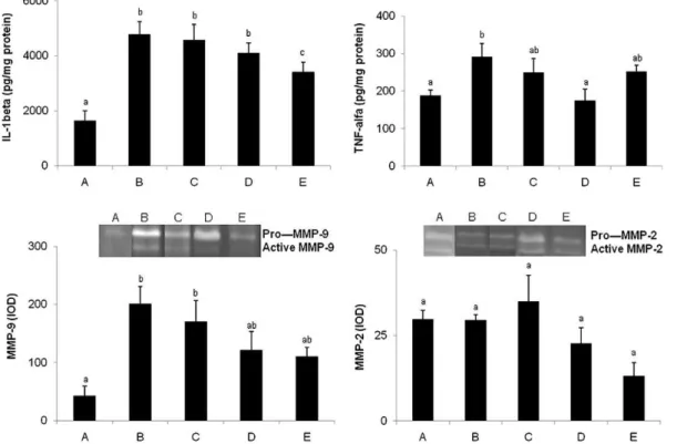
after 48 hours in comparison with non-colitic rats (data not shown) and was likely related to a reduction in food intake (data not shown) and the presence of diarrhoea in this group (Table 1). The macroscopic analysis of the colonic specimens in the colitic rats revealed the existence of severe necrosis and inflammation of the mucosa, typically extending 5.0 cm along the colon (Table 1). This inflammatory process was associated with an increase in the colonic weight/length ratio. Biochemically, the colonic damage was characterised by an increase in colonic MPO, AP (Table 2) and metalloproteinase-9 activities (Fig. 1), as well as in TNF-
a
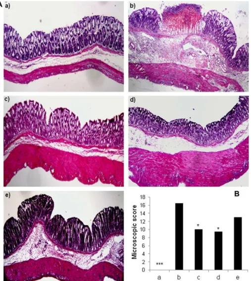
, IL-1b(Fig. 1) and MDA contents (Table 2). Furthermore, significant colo-nic GSH depletion took place in the inflamed colon (Table 2). His-tological assessment of colonic samples from the TNBS control group revealed severe transmural disruption of the normal archi-tecture of the colon, with extensive ulceration and inflammation involving all the intestinal layers of the colon, with a median score of 16.5 (Fig. 2). These samples were also characterised by severe oedema, interstitial micro-haemorrhages and diffuse leucocyte infiltration, mainly composed of neutrophils and, to a lesser extent, lymphocytes and histiocytes (Fig. 2).
The compound 4-methylesculetin showed an intestinal anti-inflammatory preventative effect in colitic rats, similar to the effects obtained with prednisolone and sulphasalazine, as dem-onstrated by a decrease in the macroscopic damage scores, in the percentage of diarrhoea and in the incidence of adhesions between the colon and adjacent organs (Table 1). This benefit was also as-sessed biochemically by the increase in GSH content and the inhi-bition in AP activity (Table 2). Additionally, only prednisolone significantly reduced TNF-
a
levels (Fig. 1). In contrast, 4-methyle-sculetin and prednisolone reduced the microscopic damage score in comparison with the non-treated colitic group (Fig. 2). Thus, the histological studies showed that treatment with 4-methylescu-letin and prednisolone promoted the recovery the intestinal cyto-architecture and a reduction in ulceration, oedema, mucus depletion and in the infiltration of inflammatory cells in the sub-mucosal layers (Fig. 2). In addition, 4-methylesculetin also reduced this infiltration into the lamina propria layer (Fig. 2). Finally, the number of apoptotic epithelial cells increased two-fold in the TNBS control group compared to the non-colitic group. However, none of the tested drugs was able to significantly reduce the number of apoptotic epithelial cells (Table 1).3.2. Colitis relapse
The histological evaluation of the colonic damage in the chronic phase of the TNBS-colitis revealed that the damage was maximal seven days after TNBS instillation and decreased gradually during the following two weeks. At this time, the colonic segments still appeared macroscopically and microscopically ulcerated and in-flamed (Table 3). The microscopic analysis also revealed leucocyte migration accompanied by mucus depletion and oedema. The inflammatory process was also demonstrated by a similar
progres-sion of the biochemical inflammatory parameters studied (Tables 4 and 5).
The administration of 4-methylesculetin, prednisolone or sul-phasalazine once the colonic damage had been induced facilitated the recovery of the inflammatory process during the following two weeks. This was demonstrated both histologically and biochemi-cally. Thus, at the first week, 4-methylesculetin significantly reduced the macroscopic damage score in comparison to the TNBS-control and apoptosis. These effects were similar to that ob-served with prednisolone and sulphasalazine treatments (Table 4). Microscopically, all treatments also reduced leucocyte infiltration and oedema in the mucosa compared with TNBS-control rats.
Biochemically, these beneficial effects were associated with a significant inhibition of MPO, AP and MMP-9 activities (Tables 4 and 5). In addition, 4-methylesculetin and prednisolone also de-creased the MDA content (Table 4) and IL-1b colonic levels (Table 5).
Colitis relapse induced by a second dose of TNBS was character-ised by a reactivation of the colonic inflammatory process, and a significant increase in the microscopic and macroscopic damage score and in the weight/length ratio (Table 3) were observed when compared to the normal evolution of colonic damage in the ani-mals that did not receive the second dose of TNBS (TNBS-control without relapse). Microscopically, the reactivation of the inflam-matory process was characterised by an increase of leucocyte infil-tration in the lamina propria and mucosa. Thus, these effects were associated with colonic oxidative damage, which caused a reduc-tion in the GSH content and an increase in the others biochemical parameters evaluated in comparison with colitic animals without relapse (Tables 4 and 5).
After the colitis relapse, the treatment with 4-methylesculetin promoted a reduction in microscopic damage score and apoptosis (Table 3). These effects were biochemically accompanied by inhibi-tion in MPO and AP activities (Table 4). The treatments with pred-nisolone and sulphasalazine were not able to significantly modify any of the microscopic or macroscopic parameters evaluated. Bio-chemically, both drugs were only able to counteract GSH content, whereas sulphasalazine also inhibited colonic AP activity (Tables 4 and 5).
3.3. Inhibition of cytokine production in cell cultures
The results obtained in these assays showed that 4-methylescu-letin dose-dependently reduced the production of the different cytokines studied (Fig. 3). In LPS-stimulated RAW264.7 macro-phages, the coumarin derivative inhibited IL-1b release signifi-cantly at a concentration of 10
l
M and achieved a complete inhibition at a concentration of 50l
M. Similarly, the incubation of 4-methylesculetin with IL-1b-stimulated Caco-2 cells reduced the production of IL-8 at a concentration of 10l
M and obtained a maximal inhibition of 70% at a concentration of 50l
M. Finally, in concanavalin A-stimulated splenocytes, this compoundTable 1
Effects of 4-methylesculetin (5 mg/Kg), prednisolone (2 mg/Kg) and sulphasalazine (50 mg/Kg) treatment on macroscopic damage score, colonic weight, incidence of diarrhoea, adherences and apoptosis in acute TNBS colitis.
Group Macroscopic damage score (0–10)1 Colonic weight (mg/cm)2 Diarrhoea (%)3 Adherence (%)3 Apoptosis/per crypt epithelial cells2
Non-colitic 0a 80.2 ± 12.17a 0a 0a 0.062 ± 0.004a
TNBS-control 8.0 (7–10)b 144.6 ± 14.04b 100b 83b 0.136 ± 0.015b
4-Methylesculetin 6.0 (3–7)c 138.0 ± 22.17bc 33a 33b 0.111 ± 0.019b
Prednisolone 6.0 (1–8)c 138.9 ± 23.98bc 33a 0a 0.087 ± 0.012a
Sulphasalazine 6.0 (3–7)c 132.8 ± 23.52bc 67bc 33a 0.089 ± 0.013a
1 Score data are expressed as the median (range).
2 Exclusion extension of lesion, Colonic weight and apoptosis data are expressed as the mean ± S.E.M. 3 Diarrhoea and adherence are expressed in percentage. Value without a common letter differp< 0.05.
inhibited either IL-2 or IFN-
c
production at a concentration of 50l
M, resulting 65% and 100% inhibition, respectively, at the high-est concentration assayed. The viability assays revealed that most of the concentrations used were devoid of any significant cytotoxic effect, with the exception of RAW264.7, in which the concentration of 100l
M of 4-methylesculetin showed an incidence of cytotoxic effects of approximately 25%.4. Discussion
This study confirms the intestinal anti-inflammatory activity of 4-methylesculetin, at a dose of 5 mg/kg, in the TNBS model of rat colitis. This natural compound showed similar efficacy to that ob-tained with either prednisolone (2 mg/kg) or sulphasalazine (50 mg/kg), two of the main drugs currently used for the treatment of human IBD. This beneficial effect was observed following the preventative dosing protocol used in the acute phase of experi-mental colitis, as well as after a curative protocol in the chronic phase.
In the chronic colitis with relapse model, the colonic damage was initially characterised by a time-dependent recuperation of inflammatory status, similar to that described previously in[11].
Two weeks after the initial insult, the colonic segments still ap-peared macroscopically ulcerated and inflamed. As expected, the second administration of TNBS resulted in a reactivation of the co-lonic damage in comparison with that observed in the control colitic group without relapse.
The treatment with 4-methylesculetin, prednisolone or sul-phasalazine showed an improvement of the intestinal inflamma-tory process in all time points studied, either before or after the second TNBS instillation. This was exhibited by a significant de-crease in the macroscopic damage score and by an inhibition in colonic MPO activity, as this enzyme activity is a marker of neu-trophil infiltration, which can be considered an index of inflam-mation damage [24]. Colonic inflammation was also characterised by an increase in AP activity, which has been attrib-uted mainly to leucocyte activity [25]. AP enzyme activity has been considered a sensitive biochemical marker of intestinal inflammation[6,26,7] and its inhibition can be interpreted as a result of the anti-inflammatory effect of a given compound. In fact, 4-methylesculetin, prednisolone and sulphasalazine inhib-ited AP activity in the first week after TNBS instillation, while 4-methylesculetin and sulphasalazine also showed this activity after the colitis relapse.
Table 2
Effects of 4-methylesculetin (5 mg/Kg), prednisolone (2 mg/Kg) and sulphasalazine (50 mg/Kg) treatment on glutathione (GSH) content, myeloperoxidase (MPO) and alkaline phosphatase (AP) activities and malondialdehyde content (MDA) in acute TNBS colitis.
Group GSH (nmol/g tissue) MPO (U/g tissue) AP (mU/mg protein) MDA (nmol/g tissue) Non-colitic 1768 ± 90.63a 201 ± 26.20a 5.44 ± 0.45a 12.99 ± 1.83a
TNBS-control 1046 ± 51.92b 2246 ± 216.97b 16.62 ± 1.43b 20.07 ± 1.29b
4-Methylesculetin 1638 ± 443.41a 1590 ± 135.53c 11.41 ± 1.55c 18.00 ± 2.29ab
Prednisolone 1698 ± 84.31a 1441 ± 218.92c 9.96 ± 1.39a 15.58 ± 2.32ab
Sulphasalazine 1695 ± 98.90a 1550 ± 88.56c 10.91 ± 1.20c 16.57 ± 1.48ab
Data are expressed as mean ± S.E.M. Means without a common letter differ,p< 0.05.
IBD is characterised by a complex inflammatory cascade responsible for the tissue destruction. In this event, matrix metalloproteinases (MMPs), a group of zinc-dependent endopep-tidases, participate in extracellular matrix degradation and remod-elling. Their enzymatic activity is regulated by tissue inhibitors of the MMPs (TIMPs); however, under pathological situations, the in-creased amount of active MMPs cannot be controlled by TIMPs, resulting in extracellular matrix breakdown and tissue injury [27]. As a result, drugs that are able to block the tissue damage-re-lated MMPs or are able to increase the tissue protection-redamage-re-lated MMPs could be considered as good candidates for the treatment of this disease. In IBD, the MMP gelatinases MMP-2 and MMP-9 has been described to display antagonistic functions[28]. Epithe-lial-derived MMP-9 is an important mediator of tissue injury in colitis in human, whereas MMP-2 protects the colon against tissue damage and maintains gut barrier function[29]. It has been sug-gested that the inhibition of intestinal MMP-9 activity may be ben-eficial in IBD[28]. In the present study, and in comparison with non colitic rats, colonic MMP-9 activity was significantly increased in control colitic rats both in the acute and chronic phases of exper-imental TNBS colitis, but not one week after colitic relapse. How-ever, colonic MMP-2 activity was only significantly inhibited one week after the first TNBS instillation. The administration of 4-methylesculetin, prednisolone or sulphasalazine to colitic rats once
colonic damage had been induced resulted in the inhibition of co-lonic MMP-9 activity seven days after TNBS administration and simultaneously maintained the protective activity of colonic MMP-2.
TNF-
a
and IL-1bare clearly involved in the intestinal inflamma-tory process by inducing the expression of different pro-inflamma-tory proteins, including MMPs[30]. It is possible that the effect of 4-methylesculetin or prednisolone on MMP-9 activity is related, at least in part, to the ability of these compounds to reduce IL-1band TNF-a
colonic levels.Because oxidative damage is involved in the pathogenesis of IBD, antioxidant compounds have been studied for the treatment of these intestinal conditions [31]. 4-methylesculetin, similar to other coumarin derivatives, has been described to possess antiox-idant and anti-inflammatory effects[32]. In a previous study, we reported the beneficial effect of 4-methylesculetin in TNBS-in-duced colitis in rats. We concluded that its activity is likely related to its antioxidant properties[13]. The results obtained in the pres-ent study confirm this observation because 4-methylesculetin im-proved the biochemical markers of colonic oxidative stress, as analysed by glutathione. Glutathione is a natural tripeptide that displays antioxidant activity, and depletion of glutathione is associated with experimental colitis [33] and MDA, the major end-product of the oxidation of polyunsaturated fatty acids. Fig. 2.Effects of different treatments in TNBS rat colits (acute protocol) in histological analyses. (A) Histological sections of colonic mucosa stained with haematoxylin and eosin: (a) non-colitic; (b)TNBS-control; (c) 4-methylesculetin (5 mg/Kg), (d) prednisolone (2 mg/Kg) and (e) sulphasalazine (50 mg/Kg). (B) Colonic microscopic damage score. Data are expressed as means ± SEM.⁄p< 0.05;⁄⁄⁄p< 0.01 vs. TNBS control group.
Glutathione is frequently measured as indicator of the ‘‘in vivo’’ lipid peroxidation and oxidative stress. In addition, the reduction in neutrophil infiltration, as detected by microscopic and biochem-ical analyses, can also contribute to these protective effects on the altered oxidative status in the inflamed colonic tissue.
It has been reported that the impairment in the epithelial bar-rier function can account for the pathophysiology of IBD, leading to a higher access of luminal agents to the intestinal tissue. Fur-thermore, this impairment is also associated with a functional loss of normal absorptive capacity and inappropriate secretion of intes-tinal fluid and electrolytes, thus promoting diarrhoea, abdominal cramps, excessive gas, and bloating[34]. One of the processes that has been proposed to be involved in the altered epithelial integrity is the increase in intestinal epithelial cell apoptosis[35]. Inhibition of cell apoptosis has been considered a symptomatic treatment
op-tion for ill patients with severe diarrhoea[34]. This inhibitory ef-fect on epithelial cell apoptosis may reduce villus atrophy and epithelial destruction [36]. The present study reveals that 4-methylesculetin, prednisolone and sulphasalazine reduced the number of intestinal epithelial cell apoptotic events across all time points studied during the chronic colitis with relapse. Different mechanisms can be involved in this effect, and one of them could be ascribed to the beneficial effects exerted by these compounds acting on the colonic oxidative status. Previously, it has been re-ported that the excessive oxidative stress to which the intestinal mucosa is subjected under inflammatory conditions might en-hance epithelial apoptosis[37]. Therefore, it has been suggested that IFN-
c
and TNF-a
, the production of which is clearly increased by the infiltrating immune-competent cells in these intestinal con-ditions, contribute to epithelial cell apoptosis in the gut[34]. In the Table 3Effects of 4-methylesculetin (5 mg/Kg), prednisolone (2 mg/Kg) and sulphasalazine (50 mg/Kg) treatment on macroscopic damage score, extension of lesion, microscopic damage score and apoptosis in chronic with relapse of TNBS colitis.
Group Macroscopic damage score (0–10)1
Colonic weight (mg/cm)2
Microscopic damage score (0–27)2
Apoptosis/per crypt epithelial cells
1 week
Non-colitic 0a 89.0 ± 7.42a 0a 0.079 ± 0.014a
TNBS-control 5.5 (5–7)b 147.5 ± 8.02b 13.0 (10–23)b 0.198 ± 0.021b
4-Methylesculetin 3.5 (3–5)c 138.3 ± 4.15b 10.5 (7–15)b 0.094 ± 0.02a
Prednisolone 2.0 (0–5)a 137.7 ± 8.39b 10.5 (7–14)b 0.109 ± 0.016a
Sulphasalazine 3.0 (2–6)c 139.1 ± 10.75b 12.0 (9–19)b 0.122 ± 0.016a
2 week
Non-colitic 0a 102.0 ± 3.89a 0a 0.070 ± 0.007
ª
TNBS-control 3.0 (2–4)b 146.3 ± 9.59b 14.0 (10–17)b 0.273 ± 0.023b
4-Methylesculetin 2.0 (2–3)a 120.1 ± 5.56a 11.5 (6–18)b 0.132 ± 0.018c
Prednisolone 2.5 (0–3)a 134.9 ± 6.14b 11.0 (9–12)b 0.140 ± 0.017c
Sulphasalazine 3.0 (2–6)c 135.1 ± 2.72b 8.0 (4–13)b 0.161 ± 0.02c
3 week
Non-colitic 0a 97.9 ± 4.13a 0a 0.103 ± 0.009ª
TNBS-control with relapse 4.0 (2–4)b 138.1 ± 13.83b 14.0 (12–18)b 0.236 ± 0.021b
TNBS-control without relapse 2.0 (1–3)c 144.5 ± 3.30b 12.0 (9–17)b 0.117 ± 0.013 ª
4-Methylesculetin 2.5 (1–3)c 151.8 ± 11.30b 7.5 (4–9)b 0.080 ± 0.008 ª
Prednisolone 2.5 (2–3)c 171.5 ± 10.21b 13.0 (8–18)b 0.117 ± 0.01 ª
Sulphasalazine 3.0 (2–5)c 171.0 ± 24.71b 12.0 (6–13)b 0.099 ± 0.011 ª
1 Score data are expressed as the median (range).
2 Colonic weight data and apoptosis are expressed as the mean ± S.E.M. Value without a common letter differ,p< 0.05.
Table 4
Effects of 4-methylesculetin (5 mg/Kg), prednisolone (2 mg/Kg) and sulphasalazine (50 mg/Kg) treatment on glutathione (GSH) content, myeloperoxidase (MPO) and alkaline phosphatase (AP) activities and malondialdehyde content (MDA) in chronic with relapse of TNBS colitis.
Group GSH (nmol/g tissue) MPO (U/g tissue) AP (mU/mg protein) MDA (nmol/g tissue)
1 week
Non-colitic 1500 ± 76.46a 109.7 ± 6.77a 7.23 ± 0.76a 12.18 ± 0.93a
TNBS-control 917 ± 26.21b 318.6 ± 51.67b 18.23 ± 4.18b 17.08 ± 1.11b
4-Methylesculetin 1016 ± 58.79c 177.5 ± 34.00a 9.96 ± 1.53a 12.99 ± 0.59a
Prednisolone 1223 ± 77.02c 128.4 ± 22.05a 6.38 ± 0.89a 12.56 ± 0.71a
Sulphasalazine 1268 ± 65.64a 154.6 ± 37.92a 7.28 ± 1.18a 17.47 ± 1.44bc
2 week
Non-colitic 2146 ± 78.71a 85.0 ± 7.70a 6.44 ± 0.95a 7.96 ± 0.55a
TNBS-control 1568 ± 123.04b 160.1 ± 24.96b 15.34 ± 2.12b 13.61 ± 1.97b
4-Methylesculetin 1715 ± 91.67b 121.7 ± 24.33ab 7.60 ± 0.44a 10.39 ± 1.13ab
Prednisolone 1875 ± 128.04b 114.7 ± 36.64ab 10.30 ± 1.20a 12.55 ± 0.98ab
Sulphasalazine 1841 ± 216.08b 100.9 ± 15.71ab 8.99 ± 1.01a 12.12 ± 1.22ab
3 week
Non-colitic 1741 ± 79.29a 95.0 ± 11.74a 6.81 ± 0.89
ª 7.53 ± 0.81a
TNBS-control with relapse 1252 ± 74.08b 200.3 ± 25.54b 11.92 ± 1.69b 10.28 ± 0.79b
TNBS-control without relapse 1436 ± 76.84ab 154.4 ± 17.53ac 7.84 ± 0.82a 10.48 ± 1.07ab
4-Methylesculetin 1471 ± 11.83ab 164.0 ± 9.16c 6.60 ± 0.69a 9.51 ± 0.46ab
Prednisolone 1693 ± 111.90ac 167.6 ± 4.62bc 8.28 ± 0.83ab 12.45 ± 1.37b
Sulphasalazine 1676 ± 122.03ac 175.9 ± 46.47bc 7.58 ± 1.36a 11.92 ± 1.51b
deregulated intestinal epithelial cells, the presence of TNF-
a
and INF-c
can promote caspase-8 activation, resulting in oxidative stress, the loss of membrane permeability and the occurrence of an apoptotic effect[36,38]. For this reason, the effects that these compounds can exert on cytokine production and release can also account to their beneficial effects in these intestinal conditions. The ability of both prednisolone[39]and sulphasalazine[40]to re-duce excess cytokine production is regulated through inhibition ofNF-
j
B activity. Of note, NF-j
B is a redox-sensitive transcription factor and is activated by oxidant stress in the inflamed intestinal mucosa[41].Since 4-methyl-esculetin was also able to inhibit cytokine pro-duction, as well as prednisolone and sulphasalazine, it is possible that its anti-inflammatory effect was also related to inhibition of the NF-
j
B activation, but new studies are necessary to elucidate this mechanism of action.Table 5
Effects of 4-methylesculetin (5 mg/Kg), prednisolone (2 mg/Kg) and sulphasalazine (50 mg/Kg) treatment on matrix metalloproteinase activity (MMP-9 and MMP-2), tumour necrosis factora(TNF-a) and interleukin 1blevels in chronic with relapse of TNBS colitis.
Group MMP-9 (IOD) MMP-2 (IOD) TNF-a(pg/mg protein) IL-1b(pg/mg protein)
1 week
Non-colitic 78.08 ± 18.54a 43.43 ± 15.82a 187.81 ± 14.53a 1370 ± 138.55a
TNBS-control 155.37 ± 31.41b 14.94 ± 5.40b 275.17 ± 21.18b 4412 ± 800.83b
4-Methylesculetin 60.25 ± 10.67a 33.82 ± 4.16ab 284.57 ± 15.54b 2055 ± 326.80a
Prednisolone 23.53 ± 3.37a 24.79 ± 4.38ab 170.82 ± 13.38a 1536 ± 291.58a
Sulphasalazine 70.17 ± 12.89a 34.03 ± 8.81ab 254.42 ± 17.80b 2289 ± 309.30a
2 week
Non-colitic 51.09 ± 3.46a 39.29 ± 5.11a 141.19 ± 16.60a 594 ± 117.60a
TNBS-control 104.29 ± 39.44b 78.63 ± 20.46a 174.81 ± 17.43a 1257 ± 225.45b
4-Methylesculetin 138.08 ± 33.13b 68.86 ± 35.01a 136.55 ± 22.59a 942 ± 126.73ab
Prednisolone 94.02 ± 26.99ab 41.58 ± 15.40a 141.55 ± 23.85a 1562 ± 244.74b
Sulphasalazine 124.96 ± 20.12b 79.46 ± 28.78a 153.45 ± 30.48a 1546 ± 226.46b
3 week
Non-colitic 70.06 ± 19.26a 37.66 ± 8.64a 65.28 ± 12.53ab 650 ± 85.93a
TNBS-control with relapse 102.09 ± 17.69a 32.95 ± 10.14a 89.73 ± 13.27ab 1045 ± 89.60bd
TNBS-control without relapse 103.58 ± 37.32a 41.92 ± 4.39a 59.00 ± 5.96a 570 ± 118.36ab
4-Methylesculetin 83.18 ± 12.76a 37.08 ± 7.12a 115.41 ± 25.70b 788 ± 149.21ab
Prednisolone 55.62 ± 6.98a 37.26 ± 5.97a 97.14 ± 10.41ab 2274 ± 133.66c
Sulphasalazine 84.21 ± 28.08a 47.99 ± 20.29a 116.24 ± 11.38b 1512 ± 164.80d
Data are expressed as mean ± S.E.M. MMP activity are expressed in densitometry as Integrated Optical Density (IOD). Means without a common letter differ,p< 0.05.
Fig. 3.In vitro effects of 4-methylesculetin on cytokine production in different cell cultures. IL-1beta in RAW264.7 cells, IL-8 in Caco-2 cells and IL-2 and IFN-cin spleen cells. (A) non stimulated cells; (B1) LPS + DMSO; (B2) IL-1 + DMSO; (B3) Concanavalin + DMSO; (C) 4-methylesculetin 1lM; (D) 4-methylesculetin 10lM; (E) 4-methylesculetin 50lM and (F) 4-methylesculetin 100lM. Results are expressed as mean ± S.E.M.⁄p< 0.05,⁄⁄p< 0.01 vs respective controls (B).
Finally, the results obtained in the ‘‘in vitro’’ assays showed that 4-methylesculetin was also able to inhibit the production of IL-8, IL-1b, IL-2 and IFN-
c
in a dose-dependent manner in different cell types involved in the intestinal immune response, i.e., epithelial cells (Caco2), macrophages (RAW264.7) and lymphocytes (mouse splenocytes). This immunomodulatory effect can account for its beneficial effect during intestinal inflammation. Considering that inhibition of IL-8, IL-1b, IL-2 and IFN-c
production contributes within vivoanti-inflammatory effects produced by 4-methylesculetin, it
is plausible to suggest that effect was derived from absorption of 4-methylesculetin. In fact, several studies shown that 4-methylescu-letin structurally-related compounds as coumarin and coumarin derivatives are prompted absorbed in small intestine, rapidly and widely distributed into tissues to produce its pharmacological ef-fects[42–45].
5. Conclusion
The intestinal anti-inflammatory activity shown by 4-methyle-sculetin in the TNBS model of colitis can be ascribed to its ability to reduce colonic oxidative stress and inhibit the production of pro-inflammatory cytokines. Moreover, 4-methylesculetin also shows a similar efficacy to that demonstrated by prednisolone or sulpha-salazine, two drugs currently used in the pharmacological treat-ment of IBD in humans. Additionally, it is interesting to note that the administration of 4-methylesculetin exerts both curative and preventive effects, showing the latter effect both when the mucosa is intact and when it is damaged and in the process of recovery.
Conflict of interest
The author declares that there are no conflicts of interest.
Acknowledgements
This work was supported by FAPESP (São Paulo Research Foun-dation) with Grant Numbers 03/09324-1, 06/55209-9 and 07/54516-7; CNPq (National Council for Scientific and Technologi-cal Development) – Brazilian Ministry of Science and Technology (Research Productivity Fellowship); the Spanish Ministry of Sci-ence and Innovation (SAF2008-02616); with funds from the Euro-pean Union; and by the Junta de Andalucia (CTS 164). N Garrido-Mesa is a predoctoral fellow of Spanish Ministry of Science and Innovation. The CIBEREHD (Centro de Investigación Biomédica en Red de Enfermedades Hepáticas y Digestivas) is funded by the Instituto de Salud Carlos III.
References
[1] G. Morrison, B. Headon, P. Gibson, Update in inflammatory bowel disease, Aust. Fam. Physician. 38 (12) (2009) 956–961.
[2] B. Romier, Y.J. Schneider, Y. Larondelle, A. During, Dietary polyphenols can modulate the intestinal inflammatory response, Nutr. Rev. 67 (7) (2009) 363– 378.
[3] J.K. Yamamoto-Furusho, Innovative therapeutics for inflammatory bowel disease, World J. Gastroenterology 13 (2007) 1893–1896.
[4] B.N. Cronstein, S.C. Kimmel, R.I. Levin, F. Martiniuk, G.A. Weissmann, A mechanism for the anti-inflammatory effects of corticosteroids: the glucocorticoid receptor regulates leukocyte adhesion to endothelial cells and expression of endothelial-leukocyte adhesion molecule 1 and intercellular adhesion molecule 1, Proc. Natl. Acad. Sci. USA 89 (1992) 9991–9995. [5] Y. Miyachi, A. Yoshioka, S. Imamura, Y. Niwa, Effect of sulphasalazine and its
metabolites on the generation of reactive oxygen species, Gut 28 (1987) 190– 195.
[6] F. Sánchez de Medina, J. Gálvez, J.A. Romero, A. Zarzuelo, Effect of quercitrin on acute and chronic experimental colitis in the rat, J. Pharm. Exp. Ther. 278 (1996) 771–779.
[7] F. Sánchez de Medina, J. Gálvez, J.A. Romero, A. Zarzuelo, Effect of quercetrin on the early stages of hapten induced colonic inflammation in the rat, Life Sci. 70 (2002) 3097–3108.
[8] J. Gálvez, T. Cruz, M.E. Crespo, M.A. Ocete, M.D. Lorente, F. Sánchez de Medina, A. Zarzuelo, Rutoside as mucosal protective in acetic acid-induced rat colitis, Planta Med. 63 (1997) 409–414.
[9] M.A. Ocete, J. Gálvez, M.E. Crespo, T. Cruz, M. González, M.I. Torres, A. Zarzuelo, Effects of morin on experimental model of acute colitis in rats, Pharmacology 57 (1998) 261–270.
[10] M.E. Crespo, J. Gálvez, T. Cruz, M.A. Ocete, A. Zarzuelo, Antiinflamatory activity of diosmin and hesperidin in rats colitis induced by TNBS, Planta Med. 65 (1999) 651–653.
[11] L.C. Di Stasi, D. Camuesco, A. Nieto, W. Vilegas, A. Zarzuelo, J. Gálvez, Intestinal anti-inflammatory activity of Paepalantine, an isocumarin isolated from the capitula of Paepalanthus bromelioides, in the trinitrobenzenesulfonic acid model of rat colitis, Planta Med. 70 (2004) 315–320.
[12] A.C. Luchini, P. Rodrigues-Orsi, S.H. Cestari, L.N. Seito, A. Witaicenis, C.H. Pellizzon, L.C. Di Stasi, Intestinal anti-inflammatory activity of coumarin and 4-hydroxycoumarin in the trinitrobenzenosulphonic acid model of rat colitis, Biol. Pharm. Bull. 37 (1) (2008) 1343–1350.
[13] A. Witaicenis, L.N. Seito, L.C. Di Stasi, Intestinal anti-inflammatory activity of esculetin and 4-methylesculetin in the trinitrobenzenesulphonic acid model of rat colitis, Chem-Biol. Interact. 186 (2010) 211–218.
[14] G.P. Morris, P.L. Beck, W. Herridge, W. Depew, M.R. Szcewczuk, J.L. Wallace, Hapten induced model of chronic inflammation and ulceration in the rat colon, Gastroenterology 96 (1989) 795–803.
[15] C.J. Bell, D.G. Gall, J.L. Wallace, Disruption of colonic electrolyte transport in experimental colitis, Am. J. Physiol. 268 (1995) G622–G630.
[16] J.E. Krawisz, P. Sharon, W.F. Stenson, Quantitative assay for acute intestinal inflammation based on myeloperoxidase activity. Assessment of inflammation in the rat and hamster model, Gastroenterology 87 (1984) 1344–1350. [17] O.A. Bessey, O.H. Lowry, M.J. Brook, Rapid colorimetric method for the
determination of alkaline phosphatase in five cubic milliliters of serum, J. Biol. Chem. 164 (1946) 321–329.
[18] M.E. Anderson, Detemination of glutathione and glutathione disulfide in biological samples, Meth. Enzymol. 113 (1985) 548–555.
[19] B. Zingarelli, C. Szabo, A.L. Salzman, Reduced oxidative and nitrosative damage in murine experimental colitis in the absence of inducible nitric oxide synthase, Gut 45 (1999) 199–209.
[20] K. Jung, H.W. Krell, B. Ortel, T. Hasan, A. Romer, D. Schnorr, S.A. Loening, M. Lein, Plasma matrix metalloproteinase 9 as biomarker of prostate cancer progression in Dunning (Copen-hagen) rats, Prostate 54 (2003) 206–211. [21] A.F. Stucchi, S. Shofer, S. Leeman, O. Materne, E. Beer, J. Mcclung, K. Shebani, F.
Moore, M. O’brien, J.M. Becker, NK-1 antagonist reduces colonic inflammation and oxidative stress in dextran sulfate-induced colitis in rats, Am. J. Physiol. Gastrointest. Liver Physiol. 279 (2000) 298–306.
[22] R. Feulgen, H. Rossenbeck, Mikroskopisch-chemischer Nachweis einer Nucleinsaure vom Typus der Thymonucleinsaure und die darauf beruhende elektive Farbung von Zellkernen in mikroskopischen Praparaten, Hoppe-Seylers’ Zeitschrift fur Physiologische Chemie 135 (1924) 203–248. [23] E. Bailón, M. Cueto-Sola, P. Utrilla, M.E. Rodríguez-Cabezas, N. Garrido-Mesa, A.
Zarzuelo, J. Xaus, J. Gálvez, M. Comalada, Butyrate in vitro immune-modulatory effects might be mediated through a proliferation-related induction of apoptosis, Immunobiology 215 (11) (2010) 863–873.
[24] L. Zheng, Z.Q. Gao, S.X. Wang, A chronic ulcerative colitis model in rats, World J. Gastroenterology 6 (1) (2000) 150-152.
[25] F. Sánchez de Medina, O. Martinez-Augustin, R. González, I. Ballester, A. Nieto, J. Gálvez, A. Zarzuelo, Induction of alkaline phosphatase in the inflamed intestine: a novel pharmacological target for inflammatory bowel disease, Biochem. Pharmacol. 68 (2004) 2317–2326.
[26] R. González, F. Sánchez de Medina, J. Gálvez, M.E. Rodriguez-Cabezas, J. Duarte, A. Zarzuelo, Dietary vitamin E supplementation protects the rat large intestine from experimental inflammation, Int. J. Vitam. Nutr. Res. 71(4) (2001) 243-250.
[27] C. Medina, M.W. Radomski, Role of matrix metalloproteinases in intestinal inflammation, J. Pharmacol. Exp. Ther. 318 (2006) 933–938.
[28] G. Pallavi, M. Vijay-Kumar, L. Wang, A.T. Gewirtz, D. Merlin, S.W. Sitaraman, Matrix metalloproteinase-9-mediated tissue injury overrides the protective effect of matrix metalloproteinase-2 during colitis, Am. J. Physiol. Gastrointest. Liver Physiol. 296 (2009) G175–G184.
[29] Q. Gao, M.J.W. Meijer, F.J.G.M. Kubben, C.F.M. Sier, L. Kruidenier, W. Duijn, M. Berg, R.A. Hogezand, C.B.H.W. Lamers, H.W. Verspaget, Expression of matrix metalloproteinases-2 and -9 in intestinal tissue of patients with inflammatory bowel disease (IBD), Dig. Liver. Dis. 37 (2005) 584–592.
[30] M.D. Sternlicht, Z. Werb, How matrix metalloproteinases regulate cell behaviour, Annu. Rev. Cell Dev. Biol. 17 (2001) 463–516.
[31] L. Kruidenier, H.W. Verspaget, Antioxidants and mucosa protectives: realistic therapeutic options in inflammatory bowel disease?, Mediat Inflamm 7 (1998) 157–162
[32] M. Payá, B. Halliwell, J.R.S. Hoult, Interactions of a series of coumarins with reactive oxygen species. Scavenging of superoxide, hypochlorous acid and hydroxyl radicals, Biochem. Pharmacol. 44 (2) (1992) 205–214.
[33] C. Loguercio, G. D’argenio, M. Delle Cave, V. Cosenza, N. Della Valle, G. Mazzacca, C. Del Vecchio Blanco, Direct evidence of oxidative damage in acute and chronic phases of experimental colitis in rats, Dig. Dis. Sci. 41 (1996) 1204–1211.
[35] M. Iwamoto, T. Koji, K. Makiyama, N. Kobayashi, P.K. Nakane, Apoptosis of crypt epithelial cells in ulcerative colitis, J. Pathol. 180 (1996) 152–159. [36] K.L. Edelblum, F. Yan, T. Yamaoka, D.B. Polk, Regulation of apoptosis during
homeostasis and disease in the intestinal epithelium, Inflamm. Bowel Dis. 12 (2006) 413–424.
[37] L. Kruidenier, I. Kuiper, C.B.H.W. Lamers, H.W. Verspaget, Intestinal oxidative damage in inflammatory bowel disease: semi-quantification, localization, and association with mucosal antioxidants, J. Pathol. 201 (2003) 28–36. [38] S.Y. Salim, J.D. Söderholm, Importance of disrupted intestinal barrier in
inflammatory bowel diseases, Inflamm. Bowel Dis. 17 (1) (2011) 362-381. [39] M.N. Göke, M. Schneider, W. Beil, M.P. Manns, Differential glucocorticoid
effects on repair mechanisms and NF-jB activity in the intestinal epithelium, Regul. Peptides 105 (2002) 203–214.
[40] C.K. Weber, S. Liptay, T. Wirth, G. Adler, R.M. Schmid, Suppression of NF-jB activity by sulfasalazine is mediated by direct inhibition of IjB kinasesaandb, Gastroenterology 119 (5) (2000) 1209–1218.
[41] G. Rogler, K. Brand, D. Vogl, S. Page, R. Hofmeister, T. Andus, R. Knuechel, P.A. Baeuerle, J. Schölmerich, V. Gross, Nuclear factor kappaB is activated in macrophages and epithelial cells of inflamed intestinal mucosa, Gastroenterology 115 (2) (1998) 357–369.
[42] L. Feng, L. Wang, X. Jiang, Pharmacokinetics, tissue distribution and excretion of coumarin components fromPsolarea corylifoliaL. in rats, Arch. Pharm. Res. 33 (2) (2010) 225–230.
[43] B.G. Lake, Coumarin metabolism, toxicity and carcinogenicity: relevance for human risk assessment, Food Chem. Toxicol. 37 (1999) 423–453.
[44] S.L. Born, A.M. Api, R.A. Ford, F.R. Lefever, D.R. Hawkins, Comparative metabolism and kinetics of coumarin in mice and rats, Food Chem. Toxicol. 41 (2003) 247–258.
[45] C.H. Yang, H.D. Braymer, P.L. Petrakis, M.R. Shetlar, S.H. Wender, Formation of scopoletin from esculin and esculetin in the rat, Arch. Biochem. Biophys. 75 (2) (1958) 538–539.