Research Article
Effect of
Helicobacter pylori
Eradication on TLR2 and TLR4
Expression in Patients with Gastric Lesions
Aline Cristina Targa Cadamuro,
1Ana Flávia Teixeira Rossi,
1Joice Matos Biselli-Périco,
1Patrícia Fucuta Pereira,
2Edla Polsinelli Bedin Mascarin Do Vale,
2Ricardo Acayaba,
3Kátia Ramos Moreira Leite,
4Eny Maria Goloni-Bertollo,
5and Ana Elizabete Silva
11Department of Biology, S˜ao Paulo State University (UNESP), S˜ao Jos´e do Rio Preto Campus, Rua Crist´ov˜ao Colombo 2265,
15054-000 S˜ao Jos´e do Rio Preto, SP, Brazil
2Base Hospital, Ambulatory of Gastrohepatology, Avenida Brigadeiro Faria Lima 5544, 15090-000 S˜ao Jos´e do Rio Preto, SP, Brazil
3Jo˜ao Paulo II Hospital, Rua Doutor Eduardo Nielsem 960, 15030-070 S˜ao Jos´e do Rio Preto, SP, Brazil
4School of Medicine, S˜ao Paulo University (USP), Avenida Dr. Arnaldo 455, 01246-903 S˜ao Paulo, SP, Brazil
5School of Medicine, FAMERP, Avenida Brigadeiro Faria Lima 5416, 15090-000 S˜ao Jos´e do Rio Preto, SP, Brazil
Correspondence should be addressed to Ana Elizabete Silva; [email protected]
Received 7 November 2014; Revised 4 March 2015; Accepted 6 March 2015
Academic Editor: Chiara De Luca
Copyright © 2015 Aline Cristina Targa Cadamuro et al. his is an open access article distributed under the Creative Commons Attribution License, which permits unrestricted use, distribution, and reproduction in any medium, provided the original work is properly cited.
Objective.Helicobacter pylori(Hp) is recognized by TLR4 and TLR2 receptors, which trigger the activation of genes involved
in the host immune response. hus, we evaluated the efect of eradication therapy on TLR2 and TLR4 mRNA and protein expression inH. pylori-infected chronic gastritis patients (CG-Hp+) and 3 months ater treatment.Methods. A total of 37 patients CG-Hp+ were evaluated. he relative quantiication (RQ) of mRNA was assessed by TaqMan assay and protein expression by immunohistochemistry.Results. Before treatment bothTLR2andTLR4mRNA in CG-Hp+ patients were slightly increased (TLR2= 1.32;TLR4= 1.26) in relation to Hp-negative normal gastric mucosa (� ≤ 0.05). Ater successful eradication therapy no signiicant change was observed (TLR2= 1.47;TLR4= 1.53;� > 0.05). In addition, thecagAandvacAbacterial genotypes did not inluence the gene expression levels, and we observed a positive correlation between the RQ values ofTLR2andTLR4, both before and ater treatment. Immunoexpression of the TLR2 and TLR4 proteins conirmed the gene expression results.Conclusion. In conclusion, the expression of bothTLR2andTLR4is increased in CG-Hp+ patients regardless ofcagAandvacAstatus and this expression pattern is not signiicantly changed ater eradication of bacteria, at least for the short period of time evaluated.
1. Introduction
heHelicobacter pylori(H. pylori) bacterium is responsible
for 5.5% of all infection-associated cancers [1] and is the
major cause of gastric cancer in consequence of chronic inlammation. Persistent gastric mucosa inlammation results in chronic gastritis and progresses through a multistep pro-cess to gastric atrophy, intestinal metaplasia, dysplasia, and
inally carcinoma [2]. he clinical consequences ofH. pylori
infection are determined by bacteria virulence genes as well as by host genetic factors such as immune response genes,
besides environmental factors [3–5]. Among the bacterial
products, the CagA (cytotoxin-associated gene A) and VacA (vacuolating cytotoxin) proteins are the major virulence factors related to the severity of gastric lesions and cell
responses [6,7].
he gastric epithelium cells provide the irst point of
contact for H. pylori adhesion through interaction with
Toll-like receptors (TLRs), responding to the infection by
activating various signaling pathways [8]. TLRs are key
regulators of both innate and adaptive immune responses, recognizing several microbial products, such as lipoproteins,
peptidoglycans, and lipopolysaccharides (LPS) [9]. he LPS
ofH. pyloriis recognized mainly not only by TLR4 [10], but Volume 2015, Article ID 481972, 9 pages
also by TLR2, which recognizes other forms that are
struc-turally diferent from those recognized by TLR4 [11]. Both
TLR2 and TLR4 are activated, ater the bacteria recognition, in cooperation with the adapter molecule MyD88, triggering the mitogen-activating protein kinase (MAPK) signaling pathway. At this point, there is a subsequent activation of
the transcription factor NF-�B, which leads to the rapid
expression of inducible nitric oxide synthase (iNOS) and proinlammatory cytokines, chemokines and their receptors,
and interleukins [12,13]. When these factors are stimulated,
they initiate a marked inlammatory response of the mucosa, characterized as chronically active gastritis, and may acquire
oncogenic potential [14,15].
So far, the strategy for prevention ofH. pylori-associated
gastric cancer has been the eradication of these bacteria, regarded as a irst-line therapy to reverse the preneoplastic
lesions and prevent malignant progression [16]. However,
treatment is not adopted for asymptomatic carriers in
devel-oping countries, due to its high cost [17]. H. pylori is
susceptible to most antibiotics, although resistance has been common, and triple or quadruple therapy consisting of two antibiotics, a proton pump inhibitor, and bismuth has lately
been used to eradicate the bacteria [18]. Unfortunately, the
eradication is not always successful, mainly due to
chemore-sistance [19]. Studies to evaluate changes in expression levels
of genes involved in the recognition of the bacteria and
the immune response of the host in patients infected byH.
pyloriare scarce, both before and ater eradication treatment. Moreover, there are no reports about the expression of TLR2 and TLR4 in gastric lesions before and ater bacterial clearance. herefore, the main goal of the present study was to evaluate, for the irst time, the mRNA and protein
expression levels of TLR2 and TLR4 in H. pylori-infected
chronic gastritis patients and the occurrence of changes in the
expression levels of these receptors ater successfulH. pylori
eradication therapy.
2. Materials and Methods
2.1. Patients. At irst, 59 patients scheduled for upper endoscopy with positive histological and molecular diagnosis for H. pyloriand not yet submitted to eradication therapy were enrolled prospectively between May 2010 and December 2012 from the Gastro-Hepatology Outpatient Clinic at the Base Hospital and the Jo˜ao Paulo II Hospital, both at S˜ao Jos´e do Rio Preto, SP, Brazil.
From each patient, gastric biopsies of the antrum region were collected for histological analyses and molecular and immunohistochemical studies. None of the individuals had taken any antibiotics, nonsteroidal anti-inlammatory drugs, or corticosteroids during the two months prior to endoscopy,
nor did they take proton pump inhibitors or H2antagonists
in the 15 days preceding the procedure. Patients with gastric cancer and infectious diseases were excluded from this study. Gastric biopsy specimens were examined histologically by a specialized pathologist for the presence of the bacteria and histopathologically classiied as supericial chronic gastritis (� = 45; mean age 44 years; 19 females and 17 males), atrophic
Table 1: Demographic and clinicopathological data ofH. pylori -positive patients with chronic gastritis.
Patients Total� = 59
Age
Mean (SD), years 48.0±15.9
Range 21–82
Gender
Male 26 (44%)
Female 33 (56%)
Drinking
Yes 19 (32%)
No 36 (61%)
Not available 4 (7%)
Smoking
Yes 21 (36%)
No 36 (61%)
Not available 2 (3%)
Histological diagnosis
Chronic gastritis 45 (76%)
Atrophic gastritis 8 (14%)
Atrophic gastritis-associated intestinal metaplasia 6 (10%) Eradication therapy
Completed treatment 37/59 (63%)
Bacteria eradication 23/37 (62%)
Bacteria noneradication 14/37 (38%)
�: number of individuals.
gastritis (� = 8; mean age 50 years; 3 females and 5 males),
and atrophic gastritis with intestinal metaplasia (� = 6; mean
age 50 years; 4 females and 2 males), according to the Sydney
system [20], constituting the so-called CG-Hp+ group. Of the
59 CG-Hp+ patients, only 37 (63%) concluded the treatment and were called completed treatment group, and 23/37 (62%) of them had the bacteria eradicated, as evidenced by
concor-dant histological and molecularH. pylori-negative diagnosis.
However, 14/37 (38%) remain infected showing histological
or molecular H. pylori-positive diagnosis (Table 1). Four
gastric biopsy specimens presented histologically normalH.
pylori-negative gastric mucosa (normal Hp- group) and were used as control (mean age 35.6 years; 3 females and 1 male). Epidemiological data of patients and controls were collected using a standard interviewer-administered questionnaire, containing questions about smoking habits, alcohol intake, previous or ongoing treatment, use of medications, previous surgeries, and family history of cancer.
he CG-Hp+ group was submitted to standard triple ther-apy consisting of amoxicillin (1 g), clarithromycin (500 mg), and omeprazole (20 mg), all given twice daily for seven days. hree months ater treatment, the individuals underwent another endoscopy for collection of gastric biopsies of the antrum region. Immediately ater collection, the biopsy
specimens were placed into RNA Later solution (Applied
and written informed consent was obtained from all partici-pating individuals.
2.2. Molecular Diagnosis for H. pylori and cagA and vacA Genotypes. DNA/RNA extraction from the gastric biopsies was performed according to the protocol accompanying
the reagent Trizol (Invitrogen) and the concentrations were
determined in a NanoDrop ND1000 spectrophotometer (hermo Scientiic). Firstly, multiplex PCR was performed,
using 100 ng of DNA in a inal volume of 25�L containing
speciic primers forH. pylorigenes such asUreAandtsaA,
besides the constitutive humanCYP1A1gene, according to
our protocol which was described in previous study [21].
Molecular diagnosis was considered positive when at least
one gene (UreAortsaA) had been ampliied. heH. pylori
-positive samples were also subjected to PCR for investigation
of polymorphisms in the sm regions of the gene vacA as
previously described [22]. Primers amplify s1 fragment of
176 bp or s2 fragment of 203 bp, while primers for “m” alleles amplify m1 fragment of 400 bp or m2 fragment of 475 bp. Positive and negative controls were used in all experiments.
2.3. TaqMan Quantitative Real Time PCR (qPCR) for TLR2 and TLR4 mRNA. Reverse transcription (RT) of total RNA was performed using a High Capacity cDNA Archive Kit (Applied Biosystems), in a total volume of 25�L, according to the manufacturer’s protocol. hen, qPCR was carried
out in aStepOnePlus Real Time PCR System 2.2.2 (Applied
Biosystems), using speciic TaqMan probes for target genes
TLR2 (assay ID Hs00610101 m1, Applied Biosystems) and
TLR4 (assay ID Hs01060206 m1, Applied Biosystems) and
two reference genes,ACTB(part number: 4352935E,Applied
Biosystems) andGAPDH (glyceraldehyde 3-phosphate dehy-drogenase) (part number: 4352934E, Applied Biosystems), used as endogenous controls according to the manufacturer’s instructions. All reactions were performed in triplicate in a
inal volume of 20�L, using 100 ng/�L cDNA and a blank to
ensure the absence of contamination. Relative quantiication
(RQ) of TLR2 and TLR4 mRNA was obtained according
to the model proposed by Livak and Schmittgen [23] and
normalized to theACTBandGAPDHreference genes and a
pool of normal Hp- samples.
2.4. Immunohistochemical Assay for TLR2 and TLR4 Proteins. Immunohistochemical analysis was performed in 14 samples from the CG-Hp+ group before and ater bacteria eradication and four samples from the normal Hp- group. Consecutive
4�m thick sections were cut from each trimmed parain
block. Deparainized tissue slides were then submitted to antigen retrieval, using a high-temperature antigen-unmasking technique. he sections were incubated with speciic primary antibodies: rabbit polyclonal antibody
anti-TLR2 (06-1119, 1 : 50 dilution; Millipore) and mouse
monoclonal anti-TLR4 (76B357.1, 1 : 200 dilution; Abcam).
hen the slides were incubated with biotinylated secondary
antibody (Picture Max Polymer Detection Kit, Invitrogen)
for 30 minutes, following the manufacturer’s protocol.
Immunostaining was done with 3,3�-diaminobenzidine
tetrahydrochloride (DAB) containing 0.005% H2O2,
coun-terstained with hematoxylin. Placenta mucosa and appendix tissue were used, respectively, as positive controls for the TLR2 and TLR4 proteins. he immunostaining was evaluated in the cytoplasm by densitometric analysis with an arbitrary scale going from 0 to 255, performed with Axio Vision sotware under a Zeiss-Axioskop II light microscope. Sixty equally distributed points were scored in each one of
the regions, and the results were expressed as mean±SE.
2.5. Statistical Analysis. Data analysis was performed using the computer sotware GraphPad Prism 5 version 5.01. he distribution of continuous data was evaluated using the D’Agostino and Pearson omnibus normality test or Shapiro-Wilk normality test. Data are presented as median and
range, as mean±standard deviation (SD), or as frequencies,
according to the data distribution. Student’st-test for paired
and unpaired data or correspondent nonparametric tests, such as the Mann-Whitney test and the Wilcoxon signed rank test, were used for comparisons between groups. To evaluate the association between relative gene expression and risk factors such as age, gender, smoking, drinking, and bacterial virulence genotypes, the Mann-Whitney test was performed.
he correlation betweenTLR2andTLR4mRNA expression
before and ater eradication therapy was analyzed using Spearman’s Correlation. For protein expression, the means obtained from the densitometry analysis were compared before and ater treatment and with the normal Hp- group using ANOVA followed by the Bonferroni test. he level of
signiicance was set at� ≤ 0.05.
3. Results
3.1. he Relative Expression of TLR2 and TLR4 mRNA Is Not Changed ater Successful Eradication herapy. Table2shows
the data regarding the relative expression levels of TLR2
and TLR4 mRNA of 37 CG-Hp+ patients who concluded
the treatment (completed treatment group), 23 CG-Hp+ patients in which the bacteria were eradicated, allowing paired analysis before and ater eradication therapy, and 14 CG-Hp+ patients in which the bacteria were noneradicated.
he relative expression levels ofTLR2andTLR4mRNA ater
normalization with theACTBandGAPDHreference genes
and comparison with normal mucosaH. pylori-negative in
all groups, either before or ater treatment, were increased
signiicantly (� < 0.05). Considering all patients that
com-pleted the treatment, no signiicant change was found ater
treatment in the relative expression levels of eitherTLR2or
TLR4mRNA (TLR2= 1.55 andTLR4= 1.64) in comparison to
the same cases before the treatment (TLR2= 1.31 andTLR4=
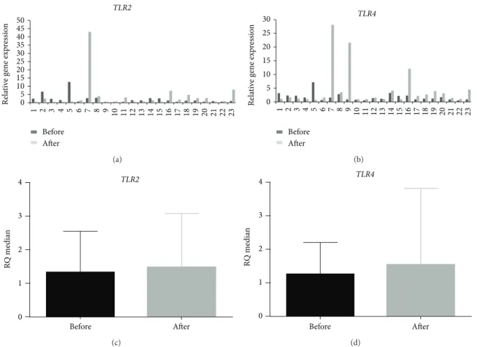
1.45). In the group that eradicated the bacteria, heterogeneity
of relative expression levels for bothTLR2andTLR4mRNAs
can be observed before and ater the treatment (Figures1(a)
and1(b)). However no signiicant diferences were observed
for both genes comparing the expression levels in this group
before and ater treatment (� = 0.533and � = 0.094for
TLR2andTLR4, resp.) (Figures1(c)and1(d)). Furthermore,
Table 2: Comparison ofTLR2andTLR4mRNA relative expression levels before and aterH. pylorieradication therapy.
Before treatment Ater treatment
� �
TLR2
Completed treatment 37 37
RQ median 1.31 1.55
Range 0.37–23.05 0.34–43.12
�value 0.291
Eradicated 23 23
RQ median 1.32 1.47
Range 0.37–12.63 0.34–43.12
�value 0.533
Noneradicated 14 14
RQ median 1.23 1.83
Range 0.66–23.05 0.37–6.36
�value 0.357
TLR4
Completed treatment 37 37
RQ median 1.45 1.64
Range 0.50–11.09 0.56–28.07
�value 0.084
Eradicated 23 23
RQ median 1.26 1.53
Range 0.50–7.17 0.64–28.07
�value 0.094
Noneradicated 14 14
RQ median 1.88 1.99
Range 0.54–11.09 0.56–9.75
�value 0.626
�: number of individuals;�: probability; RQ: relative quantiication;
statis-tical analysis by Wilcoxon signed rank test.
TLR4mRNA before and ater treatment considering only the
eradicated patients was found (before:�2= 0.85,� < 0.0001;
ater:�2= 0.55,� = 0.006).
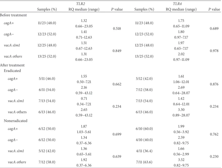
he inluence of cagA and vacA bacterial genotypes
on the gene expression levels, both before and ater
treat-ment (Table3), showed no evidenced signiicant diference
between cagA+ and cagA− genotypes (� > 0.05) for
both analyzed genes. Similarly, no signiicant diference was
observed regardingVacAsm genotype. We also evaluated the
association between relative expression levels ofTLR2 and
TLR4mRNA and the risk factors such as age, gender,
smok-ing, drinksmok-ing, and histological type of gastric lesion. None of the factors investigated showed signiicant diferences (data not shown).
3.2. TLR2 and TLR4 Protein and mRNA Relative Expressions Are Concordant. In normal mucosa, the TLR2 and TLR4 protein expression was weak or absent, mainly in the foveolar
epithelium (Figures 2(a) and 2(b)). Nevertheless, the
CG-Hp+ samples collected before the treatment showed a cyto-plasmatic, perinuclear, and focal immunostaining pattern,
mostly in the basal area of the foveolar epithelium. A strong expression in the inlammatory cells was also observed
(Figures2(c)and2(d)). Ater the eradication ofH. pylori, an
immunostaining pattern similar to the one observed before the treatment was found for both TLR2 and TLR4 proteins
(Figures2(e)and2(f)).
he mean optical densitometry values observed in the
normal Hp- group for TLR2 and TLR4 were105.6 ± 2.7and
101.4 ± 6.5, respectively. While the CG-Hp+ group before treatment presented signiicantly increased mean values for
both TLR2 (151.7±6.1) and TLR4 (132.2±4.7) in comparison
with the normal Hp- group (� = 0.020and� = 0.007, resp.).
Ater eradication of the bacteria, both TLR2 and TLR4 proteins showed a slight reduction in their mean optical
densitometry values (136.1 ± 6.1 and 122.8 ± 5.8, resp.).
However, there were no signiicant diferences between these
values before and ater treatment (� = 0.064 and � =
0.198, resp.) (Figures2(g)and2(h)), conirming the indings regarding the mRNA relative expression.
4. Discussion
In this study we investigated for the irst time the
occur-rence of alterations in the TLR2 and TLR4 mRNA and
protein expression inH. pylori-infected patients with chronic
gastritis, before and ater successful bacteria eradication treatment. Our results did not reveal signiicant changes in
the relative expression levels of eitherTLR2orTLR4mRNA
ater treatment in eradicated patients, which was conirmed by immunohistochemistry. Moreover, the mRNA expression of both receptors remained increased ater eradication ther-apy compared to the normal Hp- group, showing that the eradication of the bacteria did not normalize the expression of these receptors, at least under the conditions evaluated. Additionally, we also observed a positive correlation between
the mRNA expression values ofTLR2andTLR4conirming
thatH. pyloriactivates both receptors.
TLRs are transmembrane proteins that play a critical
role in the recognition of pathogen components [24]. LPS of
Gram-negative bacteria are recognized mainly by TLR4 and also TLR2 activating signaling pathways that culminate in an
inlammatory response [25]. It is believed that the interaction
between bacterial virulence and a genetically susceptible host is associated with more severe chronic inlammation,
which may, in the long run, lead to cancer [26]. Under
normal physiological conditions, the expression of these receptors in the mucosa of the gastrointestinal tract is low due to the action of their antagonists, such as TOLLIP
(Toll-interacting protein) and PPAR� (Peroxisome
proliferator-activated receptor) that show higher levels in order to prevent
inappropriate activation of nonpathogenic antigens [27–29].
In our study, we observed a slightly increased expression
of both TLR2 and TLR4 in CG-Hp+ patients even ater
successfulH. pylorieradication compared to the noninfected
normal mucosa. In children infected withH. pylori,
Lagunes-Servin et al. (2013) [30] found an increase in the
0
1 2 3 4 5 6 7 8 9 10 11 12 13 14 15
TLR2
16 17 18 19 20 21 22 23
Before Aer 5 10 15 20 25 30 35 40 45 50
Rela
ti
ve
gene exp
ressio
n
(a)
TLR4
1 2 3 4 5 6 7 8 9 10 11 12 13 14 15 16 17 18 19 20 21 22 23
0 5 10 15 20 25 30
Rela
ti
ve
gene exp
ressio
n
Before Aer
(b)
TLR2
RQ
me
d
ian
0 1 2 3 4
Before Aer
(c)
TLR4
RQ
me
d
ian
0 1 2 3 4
Before Aer
(d)
Figure 1: Relative expression levels ofTLR2andTLR4RNAm in the eradicated patients group with chronic gastritis before and aterH. pylori
treatment. Relative quantiication (RQ) of the mRNA expression levels of (a)TLR2and (b)TLR4per individual evaluated; RQ median of (c)TLR2and (d)TLR4mRNA before and aterH. pylorieradication. Data are presented as median and range for experiments performed in triplicate. Statistical signiicance was determined using Wilcoxon’s signed rank test.
an association with pro- and anti-inlammatory cytokines
(IL-8, TNF-�, and IL-10). hese indings conirm that H.
pylori has the ability to increase the in vivo expression of TLRs by gastric epithelial cells early during infection in children, starting a chronic and balanced inlammatory process that will continue for decades, and so may contribute
to the development of H. pylori-associated diseases later
in adulthood. Pimentel-Nunes et al. (2013) [31] observed
that, considering the diferent TLRs of normal H. pylori
-negative mucosa, the mRNAofTLR5was the most expressed,
followed by those of TLR2 and TLR4. Furthermore, these
authors foundTLR2and TLR4overexpression in intestinal
metaplasia, independent of theH. pyloristatus, and in the
dysplasia/cancer sequence. Moreover, upregulation ofTLR2
andTLR4mRNA was also observed inH. pylori-associated
normal mucosa. hese results were conirmed by immuno-histochemical analyses, which found an increase in protein
expression in H. pylori-infected normal mucosa, further
increasing in intestinal metaplasia and dysplasia/carcinoma. hese indings suggest that progressive activation of these
receptors, initially not only by H. pylori, but also by other
PAMPs (pathogen-associated molecular patterns) or DAMPs (damage-associated molecular patterns), at later stages, may
play an important role in gastric carcinogenesis and tumor
progression [31].
Upregulation ofTLR4expression responsiveness to LPS
and H. pylori in gastric cell lines has also been reported [32, 33]. H. pylori infection induced both TLR4 mRNA and protein expression in AGS cells that were dependent on bacterial load and infection duration. However, the
transfection of AGS cells with TLR4 siRNA followed by
the bacterial infection suppressed the expression of this
receptor [32]. Moreover, LPS of H. pyloriupregulate TLR4
expression via TLR2 signaling in MKN28 gastric cell lines
by the MEK1/2-ERK1/2 MAP kinase pathway [34], leading
also to an increase in cell proliferation. Conversely, previous
studies [35–37] did not observe any relevant role of TLR4
in the cellular recognition ofH. pyloriin AGC cells. hese
controversial results may be due to diferences in the lipid
A structures produced by distinctH. pyloristrains [38–40].
herefore, the interaction of the bacteria with TLR2 should also be considered, mainly ater the irst contact with the
gastric mucosa, triggering immunologic responses [41] such
as induction of IL-8 and subsequent activation of NF-�B [11].
Our study revealed no reduction of the transcript levels
(a) (b)
(c) (d)
(e) (f)
TLR2
NM CG before CG ater
M
ea
n
o
p
ti
cal den
si
to
m
etr
y (a.u
.)
0 50 100 150 200
∗
(g)
TLR4
NM CG before CG ater
M
ea
n
o
p
ti
cal den
si
to
m
etr
y (a.u
.)
0 50 100 150
∗
(h)
Table 3: Comparisons ofTLR2andTLR4mRNA expression levels according tocagAandvacAgenotypes ofH. pyloriin infected patients before and ater bacteria eradication treatment.
TLR2 TLR4
Samples (%) RQ median (range) �value Samples (%) RQ median (range) �value
Before treatment
cagA+ 11/23 (48.0) 1.32 11/23 (48.0) 1.75
0.689 0.66–23.05
0.518 0.65–11.09
cagA− 12/23 (52.0) 1.41 12/23 (52.0) 1.80
0.71–12.63 0.97–7.17
vacA s1m1 12/25 (48.0) 1.51 12/25 (48.0) 1.97
0.978 0.67–12.63
0.849 0.65–7.17
vacA others 13/25 (52.0) 1.31 13/25 (52.0) 2.02
0.66–23.05 0.97–11.09
Ater treatment Eradicated
cagA+ 5/11 (46.0) 1.55 5/12 (42.0) 1.61
0.876 0.50–7.21
0.662 1.06–12.01
cagA− 6/11 (54.0) 2.16 7/12 (58.0) 2.69
0.59–43.12 0.64–28.07
vacA s1m1 7/13 (54.0) 0.71 7/13 (54.0) 1.42
0.234
0.34–7.21 0.234 0.64–12.01
vacAothers 6/13 (46.0) 2.65 6/13 (46.0) 3.30
0.59–43.12 0.89–28.07
Noneradicated
cagA+ 6/12 (50.0) 1.87 6/10 (60.0) 1.99
0.762 1.03–5.61
0.699 0.56–3.92
cagA− 6/12 (50.0) 1.34 4/10 (40.0) 2.59
0.37–6.36 0.82–9.75
vacA s1m1 5/12 (42.0) 1.36 4/11 (36.4) 1.66
0.230 0.65–5.61
0.639 0.56–2.99
vacA others 7/12 (58.0) 1.92 7/11 (63.6) 3.52
0.37–6.36 0.82–9.75
vacAothers(s1m2, s2m2, s1 , s2 ,and m1);�value = Mann-Whitney test;� < 0.05.
showing that the successful eradication of H. pylori does
not change the expression of these receptors within a short period ater the treatment. Similarly, Garza-Gonz´alez et al.
(2008) [42] found no quantitative diferences in the TLR4
andTLR5mRNA levels either, regardless of the presence or
absence of H. pyloriin gastric epithelial cells biopsies and
AGS cells, suggesting that the mRNA levels of both receptors may not be inluenced by the infection process or at least not at the time points selected for analysis. However, in our
study, we observed higher levels ofTLR2andTLR4mRNA
and of both proteins inH. pylori-infected mucosa compared
to noninfected normal mucosa.It should however be taken
into consideration that the posttreatment time elapsed until biopsy collection which may not have been suicient for mucosal renovation and transcription level normalization.
Moreover, alterations in mRNA expression levels ater H.
pyloriinfection eradication therapy have been demonstrated, involving genes associated with cell damage, inlammation,
proliferation, apoptosis, and intestinal diferentiation [43,
44].
his study did not investigate the molecular mechanisms
involved in the inlammatory cascade induced byH. pylori
infection triggered by TLR4 and TLR2. herefore, further investigations are needed to clarify the possible involvement
of signaling pathway MyD88-MAPK-NF�B as well as the
role of PPARs (Peroxisome proliferator-activated receptors) on inhibition of pathway regulating expression of
proinlam-matory genes and stress kinase pathways [31,45,46], which
suppresses inlammation inH. pyloriinfection.
When we compared the expression levels ofTLR2 and
TLR4mRNA with risk factors and bacterial virulence
geno-types, we did not ind any association. he studies that assess
the efects ofcagAandvacAvirulence factors on the gene and
protein expression are controversial. Our results evidenced that there were no quantitative diferences in the mRNA levels
of these receptors regardless ofcagAandvacAstatus. Similar
results were reported by Garza-Gonz´alez et al. (2008) [42],
which demonstrated that the mRNA levels ofTLR4andTLR5
in gastric cells bothin vivoandin vitrowere not inluenced by
be involved in the irst steps of innate immune-recognition of H. pylori. Another study evidenced downregulation of TLRs
2 and 5 and upregulation of TLR9 by H. pylori in human
neutrophils regardless of cagPAI status and the integrity of
T4SS [47].
In conclusion, we report a discrete increase inTLR2and
TLR4 mRNA and protein expression in CG-Hp+ patients
before eradication therapy and the maintaining of this expression pattern even ater treatment, suggesting that these receptors remain expressed in the gastric mucosa even ater eradication of the bacteria, at least for the period evaluated. herefore, considering the higher risk of malignant
progres-sion in patients infected byH. pylorifor a long time, further
investigations are needed to clarify the changes in the expres-sion of other genes related with the inlammatory cascade induced by bacteria, such as those encoding cytokines and malignant transformation processes as well as the signaling pathways involved.
Conflict of Interests
he authors declare that there are no competing interests.
Acknowledgments
he authors are grateful to Dr. Sebasti˜ao Roberto Taboga and Luiz Roberto Faleiros Junior for their help with the histological sections, Isaque Santana for technical assistance during the immunohistochemical assay, and nurse Gizelda Warick Mazzale for her help in contacting patients. his study was supported by grants from the Brazilian agencies FAPESP (2012/15036-8) and CNPq (304870/2012-9).
References
[1] D. M. Parkin, “he global health burden of infection-associated cancers in the year 2002,”International Journal of Cancer, vol. 118, no. 12, pp. 3030–3044, 2006.
[2] P. Correa, “Human model of gastric carcinogenesis,” Cancer
Research, vol. 48, no. 13, pp. 3554–3560, 1988.
[3] J. G. de Oliveira and A. E. Silva, “Polymorphisms of the TLR2 and TLR4 genes are associated with risk of gastric cancer in a Brazilian population,”World Journal of Gastroenterology, vol. 18, no. 11, pp. 1235–1242, 2012.
[4] H. Xue, J. Liu, B. Lin, Z. Wang, J. Sun, and G. Huang, “A meta-analysis of interleukin-8-251 promoter polymorphism associated with gastric cancer risk,”PLoS ONE, vol. 7, no. 1, Article ID e28083, 2012.
[5] A. Lamb and L.-F. Chen, “Role of the Helicobacter pylori -induced inlammatory response in the development of gastric cancer,”Journal of Cellular Biochemistry, vol. 114, no. 3, pp. 491– 497, 2013.
[6] S. F. Moss and M. J. Blaser, “Mechanisms of disease: inlam-mation and the origins of cancer,” Nature Clinical Practice
Oncology, vol. 2, no. 2, pp. 90–97, 2005.
[7] R. M. Peek Jr. and J. E. Crabtree, “Helicobacterinfection and gastric neoplasia,”he Journal of Pathology, vol. 208, no. 2, pp. 233–248, 2006.
[8] K. T. Wilson and J. E. Crabtree, “Immunology ofHelicobacter
pylori: insights into the failure of the immune response and
perspectives on vaccine studies,”Gastroenterology, vol. 133, no. 1, pp. 288–308, 2007.
[9] S. Akira, S. Uematsu, and O. Takeuchi, “Pathogen recognition and innate immunity,”Cell, vol. 124, no. 4, pp. 783–801, 2006. [10] M. Matsuura, “Structural modiications of bacterial
lipopoly-saccharide that facilitate gram-negative bacteria evasion of host innate immunity,”Frontiers in Immunology, vol. 4, article 109, 2013.
[11] S.-I. Yokota, T. Ohnishi, M. Muroi, K.-I. Tanamoto, N. Fujii, and K.-I. Amano, “Highly-puriiedHelicobacter pyloriLPS prepara-tions induce weak inlammatory reacprepara-tions and utilize Toll-like receptor 2 complex but not Toll-like receptor 4 complex,”FEMS
Immunology and Medical Microbiology, vol. 51, no. 1, pp. 140–
148, 2007.
[12] F. Re and J. L. Strominger, “Heterogeneity of TLR-induced responses in dendritic cells: from innate to adaptive immunity,”
Immunobiology, vol. 209, no. 1-2, pp. 191–198, 2004.
[13] S.-Z. Ding, A. M. Torok, M. F. Smith Jr., and J. B. Goldberg, “Toll-like receptor 2-mediated gene expression in epithelial cells duringHelicobocter pyloriinfection,”Helicobacter, vol. 10, no. 3, pp. 193–204, 2005.
[14] S. Akira and K. Takeda, “Toll-like receptor signalling,”Nature
Reviews Immunology, vol. 4, no. 7, pp. 499–511, 2004.
[15] M. Fukata and M. T. Abreu, “Pathogen recognition receptors, cancer and inlammation in the gut,” Current Opinion in
Pharmacology, vol. 9, no. 6, pp. 680–687, 2009.
[16] M. F. Tsan, “Toll-like receptors, inlammation and cancer,”
Seminars in Cancer Biology, vol. 16, no. 1, pp. 32–37, 2006.
[17] M.-S. Wu, L.-P. Chow, J.-T. Lin, and S.-H. Chiou, “Proteomic identiication of biomarkers related to Helicobacter pylori -associated gastroduodenal disease: challenges and opportuni-ties,”Journal of Gastroenterology and Hepatology, vol. 23, no. 11, pp. 1657–1661, 2008.
[18] M. Asaka, M. Kato, S.-I. Takahashi et al., “Guidelines for the management of Helicobacter pylori infection in Japan: 2009 revised edition,”Helicobacter, vol. 15, no. 1, pp. 1–20, 2010. [19] P. Malfertheiner, F. Megraud, C. A. O’Morain et al.,
“Management ofHelicobacter pyloriinfection—the Maastricht IV/Florence consensus report,”Gut, vol. 61, no. 5, pp. 646–664, 2012.
[20] M. F. Dixon, R. M. Genta, J. H. Yardley et al., “Classiication and grading of Gastritis: the updated Sydney system,”he American
Journal of Surgical Pathology, vol. 20, no. 10, pp. 1161–1181, 1996.
[21] A. F. T. Rossi, M. C. Duarte, A. B. Poltronieri et al., “Deregu-lation of annexin-A1 and galectin-1 expression in precancerous gastric lesions: intestinal metaplasia and gastric ulcer,”
Media-tors of Inlammation, vol. 2014, Article ID 478138, 11 pages, 2014.
[22] L. L. Gatti, E. K. Fagundes E Souza, K. R. Leite et al., “cagA vacA alelles and babA2 genotypes ofHelicobacter pyloriassociated with gastric disease in Brazilian adult patients,” Diagnostic
Microbiology and Infectious Disease, vol. 51, no. 4, pp. 231–235,
2005.
[23] K. J. Livak and T. D. Schmittgen, “Analysis of relative gene expression data using real-time quantitative PCR and the
2−ΔΔ��method,”Methods, vol. 25, no. 4, pp. 402–408, 2001.
[24] R. Medzhitov and C. Janeway Jr., “innate immunity,”he New
England Journal of Medicine, vol. 343, no. 5, pp. 338–344, 2000.
[25] M. R. Amieva and E. M. El-Omar, “Host-bacterial interactions
inHelicobacter pyloriinfection,”Gastroenterology, vol. 134, no.
[26] J. C. Machado, C. Figueiredo, P. Canedo et al., “A proinlam-matory genetic proile increases the risk for chronic atrophic gastritis and gastric carcinoma,”Gastroenterology, vol. 125, no. 2, pp. 364–371, 2003.
[27] P. Pimentel-Nunes, J. B. Soares, R. Roncon-Albuquerque Jr., M. Dinis-Ribeiro, and A. F. Leite-Moreira, “Toll-like receptors as therapeutic targets in gastrointestinal diseases,”Expert Opinion
on herapeutic Targets, vol. 14, no. 4, pp. 347–368, 2010.
[28] G. Melmed, L. S. homas, N. Lee et al., “Human intestinal epithelial cells are broadly unresponsive to toll-like receptor 2-dependent bacterial ligands: Implications for host-microbial interactions in the gut,”he Journal of Immunology, vol. 170, no. 3, pp. 1406–1415, 2003.
[29] F. Y. Liew, D. Xu, E. K. Brint, and L. A. J. O’Neill, “Negative regulation of toll-like receptor-mediated immune responses,”
Nature Reviews Immunology, vol. 5, no. 6, pp. 446–458, 2005.
[30] H. Lagunes-Servin, J. Torres, C. Maldonado-Bernal et al., “Toll-like receptors and cytokines are upregulated duringHelicobacter
pyloriinfection in children,”Helicobacter, vol. 18, no. 6, pp. 423–
432, 2013.
[31] P. Pimentel-Nunes, N. Gonc¸alves, I. Boal-Carvalho et al., “
Heli-cobacter pyloriinduces increased expression of toll-like
recep-tors and decreased toll-interacting protein in gastric mucosa that persists throughout gastric carcinogenesis,”Helicobacter, vol. 18, no. 1, pp. 22–32, 2013.
[32] B. Su, P. J. M. Ceponis, S. Lebel, H. Huynh, and P. M. Sherman,
“Helicobacter pyloriactivates Toll-like receptor 4 expression in
gastrointestinal epithelial cells,”Infection and Immunity, vol. 71, no. 6, pp. 3496–3502, 2003.
[33] D.-Y. Lu, H.-C. Chen, M.-S. Yang et al., “Ceramide and toll-like receptor 4 are mobilized into membrane rats in response to
Helicobacter pyloriinfection in gastric epithelial cells,”Infection
and Immunity, vol. 80, no. 5, pp. 1823–1833, 2012.
[34] S.-I. Yokota, T. Okabayashi, M. Rehli, N. Fujii, and K.-I. Amano,
“Helicobacter pylori lipopolysaccharides upregulate toll-like
receptor 4 expression and proliferation of gastric epithelial cells via the MEK1/2-ERK1/2 mitogen-activated protein kinase pathway,”Infection and Immunity, vol. 78, no. 1, pp. 468–476, 2010.
[35] S. Maeda, M. Akanuma, Y. Mitsuno et al., “Distinct mechanism
ofHelicobacter pylori-mediated NF-kappa B activation between
gastric cancer cells and monocytic cells,”he Journal of
Biologi-cal Chemistry, vol. 276, no. 48, pp. 44856–44864, 2001.
[36] F. B¨ackhed, B. Rokbi, E. Torstensson et al., “Gastric mucosal recognition ofHelicobacter pyloriis independent of Toll-like receptor 4,”he Journal of Infectious Diseases, vol. 187, no. 5, pp. 829–836, 2003.
[37] J.-M. Otte, H. M. Neumann, S. Brand, H. Schrader, W. E. Schmidt, and F. Schmitz, “Expression of beta-defensin 4 is increased in human gastritis,” European Journal of Clinical
Investigation, vol. 39, no. 2, pp. 126–138, 2009.
[38] T. Kawahara, S. Teshima, A. Oka, T. Sugiyama, K. Kishi, and K. Rokutan, “Type I Helicobacter pylori lipopolysaccharide stimulates toll-like receptor 4 and activates mitogen oxidase 1 in gastric pit cells,”Infection and Immunity, vol. 69, no. 7, pp. 4382–4389, 2001.
[39] S. Ishihara, M. A. K. Rumi, Y. Kadowaki et al., “Essential role of MD-2 in TLR4-dependent signaling duringHelicobacter pylori -associated gastritis,”he Journal of Immunology, vol. 173, no. 2, pp. 1406–1416, 2004.
[40] Y. Hirata, T. Ohmae, W. Shibata et al., “MyD88 and TNF receptor-associated factor 6 are critical signal transducers in
Helicobacter pylori-infected human epithelial cells,”he Journal
of Immunology, vol. 176, no. 6, pp. 3796–3803, 2006.
[41] M. F. Smith Jr., A. Mitchell, G. Li et al., “Toll-like receptor (TLR) 2 and TLR5, but not TLR4, are required forHelicobacter pylori -induced NF-kappaB activation and chemokine expression by epithelial cells,”he Journal of Biological Chemistry, vol. 278, no. 35, pp. 32552–32560, 2003.
[42] E. Garza-Gonz´alez, V. Bocanegra-Garc´ıa, F. J. Bosques-Padilla, J. P. Flores-Guti´errez, F. Moreno, and G. I. Perez-Perez, “mRNA levels of TLR4 and TLR5 are independent ofH pylori,”World
Journal of Gastroenterology, vol. 14, no. 34, pp. 5306–5310, 2008.
[43] C. J. Tsai, R. Herrera-Goepfert, R. J. Tibshirani et al., “Changes of gene expression in gastric preneoplasia following
Helicobac-ter pylorieradication therapy,”Cancer Epidemiology Biomarkers
and Prevention, vol. 15, no. 2, pp. 272–280, 2006.
[44] S. S. Kim, P. Meitner, T. A. Konkin, Y. S. Cho, M. B. Resnick, and S. F. Moss, “Altered expression of Skp2, c-Myc and p27 proteins but not mRNA aterH. pylorieradication in chronic gastritis,”
Modern Pathology, vol. 19, no. 1, pp. 49–58, 2006.
[45] M. Muzio, G. Natoli, S. Saccani, M. Levrero, and A. Mantovani, “he human toll signaling pathway: divergence of nuclear factor�b and jnk/sapk activation upstream of tumor necrosis factor receptor-associated factor 6 (TRAF6),” he Journal of
Experimental Medicine, vol. 187, no. 12, pp. 2097–2101, 1998.
[46] J.-M. Lee, S. S. Kim, and Y.-S. Cho, “he role of PPAR�in
Helicobacter pyloriinfection and gastric carcinogenesis,”PPAR
Research, vol. 2012, Article ID 687570, 6 pages, 2012.
Submit your manuscripts at
http://www.hindawi.com
Stem Cells
International
Hindawi Publishing Corporation
http://www.hindawi.com Volume 2014
Hindawi Publishing Corporation
http://www.hindawi.com Volume 2014
INFLAMMATION
Hindawi Publishing Corporation
http://www.hindawi.com Volume 2014
Behavioural
Neurology
Endocrinology
International Journal of Hindawi Publishing Corporationhttp://www.hindawi.com Volume 2014
Hindawi Publishing Corporation
http://www.hindawi.com Volume 2014
Disease Markers
Hindawi Publishing Corporation
http://www.hindawi.com Volume 2014 BioMed
Research International
Oncology
Journal ofHindawi Publishing Corporation
http://www.hindawi.com Volume 2014
Hindawi Publishing Corporation
http://www.hindawi.com Volume 2014
Oxidative Medicine and Cellular Longevity
Hindawi Publishing Corporation
http://www.hindawi.com Volume 2014
PPAR Research
The Scientiic
World Journal
Hindawi Publishing Corporation
http://www.hindawi.com Volume 2014
Immunology Research Hindawi Publishing Corporation
http://www.hindawi.com Volume 2014 Journal of
Obesity
Journal ofHindawi Publishing Corporation
http://www.hindawi.com Volume 2014
Hindawi Publishing Corporation
http://www.hindawi.com Volume 2014
Computational and Mathematical Methods in Medicine
Ophthalmology
Journal ofHindawi Publishing Corporation
http://www.hindawi.com Volume 2014
Diabetes Research
Journal ofHindawi Publishing Corporation
http://www.hindawi.com Volume 2014
Hindawi Publishing Corporation
http://www.hindawi.com Volume 2014 Research and Treatment
AIDS
Hindawi Publishing Corporation
http://www.hindawi.com Volume 2014
Gastroenterology Research and Practice
Hindawi Publishing Corporation
http://www.hindawi.com Volume 2014
Parkinson’s
Disease
Evidence-Based Complementary and Alternative Medicine
Volume 2014