A B S T R A C T A B S T R A C T A B S T R A C T A B S T R A C T A B S T R A C T
The authors address the value of endoscopic retrograde cholangiopancreatography, ultrasonography, computed tomography, magnetic resonance imaging and endoscopic ultrasound for the diagnosis of cystic and solid neoplasias of the pancreas, demonstrating that each of them is of great importance to undoubtedly increase the diagnostic accuracy of the biliopancreatic system diseases. The best method for each of several tumors is then determined.
Key words Key words Key words Key words
Key words: Diagnostic imaging. Neoplasias. Cystic neoplasias, mucinous and serous. Pancreas.
hypoechoic areas in about 97% of cases, about 3% being isoechoic in relation to the sorrounding parenchyma. The lesion is heterogeneous, with imprecise limits.
Pancreatic adenocarcinoma is usually seen at CT as a focal, poorly defined, heterogeneous and hypoattenuated area that enhances less than the adjacent normal pancreatic parenchyma. The sensitivity of CT in detecting it as a hypoattenuated injury is directly related to the technique applied, requiring thin-cut images (3-5mm) obtained during rapid administration of iodinated contrast, so that small tumors can be identified. Helical CT scanners are recommended because they allow studies on arterial and portal phases of the administration of intravenous contrast medium and do not suffer interference from respiratory movements. MRI with fat suppression, on its turn, tends to significantly improve the sensitivity of the diagnosis of tumors of the pancreas (Figure 11).
EE is the technique of choice for suspected focal lesions in the pancreas. EE-FNA allows discarding the diagnosis of tumor of the pancreas with a sensitivity of 85-95% and specificity of around 100% for the diagnosis of malignant tumors when performed by an experienced physician24. However, it displays a lower negative predictive
value (between 20 and 50%) for the diagnosis of malignancy and therefore a negative biopsy does not rule out the existence of cancer24 (Figure 12).
Work performed in the Faculty of Medicine of Ribeirão Preto – University of São Paulo, São Paulo – SP, Brazil.
1. Professor, Department of Surgery and Anatomy, Faculty of Medicine of Ribeirão Preto – University of São Paulo; Staff, Endoscopy and Ecoendoscopy, Nove de Julho Hospital, São Paulo – SP, Brazil; 2. Affiliate Professor, Department of Diagnostic Imaging DDI, São Paulo Federal University – UNIFESP, São Paulo – SP, Brazil; 3. Staff, Second General Surgery Clinic, Rio de Janeiro State Servers Hospital – Rio de Janeiro – RJ, Brazil.
SOLID NEOPLASIAS
SOLID NEOPLASIAS
SOLID NEOPLASIAS
SOLID NEOPLASIAS
SOLID NEOPLASIAS
Ductal adenocarcinoma Ductal adenocarcinoma Ductal adenocarcinoma Ductal adenocarcinoma Ductal adenocarcinoma
This tumor is the most commonly found. It appears at US and EE as a focal, usually hypoechoic, rounded lesion, with irregular or undefined borders. The smaller the size, the easier its identification due to its greater clarity in relation to the adjacent parenchyma, and it does not escape the field of view of the device22. The differential
diagnosis between pancreatitis and a focus of a malignant tumor of the pancreas is difficult; however, the EE has a high negative predictive value for the diagnosis of pancreatic cancer. The diagnosis of a tumor on chronic pancreatitis, on its turn, remains a challenge for the examiner23. In any
case, the cytology obtained by EE-FNA may provide definitive information for the differential diagnosis.
EE is considered the best technique for the diagnosis of pancreatic tumors less than 3cm in diameter, which are the best candidates for resection. Its sensitivity is superior to US, CT and MRI and equal to the ERCP, but without its invasive nature25. In addition, EE is widely used for tumor staging, with
excellent results, especially in lesions smaller than 4 cm, as it can very accurately identify vascular invasion, both of the por-tal trunk and the arterial axle, even in potentially resectable patients identified by TC26. EE-FNA also enables the treatment
of uncontrollable pain caused by this type of tumor by injecting alcohol for celiac plexus neurolysis27.
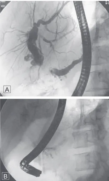
In the final line of the algorithm of unresectable pancreatic head tumors (Figure 13) associated with biliary
or duodenal obstruction, ERCP is indicated, as it enables the insertion of self-expandable metal prostheses in the common bile duct or duodenum28 (Figure 14).
Functioning neuroendocrine neoplasia Functioning neuroendocrine neoplasia Functioning neuroendocrine neoplasia Functioning neuroendocrine neoplasia Functioning neuroendocrine neoplasia There are several islet cell tumors. Some are functioning and others not. They represent benign adenomas and malignant pancreatic tumors and are usually small. Functional tumors, as a result of their intense hormonal activity, produce symptoms despite their small size, which makes them more difficult to identify. The nonfunctioning tumors are usually larger and easier to be identified29. In general, neuroendocrine tumors are
homogeneous, solid and often hypoechoic, while some are moderately echogenic. Calcifications and fluid spaces can be seen in larger lesions. The tumors are spherical with sharp edges, homogeneous and grow slow. The solid masses are more likely to be functional, and the ones with necrotic liquid component, less29.
The larger tumors are easily identified by both CT and MRI. However, most of these tumors are small, so it is essential to use a proper technique. Very thin cuts and acquisition of early arterial phase are essential for identification. MRI also offers the possibility to identify hypointense nodules in T1 sequences, and hyperintense on T2.
Insulinoma Insulinoma Insulinoma Insulinoma Insulinoma
Insulinoma is the most common functional neuroendocrine tumor (60%). Its malignancy rate is 10%30.
Clinically, patients experience fasting hypoglycemia and inappropriately elevated plasma insulin levels29 (Figure 15).
In 70% of patients there is a solitary adenoma, in 10%
Figure 11 Figure 11 Figure 11 Figure 11
Figure 11 - MRI: (A) sagital and (B) coronal sections, showing area of a tumor located in the cephalic portion of the pancreas.
Figure 12 Figure 12 Figure 12 Figure 12
Figure 12 - Echoendoscopy image of the case of the previous figure. Note hypoechoic, heterogeneous nodular area, with poorly defined borders, measuring 2x2cm. This lesion invaded the splenomesenteric confluence.
there are multiple adenomas and, in 10%, metastases. The lesions may be tiny or reach 1500g. Nearly 90% of insulinomas are less than 2cm and their small size makes them poorly palpable during surgery. Their occur more frequently in the body and tail of the pancreas, where the concentration of Langerhans islets is larger31. EE-FNA is an
excellent method for the accurate diagnosis of these nodules. Its sensitivity rate is around 80% for definite diagnosis32,33 (Figura16).
Gastrinoma Gastrinoma Gastrinoma Gastrinoma Gastrinoma
Its occurrence reaches 18% of the neuroendocrine tumors. The malignancy rate of gastrinomas varies from 25% to 60%. In patients with Zollinger-Ellison the lesions are gastrinomas. They are associated with gastric hypersecretion and peptic ulcer. Most of them are
Figure 15 Figure 15Figure 15
Figure 15Figure 15 - Patient with MEN I syndrome. MR image showing a hypointense rounded nodule of about 2 cm. There was doubt about the existence of splenosis. Figure 14
Figure 14Figure 14
Figure 14Figure 14 - Radiologic image after the insertion of two prostheses, one into the common bile duct and one into the duodenum. The patient had advanced cancer of the pancreas with invasion of adjacent structures (uT4N1Mx).
Figure 13 Figure 13Figure 13 Figure 13
Figure 13 - (A) After injection of contrast into the MPD, which is dilated, and into the bile duct, which is also increased in diameter, we noted a dual sign of stenosis, which confirmed the finding of endoscopic ultrasound for advanced pancreatic cancer. (B) Two 10 F plastic prostheses were inserted.
pancreatic or peripancreatic, with 13% located in the duodenum, and more than 60% of these lesions are malignant34 (Figure 17).
Nonfunctioning neuroendocrine neoplasia Nonfunctioning neuroendocrine neoplasia Nonfunctioning neuroendocrine neoplasia Nonfunctioning neuroendocrine neoplasia Nonfunctioning neuroendocrine neoplasia The nonfunctioning tumors represent 15% to 33% of all tumors in this category. They are easy to spot because they reach a larger size before causing symptoms. Their size range is generally 10 to 20 cm, usually not detected until they reach a larger size. They are usually solitary and cause abdominal pain (36%), jaundice (28%) or a palpable mass. Most of these tumors are malignant (60 to 92%). There are certain characteristics that seem relevant to these tumors: large size, more than 10cm in diameter, and hypoechogenicity, with more or less necrotic areas29 (Figure 18).
A
diagnosis of metastasis should be considered if the lesions are found within the pancreas in patients with known primary tumor. The incidence of pancreatic metastasis affects 8.4% of patients with lung tumors, 19% with breast cancer and 37.5% with malignant melanoma. Therefore, the following primary tumors should be investigated when there is pancreatic metastasis: melanoma, lung, breast and renal cancer, hepatocellular carcinoma and sarcoma35.
Pancreatic lymphoma Pancreatic lymphoma Pancreatic lymphoma Pancreatic lymphoma Pancreatic lymphoma
Intra-abdominal lymphomas may also involve the pancreas, producing granular hypoechoic masses. The su-perior mesenteric vessels can be dislocated anteriorly, not posteriorly, as seen in patients with primary pancreatic tumors. Again, the definitive diagnosis is made by EE- FNA36
(Figure 21).
Figure 16 Figure 16 Figure 16 Figure 16
Figure 16 - Same case as the previous figure, submitted to EE-FNA (A). The result of pathology revealed the presence of a neuroendocrine tumor, confirmed by surgery (B).
Metastatic Pancreatic Disease Metastatic Pancreatic Disease Metastatic Pancreatic Disease Metastatic Pancreatic Disease Metastatic Pancreatic Disease
Direct invasion of surrounding organs by tumors may also appear as primary pancreatic mass, whose echotexture is hypoechoic. This may occur with gastric, colonic, duodenal and biliary tumors. Since the lesion cannot be distinguished from a primary car-cinoma, the diagnosis should be made by EE-FNA. The pancreas is rarely involved by metastatic disease from other primary tumors, but when the invasion happens, it usually does through direct invasion. Due to their small size and paucity of clinical symptoms, pancreatic metastases are not frequently diagnosed. In general, they appear as homogeneous, solid lesions displaying mass effect, with a more hypoechoic internal structure (Figure 19), or even as hypervascular nodules occupying the pancreas of a patient with metastatic clear cell carcinoma of the kidney (Figure 20). The
Figure 17 Figure 17 Figure 17 Figure 17
Figure 17 - Echoendoscopy images from two different gastrinoma cases. In (A) the injury was in the back of the body of the pancreas and in (B) the tumor was in the uncinate process.
A
B
A
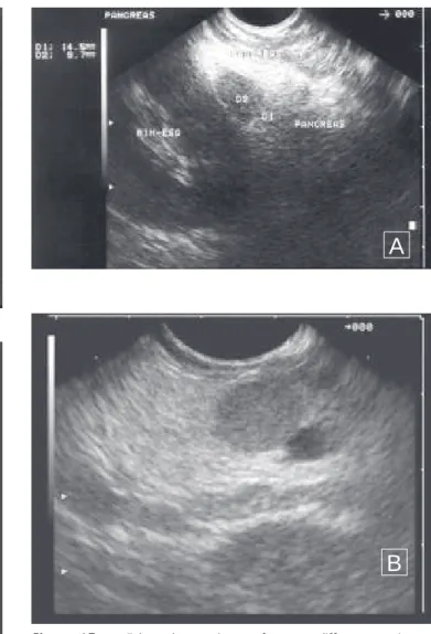
Figure 18 Figure 18Figure 18 Figure 18
Figure 18 - Patient with a nonfunctioning neuroendocrine tumor. (A) CT scan showing a hypodense nodule located in the cephalic portion of the pancreas, (B) endoscopic ultrasound image confirmed the CT findings and the biopsy revealed neuroendocrine tumor; In (C) image of the surgical specimen.
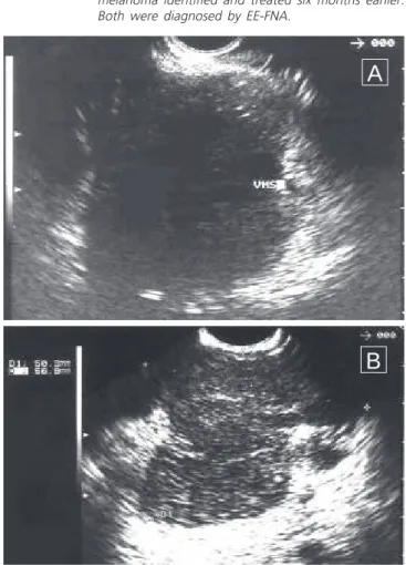
Figure 19 Figure 19Figure 19
Figure 19Figure 19 - (A) pancreatic metastasis of a colon tumor operated three years before; (B) metastasis from torso melanoma identified and treated six months earlier. Both were diagnosed by EE-FNA.
B
C
Figure 20 -Figure 20 -Figure 20 Figure 20
-Figure 20 - Two cases of pancreatic lymphoma diagnosed by EE-FNA. (A) hypoechoic lesion, heterogeneous, imprecise limits, the major was at the head of the pancreas, (B) hypoechoic lesion, heterogeneous, imprecise limits of 5.6 x5, 0cm, located in the body of the pancrea.
B
A
REFERÊNCES
REFERÊNCES
REFERÊNCES
REFERÊNCES
REFERÊNCES
22. Fernández-Esparrach G, Ginès A, Pellisé M, Bordas JM. Role of endoscopic ultrasonography in the study of extrahepatic cholestasis. Gastroenterol Hepatol 2002; 25(10):633-8.
23. Ardengh JC, Lopes CV, Campos AD, Pereira de Lima LF, Venco F, Módena JL. Endoscopic ultrasound and fine needle aspiration in chronic pancreatitis: differential diagnosis between pseudotumoral masses and pancreatic cancer. JOP 2007; 8(4):413-21.
24. Ardengh JC, Coelho DE, Coelho JF, de Lima LF, dos Santos JS, Módena JL. Single-step EUS-guided endoscopic treatment for sterile pancreatic collections: a single-center experience. Dig Dis 2008; 26(4):370-6.
25. Ardengh JC, Malheiros CA, Rahal F, Pereira V, Ganc AJ. Microlithiasis of the gallbladder: role of endoscopic ultrasonography in patients with idiopathic acute pancreatitis. Rev Assoc Med Bras 2010; 56(1):27-31.
26. Ardengh JC, Malheiros CA, Pereira V, Coelho DE, Coelho JF, Rahal F. Endoscopic ultrasound-guided fine-needle aspiration using helical computerized tomography for TN staging and vascular injury in operable pancreatic carcinoma. JOP 2009; 10(3):310-7. 27. Levy MJ, Topazian MD, Wiersema MJ, Clain JE, Rajan E, Wang KK,
et al. Initial evaluation of the efficacy and safety of endoscopic ultrasound-guided direct Ganglia neurolysis and block. Am J Gastroenterol 2008; 103(1):98-103.
28. Baron TH, Kozarek RA. Preoperative biliary stents in pancreatic cancer—proceed with caution. N Engl J Med 2010; 362(2):170-2. 29. Davies K, Conlon KC. Neuroendocrine tumors of the pancreas.
Curr Gastroenterol Rep 2009; 11(2):119-27.
30. Ardengh JC, Rosenbaum P, Ganc AJ, Goldenberg A, Lobo EJ, Malheiros CA, et al. Role of EUS in the preoperative localization of insulinomas compared with spiral CT. Gastrointest Endosc 2000; 51(5):552-5.
R E S U M O R E S U M O R E S U M O R E S U M O R E S U M O
Os autores fazem uma revisão considerando o valor da colangiopancreatografia endoscópica retrógrada, da ultrassonografia, da tomografia computadorizada, da ressonância magnética e da ecoendoscopia para o diagnóstico das neoplasias císticas e sólidas do pâncreas, demonstrando que cada um deles tem grande importância para aumentar, de forma inconteste, a acurácia diagnóstica das doenças do sistema biliopancreático. determinando qual o melhor método para cada um dos diversos tumores.
Descritores: Descritores: Descritores: Descritores:
Descritores: Diagnóstico por imagem. Neoplasias. Neoplasias císticas mucinosas e serosas. Pâncreas.
31. Mathur A, Gorden P, Libutti SK. Insulinoma. Surg Clin North Am 2009; 89(5):1105-21.
32. Ardengh JC, de Paulo GA, Ferrari AP. EUS-guided FNA in the diagnosis of pancreatic neuroendocrine tumors before surgery. Gastrointest Endosc 2004; 60(3):378-84.
33. Ardengh JC, Valiati LH, Geocze S. Identification of insulinomas by endoscopic ultrasonography. Rev Assoc Med Bras 2004; 50(2):167-71.
34. Price TN, Thompson GB, Lewis JT, Lloyd RV, Young WF. Zollinger-Ellison syndrome due to primary gastrinoma of the extrahepatic biliary tree: three case reports and review of literature. Endocr Pract 2009; 15(7):737-49.
35. DeWitt J, Jowell P, Leblanc J, McHenry L, McGreevy K, Cramer H, et al. EUS-guided FNA of pancreatic metastases: a multicenter experience. Gastrointest Endosc 2005; 61(6):689-96.
36. Ardengh JC, Lopes CV, de Lima LF, de Oliveira JR, Venco F, Santo GC, et al. Diagnosis of pancreatic tumors by endoscopic ultrasound-guided fine-needle aspiration. World J Gastroenterol 2007; 13(22):3112-6.
Received 15/01/2010
Accepted for publication 15/02/2010 Conflict of interest: none
Source of funding: none
How to cite this article: How to cite this article: How to cite this article: How to cite this article: How to cite this article:
Ardengh JC, Goldman SM, Lima Filho ER. Current role of imaging methods in the diagnosis of cystic solid pancreas neoplasms - part II. Rev Col Bras Cir. [periódico na Internet] 2011; 38(3). Disponível em URL: http://www.scielo.br/rcbc