344
Radiol Bras. 2017 Set/Out;50(5):338–348 Letters to the EditorDiffuse plasmacytoma of the pancreas: a rare entity
Dear Editor,
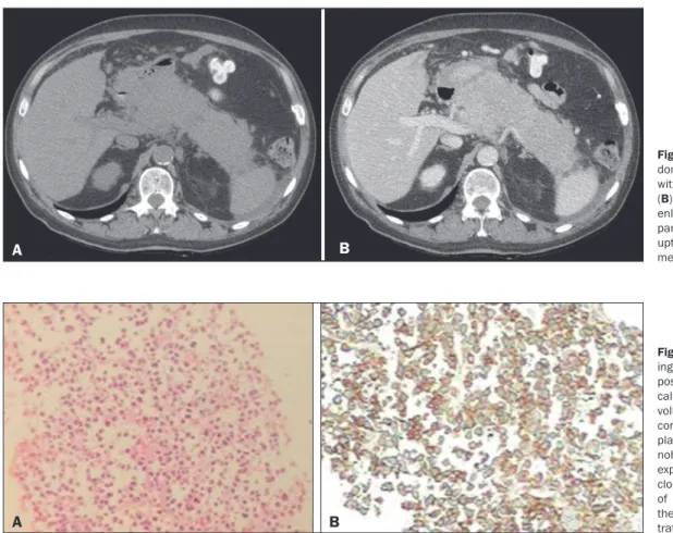
A 50-year-old male patient, diagnosed with multiple myeloma 10 months prior and undergoing chemotherapy, presented to the emergency department with abdominal pain. Laboratory tests revealed slightly elevated pancreatic enzymes. Subsequently, contrast-enhanced computed tomography (CT) of the abdomen showed diffuse, marked enlargement of the pancreatic paren-chyma, with homogeneous uptake of the iodinated contrast me-dium in the portal phase (Figure 1). The initial working diagno-sis was acute pancreatitis. However, the expected clinical, bio-chemical, and radiological improvement did not occur. We chose to perform CT-guided biopsy, and the histopathological analysis of the biopsy sample revealed a malignant neoplasm composed of loosely cohesive atypical cells, with hyperchromatic, volumi-nous, eccentric nuclei, consistent with a diagnosis of plasma cell neoplasm (Figure 2A). A complementary immunohistochemical study revealed expression of CD138, together with monoclonal immunoglobulin deposits of kappa light chain, conirming the diagnosis of pancreatic iniltration by plasmacytoma (Figure 2B).
Multiple myeloma is characterized by proliferation of ma-lignant plasma cells originating from the bone marrow and ac-counts for 10% of all hematological malignancies. Extramedul-lary plasmacytoma accounts for 5% of all plasma cell tumors and primarily affects males, the mean age at presentation being approximately 55 years. They can be primary, occurring as soli-tary masses without bone marrow involvement, or secondary, occurring as part of a multiple myeloma, the latter being the more common presentation(1–4). The most common site of ex-tramedullary involvement is the upper respiratory tract (80%);
however, other sites, such as the gastrointestinal tract, genito-urinary tract, reticuloendothelial system, thyroid, lungs, skin, and testicles, can also be involved(4).
There have been few reports of extramedullary plasma-cytoma affecting the pancreas. Of the approximately 25 cases described, most have involved a focal mass and only one has involved diffuse iniltration of the pancreas(2–6), ours therefore representing only the second such case reported. The most com-mon site of presentation is the pancreatic head, in most cases resulting in abdominal pain and obstructive jaundice(1–4). The radiological indings of pancreatic plasmacytoma are not highly speciic. In the focal presentation, the solid mass is homoge-neous or heterogehomoge-neous, multilobulated, with variable enhance-ment(1); in the one previously reported case with a diffuse pre-sentation, there was diffuse volumetric enlargement of the pan-creas with lobulated contours and predominantly homogenous uptake in the portal phase(6), similar to what was observed in the case reported here.
Although CT is the method of choice for the investiga-tion of pancreatic plasmacytoma, it is not capable of excluding diseases such as adenocarcinoma, lymphoma, and metastasis, histopathology therefore being fundamental for the diagnosis(3). In the case reported here, given the diffuse presentation, the main diagnostic hypotheses were pancreatitis and lymphoma. Lymphoma was excluded because of the clinical and laboratory indings, which indicated that pancreatitis was the most likely diagnosis. However, based on the history of multiple myeloma and the persistence of symptoms, the possibility of pancreatic iniltration by plasmacytoma was considered. Treatment for ex-tramedullary plasmacytoma involves the combination of local radiation, chemotherapy, and, in selected cases, surgery(4).
Figure 2. A: Histopathology show-ing malignant neoplasm com-posed of loosely cohesive atypi-cal cells, with hyperchromatic, voluminous, eccentric nuclei, consistent with a diagnosis of plasma cell neoplasm. B: Immu-nohistochemistry showing CD138 expression, together with mono-clonal immunoglobulin deposits of kappa light chain, conirming the diagnosis of pancreatic inil -tration by plasmacytoma.
B
A
Figure 1. Axial CT scans of the ab-domen, without contrast (A) and with contrast in the portal phase (B), showing diffuse, marked enlargement of the pancreatic parenchyma, with homogeneous uptake of the iodinated contrast medium.
345
Radiol Bras. 2017 Set/Out;50(5):338–348Letters to the Editor
http://dx.doi.org/10.1590/0100-3984.2016.0052
Camila Soares Moreira de Sousa1, Carla Lorena Vasques Mendes de Miranda1, Marcelo Coelho Avelino2, Breno Braga Bastos3, Ilan Lopes Leite Mendes1
1. Med Imagem – Radiologia, Teresina, PI, Brazil. 2. Hospital de Urgência de Teresina Prof. Zenon Rocha, Teresina, PI, Brazil. 3. UDI 24 horas – Radiologia, Teresina, PI, Brazil. Mailing address: Dra. Camila Soares Moreira de Sousa. Med Imagem – Radiologia. Rua Paissandu, 1862, Centro. Teresina, PI, Brazil, 64001-120. E-mail: [email protected].
Plasmacytoma of the pancreas is a rare entity and contin-ues to be the subject of many studies. In patients with multiple myeloma and focal or diffuse enlargement of the pancreas, the hypothesis of plasmacytoma should be considered, thus avoid-ing delayed diagnosis.
REFERENCES
1. Hue SS, Azhar R. Plasmacytoma of the pancreas: an unusual manifestation of multiple myeloma. Singapore Med J. 2013;54:e105–7.
2. Smith A, Hal H, Frauenhoffer E. Extramedullary plasmacytoma of the
pancreas: a rare entity. Case Rep Radiol. 2012;2012:798264.
3. Hatem M, So B, Gray R, et al. Plasmocytoma presented as pancreatic
head mass. Radiol Case Rep. 2015;10:81–7.
4. Pallavi R, Ravella PM, Popescu-Martinez A. An unusual pancreatic
mass: a case report and literature review. Transl Gastrointest Cancer. 2014;3:106–10.
5. Hiller N, Goitein O, Ashkenazi YJ. Plasmacytoma of the pancreas. Isr Med Assoc J. 2004;6:704–5.
6. Wilson TE, Korobkin M, Francis IR. Pancreatic plasmacytoma: CT indings. AJR Am J Roentgenol. 1989;152:1227–8.
A rare case of pneumorrhachis accompanying spontaneous pneumomediastinum
Dear Editor,
A 7-year-old female with dyspnea and edema of the neck, accompanied by a cough, was treated at another facility, where anti-inlammatory drugs and an inhaler were prescribed. The patient evolved to worsening of the dyspnea and cough, in ad-dition to intercostal retraction and increased neck volume. She presented to our facility in satisfactory general health. On physical examination, the oropharynx showed no alterations, al-though there was bilateral edema of the neck and periorbital area, together with diminished breath sounds, sparse wheezing, respiratory rate of 30 breaths/min, intercostal retraction, and subcutaneous crackles on anterior/posterior thoracic palpation, without Hamman’s sign. A chest X-ray obtained at admission (Figure 1) showed pneumomediastinum and extensive subcuta-neous emphysema. She underwent computed tomography (CT) of the chest (Figure 2), which revealed pneumorrhachis, a rare inding. The patient remained in the hospital for ive days under supportive care, and there was complete remission of symptoms.
Spontaneous pneumomediastinum, also known as Ham-man’s syndrome, is an uncommon condition in medical prac-tice, occurring in approximately 1/30,000 hospital admissions(1)
and in only 1% of asthma cases(2). Its main causes are intense physical exercise, labor (of childbirth), pulmonary barotrauma, diving to great depths, severe paroxysmal coughing, vomiting, asthma, inhalation of narcotics, bronchial asthma, and a slender body type(1,2).
The pathophysiological hallmark of Hamman’s syndrome is alveolar overdistension and rupture, which results from high intra-alveolar pressure, low perivascular pressure, or both. After the initial event, the air freely penetrates the mediastinum during the respiratory cycle, in order to balance the pressure gradients(3,4).
Figure 1. Posteroanterior chest X-ray showing pneumomediastinum (arrow-heads), together with extensive subcutaneous emphysema in the supraclavicu-lar and axilsupraclavicu-lary regions (arrows).
Figure 2. CT of the chest in the axial (A) and sagittal (B) planes showing pneu-morrhachis (arrows) and mediastinal emphysema (arrowheads).