Distinct Clinicopathological Patterns of
Mismatch Repair Status in Colorectal Cancer
Stratified by
KRAS
Mutations
Wenbin Li☯, Wenxue Zhi☯, Shuangmei Zou, Tian Qiu, Yun Ling, Ling Shan, Susheng Shi‡
*, Jianming Ying‡*
Department of pathology, Cancer Hospital, Chinese Academy of Medical Sciences & Peking Union Medical College, Beijing, China
☯These authors contributed equally to this work. ‡These authors also contributed equally to this work.
*[email protected](JY);[email protected](SS)
Abstract
In sporadic colorectal cancer (CRC), theBRAFV600Emutation is associated with deficient mismatch repair (MMR) status and inversely associated with to KRAS mutations. In contrast to deficient MMR (dMMR) CRC, data on the presence ofKRASoncogenic mutations in pro-ficient MMR (pMMR) CRC and their relationship with tumor progression are scarce. We therefore examined the MMR status in combination withKRASmutations in 913 Chinese patients and correlated the findings obtained with clinical and pathological features. The MMR status was determined based on detection of MLH1, MSH2, MSH6 and PMS2 expression.KRASmutation and dMMR status were detected in 36.9% and 7.5% of cases, respectively. Four subtypes were determined by MMR andKRASmutation status:KRAS (+)/pMMR (34.0%),KRAS(+)/dMMR (2.9%),KRAS(-)/pMMR (58.5%) andKRAS(-)/dMMR (4.6%). A higher percentage of pMMR tumors withKRASmutation were most likely to be female (49.0%), proximal located (45.5%), a mucinous histology (38.4%), and to have in-creased lymph node metastasis (60.3%), compared with pMMR tumors withoutBRAFV600E andKRASmutations (36.0%, 29.3%, 29.4% and 50.7%, respectively; allP<0.01). To the contrary, compared with those withKRAS(-)/dMMR tumors, patients withKRAS(+)/dMMR tumors demonstrated no statistically significant differences in gender, tumor location, pT depth of invasion, lymph node metastasis, pTNM stage, and histologic grade. This study re-vealed that specific epidemiologic and clinicopathologic characteristics are associated with MMR status stratified byKRASmutation. Knowledge of MMR andKRASmutation status may enhance molecular pathologic staging of CRC patients and metastatic progression in CRC can be estimated based on the combination of these biomarkers.
OPEN ACCESS
Citation:Li W, Zhi W, Zou S, Qiu T, Ling Y, Shan L, et al. (2015) Distinct Clinicopathological Patterns of Mismatch Repair Status in Colorectal Cancer Stratified byKRASMutations. PLoS ONE 10(6): e0128202. doi:10.1371/journal.pone.0128202
Academic Editor:Hiromu Suzuki, Sapporo Medical University, JAPAN
Received:February 3, 2015
Accepted:April 24, 2015
Published:June 4, 2015
Copyright:© 2015 Li et al. This is an open access article distributed under the terms of theCreative Commons Attribution License, which permits unrestricted use, distribution, and reproduction in any medium, provided the original author and source are credited.
Data Availability Statement:All relevant data are within the paper.
Funding:This work was supported by a grant from Youth Backbone Program (to Jianming Ying) of Cancer Hospital, CAMS, Beijing, China, the Natural Basic Research Program of China (973 program 2014CB542002), and the National Natural Science Foundation of China (81401984). The funders had no role in study design, data collection and analysis, decision to publish, or preparation of the manuscript.
Introduction
Colorectal cancer (CRC) is a heterogenous disease evolving from diverse genetic pathways and an accurate assessment of cancer based on tumor features would permit personalized cancer treatment [1,2,3]. Currently, anatomic and pathologic staging is still the most accurate predic-tor of patient outcome [4]. The discovery and validation of genetic markers determining the ef-ficiency of metastatic progression of CRC is therefore an important area of research, with the potential value of defining the subset of patients at highest or lowest risk of relapse. One of the promising molecular markers investigated in CRC is the presence of tumor microsatellite insta-bility (MSI) [5,6,7,8].
CRC is generally divided into two well-known molecular pathways, including the chromo-somal instability (CIN) pathway and the microsatellite instability (MSI) pathway. MSI is the re-sult of deficient DNA mismatch repair (dMMR) [9]. A germline mutation in one of the MMR genes, includingMLH1,MSH2,MSH6orPMS2, is the cause of dMMR in patients with Lynch syndrome, which is an inherited disorder that increases the risk of developing CRC [10,11]. De-ficient MMR is also observed in 10% to 20% of patients with sporadic CRC, of which the majori-ty of dMMR tumors are due to hypermethylation ofMLH1gene promoter, withMSH2and MSH6accounting for a smaller percentage [5]. Sporadic dMMR tumors, but not Lynch syn-drome, frequently carry the activating somatic V600E mutation in the exon 15 of theBRAF on-cogene [12,13,14]. Both sporadic and Lynch syndrome-associated tumors with dMMR status have distinct clinicopathologic features, such as preferential location in the proximal colon, prominent lymphocytic infiltrate, mucinous or signet ring differentiation, and association with a favorable prognosis. Data from the PETACC-3 trail reported that tumor specimens with dMMR status are more common in stage II disease than in stage III disease (22%vs12%, P<0.001) and with a percentage of 3.5% in stage IV tumors. These results indicate that dMMR tumors have a decreased likelihood to metastasize and suggest a more favorable outcome [9,15].
The Ras/Raf/MEK/ERK kinase cascade is involved in the control of cell proliferation, cell survival and invasion in CRC cancer cells [16].KRASis mutated in 35%-40% CRC and muta-tion of theKRASprotooncogene is an early event in development of these cancers, exerting a strong influence on the growth of colonic polyps and early cancers [17,18]. Robust evidence suggests the predictive value ofKRASmutation in metastatic CRC treated with anti-EGFR tar-geted therapy [19,20]. However, the clinical significance ofKRASmutation as a prognostic marker is controversial. Some studies reported no association with survival, whereas others suggested that patients withKRASmutated tumors have poorer outcome for any mutation sub-type [21,22,23,24].
The association of MMR status,KRASandBRAFmutations on clinical outcome are fre-quently documented. However, development of a more accurate prediction on clinical outcome using biomarker combinations remains a worthy area of investigation. Furthermore, in con-trast to dMMR CRC, data on the presence ofKRASoncogenic mutations in proficient MMR (pMMR) CRC and their relationship with tumor progression are scarce. The aim of this study was to evaluate the prognostic role of MMR status in combination withKRASmutations in 913 Chinese patients and characterize the specified subtypes with respect to clinicopathologic features.
Materials and Methods
Study population
were retrospectively collected from the Department of Pathology, Cancer Hospital, Chinese Academy of Medical Sciences, Beijing, China from 2011 to 2013. A central pathology review was performed. Stratification factors included: number of metastatic regional lymph nodes (N1: 1–3vsN2:4), histologic grade (G1-2: well/moderately differentiatedvsG3: poorly differentiat-ed/ undifferentiated), tumor diameter, pT classification, histological subtype, tumor location, tumor size as well as the pTNM stage. The pTNM staging system of the 7th edition AJCC cancer staging was used. Evaluation of M stage was mainly according to confirmed pathological results and/or radiological data. Proximal tumor site included cecum, ascending, hepatic flexure and transverse colon; distal site included splenic flexure, descending and sigmoid colon. Mucinous differentiation in the tumor was defined by the presence of pools of extracellular mucin-contain-ing clusters of carcinomatous cells. When>50% of analyzed tumor demonstrated mucinous differentiation, the tumor was classified as mucinous carcinoma. The study was approved by the Institute Review Board of the Cancer Hospital, Chinese Academy of Medical Sciences. Each par-ticipant signed an Institutional Review Board approved informed consent in accordance with current guidelines.
KRAS
and
BRAF
V600Emutation analysis
Assessment ofKRASandBRAFV600E mutational status was performed in the Molecular Pa-thology Laboratory of the Department of PaPa-thology, Cancer Hospital, CAMS, using appropri-ate quality control procedures. Mutation status was determined using genomic DNA extracted from macrodissected formalin-fixed, paraffin-embedded tumor tissue. BothKRAS(codons 12 and 13) andBRAF(p.V600E) mutation tests were performed using a multiplex allele-specific PCR-based assay (ACCB, Beijing, China), together with the Stratagene Mx3000P (Agilent Technologies Inc, Santa Clara, CA), which assesses seven different potential mutations in KRAScodons 12 and 13 (Gly12Ala,Gly12Asp,Gly12Arg,Gly12Cys,Gly12Ser,Gly12Val, and Gly13Asp). NeitherKRASnorBRAFV600Emutated tumors were designated as wild-type KRASsubtype.
DNA mismatch repair proteins expression
MMR protein (MLH1, PMS2, MSH2 and MSH6) expression was performed as a routine prac-tice in our pathological department. All samples were stained in an autostainer (Autostainer Link 48, Dako, Denmark). Fourμm thick tissue sections were deparaffinized in xylene,
rehy-drated in graded alcohol and washed in distilled water. Ready-to-use primary mouse monoclo-nal antibodies included MLH1 antibody (ES05, Dako) and MSH2 antibody (FE11, Dako). Ready-to-use primary rabbit monoclonal antibodies included MSH6 antibody (EP49, Dako) and PMS2 antibody (EP51, Dako). MMR protein loss was defined as absence of nuclear stain-ing in tumor cells but positive nuclear stainstain-ing in normal colonic epithelial cells and lympho-cytes. Tumors were designated as dMMR status if loss of at least one MMR protein was detected and pMMR if all proteins were intact.
MLH1
promoter methylation analysis
All DNA specimens were subjected to bisulfite modification using the EZ DNA Methylation Kit (Zymo Research, CA, USA) according to the manufacturer instructions. Oneμg of genomic
DNA from each sample was bisulfite converted and eluted in 18μl elution buffer.
MLH1promoter. MSP PCR primer specificity was confirmed as they did not amplify non-bisul-phite-treated genomic DNA templates, and the MSP products of several primary tumors have been confirmed by direct sequencing with BigDye v3.1 (Applied Biosystems), indicating that our MSP system is specific.
Statistical analysis
The primary objective of this study was to identify distinct clinicopathologic features associated with MMR status andKRASmutation. Logistic regression models were used to detect associa-tions of these characteristics with each of theKRASmutations and MMR status. Kruskal-Wallis andχ2(or Fisher’s exact) tests were used to compare continuous and categorical variables,
re-spectively. Univariate logistic regression models were used to further categorize and define the final covariables used for multivariable analysis. Statistical tests were two-sided, andPvalues of 0.05 were considered significant. For multiple comparisons, Bonferroni-adjustedPvalues were reported for the differences betweenKRASmutation and MMR status (α= 0.05/6). Statistically
significant characteristics based on univariate models were then included in multivariable models using stepwise and backwards model selection procedures. Odds ratios and their 95% confidence intervals were calculated. Statistics were carried out using SPSS software (version 16.0 of SPSS, Chicago, IL, USA).
Results
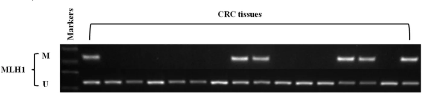
A total of the 913 cases were evaluated by immunohistochemistry (IHC) for the presence or ab-sence of MLH1, MSH2, MSH6 and PMS2 protein expression. Of the 69 cases (69/913, 7.5%) with dMMR, 49 had an absence of protein expression for MLH1/PMS2, 9 for MSH2/MSH6, 5 for MSH6 and 6 for PMS2 alone. In order to distinct Lynch syndrome-related CRC from spo-radic MSI cancers, we performed theMLH1promoter methylation study. Among 49 cases with an absence of protein expression for MLH1/PMS2, 32.6% (16/49) cases hadMLH1 pro-moter methylation. Of them,KRASmutations inMLH1methylated sporadic MSI tumors were 25% (4/16) and the mutation frequency was much lower than that of overall mutations, 36.9% (337/913) (Fig 1).
Patient and tumor characteristics with respect to the MMR status were shown inTable 1. The mean age at presentation for dMMR tumors was 53.3±12.7 years, which was younger than that of pMMR tumors (P= 0.003). Overall, tumors with dMMR were more frequently located on the proximal side of the colon (72.4%vs18.9%,P<0.0001) and were more likely to be poorly differentiated (34.8%vs18.0%,P<0.001), compared with pMMR tumors. In addition, tumors with dMMR were also significantly associated with mucinous histological subtype (63.8%vs32.7%,P<0.0001) and reduced lymph node metastasis (39.1%vs54.6%,P= 0.01). There were no statistically significant differences in gender and pT stage.
Fig 1. Representive MSP (Methylation-specific PCR) results ofMLH1methylation in colorectal cancer with loss of MLH1/PMS2 protein expression.
Mutations inKRASandBRAFV600Ewere mutually exclusive. There were 6 cases with dMMR tumors that harboredBRAFV600Emutations and these cases were excluded from the analysis.KRASmutations in codons 12 and 13 were observed in 36.9% (337/913) of all tumors. A higher frequency ofKRASmutations were detected in dMMR tumors (27/69, 39.1%) com-pared with pMMR tumors (310/844, 36.7%), although this difference did not reach statistical significance. Of the 27 dMMR andKRAS-mutated tumors, 16 cases were defined with loss of MLH1/PMS2, 7 with MSH2/MSH6 and 4 with PMS2 alone.
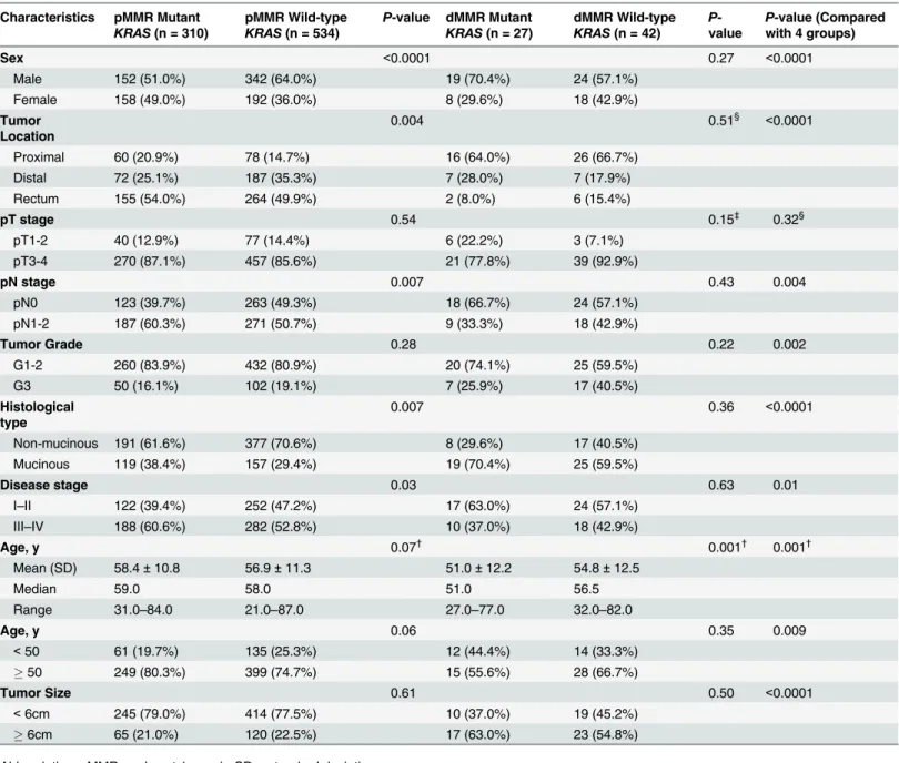
Patient and tumor characteristics with respect to both the MMR andKRASmutation status were summarized inTable 2. Of the 913 cases where both the MMR andKRASmutation status were defined, 27 (2.9%) cases with dMMR wereKRAS(+) and 42 (4.6%) wereKRAS(-), Table 1. Distributions of clinicopathologic characteristics by MMR status.
Characterics pMMR (n = 844) dMMR (n = 69) P-value
Sex 0.54
Male 494 (58.5%) 43 (62.3%)
Female 350 (41.5%) 26 (37.7%)
Tumor Location <0.0001
Proximal colon 158 (18.9%) 42 (72.4%)
Distal colon 259 (31.0%) 14 (24.1%)
Rectum 419 (50.1%) 2 (3.4%)
pT stage 0.06
pT1-2 117 (13.9%) 4 (5.8%)
pT3-4 727 (86.1%) 65 (94.2%)
pN stage 0.01
pN0 383 (45.4%) 42 (60.9%)
pN1-2 461 (54.6%) 27 (39.1%)
Tumor Grade 0.001
G1-2 692 (82.0%) 45 (65.2%)
G3 152 (18.0%) 24 (34.8%)
Histological type <0.0001
Mucinous 276 (32.7%) 44 (63.8%)
Non-mucinous 568 (67.3%) 25 (36.2%)
Disease stage 0.02
I–II 374 (44.3%) 41 (59.4%)
III–IV 470 (55.7%) 28 (40.6%)
Age, y 0.003†
Mean (SD) 57.5±11.1 53.3±12.7
Median 58.0 55.0
Range 21.0–87.0 27.0–82.0
Age, y 0.008
<50 197 (23.3%) 26 (37.7%)
50 647 (76.7%) 43 (62.3%)
Tumor Size <0.0001
<6cm 658 (78.0%) 29 (42.0%)
6cm 186 (21.0%) 40 (58.0%)
Abbreviations: MMR = mismatch repair; SD = standard deviation. †
Two-sided Kruskal Wallis test Others are two-sidedχ2test.
whereas 310 (34.0%) cases with pMMR wereKRAS(+) and 534 (58.5%) wereKRAS(-). Among these four groups, significant differences were observed for gender {KRAS(+)/dMMR cases more likely to be male,P<0.0001}, age {KRAS(+)/dMMR more likely to have a younger age at diagnosis of disease,P= 0.0001}, grade {KRAS(-)/dMMR more likely to have lower Table 2. Clinicopathological characteristics byKRASmutation status and MMR status.
Characteristics pMMR Mutant
KRAS(n = 310)
pMMR Wild-type
KRAS(n = 534)
P-value dMMR Mutant
KRAS(n = 27)
dMMR Wild-type
KRAS(n = 42)
P -value
P-value (Compared with 4 groups)
Sex <0.0001 0.27 <0.0001
Male 152 (51.0%) 342 (64.0%) 19 (70.4%) 24 (57.1%)
Female 158 (49.0%) 192 (36.0%) 8 (29.6%) 18 (42.9%)
Tumor Location
0.004 0.51§ <0.0001
Proximal 60 (20.9%) 78 (14.7%) 16 (64.0%) 26 (66.7%)
Distal 72 (25.1%) 187 (35.3%) 7 (28.0%) 7 (17.9%)
Rectum 155 (54.0%) 264 (49.9%) 2 (8.0%) 6 (15.4%)
pT stage 0.54 0.15‡ 0.32§
pT1-2 40 (12.9%) 77 (14.4%) 6 (22.2%) 3 (7.1%)
pT3-4 270 (87.1%) 457 (85.6%) 21 (77.8%) 39 (92.9%)
pN stage 0.007 0.43 0.004
pN0 123 (39.7%) 263 (49.3%) 18 (66.7%) 24 (57.1%)
pN1-2 187 (60.3%) 271 (50.7%) 9 (33.3%) 18 (42.9%)
Tumor Grade 0.28 0.22 0.002
G1-2 260 (83.9%) 432 (80.9%) 20 (74.1%) 25 (59.5%)
G3 50 (16.1%) 102 (19.1%) 7 (25.9%) 17 (40.5%)
Histological type
0.007 0.36 <0.0001
Non-mucinous 191 (61.6%) 377 (70.6%) 8 (29.6%) 17 (40.5%)
Mucinous 119 (38.4%) 157 (29.4%) 19 (70.4%) 25 (59.5%)
Disease stage 0.03 0.63 0.01
I–II 122 (39.4%) 252 (47.2%) 17 (63.0%) 24 (57.1%)
III–IV 188 (60.6%) 282 (52.8%) 10 (37.0%) 18 (42.9%)
Age, y 0.07†
0.001†
0.001†
Mean (SD) 58.4±10.8 56.9±11.3 51.0±12.2 54.8±12.5
Median 59.0 58.0 51.0 56.5
Range 31.0–84.0 21.0–87.0 27.0–77.0 32.0–82.0
Age, y 0.06 0.35 0.009
<50 61 (19.7%) 135 (25.3%) 12 (44.4%) 14 (33.3%)
50 249 (80.3%) 399 (74.7%) 15 (55.6%) 28 (66.7%)
Tumor Size 0.61 0.50 <0.0001
<6cm 245 (79.0%) 414 (77.5%) 10 (37.0%) 19 (45.2%)
6cm 65 (21.0%) 120 (22.5%) 17 (63.0%) 23 (54.8%)
Abbreviations: MMR = mismatch repair; SD = standard deviation. †
Two-sided Kruskal Wallis test ‡Two-sided
χ2test with continuity correction §Fischer’s exact test
Others are two-sidedχ2test
APvalue for significance was adjusted for multiple hypothesis testing toP= 0.05/6 = 0.0083. Thus, aPvalue between 0.05 and 0.0083 should be regarded as of borderline significance.
grade disease,P= 0.002}, tumor location {KRAS(+) andKRAS(-)/dMMR cases more likely to be located in the proximal colon,P<0.0001}and lymph node metastasis {KRAS(+)/pMMR cases more likely to have higher pN stage,P= 0.004}. However, no differences were noted in the pT stage among these four groups.
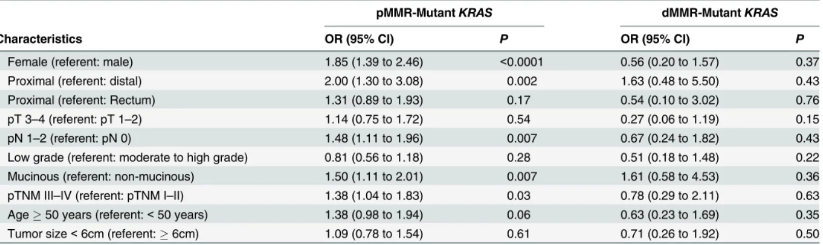
When compared with those withKRAS(-)/pMMR tumors, patients withKRAS(+)/pMMR tumors were most likely to be female (49.0%vs36.0%; OR = 1.85; 95% CI = 1.39 to 2.46; P<0.0001), to be proximal located (45.5%vs29.3%; OR = 2.00; 95% CI = 1.30 to 3.08;
P= 0.002), to have a mucinous histology (38.4%vs29.4%; OR = 1.50; 95% CI = 1.11 to 2.01; P= 0.007), and to have increased lymph node metastasis (60.3%vs50.7%; OR = 1.48; 95% CI = 1.11 to 1.96;P= 0.007) (Table 3). To the contrary, compared with those withKRAS (-)/dMMR tumors, patients withKRAS(+)/dMMR tumors demonstrated no statistically signifi-cant differences in gender, tumor location, pT depth of invasion, lymph node metastasis, pTNM stage, and histologic grade. However, the mean age at presentation forKRAS(+)/dMMR tumors was 51.051.051.0 presentation for s,n, than that ofKRAS(-)/dMMR tumors (P= 0.001).
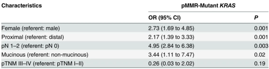
In the analysis using multivariable logistic regression models, we reviewed clinicopatholog-ic characteristclinicopatholog-ics inTable 4. As shown multivariably, tumors withKRAS(+)/pMMR were sta-tistically associated with proximal location, mucinous histology and increased lymph node metastasis.
Discussion
Defining tumor subtypes of CRC based on pathway-driven alterations has the potential to im-prove prognostication and guide targeted therapy. Distinct clinical and pathological features of CRC with different MMR status have long been identified [6,26,27,28]. In this study, we dem-onstrated molecular and clinicopathological features of pMMR and dMMR tumors stratified byKRASmutation status in a large cohort of consecutive Chinese CRC patients. Proficient MMR tumors that were nonmutated forKRASandBRAFV600Ewere the most prevalent subtype and represented 58.5% (534/913) of our study cohort. Compared with this subtype, patients Table 3. Univariate logistic regression model associations betweenKRASmutation status and MMR status.
pMMR-MutantKRAS dMMR-MutantKRAS
Characteristics OR (95% CI) P OR (95% CI) P
Female (referent: male) 1.85 (1.39 to 2.46) <0.0001 0.56 (0.20 to 1.57) 0.37
Proximal (referent: distal) 2.00 (1.30 to 3.08) 0.002 1.63 (0.48 to 5.50) 0.43
Proximal (referent: Rectum) 1.31 (0.89 to 1.93) 0.17 0.54 (0.10 to 3.02) 0.76
pT 3–4 (referent: pT 1–2) 1.14 (0.75 to 1.72) 0.54 0.27 (0.06 to 1.19) 0.15
pN 1–2 (referent: pN 0) 1.48 (1.11 to 1.96) 0.007 0.67 (0.24 to 1.82) 0.43
Low grade (referent: moderate to high grade) 0.81 (0.56 to 1.18) 0.28 0.51 (0.18 to 1.48) 0.22
Mucinous (referent: non-mucinous) 1.50 (1.11 to 2.01) 0.007 1.61 (0.58 to 4.53) 0.36
pTNM III–IV (referent: pTNM I–II) 1.38 (1.04 to 1.83) 0.03 0.78 (0.29 to 2.11) 0.63
Age50 years (referent:<50 years) 1.38 (0.98 to 1.94) 0.06 0.63 (0.23 to 1.69) 0.35 Tumor size<6cm (referent:6cm) 1.09 (0.78 to 1.54) 0.61 0.71 (0.26 to 1.92) 0.50
CI = confidence interval; dMMR = deficient mismatch repair; pMMR = proficient mismatch repair; OR = odds ratio. ‡
= Two-sidedχ2test with continuity correction §Fischer’s exact test
Others are two-sidedχ2test.
APvalue for significance was adjusted for multiple hypothesis testing to P = 0.05/6 = 0.0083. Thus, aPvalue between 0.05 and 0.0083 should be regarded as of borderline significance.
withKRAS(+)/pMMR tumors were more common in the proximal colon and to have a mucin-ous histology. Most importantly, patients withKRAS(+)/pMMR tumors showed increased lymph node metastasis among four subtypes and may have worse survival rate.
Consistent with the previous findings, our data suggest that tumors with dMMR status often exhibit poor differentiation, mucinous cell type, proximal location and reduced lymph node metastasis. In addition to its role in identifying unique pathological features of CRC, dMMR status has also been used as a prognostic marker and the guidance for Fluorouracil-based adjuvant chemotherapy [8]. Recent evidence indicated that CRC could be further classi-fied into five prespeciclassi-fied subtypes using a biomarker combination ofKRASandBRAFV600E mutations, MMR status andMLH1methylation with statistically significant differences in clinicopathologic features and patient survival rates [29]. Thus, a biomarker-based classifier provides important prognostic information in CRC with implications for patient management. Evidence from other reports supported the idea thatKRASmutation and MMR status are ge-netic markers that arise early and remain biologically relevant throughout all stages of tumor progression [30,31]. In addition,KRASmutations found in primary tumors are preserved in recurrences and metastases. Consequently, we evaluated the prespecified tumor subtypes with respect to clinicopathologic features in biomarker combinations ofKRASmutations and MMR status.
Deficient MMR cancers typically originate in the proximal colon [15]. As expected, the vast majority of dMMR tumors in this study (72.4%) were from the proximal colon and this distri-bution was not influenced byKRASmutations. Although most of the pMMR tumors were not likely to be proximal located, it is interesting that when pMMR tumors stratified byKRAS mu-tations,KRASmutant tumors (20.9% proximal) were more likely to be proximal compared to KRAS(-)/pMMR tumors (14.7% proximal). Traditionally, colon cancers developed in the prox-imal bowel often created an environment in which CIMP (CpG Island Methylator Phenotype) is more likely to arise, and this synergizes withBRAFmutation to allow progression of serrated polyps [32]. However, recent evidence suggested thatKRASmutations could also be found in CIMP high and CIMP low tumors which were often located in the proximal colon [33]. This indicated that there were more comprehensive mechanisms underlying the location of colon cancer and the mutational profiles. Mucinous carcinoma is diagnosed when at least 50% of the tumor comprises secretory mucin and is often associated with dMMR status and serrated ade-nocarcinoma [34]. This is consistent with our finding that dMMR tumors demonstrated more mucinous differentiation than pMMR tumors (63.8%vs32.7%). However, when stratified by KRASmutation status, we observed that pMMR tumors with mutantKRASphenotype exhib-ited more mucinous differentiation than wild typeKRASsubtype (38.4%vs29.4%). This is Table 4. Multivariate logistic regression model associations between patient, tumor andKRASor
BRAFV600E
mutation status.
Characteristics pMMR-MutantKRAS
OR (95% CI) P
Female (referent: male) 2.73 (1.69 to 4.85) 0.001
Proximal (referent: distal) 2.17 (1.39 to 3.33) 0.001
pN 1–2 (referent: pN 0) 4.95 (2.84 to 6.38) 0.003
Mucinous (referent: non-mucinous) 3.44 (1.11 to 7.47) 0.02
pTNM III–IV (referent: pTNM I–II) 0.26 (0.03 to 2.02) 0.19
CI = confidence interval; pMMR = proficient mismatch repair; OR = odds ratio.
largely becauseKRASmutation is not only linked to conventional adenomas but also associated with serrated adenomas in the development of colorectal cancer [35].
A significant association was found between the presence of lymph node metastases and pMMR tumors stratified byKRASmutation status. Our findings revealed that pMMR tumors withKRASmutation demonstrated more positive lymph nodes and pTNM III-IV stage of dis-ease than tumors withKRAS(-)/pMMR status. This is consistent with findings from a smaller report, which demonstrated that the frequency ofKRASmutations was higher in pMMR lymph node positive tumors as compared to pMMR lymph node negative tumors [36,37]. Our results indicate that the majority of pMMR tumors neededKRASmutation to be able to metastasize and this activation was crucial for neoplastic cells to acquire invasive potential. Mutations inKRASoncogene lead to alterations in encoded amino acids adjacent to the GTP binding pocket and reduced the GTPase activity ofKRASprotein after guanine nucleotide acti-vating protein (GAP) binding [38]. Bothin vitroandin vivoexperimental models, transfection of mutated, constitutively active forms ofKRASoncogene into previously noncancerous cells can lead to invasive and metastatic phenotypes. Ectopic expression of activeKRASin the mu-rine NIH 3T3 fibroblast cell line resulted in increased invasion and acquisition of metastatic properties [39]. Using tail vein injection of transformed cells,in vivomodels were observed by liver and lung metastasis [40]. In addition to the evidence obtained from cell and animal experiments, clinical studies have also displayed significant lymph node metastasis inKRAS (+)/pMMR tumors [36,41]. Gene expression profiling reveals that genes involoving epithelial mesenchymal transition and matrix remodeling that can facilitate tumor invasion and metasta-sis are up-regulated in mutantKRAS-pMMR tumors [42]. Consequently,KRASoncogenic activation was shown to be an important mediator of tumor cell invasion and metastasis in pMMR tumors.
The frequency ofKRASmutations in Lynch syndrome-related CRC and sporadic CRC is al-most the same. However,KRASmutations are significantly more frequent in Lynch syndrome-related CRC than that in sporadic MSI-H CRCs [37,43]. Lynch syndrome-related CRC tend to be early-onset and proximal location. So this may explain the younger age and proximal loca-tion observed inKRAS+/MSI tumors. Despite these positive findings, our study has some limi-tations. First, because this is a retrospectively study, it is hard to collect the blood or saliva sample from patients to detect germline mutations to further distinguish the Lynch syndrome-related CRC from sporadic cancer. So we could not calculate the precise frequency ofKRAS mutations in hereditary CRCs, however, it is sure that the Lynch syndrome-related CRC in our study showed preferentiallyKRASmutations. Second, we did not examine other less common mutations inKRAScodons 61, 117 and 146, which also contributed to the oncogenic transfor-mation of tumor cells.
This study suggests that specific epidemiologic and clinicopathologic characteristics are as-sociated with MMR status stratified byKRASmutation in CRC. Knowledge of MMR and KRASmutation status may enhance molecular pathologic staging of CRC patients and meta-static progression can also be estimated based on the combination of these biomarkers. Valida-tion of addiValida-tional genetic biomarkers will help to refine management decisions for individual patients based on tumor biology. Importantly, this may also aid the development of novel ther-apeutic targets to aid treatment of these aggressive cancers.
Acknowledgments
China (973 program 2014CB542002) and the National Natural Science Foundation of China (81401984).
Author Contributions
Conceived and designed the experiments: JMY SSS. Performed the experiments: WBL WXZ YL LS SMZ. Analyzed the data: WBL SMZ. Contributed reagents/materials/analysis tools: QT WXZ. Wrote the paper: WBL JMY.
References
1. Fearon ER (2011) Molecular genetics of colorectal cancer. Annu Rev Pathol 6: 479–507. doi:10.1146/ annurev-pathol-011110-130235PMID:21090969
2. Grady WM, Carethers JM (2008) Genomic and epigenetic instability in colorectal cancer pathogenesis. Gastroenterology 135: 1079–1099. doi:10.1053/j.gastro.2008.07.076PMID:18773902
3. Jass JR (2007) Classification of colorectal cancer based on correlation of clinical, morphological and molecular features. Histopathology 50: 113–130. PMID:17204026
4. Siegel R, Desantis C, Jemal A (2014) Colorectal cancer statistics, 2014. CA Cancer J Clin 64: 104– 117. doi:10.3322/caac.21220PMID:24639052
5. Sinicrope FA, Sargent DJ (2012) Molecular pathways: microsatellite instability in colorectal cancer: prognostic, predictive, and therapeutic implications. Clin Cancer Res 18: 1506–1512. doi:10.1158/ 1078-0432.CCR-11-1469PMID:22302899
6. Ribic CM, Sargent DJ, Moore MJ, Thibodeau SN, French AJ, Goldberg RM, et al. (2003) Tumor micro-satellite-instability status as a predictor of benefit from fluorouracil-based adjuvant chemotherapy for colon cancer. N Engl J Med 349: 247–257. PMID:12867608
7. Sinicrope FA, Mahoney MR, Smyrk TC, Thibodeau SN, Warren RS, Bertagnolli MM, et al. (2013) Prog-nostic impact of deficient DNA mismatch repair in patients with stage III colon cancer from a random-ized trial of FOLFOX-based adjuvant chemotherapy. J Clin Oncol 31: 3664–3672. doi:10.1200/JCO. 2013.48.9591PMID:24019539
8. Sinicrope FA, Foster NR, Thibodeau SN, Marsoni S, Monges G, Labianca R, et al. (2011) DNA mis-match repair status and colon cancer recurrence and survival in clinical trials of 5-fluorouracil-based ad-juvant therapy. J Natl Cancer Inst 103: 863–875. doi:10.1093/jnci/djr153PMID:21597022
9. Popat S, Hubner R, Houlston RS (2005) Systematic review of microsatellite instability and colorectal cancer prognosis. J Clin Oncol 23: 609–618. PMID:15659508
10. Umar A, Risinger JI, Hawk ET, Barrett JC (2004) Testing guidelines for hereditary non-polyposis colo-rectal cancer. Nat Rev Cancer 4: 153–158. PMID:14964310
11. Salovaara R, Loukola A, Kristo P, Kaariainen H, Ahtola H, Eskelinen M, et al. (2000) Population-based molecular detection of hereditary nonpolyposis colorectal cancer. J Clin Oncol 18: 2193–2200. PMID:
10829038
12. Davies H, Bignell GR, Cox C, Stephens P, Edkins S, Clegg S, et al. (2002) Mutations of the BRAF gene in human cancer. Nature 417: 949–954. PMID:12068308
13. Wang L, Cunningham JM, Winters JL, Guenther JC, French AJ, Boardman LA, et al. (2003) BRAF mu-tations in colon cancer are not likely attributable to defective DNA mismatch repair. Cancer Res 63: 5209–5212. PMID:14500346
14. Nagasaka T, Koi M, Kloor M, Gebert J, Vilkin A, Nishida N, et al. (2008) Mutations in both KRAS and BRAF may contribute to the methylator phenotype in colon cancer. Gastroenterology 134: 1950–1960, 1960 e1951. doi:10.1053/j.gastro.2008.02.094PMID:18435933
15. Jass JR, Do KA, Simms LA, Iino H, Wynter C, Pillay SP, et al. (1998) Morphology of sporadic colorectal cancer with DNA replication errors. Gut 42: 673–679. PMID:9659163
16. Perkins G, Pilati C, Blons H, Laurent-Puig P (2014) Beyond KRAS status and response to anti-EGFR therapy in metastatic colorectal cancer. Pharmacogenomics 15: 1043–1052. doi:10.2217/pgs.14.66
PMID:24956256
17. Lengauer C, Kinzler KW, Vogelstein B (1997) Genetic instability in colorectal cancers. Nature 386: 623–627. PMID:9121588
18. Karapetis CS, Khambata-Ford S, Jonker DJ, O'Callaghan CJ, Tu D, Tebbutt NC, et al. (2008) K-ras mu-tations and benefit from cetuximab in advanced colorectal cancer. N Engl J Med 359: 1757–1765. doi:
19. Normanno N, Tejpar S, Morgillo F, De Luca A, Van Cutsem E, Ciardiello F. (2009) Implications for KRAS status and EGFR-targeted therapies in metastatic CRC. Nat Rev Clin Oncol 6: 519–527. doi:
10.1038/nrclinonc.2009.111PMID:19636327
20. Moosmann N, Heinemann V (2007) Cetuximab in the treatment of metastatic colorectal cancer. Expert Opin Biol Ther 7: 243–256. PMID:17250462
21. Samowitz WS, Curtin K, Schaffer D, Robertson M, Leppert M, Slattery ML. (2000) Relationship of Ki-ras mutations in colon cancers to tumor location, stage, and survival: a population-based study. Cancer Epidemiol Biomarkers Prev 9: 1193–1197. PMID:11097226
22. Rosty C, Young JP, Walsh MD, Clendenning M, Walters RJ, Pearson S, et al. (2013) Colorectal carci-nomas with KRAS mutation are associated with distinctive morphological and molecular features. Mod Pathol 26: 825–834. doi:10.1038/modpathol.2012.240PMID:23348904
23. Zlobec I, Kovac M, Erzberger P, Molinari F, Bihl MP, Rufle A, et al. (2010) Combined analysis of specific KRAS mutation, BRAF and microsatellite instability identifies prognostic subgroups of sporadic and he-reditary colorectal cancer. Int J Cancer 127: 2569–2575. doi:10.1002/ijc.25265PMID:20162668
24. Yoon HH, Tougeron D, Shi Q, Alberts SR, Mahoney MR, Nelson GD, et al. (2014) KRAS codon 12 and 13 mutations in relation to disease-free survival in BRAF-wild-type stage III colon cancers from an adju-vant chemotherapy trial (N0147 alliance). Clin Cancer Res 20: 3033–3043. doi:10.1158/1078-0432. CCR-13-3140PMID:24687927
25. Zhao H, Li Q, Wang J, Su X, Ng KM, Qiu T, et al. (2012) Frequent epigenetic silencing of the folate-metabolising gene cystathionine-beta-synthase in gastrointestinal cancer. PLoS One 7: e49683. doi:
10.1371/journal.pone.0049683PMID:23152928
26. Campbell PT, Jacobs ET, Ulrich CM, Figueiredo JC, Poynter JN, McLaughlin JR, et al. (2010) Case-control study of overweight, obesity, and colorectal cancer risk, overall and by tumor microsatellite in-stability status. J Natl Cancer Inst 102: 391–400. doi:10.1093/jnci/djq011PMID:20208017
27. Sargent DJ, Marsoni S, Monges G, Thibodeau SN, Labianca R, Hamilton SR, et al. (2010) Defective mismatch repair as a predictive marker for lack of efficacy of fluorouracil-based adjuvant therapy in colon cancer. J Clin Oncol 28: 3219–3226. doi:10.1200/JCO.2009.27.1825PMID:20498393
28. Sinicrope F, Foster NR, Sargent DJ, Thibodeau SN, Smyrk TC, O'Connell MJ, et al. (2010) Model-based prediction of defective DNA mismatch repair using clinicopathological variables in sporadic colon cancer patients. Cancer 116: 1691–1698. doi:10.1002/cncr.24913PMID:20186699
29. Sinicrope FA, Shi Q, Smyrk TC, Thibodeau SN, Dienstmann R, Guinney J, et al. (2015) Molecular Markers Identify Subtypes of Stage III Colon Cancer Associated With Patient Outcomes. Gastroenterol-ogy 148: 88–99. doi:10.1053/j.gastro.2014.09.041PMID:25305506
30. Nash GM, Gimbel M, Cohen AM, Zeng ZS, Ndubuisi MI, Nathanson DR, et al. (2010) KRAS mutation and microsatellite instability: two genetic markers of early tumor development that influence the progno-sis of colorectal cancer. Ann Surg Oncol 17: 416–424. doi:10.1245/s10434-009-0713-0PMID:
19813061
31. Asaka S, Arai Y, Nishimura Y, Yamaguchi K, Ishikubo T, Yatsuoka T, et al. (2009) Microsatellite insta-bility-low colorectal cancer acquires a KRAS mutation during the progression from Dukes' A to Dukes' B. Carcinogenesis 30: 494–499. doi:10.1093/carcin/bgp017PMID:19147861
32. French AJ, Sargent DJ, Burgart LJ, Foster NR, Kabat BF, Goldberg R, et al. (2008) Prognostic signifi-cance of defective mismatch repair and BRAF V600E in patients with colon signifi-cancer. Clin Cancer Res 14: 3408–3415. doi:10.1158/1078-0432.CCR-07-1489PMID:18519771
33. The Cancer Genome Altas Network. (2012) Comprehensive molecular characterization of human colon and rectal cancer. Nature 487: 330–337. doi:10.1038/nature11252PMID:22810696
34. Whitehall VL, Wynter CV, Walsh MD, Simms LA, Purdie D, Pandeya N, et al. (2002) Morphological and molecular heterogeneity within nonmicrosatellite instability-high colorectal cancer. Cancer Res 62: 6011–6014. PMID:12414620
35. Ogino S, Chan AT, Fuchs CS, Giovannucci E (2011) Molecular pathological epidemiology of colorectal neoplasia: an emerging transdisciplinary and interdisciplinary field. Gut 60: 397–411. doi:10.1136/gut. 2010.217182PMID:21036793
36. Oliveira C, Velho S, Moutinho C, Ferreira A, Preto A, Domingo E, et al. (2007) KRAS and BRAF onco-genic mutations in MSS colorectal carcinoma progression. Oncogene 26: 158–163. PMID:16953233
37. Oliveira C, Westra JL, Arango D, Ollikainen M, Domingo E, Ferreira A, et al. (2004) Distinct patterns of KRAS mutations in colorectal carcinomas according to germline mismatch repair defects and hMLH1 methylation status. Hum Mol Genet 13: 2303–2311. PMID:15294875
39. Campbell PM, Der CJ (2004) Oncogenic Ras and its role in tumor cell invasion and metastasis. Semin Cancer Biol 14: 105–114. PMID:15018894
40. Al-Mulla F, MacKenzie EM (2001) Differences in in vitro invasive capacity induced by differences in Ki-Ras protein mutations. J Pathol 195: 549–556. PMID:11745690
41. Roth AD, Tejpar S, Delorenzi M, Yan P, Fiocca R, Klingbiel D, et al. (2010) Prognostic role of KRAS and BRAF in stage II and III resected colon cancer: results of the translational study on the PETACC-3, EORTC 40993, SAKK 60–00 trial. J Clin Oncol 28: 466–474. doi:10.1200/JCO.2009.23.3452PMID:
20008640
42. Marisa L, de Reynies A, Duval A, Selves J, Gaub MP, Vescovo L, et al. (2013) Gene expression classi-fication of colon cancer into molecular subtypes: characterization, validation, and prognostic value. PLoS Med 10: e1001453. doi:10.1371/journal.pmed.1001453PMID:23700391