HCT116 Colorectal Cancer Cells
Carrie Luu1, Eileen L. Heinrich1, Marjun Duldulao1, Amanda K. Arrington1, Marwan Fakih2, Julio Garcia-Aguilar3*, Joseph Kim1*
1Division of Surgical Oncology, Department of Surgery, City of Hope Comprehensive Cancer Center, Duarte, California, United States of America,2Department of Medical Oncology and Experimental Therapeutics, City of Hope Comprehensive Cancer Center, Duarte, California, United States of America,3Department of Surgery, Memorial Sloan-Kettering Cancer Center, New York, New York, United States of America
Abstract
Recent reports have indicated thatKRASandTP53 mutations predict response to therapy in colorectal cancer. However, little is known about the relationship between these two common genetic alterations. Micro-RNAs (miRNAs), a class of noncoding RNA implicated in cellular processes, have been increasingly linked toKRASand TP53. We hypothesized that lethal-7a (let-7a) miRNA regulatesKRASthroughTP53. To investigate the relationship betweenKRAS, TP53, and let-7a, we used HCT116KRASmuthuman colorectal cancer cells with four different genotypic modifications inTP53(TP532/2, TP53+/2, TP53mut/+,andTP53mut/2). Using these cells we observed that K-Ras activity was higher in cells with mutant or knocked out TP53alleles, suggesting that wild-typeTP53may suppress K-Ras activity. Let-7a was present in HCT116KRASmutcells, though there was no correlation between let-7a level andTP53genotype status. To explore how let-7a may regulate K-Ras in the different TP53 genotype cells we used let-7a inhibitor and demonstrated increased K-Ras activity across all TP53, thus corroborating prior reports that let-7a regulates K-Ras. To assess potential clinical implications of this regulatory network, we examined the influence ofTP53genotype and let-7a inhibition on colon cancer cell survival following chemoradiation therapy (CRT). We observed that cells with complete loss of wild-typeTP53 alleles (2/2 or2/mut) were resistant to CRT
following treatment with 5-fluorouracil and radiation. Further increase in K-Ras activity with let-7a inhibition did not impact survival in these cells. In contrast, cells with single or double wild-typeTP53alleles were moderately responsive to CRT and exhibited resistance when let-7a was inhibited. In summary, our results show a complex regulatory system involvingTP53, KRAS, and let-7a. Our results may provide clues to understand and target these interactions in colorectal cancer.
Citation:Luu C, Heinrich EL, Duldulao M, Arrington AK, Fakih M, et al. (2013)TP53and Let-7a micro-RNA Regulate K-Ras Activity in HCT116 Colorectal Cancer Cells. PLoS ONE 8(8): e70604. doi:10.1371/journal.pone.0070604
Editor:Alfredo Fusco, Consiglio Nazionale delle Ricerche (CNR), Italy ReceivedMarch 4, 2013;AcceptedJune 21, 2013;PublishedAugust 1, 2013
Copyright:ß2013 Luu et al. This is an open-access article distributed under the terms of the Creative Commons Attribution License, which permits unrestricted use, distribution, and reproduction in any medium, provided the original author and source are credited.
Funding:The authors have no support or funding to report.
Competing Interests:The authors have declared that no competing interests exist.
* E-mail: [email protected] (JGA); [email protected] (JK)
Introduction
Colorectal cancer (CRC) is the 4th most common cancer and the 2ndmost common cause of cancer-related death in the US [1]. Recent advances that have improved outcomes in CRC have included the identification of accurate prognostic and predictive molecular biomarkers [2–4]. These have included genetic alterations inKRAS and TP53, which are frequently detected in patients with CRC.KRASmutations are present in approximately 30–50% of CRCs, but also in 17–25% of all human tumors [2,5– 7]. Similarly,TP53alterations are common in patients with CRC (nearly 50%) [8]. Both prognostic and predictive roles have been identified for both genes [9]. Our own group and others has recently shown that detection of concurrent KRAS and TP53 mutations, with an incidence of 5% to 20% in CRC patients, correlated with resistance to neoadjuvant chemoradiation therapy (CRT) in patients with rectal cancer [10–12]. Despite the frequency of these mutations in CRC, little is known about interactions between the two genes.
The link between these two frequently altered genes in CRC may lie in micro-RNA (miRNA), a class of non-coding RNA which function in transcriptional regulation and more specifically
may influence the regulation of cell proliferation and apoptosis [13,14]. Recent reports have suggested that the tumor suppressor activity of miRNA lethal 7a (let-7a) may be due to its association withKRAS and that inhibition of tumor growth may occur by suppression of K-Ras expression by let-7a [15,16]. Emerging clinical data suggest that intra-tumor let-7a expression correlates with tumor response and overall survival in metastatic colorectal cancer patients receiving epidermal growth factor (EGFR) targeting agents in bothKRAS wild-type and mutant populations [17]. Additional studies have speculated that the role ofTP53in DNA repair and apoptosis may in part be regulated by miRNAs, including let-7a [18,19]. Therefore, a complex regulatory network forTP53andKRASmay be linked by let-7a.
between let-7a level andTP53genotype. Nonetheless, changes in K-Ras activity were regulated by let-7a. This first report ofTP53 and let-7a regulation of K-Ras activity provides clues to better understand the complex interaction betweenTP53andKRAS.
Materials and Methods
Cell Culture
The human CRC cell line HCT116, harboring a single mutant KRAS allele and double wild-type TP53 (TP53+/+) alleles, was modified into four stable cell lines with differentTP53genotypes (TP532/2, TP53+/2, TP53mut/+,andTP53mut/2). The parent and
modified cell lines were kindly provided by Dr. Bert Vogelstein (Johns Hopkins University; Baltimore, MD) [20]. Mutant and knockout alleles were both used to assess potential differences between the two alleles, as suggested in prior reports [21]. All cell lines were assessed by DNA extraction, polymerase chain reaction (PCR), and sequencing for KRAS and TP53 gene mutations to verify genotypes. Cells were maintained in McCoy 5A medium (Irvine Scientific; Santa Ana, CA) with 10% fetal bovine serum (Omega Scientific; Tarzana, CA) and 1% penicillin-streptomycin-glutamine (Invitrogen; Carlsbad, CA) at 5% CO2at 37uC.
Immunoblotting
Cell lysates were collected using RIPA buffer (Invitrogen; Carlsbad, CA) plus protease inhibitors (Thermo Scientific; Rock-ford, IL). Twenty micrograms of protein were separated on 12% SDS-polyacrylamide gels and transferred onto PVDF membranes (Millipore; Bedford, MA). The membranes were blocked for 1 h with 5% non-fat dry milk and probed overnight with primary antibodies. After washing, membranes were labeled with horse-radish peroxidase (HRP)-conjugated secondary antibodies (BioRad; Hercules, CA). Membranes were developed using a chemiluminescent substrate (Amersham Pharmacia; Piscataway, NJ) and imaged. Antibodies used were: anti-K-Ras (Millipore) and anti-GAPDH (Cell Signaling; Danvers, MA).
K-Ras Activity
K-Ras activity was measured using the Ras Activation ELISA Assay Kit (Millipore). In brief, cell lysates were incubated with Raf-1 Ras Binding Domain (RBD)-agarose. Raf-1-RBD was used to capture the active GTP-bound K-Ras protein, which was then detected by the addition of an anti-K-Ras-antibody (Millipore). An HRP-conjugated secondary antibody was then added. A lumi-nometer was used to measure the signals after addition of a chemiluminescent reagent (Perkin-Elmer; Shelton, CT). Since assays were performed to assess the relative changes in K-Ras activity among the differentTP53genotypes, K-Ras activity from the parental TP53+/+ line was set as 1. For assays with let-7a inhibitor, each genotype acted as its own control. Three independent assays were performed for each cell line, and the mean absorbance6the standard deviation (SD) was plotted for each line.
Reverse Transcription
RT-PCR was performed using the Taqman H miRNA assay (Invitrogen) following the manufacturer’s protocol. Briefly, reverse transcription was performed using the provided Multiscribe RTTM Reagent master mix plus cell lysates and the let-7a specific primers (Invitrogen). Reactions were performed in a thermal cycler at 16uC for 30 min, 42uC for 30 min, and 85uC for 5 min.
Real Time-PCR
RT-PCR was performed using the TaqmanHmiRNA Cells to CTTMKit (Invitrogen) according to the manufacturer’s protocol. Briefly, the RT product was added to the PCR cocktail (TaqmanH
miRNA assay reagent plus provided TaqmanH master mix) and the RT-PCR reactions were run at 95uC for 10 min, followed by 40 cycles of 15 sec at 95uC and 1 min at 60uC in the Applied Biosystems 7500 fast RT-PCR system (Foster City, CA). Each sample was assayed in triplicate.
Let-7a Inhibitor
Let-7a was inhibited by miRIDIAN miRNA inhibitors (Dharmacon; Lafeyette, CO) following the manufacturer’s proto-col. Briefly, HCT116 cells (36105) were plated in 6-well plates and incubated overnight. They were then transfected with 100 nM of miRIDIAN miRNA inhibitor for 24 h. Cells were incubated for an additional 48 h prior to being utilized in assays.
Chemoradiation Therapy
HCT116 cells were treated with CRT according to an established protocol [22]. After a total of 72 h incubation with let-7a inhibitor, cells were plated in 6-well plates and incubated overnight. Cells were then treated with 4 nM 5-Fluorouracil (5-FU) (Sigma Aldrich; St. Louis, MO) for 16 h after which they were exposed to 4 Gy of radiation. Drug exposure was halted 6 h after radiation treatment by medium exchange. Cells that had not been treated with let-7a inhibitor were similarly treated with CRT.
Determination of Cell Viability
Cell viability was assessed using an ATP-based assay (Cell Titer-Glo; Promega; Madison, WI) as per manufacturer’s protocol. Briefly, HCT116 cells (56103) were plated in 96-well plates and
incubated overnight. Cell Titer-Glo reagent was added and cell lysis induced. Luminescence was recorded with a luminometer. Three independent experiments were performed. Data shown represent the average viability, as a percent of the viability of untreated cells.
Statistical Analysis
Statistical analysis was performed on Microsoft excel software (Microsoft; Redmond, WA). Each data point represents the average value of three independent experiments. Control and treatment conditions were compared by Student’s t-test and differences among cell lines were compared by analysis of variance (ANOVA). P value,0.05 was considered statistically significant.
Results
K-Ras Protein Level is not Dependent onTP53Mutation Status
To determine the effect of differentTP53genotypes on K-Ras expression, western blot assay was performed to compare protein levels. Our results show that K-Ras expression levels were not influenced by the differentTP53genotypes (Figure 1A) suggesting thatTP53does not regulate K-Ras protein expression.
Effect ofTP53 Mutation Status on K-Ras Activity
The effect of TP53genotype on K-Ras activity was measured using a Raf pull-down assay. We discovered that K-Ras activity was lowest inTP53+/+cells. Increased K-Ras activity was most pronounced inTP53mut/2, followed byTP532/2,TP53+/mut
, and TP532/+ (44%, 32%, 20%, and 10% increases, respectively, p,0.05) (Figure 1B). These results show that K-Ras activity was
lowest in cells with wild-type TP53alleles; and the highest levels were identified in cells with homozygous loss of the wild-type TP53 alleles.
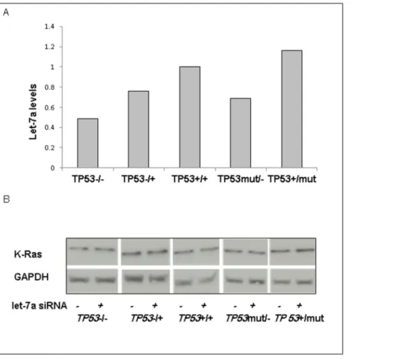
Let-7a is Expressed in All HCT116 Lines
Let-7a expression in HCT116 cell lines was measured by qPCR. Let-7a expression was detected in all cell lines (Figure 2A). There was no pattern of change in let-7a levels which corresponded to alterations inTP53genotype.
Let-7a does not Regulate K-Ras Protein Levels
Let-7a gene expression was inhibited.90% by qPCR. K-Ras protein levels were assessed across the TP53 genotypes in the presence or absence of let-7 inhibition. K-Ras protein levels were not altered by let-7a inhibition (Figure 2B).
Let-7a Negatively Regulates K-Ras Activity
The effect of let-7a inhibition on K-Ras activity was also determined. With let-7a inhibition, K-Ras activity increased across all TP53 genotypes (Figure 3). We found that K-Ras activity increased by 50% to 112% compared to let-7a expressing cells (p,0.05). The percent increase in K-Ras activity across all cell lines was not clearly associated withTP53genotype and whether knockout or mutant alleles were present.
Cells with Wild-typeTP53Alleles are Sensitive to CRT and let-7a Inhibition Decreases Response to CRT
Cells harboring different TP53 genotypes were treated with CRT and cell viability was measured (Figure 4). Cells lacking any wild-type TP53 allele were resistant to CRT, whereas cells harboring a single wild-typeTP53allele exhibited approximately 50% cell death. CRT treatment was performed again with let-7a inhibition, which decreased CRT-induced cell death by nearly 20% in cells harboring at least one wild-typeTP53allele (p,0.05). Cells with homozygous loss of wild-typeTP53alleles (2/2andmut/
2), which correlated with highest K-Ras activity (see Figure 1B),
exhibited no change in cell viability regardless of changes in let-7a expression levels.
Discussion
KRASandTP53are common somatic mutations with predictive and prognostic value in patients with CRC. The detection of select KRASmutations has been associated with increased risk of disease relapse and death [23] and has predicted resistance to anti-EGFR therapies in CRC [9]. Similarly, selectTP53alterations have been associated with poor outcomes in CRC [8]. We have also reported that the concurrent detection of TP53and KRAS mutations in patients receiving neoadjuvant CRT for rectal cancer predicted failure of pathologic complete response [10]. In this investigation, we sought to better understand a possible interaction between the Figure 1. K-Ras expression is not dependent onTP53mutational status but K-Ras activity is increased in all cells compared toTP53+/ +cells.A) HCT116 cells with the noted variations inTP53mutation status were collected and lysed for western blotting for K-Ras expression. GAPDH
was used as a loading control. B) K-Ras activity was measured in all cell lines using a Raf pull-down ELISA assay. Results shown represent the average of 3 independent experiments performed in triplicate relative toTP53+/+cells, p,0.05. Error bars show standard deviation (SD).
two genes. We identified let-7a as a potential link betweenTP53 andKRAS. Prior characterization of let-7a has revealed its role in
controlling cell cycle progression and division in human lung and colon cancers [24,25]. Moreover, Johnson et al. [15] discovered Figure 2. Let-7a is expressed in all cell lines and inhibition of let-7a does not affect K-Ras protein levels.A) Let-7a expression was measured via qRT-PCR. Data shown represents a single qRT-PCR performed in triplicate. B) Cells were transfected with 100 nM of let-7a inhibitor for 24 h. Protein for control and for let-7a inhibited cells were collected for immunoblotting for K-Ras expression. GAPDH served as a loading control. doi:10.1371/journal.pone.0070604.g002
Figure 3. K-Ras activity is increased in cells with let-7a inhibition.Cell lysates were collected following 72 h incubation and subjected to a Raf-pull down assay which measures K-Ras activity. Data shown represents three experiments done in triplicate. Error bars show SD. *p,0.05. doi:10.1371/journal.pone.0070604.g003
that let-7 negatively controlledKRASexpression in lung cancer. In addition, Ruzzo et al. [17] reported an improved overall survival in patients with elevated let-7a within aKRASmutant colorectal cancer population treated with anti-EGFR therapy, suggesting a tumor inhibitory role for let-7a in KRAS-activated tumors.TP53 has been known to transcriptionally regulate the expression of miRNAs [26,27]. Therefore, we theorized the existence of a series of regulatory steps fromTP53to let-7a toKRAS. We discovered that oncogenic K-Ras activity was regulated byTP53 genotype and let-7a; and changes in both influenced response to CRT.
In our study we utilized CRC cells harboring various TP53 genotypes. These cells were derived from a parental cell line harboring the gain of functionKRASmutation located on codon 13 [28,29]. There is rationale for utilizing cells with knockout and mutantTP53alleles in our study. Despite the common loss of wild-typeTP53alleles, the knockout and mutantTP53alleles are not alike. Complete loss ofTP53alleles results in corresponding loss of tumor suppressor activity while mutantTP53alleles may express p53 proteins that lose tumor suppressor activity but also gain oncogenic properties [21]. However, in this investigation we were unable to find consistent differences between KO and mutant TP53 alleles with regards to K-Ras activity at baseline and in response to let-7a inhibition, nor in response to CRT.
As noted previously, we observed changes in K-Ras activity with inhibition of let-7a expression. However, we did not observe corresponding changes in K-Ras protein expression, which conflicts with prior reports demonstrating let-7 miRNA regulation of K-Ras expression [30,31]. Nevertheless, our findings are consistent with established regulatory mechanisms of miRNA. Indeed, miRNA can regulate genes through a variety of mechanisms from mRNA translation to stability in the cytoplasm to cell cycle activity [14,24]. The differences between our results and those of prior reports could also be due to differences between species or cell lines utilized. Thus, while K-Ras expression remained unchanged by let-7a inhibition, our experiments showed that K-Ras activity was affected by let-7a. In addition, cells harboring homozygous wild-typeTP53alleles expressed the lowest K-Ras activity, which suggests that wild-type TP53 alleles may suppress K-Ras activity. Furthermore, our results are consistent with a report in pancreatic cancer, showing an increase in K-Ras
activity when mutantKRAS was paired with mutantTP53[32]. Finally, K-Ras activity clearly increased when let-7a was inhibited regardless ofTP53genotype, further suggesting let-7a regulation of K-Ras activity.
Chemotherapy and radiation are important components of the multimodal management of surgical patients with rectal cancer; and neoadjuvant CRT has been associated with downstaging of disease and decreased local recurrence [33,34]. In our investiga-tion, we discovered that cells lacking wild-typeTP53alleles were resistant to CRT and exhibited no cell death when treated with CRT. In contrast, cells with at least one wild-type allele (TP53+/+, TP53 mut/+, and TP532/+) had approximately 50% cell death when treated with CRT. These results are consistent with the results from our prior clinical trials wherein patients with double mutations (KRAS and TP53) failed to demonstrate a pathologic complete response after treatment with neoadjuvant CRT [10]. When let-7a expression was inhibited (leading to increased K-Ras activity), cells lacking wild-type TP53 alleles (TP532/2and TP53mut/2) had 100% cell survival. However, in cells harboring at least one wild-typeTP53allele (previously showing some cell death), cell survival increased in response to let-7a inhibition. These results have two important implications: (1)TP53genotype influences K-Ras activity and may predict response to CRT and (2) let-7a expression levels influence K-Ras activity and may alter response to CRT.
In summary, we provide insight into potential mechanisms linking KRAS, TP53, and let-7a. Although double mutants are relatively rare, the condition appears to have a poor phenotype with resistance to CRT. Our results contribute to a better understanding of the connection between KRAS and TP53. Knowledge of the pathways linkingKRAS,TP53, and let-7a may provide greater insight into the mechanisms driving the poor phenotype observed in certain mutations in CRC. While we acknowledge that the limitations of our study include the lack of in vivomodeling and the limitations of one cell line, our focus was on the investigation of the potential cross-talk between let-7a and p53, which we clearly demonstrate in our model. It is feasible that this interaction may be cell line specific or may depend on other aberrant pathways in HCT-116 (MSI-H, PIK3 mutant). Future work will focus on investigating the potential mechanisms of Figure 4. After let-7a inhibition, cell viability is partially rescued in CRT-sensitive cells.Cells with varyingTP53status+/2let-7a inhibitor were treated with 5-FU followed by radiation. Cell viability was measured. Data shown represent the average viability as a percent of untreated cells. *p,0.05, **p.0.05.
interaction between let-7a and p53 across different colorectal cell lines and with different molecular profiles.
Author Contributions
Conceived and designed the experiments: JGA JK. Performed the experiments: MD. Analyzed the data: CL ELH MD AKA MF JGA JK. Contributed reagents/materials/analysis tools: JK JGA. Wrote the paper: CL ELH MD AKA MF JGA JK.
References
1. Siegel R, DeSantis C, Virgo K, Stein K, Mariotto A, et al. (2012) Cancer treatment and survivorship statistics, 2012. CA Cancer J Clin 62: 220–241. 2. Bell SM, Scott N, Cross D, Sagar P, Lewis FA, et al. (1993) Prognostic value of
p53 overexpression and c-Ki-ras gene mutations in colorectal cancer. Gastroenterology 104: 57–64.
3. Lumachi F, Orlando R, Marino F, Chiara GB, Basso SM (2012) Expression of p53 and Ki-67 as prognostic factors for survival of men with colorectal cancer. Anticancer Res 32: 3965–3967.
4. Umeda Y, Nagasaka T, Mori Y, Sadamori H, Sun DS, et al. (2013) Poor prognosis of KRAS or BRAF mutant colorectal liver metastasis without microsatellite instability. J Hepatobiliary Pancreat Sci 20: 223–233. 5. Tie J, Lipton L, Desai J, Gibbs P, Jorissen RN, et al. (2011) KRAS mutation is
associated with lung metastasis in patients with curatively resected colorectal cancer. Clin Cancer Res 17: 1122–1130.
6. De Roock W, Piessevaux H, De Schutter J, Janssens M, De Hertogh G, et al. (2008) KRAS wild-type state predicts survival and is associated to early radiological response in metastatic colorectal cancer treated with cetuximab. Ann Oncol 19: 508–515.
7. Vogelstein B, Fearon ER, Hamilton SR, Kern SE, Preisinger AC, et al. (1988) Genetic alterations during colorectal-tumor development. N Engl J Med 319: 525–532.
8. Russo A, Bazan V, Lacopetta B, Kerr D, Soussi T, et al. (2005) The TP53 colorectal cancer international collaborative study on the prognostic and predictive significance of p53 mutation: influence of tumor site, type of mutation, and adjuvant treatment. J Clin Oncol 23: 7518–7528.
9. Lievre A, Bachet JB, Le Corre D, Boige V, Landi B, et al. (2006) KRAS mutation status is predictive of response to cetuximab therapy in colorectal cancer. Cancer Res 66: 3992–3995.
10. Garcia-Aguilar J, Chen Z, Smith DD, Li W, Madoff RD, et al. (2011) Identification of a biomarker profile associated with resistance to neoadjuvant chemoradiation therapy in rectal cancer. Ann Surg 254: 486–492.
11. Smith G, Carey FA, Beattie J, Wilkie MJV, Lightfoot TJ, et al. (2002) Mutations in APC, Kirsten-ras, and p53–alternative genetic pathways to colorectal cancer. Proc Natl Acad Sci USA 99: 9433–9438.
12. Tortola S, Marcuello E, Gonza´lez I, Reyes G, Arribas R, et al. (1999) p53 and K-ras gene mutations correlate with tumor aggressiveness but are not of routine prognostic value in colorectal cancer. J Clin Oncol 17: 1375–1375. 13. Zhang B, Pan X, Cobb GP, Anderson TA (2007) microRNAs as oncogenes and
tumor suppressors. Dev Biol 302: 1–12.
14. Filipowicz W, Bhattacharyya SN, Sonenberg N (2008) Mechanisms of post-transcriptional regulation by microRNAs: are the answers in sight? Nat Rev Genet 9: 102–114.
15. Johnson SM, Grosshans H, Shingara J, Byrom M, Jarvis R, et al. (2005) RAS is regulated by the let-7 microRNA family. Cell 120: 635–647.
16. He X, Chen J, Zhang Z, Li C, Peng Q, et al. (2010) The let-7a microRNA protects from growth of lung carcinoma by suppression of k-Ras and c-Myc in nude mice. J Cancer Res Clin Oncol 136: 1023–1028.
17. Ruzzo A, Graziano F, Vincenzi B, Canestrari E, Perrone G, et al. (2012) High let-7a microRNA levels in KRAS-mutated colorectal carcinomas may rescue anti-EGFR therapy effects in patients with chemotherapy-refractory metastatic disease. Oncologist 17: 823–829.
18. Saleh AD, Savage JE, Cao L, Soule BP, Ly D, et al. (2011) Cellular stress induced alterations in microRNA let-7a and let-7b expression are dependent on p53. PLoS One 6: e24429.
19. Bommer GT, Gerin I, Feng Y, Kaczorowski AJ, Kuick R, et al. (2007) p53-mediated activation of miRNA34 candidate tumor-suppressor genes. Curr Biol 17: 1298–1307.
20. Bunz F, Dutriaux A, Lengauer C, Waldman T, Zhou S, et al. (1998) Requirement for p53 and p21 to sustain G2 arrest after DNA damage. Science 282: 1497–1501.
21. Rivlin N, Brosh R, Oren M, Rotter V (2011) Mutations in the p53 tumor suppressor gene: Important milestones at the various steps of tumorigenesis. Genes Cancer 2: 466–474.
22. Spitzner M, Emons G, Kramer F, Gaedcke J, Rave-Fra¨nk M, et al. (2010) A gene expression signature for chemoradiosensitivity of colorectal cancer cells. Int J Radiat Oncol Biol Phys 78: 1184–1192.
23. Bazan V, Migliavacca M, Zanna I, Tubiolo C, Grassi N, et al. (2002) Specific codon 13 K-ras mutations are predictive of clinical outcome in colorectal cancer patients, whereas codon 12 K-ras mutations are associated with mucinous histotype. Ann Oncol 13: 1438–1446.
24. Johnson CD, Esquela-Kerscher A, Stefani G, Byrom M, Kelnar K, et al. (2007) The let-7 microRNA represses cell proliferation pathways in human cells. Cancer Res 67: 7713–7722.
25. Akao Y, Nakagawa Y, Naoe T. (2006) let-7 microRNA functions as a potential growth suppressor in human colon cancer cells. Biol Pharm Bull 29: 903–906. 26. He L, He X, Lowe SW, Hannon GJ (2007) microRNAs join the p53
network-another piece in the tumour-suppression puzzle. Nat Rev Cancer 7: 819–822. 27. Raver-Shapira N, Marciano E, Meiri E, Spector Y, Rosenfeld N, et al. (2007)
Transcriptional activation of miR-34a contributes to p53-mediated apoptosis. Mol Cell 26: 731–743.
28. Bunz F, Hwang PM, Torrance C, Waldman T, Zhang Y, et al. (1999) Disruption of p53 in human cancer cells alters the responses to therapeutic agents. J Clin Invest 104: 263–270.
29. Tazawa H, Tsuchiya N, Izumiya M, Nakagama H. (2007) Tumor-suppressive miR-34a induces senescence-like growth arrest through modulation of the E2F pathway in human colon cancer cells. Proc Natl Acad Sci USA 104: 15472– 15477.
30. Han Z, Yang Q, Liu B, Wu J, Li Y, et al. (2012) MicroRNA-622 functions as a tumor suppressor by targeting K-Ras and enhancing the anticarcinogenic effect of resveratrol. Carcinogenesis 33: 131–139.
31. Levy R, Biran A, Poirier F, Raz A, Kloog (2011) Y Galectin-3 mediates cross-talk between K-Ras and Let-7c tumor suppressor microRNA. PLoS One 6: e27490.
32. Ji B, Tsou L, Wang H, Gaiser S, Chang DZ, et al. (2009) Ras activity levels control the development of pancreatic diseases. Gastroenterology 137: 1072– 1082.e6.
33. Garcı´a-Aguilar J, Hernandez de Anda E, Sirivongs P, Lee S-H, Madoff R, et al. (2003) A pathologic complete response to preoperative chemoradiation is associated with lower local recurrence and improved survival in rectal cancer patients treated by mesorectal excision. Dis Colon Rectum 46: 298–304. 34. Janjan NA, Crane C, Feig BW, Cleary K, Dubrow R, et al. (2001) Improved
overall survival among responders to preoperative chemoradiation for locally advanced rectal cancer. Am J Clin Oncol 24: 107–112.