Yeon Jung Kim
Avaliação do efeito da Artin M no processo
de reparação em mucosa mastigatória.
Yeon Jung Kim
Avaliação do efeito da Artin M no processo
de reparação em mucosa mastigatória.
Tese apresentada ao Programa de Pós-Graduação em Odontologia, área de Periodontia, da Faculdade de Odontologia de Araraquara da Universidade Estadual Paulista para a obtenção do título de Doutor em Odontologia.
Orientador: Prof. Dr. Joni Augusto Cirelli
Kim, Yeon Jung
Avaliação do efeito da Artin M no processo de reparação em mucosa mastigatória / Yeon Jung Kim . – Araraquara: [s.n.], 2011. 110 f. ; 30 cm.
Tese (Doutorado) – Universidade Estadual Paulista, Faculdade de Odontologia
Orientador: Prof. Dr. Joni Augusto Cirelli
1. Mucosa bucal 2. Cicatrização 3. Periodontia 4. Glicoproteínas I. Título
YEON JUNG KIM
AVALIAÇÃO DO EFEITO DA ARTIN M NO PROCESSO DE REPARAÇÃO EM MUCOSA MASTIGATÓRIA
COMISSÃO JULGADORA
TESE PARA OBTENÇÃO DO GRAU DE DOUTOR
Presidente e Orientador Prof. Dr. Joni Augusto Cirelli
2º Examinador Profa. Dra. Rosemary Adriana Chiérici Marcantonio 3º Examinador Profa. Dra. Maria Cristina Roque Antunes Barreira 4º Examinador Prof. Dr. Rodrigo Otávio Citó César Rêgo
5º Examinador Prof. Dr. Sérgio Luis Scombatti de Souza
Dedicatória
À Deus,
pela sua presença constante na minha vida e pelos sonhos que me permitiu realizar
Aos meus pais,
Myoung Hyun Kim
eHyun Suk Kim
,Pelo amor incondicional, dedicação absoluta e pelos exemplos de vida, que me fizeram fortes para que pudesse vencer todos os obstáculos desta caminhada.
Agradecimentos especiais
Aos meus irmãos, Young Rae e Young Hyo,
Pelo companheirismo, por todo apoio e por estarem presentes em todas as minhas conquistas.
A minha sobrinha Julia,
Pelo amor que me faz sentir, por cada momento que posso presenciar a pureza de seus sorrisos, por me fazer uma pessoa mais feliz.
Aos meus familiares,
Pelo carinho e apoio prestados em todos os momentos da minha vida.
Agradecimentos
À Faculdade de Odontologia de Araraquara- Unesp, na pessoa de seu Diretor, Prof. Dr. José Cláudio Martins Segala e Vice-Diretora, Prof. Dra. Andrea Montandon, que me acolhe desde 2000.
Ao meu orientador Prof. Dr. Joni Augusto Cirelli, pelos ensinamentos, confiança em meu trabalho, disponibilidade e a contribuição durante a realização do curso de doutorado.
À Profa. Dra. Maria Cristina Antunes Roque-Barreira, aos alunos de pós-graduação especialmente Fernanda Carvalho e Marina Conrado
e aos funcionários do Departamento de Biologia Celular e Molecular da Faculdade de Medicina de Ribeirão Preto – USP pelo carinho que me receberam, pela colaboração e condições oferecidas durante a realização desta pesquisa.
Aos amigos do laboratório especialmente João, Andressa, Pedro e
Letícia, que participaram ativamente deste trabalho.
Aos grandes amigos que me apoiaram durante todo esse tempo:
Andreza, Alessandra, Roberta, Carina, Ana Paula, Juliê, Ana Elisa, Lílian, Aline, Fer Lessa, Rafael, Lícia, Fábio, Leda, Paty, pela amizade sincera, pelo convívio agradável e por momentos de descontração durante esses anos.
A todos meus amigos de longa data que pacientemente souberam entender minha ausência.
.
A Prof. Dra. Andréa Gonçalves, pelo apoio e além de tudo pela amizade tão especial.
A todos os meus colegas de curso de pós-graduação: Livia, Michele, Naná, Rubão, Roberta, Shelon, Sabrina, Andrés, Wagner, Rodrigo,
Marina, Giovana, Li Finoti, pela troca de conhecimentos e convívio agradável.
Aos Docentes da Disciplina de Periodontia e de outras que se dedicaram às disciplinas durante o curso de Doutorado: Dr. Carlos Rossa Junior, Dr. Elcio Marcantonio Junior, Dr. José Eduardo Cezar Sampaio, Dr. Luís Carlos Spolidorio, Dra. Rosemary Adriana Chierici Marcantonio, Dra. Silvana Regina Perez Orrico. Pela excelente formação e extrema competência, pela disponibilidade e convivência e por contribuírem de maneira especial com a minha formação profissional.
À Regina Lúcia pela paciência, cooperação e empenho para que tudo seja da forma mais organizada possível.
Aos funcionários da disciplina de Periodontia, especialmente
Claudinha, Zezé e D. Maria do Rosário, pela disponibilidade e compreensão que possibilitaram a realização desse trabalho.
Aos funcionários da do departamento de Diagnóstico e Cirurgia, Toninho e Thelma, pela atenção e disponibilidade.
Aos funcionários da Seção de Pós-Graduação Mara, Rosângela,
Aos funcionários da Biblioteca, por toda atenção e auxílios prestados.
À Fundação de Amparo à Pesquisa do Estado de São Paulo - Fapesp,
Coordenação e Aperfeiçoamento de Pessoal de Nível Superior - CAPES e ao
Conselho Nacional de Desenvolvimento Científico e Tecnológico - CNPq pelo apoio financeiro.
Epígrafe
“A mente que se abre a uma nova idéia jamais voltará ao
seu tamanho original. “
Dados Curriculares
YEON JUNG KIM
NASCIMENTO 11 de julho de 1980 –Seul – Coréia do Sul.
FILIAÇÃO Myoung Hyun Kim
Hyun Suk Kim Lee
2000/2003 Curso de Graduação em Odontologia pela Faculdade de
Odontologia de Araraquara - UNESP.
2004/2005 Estágio de Atualização junto à disciplina de Periodontia na
Faculdade de Odontologia de Araraquara – UNESP.
2005/2006 Curso de Especialização em Periodontia na Associação
Paulista de Cirurgiões Dentistas - APCD
2006/2008 Curso de Pós-Graduação em Odontologia, Área de
Periodontia, nível de Mestrado, na Faculdade de Odontologia de Araraquara – UNESP.
2008/2011 Curso de Pós-Graduação em Odontologia, Área de
SUMÁRIO
LISTA DE ABREVIATURA ... 12
RESUMO ... 13
ABSTRACT ... 15
1 INTRODUÇÃO ... 17
2 PROPOSIÇÃO ... 28
3 CAPÍTULOS ... 30
3.1 Capítulo 1 ... 31
3.2 Capítulo 2 ... 52
4 CONSIDERAÇÕES FINAIS ... 75
5 CONCLUSÃO ... 80
6 REFERÊNCIAS ... 82
7 APÊNDICE ... 94
7.1 Metodologia ... 95
8 ANEXOS... 107
8.1 Aprovação do Comitê de Ética- Estudo1 ... 108
LISTA DE ABREVIATURAS
EGF: Fator de crescimento epidermal FGF: Fator de crescimento de fibroblasto
bFGF: Fator de crescimento de fibroblasto básico IFN-β: interferon - beta
IGF: Fator de crescimento semelhante à insulina IL-1: Interleucina - 1
IL-1α: Interleucina – 1 alfa IL-1β: Interleucina – 1 beta IL-6: Interleucina - 6 IL-8: Interleucina - 8
MMP: Metaloproteinase da matriz MMP-1: Metaloproteinase da matriz - 1 MMP-2: Metaloproteinase da matriz - 2 MMP-8: Metaloproteinase da matriz - 8 MMP-9: Metaloproteinase da matriz - 9
PCNA: Antígeno nuclear de proliferação celular PDGF: Fator de crescimento derivado de plaquetas PGE2: Prostaglandina E2
TGF:fator de crescimento de transformação
TGF-β: Fator de crescimento de transformação - beta TLR2: Receptor tipo Toll 2
TNFα: Fator de necrose tumoral - alfa
Kim YJ. Avaliação do efeito da Artin M no processo de reparação em mucosa mastigatória [Tese de Doutorado]. Araraquara: Faculdade de Odontologia da UNESP; 2011.
RESUMO
Artin M é uma lectina purificada de sementes de Artocarpus heterophyllus
que, recentemente, mostrou-se capaz depromover aceleração da cicatrização de lesões por queimadura de pele ou por abrasão da córnea em ratos e coelhos. O objetivo desta tese foi avaliar os efeitos da Artin M no processo de reparação da mucosa mastigatória bucal, por meio de modelos de estudo in vivo. Primeiro estudo: Três feridas cirúrgicas circulares de 6 mm de diâmetro foram criadas na mucosa palatina de 20 cães, e divididas aleatoriamente em 3 grupos de acordo com os tratamentos: C – controle (não tratados), A - Artin M, V - veículo. Quatro animais de cada grupo foram sacrificados após 2, 4, 7, 14 e 21 dias pós-tratamento e suas maxilas analisadas clinicamente quanto ao padrão de cicatrização, seguido de análise histológica, imuno-histoquímica para antígeno nuclear de proliferação celular (PCNA) e atividade de mieloperoxidase. Clinicamente, o grupo A demonstrou melhor cicatrização em todos os períodos quando comparada aos outros grupos (p<0,05). A análise histológica mostrou ter havido no grupo A maior estimulação na produção de fibras colágenas, maturação do tecido de granulação e organização do epitélio. A imunolocalização de PCNA mostrou uma maior tendência na proliferação celular em lesões do grupo A principalmente nos dias iniciais (p<0,05). O influxo de neutrófilos mostrou-se estatisticamente aumentado no grupo A quando comparado aos outros grupos nos dias 2 e 4 (p<0,05). Conclui-se que a Artin M promoveu aceleração na reparação das feridas na mucosa mastigatória em cães, via recrutamento de neutrófilos e indução da proliferação celular.
tecidual. Portanto, tendo em vista os resultados obtidos no primeiro modelo visamos avaliar a resposta ao tratamento tópico do gel de Artin M na mucosa mastigatória de rato.
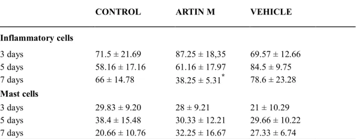
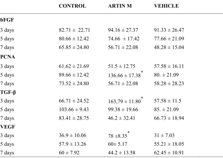
Segundo estudo: Feridas cirúrgicas circulares de 4 mm de diâmetro foram criados na mucosa palatina de 72 ratos Wistar, divididos aleatoriamente de acordo com o tratamento realizado nas lesões em 3 grupos: C – controle (não tratados), A - Artin M, V - veículo. Após 3, 5 e 7 dias pós-tratamento, 8 animais de cada grupo foram sacrificados e suas hemimaxilas foram divididas para realização da análise histológica; imuno-histoquímica para PCNA, bFGF, VEGF e TGF-β; expressão protéica das citocinas pró-inflamatórias IL-1, IL-6 e TNFα, e dos fatores de crescimento VEGF e TGF-β por ELISA. A análise histológica mostrou que no grupo A houve estimulação na regeneração e organização do epitélio, na produção de fibras colágenas e maturação do tecido de granulação e diminuição estatisticamente significante no infiltrado inflamatório no dia 7 quando comparado aos outros grupos (p<0,05). A análise imuno-histoquímica mostrou maior expressão de TGF-β e VEGF no dia 3 (p<0,05) e maior proliferação celular no dia 5 (p<0,05) no grupo A. Além disso, maior expressão protéica de TGF-β e VEGF nas lesões tratadas pelo Artin M foi confirmada pela ELISA no dia 3 (p<0,05). Diante dos resultados obtidos, concluiu-se que a aplicação tópica de gel de Artin M promoveu a reparação da mucosa mastigatória de ratos através estimulação na produção de TGF-β e VEGF, proliferação celular, deposição de colágeno e reepitelização.
Kim YJ. Evaluation of Artin M on wound healing in palatal mucosa [Tese de Doutorado]. Araraquara: Faculdade de Odontologia da UNESP; 2011.
ABSTRACT
Artin M, lectin from Artocarpus heterophyllus seeds, has been demonstrated to stimulate recruitment and activation of neutrophil and mast cells. Furthermore, it has been shown to accelerate the process of wound healing on burn injuries and corneal epithelial lesions in rats and rabbits, respectively. The aim of this study was to evaluate the effects of Artin M on wound healing in palatal mucosa in two animal models.
It is well known that cytokines and growth factors released from different cells coordinate the healing process. Therefore, based on previous results we evaluated the effects of topical application of Artin M on wound healing in palatal mucosa of rats.
Study 2: Seventy-two rats were divided into three groups according to one of the treatment assigned: C– Control (non-treated), A- Artin M gel, and V– Vehicle (carboxymethylcellulose 3% gel). A 4 mm full thickness wound was surgically created on the palatal mucosa of each animal. Eight animals per group were sacrificed at 3, 5, and 7 days post-surgery. Maxillary biopsies were harvested and divided to perform histological analysis, imunohistochemical analysis for bFGF, PCNA TGF-β and VEGF and ELISA assay to evaluate levels of IL-1, IL-6, TNFα, TGF-β and VEGF. Histological features showed faster reepithelization in group A, significant decrease in inflammatory cells infiltration by day 7 (p<0.05) and increased collagen fiber formation resulting in faster maturation of granular tissue compared to the other groups. Also, Artin M significantly induced cells proliferation at day 5 (p<0.05). Significantly higher expression of TGF-β and VEGF were observed in group A compared to C at day 3 (p<0.05). In conclusion, the single application of Artin M gel has shown to be effective in the healing of oral mucosa wound in rats by enhancing TGF-β and VEGF release, cell proliferation, reepithelization, collagen deposition and arrangement of collagen fibers. These findings may partially explain potential pharmacological actions of Artin M on promoting wound healing process.
1 INTRODUÇÃO
Na Periodontia e na Implantodontia, as cirurgias mucogengivais têm sido amplamente utilizadas para devolver saúde, função e estética ao paciente. Esses procedimentos têm apresentado alto índice de sucesso devido a boa previsibilidade e prognóstico favorável 46. Dentre eles, as técnicas de enxertos de tecido mole - enxerto gengival livre e enxerto de tecido conjuntivo – são indicados, para reconstrução de áreas de recessão gengival, perda de papila interdental, deficiência no rebordo alveolar, selamento alveolar em implantação imediata e em conjunto com a técnica de regeneração óssea guiada 54,58,78.
Na técnica de enxerto de tecido mole, há a necessidade de uma área doadora, que deve apresentar gengiva ceratinizada em espessura suficiente para possibilitar a remoção do enxerto, o qual será removido e transferido para outra região, mediante perda total de continuidade com a sua área doadora 58. A irrigação sangüínea que viabilizará sua sobrevivência se fará a partir de neoformação vascular proveniente da área receptora 66. As opções de áreas doadoras de enxertos gengivais incluem regiões retromolar, rebordos edêntulos e a mucosa mastigatória do palato, principalmente na região de molares e pré-molares 89. As vantagens da utilização dos enxertos gengivais residem em índices elevados de sucesso, tratando-se de um tecido autógeno e de obtenção relativamente fácil. Entre as desvantagens, estão: a necessidade de segundo sítio cirúrgico, desconforto pós-operatório, cicatrização por segunda intenção, possibilidade de infecção e necrose 55,89.
19
Na medicina, terapias com corticosteróides, antimicrobianos, antiinflamatórios têm sido sugeridas e utilizadas visando acelerar a reparação para tratamento de feridas cutâneas em geral 65. No entanto, em vista dos efeitos adversos e na busca de propriedades adicionais, nos últimos anos, tem se intensificado pesquisas com produtos naturais como alternativas. Vários estudos têm demonstrado efeitos positivos dos derivados de plantas como Centella asiática, Morinda citrifolia, Chamomilla recutita, Aloe vera, entre outros, na reparação de feridas atuando na regulação indireta de mediadores celulares em alguma fase da reparação 6,31,65,74,99.
Reparação
A reparação consiste na sequência de eventos moleculares que são divididos didaticamente em quatro fases: inflamação, proliferação, remodelação e contração, ocorrendo na realidade uma transição gradual e contínua durante o processo 19,30,61.
A fase inflamatória inicia-se logo após a lesão, por um período de um a cinco dias, envolvendo fenômenos vasculares, de coagulação, de migração celular e de liberação de citocinas e fatores de crescimento 19. Inicialmente, na ferida, os vasos lesionados sofrem contração, seguida de agregação plaquetária, resultando na formação de coágulo. No espaço perivascular, as plaquetas extravasadas aderem-se ao colágeno, sofrendo degranulação e liberação de fatores de crescimento como fator de crescimento derivado de plaquetas (PDGF), fator de crescimento de transformação (TGF), fator de crescimento epidermal (EGF), fator de crescimento de fibroblastos (FGF) e fator de crescimento semelhante à insulina (IGF) 30,49,102.
20
Macrófagos residentes, bem como macrófagos derivados de monócitos recém-atraídos para o local, são ativados e desempenham papel essencial na formação e remodelação do tecido, sendo responsáveis pela fagocitose, recrutamento de novas células inflamatórias, ativação celular e angiogênese, atividades que decorrem da liberação de quimiocinas, citocinas e diversos fatores de crescimento, como Interleucina – 8 (IL-8), fator de necrose tumoral (TNF), EGF, PDGF, TGF, FGF 30,49. Os mastócitos também desempenham papel importante na reparação tecidual, principalmente por liberação de fatores contidos em seus grânulos, como triptase, heparina, IL-8, PDGF, TGF, bFGF VEGF que ampliam o recrutamento de neutrófilos, angiogênese, proliferação de fibroblastos e queratinócitos 4,61. A importância dessa fase foi ressaltada por Devalajara et al.,25 em seu estudo, os camundongos knockout para gene CXCR2 apresentaram recrutamento deficiente de neutrófilos e macrófagos para a local da ferida, angiogênese reduzida, e atraso na reparação, demonstrando que modificações da resposta inflamatória alteram o curso da reparação.
Dentre as enzimas proteolíticas envolvidas na reparação tecidual, as metaloproteinases da matriz (MMPs), principais responsáveis pela degradação de componente da matriz extracelular como colágeno, gelatina e elastina atuam em momentos distintos do processo 41. Maior expressão das MMPs foi detectada nos tecidos em reparação 104. Na fase inflamatória, MMPs regulam a atividade de quimiocinas, extravasamento de leucócitos e migração celular 41. Durante a angiogênese, promovem a ruptura da camada basal vascular e a proliferação endotelial, sendo essenciais na reepitelização e no remodelamento do tecido 57.
21
A angiogênese consiste na formação de novos vasos sanguíneos a partir de vasos pré-existentes e é regulada por fatores de crescimento, citocinas pró-angiogênicas e inibidores da neovascularização 57. Nesse processo, em resposta a fatores quimiotáticos liberados devido à descontinuidade da rede vascular, inicia-se a migração de células endoteliais ao local. Em inicia-seguida ocorre a proliferação endotelial, atribuída à ação de fatores de crescimento como FGF, TGF-β, EGF e fator de crescimento vascular endotelial (VEGF). Dentre os fatores de crescimento que participam no restabelecimento da vascularização, o VEGF, liberado por plaquetas e macrófagos, é principal estimulador na proliferação endotelial e formação de novos vasos 12,57. Por fim, ocorrem alinhamento e organização das células para formação do tubo capilar 29.
As repetições sucessivas de lise, síntese e direcionamento formam fibras de colágeno maiores, resultando numa orientação paralela às forças aplicadas e aumentando a sua resistência 29,90. Essa é a fase de maturação e remodelação da ferida. O remodelamento da matriz extracelular é mediado principalmente por colagenases (MMP-1 e MMP-8) e gelatinases (MMP-2 e MMP-9), controladas por IL-1, TNFα, PDGF, TGF, EGF, provenientes de fibroblastos e células epiteliais 41,104. Esse processo inicia-se a partir do terceiro dia e pode perdurar por meses.
A fase de contração de ferida contribui para fechamento de lesões, principalmente quando há reparação por segunda intenção. Esse fenômeno inicia- se aproximadamente no 4o dia e dura por um período de 15 dias. O movimento é centrípeto, isto é, ocorre das margens para o centro, processo esse que é desejável, por reduzir o risco de infecção, no entanto, pode resultar em formação excessiva de tecido cicatricial, trazendo prejuízos estéticos33. TGF-β é considerado o principal modulador dessa fase por induzir a contração da matriz extracelular e dos miofibroblastos. Entretanto, também foi demonstrado ser responsável na formação de queloides 11. Uma lesão pode ser considerada reparada depois de concluída a remodelação e contração da matriz extracelular 39,102.
22
utilização terapêutica nesse processo 1,3,68. A avaliação dos tecidos gengivais humanos demonstrou a expressão de receptores para TGF-β, IGF, PDGF nos tecidos em reparação em relação aos tecidos controle 85. Oda et al. verificaram que a aplicação tópica de bFGF nas feridas no palato de ratos acelerou a formação de tecido de granulação, reepitelização e maturação de colágeno 80. A aplicação de TGF isoladamente ou associada ao PDGF e/ou EGF estimulou maior deposição de colágeno em ratos com alterações nos padrões de reparação induzidas por diabetes ou decorrentes do uso de corticóides 11.
Fatores de crescimento na reparação
Fatores de crescimentos são polipeptídeos biologicamente ativos que, quando se ligam aos receptores específicos, desencadeiam complexa integração dos sinais que coordenam diversos processos celulares, tais como crescimento, diferenciação e∕ou metabolismo, que são fundamentais para a regulação celular durante o processo reparacional 11,12,19,30,49,90.
23
FGF
Dentre os diversos fatores estudados, a família do FGF tem sido relacionada com atividades biológicas importantes na reparação de feridas 72,80, particularmente FGF-2 ou bFGF, produzido principalmente por fibroblastos, macrófagos, mastócitos, células endoteliais, queratinócitos e condrócitos 9. Este fator de crescimento estimula a migração e proliferação de fibroblastos, migração e atividade da colagenase nas células endoteliais, participando na formação do tecido de granulação, reepitelização e remodelamento da ferida 9,79.
Estudos in vitro tem demonstrado que bFGF estimula a quimiotaxia e mitose das células do ligamento periodontal e a proliferação de células endoteliais72. Oda et al. observou que a aplicação tópica de bFGF nas feridas no palato de ratos acelerou a formação de tecido de granulação, reepitelização e maturação de colágeno 80. Em outro estudo semelhante, a aplicação de bFGF recombinante humano estimulou maior proliferação de fibroblastos gengivais de coelho in vitro, demonstrado pelo análise de síntese de DNA, de maneira dose-dependente, além de promover, in vivo, completa reepitelização de úlceras orais em período inferior às lesões não tratadas com este fator 35.
Outra função muito estudada do bFGF também é a capacidade de regular a angiogênese in vivo e in vitro, induzindo a quimiotaxia e proliferação de células endoteliais 79. Gualandris et al.,44 identificaram bFGF como um fator pró-angiogênico, ao isolarem o mesmo da placenta humana, e demonstrarem in vivo sua capacidade de estimular a proliferação celular, avaliada pela síntese de DNA, e a migração de células endoteliais. Num outro estudo, bFGF aumentou a neovascularização entre o enxerto e a área receptora e, consequentemente, elevou a taxa de sucesso dos enxertos 96.
24
para o gene do bFGF apresentaram uma redução de deposição de colágeno e atrasos na reepitelização 40.
TGF-β
Membros da família do TGF-β foram descritos primeiramente na década de 80, englobando TGF-β1-3, proteínas morfogenéticas do osso (BMPs) e ativinas 102. Incialmente o TGF-β foi associado ao recrutamento de células inflamatórias, angiogênese e síntese de colágeno. Portanto, desempenha um papel fundamental na formação de tecido de granulação em feridas em reparação. É produzido principalmente pelas plaquetas, mas também é secretado pelos macrófagos, fibroblastos e queratinócitos. É quimiotático para células inflamatórias e fibroblastos. Estimula a produção de colágeno, a diferenciação de fibroblastos em miofibroblastos, coordenando a reorganização das fibras colágenas e, conseqüentemente, a fase de contração 9. É um aos ligantes dos receptores tirosina cinase, similarmente ao PDGF, FGF e VEGF, uma vez ligado ao receptor TGF-β, ativa a sinalização intracelular pela via de proteínas Smad 9,109.
Estudos têm associado TGF-β com a formação de cicatrizes. Estudos in vitro demonstraram o efeito estimulatório do TGF-β sobre a deposição de colágeno, e a formação de tecido cicatricial e de quelóides. Assim, TGF-β tem sido muito investigado em estudos relacionados à deficiência de reparação de feridas 11,43,90.
Em relação aos tecidos periodontais, Parkar et al.85, avaliaram biópsias de tecidos gengivais humanos, 6 semanas após a realização do procedimento de regeneração tecidual guiada e observaram maior expressão aumentada de receptores de TGF-β nas áreas regeneradas. Esse resultado indica que TGF-β seja importante para a regeneração periodontal, papel coerente com suas atividades de induzir proliferação celular e produção de matriz extracelular, indicando que esse desempenha ação importante em regeneração periodontal.
25
histologicamente maior influxo de células mononucleares e maior número fibroblastos do que o controle, acompanhado de um aumento na síntese de colágeno. Além disso, o tecido tratado mostrou-se 200% mais resistente à tensão do que o não tratado. Com relação ao veículo, o uso de uma suspensão de colágeno bovino foi mais eficaz quando comparado à solução salina, evidenciando a importância de liberação lenta. Isso porque esse procedimento prolonga a exposição local, consequentemente, à ação da substância no local.
Em um estudo com camundongos knockout para o gene TGF-β, a maioria dos animais não sobreviveu durante a embriogênese, indicando a fundamental participação do TGF-β em diversos processos biológicos além do reparo tecidual como desenvolvimento embrionário e função endócrina. Já em outro estudo, camundongos deficientes para o gene Smad3 apresentaram diminuição de infiltrado inflamatório e também diminuição na deposição de colágeno; no entanto, não apresentaram alteração no curso da reparação 67.
VEGF
A revascularização é de grande importância no processo de resolução das feridas. O VEGF é o principal agente pró-angiogênico, responsável pela migração, proliferação e organização das células endoteliais. Além disso, o VEGF atua na formação de estruturas vasculares (sprouting) pelas células endoteliais que consiste em degradação da membrana basal, ativação, proliferação, invasão e formação do lúmen promovendo formação de novos vasos sanguíneos38. O VEGF é uma glicoproteína homodimérica de 45kDa, ligante de heparina. A sua importância no organismo é evidente em animais geneticamente modificados para o gene do VEGF, que sofrem mortes intra-uterinas na ausência de, pelo menos, um dos alelos 16. A potente ação angiogênica do VEGF, atualmente, fez com que ele se tornasse fatores de crescimentos mais estudados. A modulação da angiogênese representa grande interesse não somente no processo reparacional, mas também para doenças isquêmicas e em terapias antitumorais.
26
vezes mais rápida do que em animais não tratados. In vitro, verificou-se que VEGF tem uma ação direta sobre queratinócitos, acelerando sua migração e a re-epitelizacão da ferida.
A produção de VEGF é estimulada por TGF-β, prostaglandina E2 (PGE2) e interleucinas (IL) IL-1α e IL-6, mediadores que são produzidos. Dentre eles expressos por fibroblastos in vitro, sendo o TGF-β um potente indutor, estimulando 6 vezes mais a secreção de VEGF quando comparado a IL-1 ou PDGF 12. Cohen et al.20, investigaram se as citocinas como IL-6 , TNFα e IFN-β envolvidas na inflamação, reparação e progressão tumoral influenciariam na angiogênese. Através da técnica de Northern Blot, demonstraram a expressão de mRNA de VEGF induzida pela IL-6 em cultura de células da carcinoma epidermóide humana, evidenciando a regulação do VEGF via IL-6.
Na tentativa de desenvolver um dispositivo de liberação lenta, Koch et al.56, associaram o VEGF recombinante humano com matrizes de colágeno bovino e observaram que a incorporação do fator de crescimento aumentou a proliferação de células endoteliais do cordão umbilical humano no dia 7 e quando essas matrizes foram implantadas em embriões de galinha mostraram formação de micro vasos, indicando o potencial promissor da associação do potente fator pró-angiogênico com biomateriais.
Artin M
Artin M é uma lectina ligante a D-manose, isolada de sementes de
Artocarpus heterophyllus Lam. Syn. A. integrifolia L.f.. A molécula é tetramérica, constituída de cadeias polipeptídicas não glicosiladas de 16kDa 86,91,100.
27
Artin M induz a migração de neutrófilos por mecanismo haptotático, resultante da interação concomitante com laminina da matriz extracelular e receptores glicosilados, como CXCR2, da superfície celular 93. Essa propriedade depende parcialmente da ativação e degranulação dos mastócitos, também induzidas pela lectina 71. A ligação de Artin M ao receptor CXCR2 de neutrófilos é seguida de sinalização celular por via dependente da proteína G 86.
Ao ligar-se a glicanas associadas a TLR2 da superfície de macrófagos e células dendríticas, Artin M induz a produção de interleucina 12 (IL-12), e promove respostas Th1. Por essa ação imunomoduladora, Artin M tem se mostrado capaz de conferir resistência a camundongos contra patógenos de parasitismo intracelular 84.
Artin M foi utilizada no tratamento tópico de lesões provocadas por calor úmido no dorso de rato promoveu, em 24h a aceleração da cicatrização e redução da necrose86. Num estudo com coelhos, observou-se que a utilização de Artin M provocou aceleração na re-epitelização das lesões por abrasão da córnea em comparação a lesões não tratadas. Segundo os autores, este resultado seria um efeito da ação direta do Artin M na proliferação do epitélio da córnea18.
2 PROPOSIÇÃO
O objetivo geral deste estudo é avaliar os efeitos da Artin M no processo de reparação da mucosa mastigatória bucal, por meio de modelos de estudo in vivo. Sendo assim, os objetivos específicos deste estudo são:
1. Avaliar o efeito da aplicação do gel de Artin M no processo de reparação da mucosa mastigatória em palato de cães, por meio de análise clínica, histológica, imuno-histoquímica e atividade de mieloperoxidase.
3 CAPÍTULOS
3.1 Capítulo 1
Lectin Artin M improves wound healing in palatal mucosa in dogs.
Kim YJ, da Silva VC, Conrado MA, Spolidório LC, Roque-Barreira MCA, Cirelli JA.
32
Lectin Artin M improves wound healing in palatal mucosa in dogs.
Yeon J. Kim*, Vanessa C. da Silva†, Marina C. A. V. Conrado‡, Luis C. Spolidório§, Maria C. A. Roque-Barreira‡, Joni A. Cirelli*.
1.Yeon J. Kim*, M.Sc., UNESP - Univ. Estadual Paulista, School of Dentistry. 2.Vanessa C. da Silva†, Ph.D., UFMA - Federal University of Maranhão, School of Dentistry at São Luís.
3.Marina C. A. V. Conrado‡, M.Sc., School of Medicine of Ribeirao Preto, University of Sao Paulo.
4.Luis C. Spolidório§, Ph.D., UNESP - Univ. Estadual Paulista, School of Dentistry.
5.Maria C. A. Roque-Barreira‡, Ph.D., School of Medicine of Ribeirao Preto, University of Sao Paulo.
6.Joni A. Cirelli*, Ph.D., UNESP - Univ. Estadual Paulista, School of Dentistry
Key words: wound healing, palate, mouth mucosa, periodontics,
glycoproteins.
Correspondence Address:
Joni Augusto Cirelli (corresponding author)
UNESP - Univ. Estadual Paulista, School of Dentistry, Department of Diagnosis and Surgery, Division of Periodontology.
Address: Rua Humaita, 1680, Centro Araraquara, SP, 14801-903, Brazil
Phone Number: 55-16-33016375
* UNESP - Univ. Estadual Paulista, School of Dentistry, Department of Diagnosis and Surgery, Division of Periodontology, Araraquara, São Paulo, Brazil.
† UFMA - Federal University of Maranhão, School of Dentistry at São Luís , Department of Dentistry II, São Luís, MA, Brazil.
‡ Department of Cellular and Molecular Biology, School of Medicine of Ribeirao Preto, University of Sao Paulo, Ribeirao Preto, São Paulo, Brazil.
33
Fax Number: 55-16-33016369
Email: [email protected]
Sources of support: This study was supported by Coordenação de
Aperfeiçoamento de Pessoal de Nível Superior - CAPES and Fundação de Amparo à Pesquisa do Estado de São Paulo – FAPESP, Grant # 2006/60642-2. The authors declare that they have no conflict of interests in this study.
Word Count: 2602
Number of figures: 5
Number tables: 0
Runnig title: Artin M improves wound healing.
34
Abstract
Backgrounds: To evaluate the effects of Artin M on wound healing in palatal mucosa in dogs.
Methods: Three full thickness wounds of 6 mm diameter were surgically created in the hard palate mucosa of twenty dogs. The wounds of each animal were randomly divided into three groups according to one of the treatment assigned: A- Artin M gel, V– Vehicle (carboxymethylcellulose 3% gel) and C– Control. Four animals per group were sacrificed at 2, 4, 7, 14 and 21 days post-surgery. Wounded areas were photographed and scored for macroscopic healing evaluation. Lesion tissues were harvested and used for descriptive histological analysis, PCNA immunohistochemistry and measurement of myeloperoxidase activity.
Results: Clinical analysis showed accelerated wound closure in group A in comparison to other groups in all periods. Histological features showed enhanced reepithelization and collagen fiber formation resulting in faster maturation of granulation tissue in group A compared to other groups, by day 14. Treatment with Artin M significantly induced cells proliferation and increased volumetric density of fibroblasts at day 2 and 4 (p<0.05). Neutrophils infiltration in A group was significantly higher than other groups (p<0.05) at the same time-points.
35
INTRODUCTION
Palatal masticatory mucosa has been extensively used as a tissue donor area for free soft tissue grafts aiming correction of periodontal and peri-implant soft tissue deficits and deformities24,47. Although this surgical technique has high predictability and favorable prognosis, it has the disadvantage of the creation of a second wound which depending on the size of the graft removed, may have a long healing process period, causing pain and discomfort to the patient. Also, the donor area is subject to infection or necrosis, events that can delay even more the healing process 55,89.
The current therapies for palatal wound have been based on mechanical protection to prevent contact injuries and facilitate primary wound closure as monofilament suture and periodontal dressings48,51,88,94,105. The development of new drugs with potential to accelerate the healing process on palatal wound lesions has been investigated 35,80,82,94.
Artin M (previously known as KM+ and Artocarpin) 86 a lectin from
Artocarpus heterophyllus, binds D-mannose and exhibits high specificity for the
trimannoside Man1-3[Man1-6]Man, present in the core of N-glycans 69. Artin M possesses many relevant biological properties. It acts on neutrophils, inducing haptotactic migration and phenotypic and functional changes, which include intracellular tyrosine phosphorylation, shedding of L-selectin, release of
inflammatory mediators, phagocytic and cell-killing activities, and increased expression of TLR237,103. Furthermore, an amplification loop for in vivo Artin M inflammatory activity is provided by induction of mast cell degranulation 71. Artin M stimulates macrophage and dendritic cells to release IL-12, thereby establishing
in vivo Th1 immunity and conferring protection against several intracellular pathogens 21,84,101. Artin M also accelerates wound healing and epithelial tissue regeneration18.
36
MATERIAL AND METHODS
Preparation of Artin M
Artin M was extracted from Artocarpus heterophyllus seeds and purified by sugar affinity chromatography as previously described by Santos-de-Oliveira et al.1994 93. Artin M 0.001% was prepared using carboximetilcelulose 3% as a vehicle gel.
Experimental Animals
This study was approved by the Ethics Committee on Animal Experimentation of the São Paulo State University, School of Dentistry at Araraquara, SP, Brazil. A total of 20 mongrel dogs, weighing between 15 and 20 kg, were used for this study. For all clinical procedures, animals were pre anesthetized using levopremazin chloritateǁ 1mg/kg intramuscularly (IM), and further they were anaesthetized with sodium thiobarbiturate¶ 25 mg/kg intravenously (IV) and kept on intravenous hydration with a 0.9% physiological solution during surgery.A local infiltration of 2% lidocaine with norepinephrine (1:100000) was administered for hemostasis and to diminish pain.
Wound creation and treatment
Scaling and tooth prophylaxis using rubber cup and prophylactic toothpaste were initially performed in the entire mouth. After a week, upper maxillary impressions were taken with a silicone-based material#. A tooth supported acrylic guide was made for each dog to orientate the creation of the wounds during surgical procedures. Three circular full-thickness excisional wound of 6 mm diameter were created by a surgical punch, in the palatal region adjacent to the first and second pre-molars, maintaining a distance of 10 mm between wounds (Fig. 1).
Each palatal wound was randomly distributed to one of the experimental groups according to treatment: A – Artin M gel, V - carboxymethylcellulose 3% gel vehicle and C – coagulum (no treatment). Each dog had a representative
ǁ Neozine, Aventis Pharma Ltd., São Paulo, SP, Brazil ¶ Tiopental, Abbott Laboratories, São Paulo, SP, Brazil
37
sample of each treatment. Afterwards, Transpore tape** was sutured with 4-0 silk†† covering wounds in order to maintain the material and minimize mechanic trauma.
Post- Surgical Procedure
Immediately after surgeries, each animal received 10 ml of hepatic protector‡‡ IV and analgesic ketoprofen§§ 2mg/kg IM for 3 days. The animals were observed daily for any clinical abnormality and they received soft diet through the first week post-surgery.
At each experimental time-point: 2, 4, 7, 14 and 21 days after treatment, four dogs were euthanized by lethal dose of IV injection of sodium thiobarbiturate¶ and maxillary tissues were harvested.
Clinical Analysis
Wound closure and epithelium restoration were assessed visually by means of a picture taken at the end of each experimental time-point, before sacrifice. Clinical evaluation was based on wound healing scores representing stages of absence up to complete filling of wound with or without reepitelization. Images showing empty wound were classified as index 0; partial filling wounds as index 1; complete filling with none visual reepithelialization as index 2; complete filling with partial reepithelialization as index 3 and complete filling and total reepithelialization as index 4 (Fig. 2A). All images were examined by a trained and blind examiner.
Histopathological analysis
Before tissue harvesting, the diameter of each perforation of the tooth supported acrylic guide was increased to 9 mm to guarantee a safe margin around in the excision around the wound. The tissues were removed using a 15C bladeǁ ǁ with the acrylic guide in position. Half of the tissue was stored at -80o C for myeloperoxidase analysis and the other half was processed for histology.
** Transpore, 3M, St. Paul, MN, USA
38
The specimens were fixed in 10% formalin for 48h for routine histological processing and paraffin embedding. Histological serial sections, 4 and 5 µm thick, were cut in the sagital plane through the entire epithelial-connective tissue extension, representing the central and internal portions of the wounds besides external area of the security margin. Haematoxylin and Eosin (HE) and Masson trichrome staining were used.
Volume densities of fibroblasts (Vf) were estimated as previously described 107. The count was performed with the help of a light microscope¶¶, using oil immersion (magnification x1000). A square lattice of 25 points was projected into the microscope ocular, with the use of microvid system##, which connected the microscope to a computer. For each animal, 5 sections were selections and 25 points were counted in each section. Vf was expressed as percentages of the total points counted.
Immunohistochemical Analysis
Immunohistochemical staining against Proliferating Cell Nuclear Antigen (PCNA) was performed on 4 um sections mounted on silanized slides***. Briefly, tissues were deparaffinized and rehydrated. Tissue sections were incubated with 0.5% trypsin for 20 min at 37oC for antigen retrieval and treated with 3% hydrogen peroxidase in methanol for 30 min to block endogenous peroxidase activity. After, sections were incubated with 3% bovine serum albumin in Phosphate buffered saline (PBS)††† for 30 min at room temperature to block nonspecific protein binding, followed by incubation overnight with monoclonal primary anti- PCNA antibody‡‡‡ at 1:100 dilution. Then, sections were incubated with biotinylated immunoglobulin§§§, the reaction product was detected with an avidin biotin peroxidase complex§§§ and diaminobenzidine was used as a chromogen substrate. Following, sections were counterstaining with Carrazi’s
¶¶ Carl Zeiss, São Paulo, SP, Brazil
## Cambridge Instruments, Buffalo, NY, USA
*** DAKO A/S, Glostrup, Denmark
††† Sigma-Aldrich co, St. Louis, MO, USA
‡‡‡Anti-PCNA, MAB4078, Millipore, Billerica, MA, USA
§§§ ABC kit DAKO A/S, Glostrup, Denmark
39
hematoxylin and mounted with permount. Positive and negative controls were used. Wound areas were examined by using light microscopeǁǁǁ (magnification x40). Two representative fields from border and center of the defect from each section were chosen and PCNA immunoreactive cell nuclei were counted by a blind examiner.
Myeloperoxidase activity
The measurement of myeloperoxidase (MPO) activity was performed to quantify neutrophils accumulation in the palatal tissue. Tissue previously stored at -80oC were thawed, suspended and homogenized in 1mL of PBS at 13000 rpm and centrifuged at 3000 rpm for 15 min at 4oC. The material was suspended in 500 uL of 0.5% hexadecyltrimethylammonium bromide in 50 mM potassium phosphate buffer, pH 6.0, to solubilize MPO. After centrifuging at 3000 rpm for 15 min at 4oC, 50 ul of the supernatant from each sample, 50 uL of 3,3-5,5-tetramethylbenzidine in dimethylsulfoxide, 25 uL of 3% hydrogen peroxide (H2O2) were placed in each well at 96 well plate and incubated for 5 min at 37 oC. The reaction was stopped by adding 25uL of H2SO4. The absorbance was measured colorimetrically at 450 nm on spectrophotometer¶¶¶.
Statistical Analysis
All data were analyzed using the GraphPad Prism software 4.0###. One-way ANOVA followed by Tukey’s post hoc test were performed to determine the presence of any significant difference between groups for all the analyses. P-values less than 0.05 were considered statistically significant.
RESULTS
The application of Artin M has shown to be effective in the healing of oral mucosa wound, resulting in significant improvement of all parameters evaluated. Clinical analysis revealed that Artin M accelerated mucosal healing from 2 to 7
ǁǁǁ LEICA microsystem GmbH, Wetzlar, Germany ¶¶¶ Biotek, Winooski, VT, USA
40
days, when compared to other groups, reaching statistical significance from the other groups at 7 days. At this time-point, group A was the only one to present complete filling and total reepithelialization index (4). At 14 days, one lesion from C group still presented index (3) while 100% of A and V groups presented index (4). At 21 days, there were no differences among groups which showed complete filling and total reepithelialization (Fig.2B).
Histological analysis revealed necrotic tissue associated to polymorphonuclear cells (PMNs) and vascular tissues in all groups at 2 days. Artin M group presented discontinuity epithelial tissue while other groups did not present epithelial migration into wounds. At 4 days, discontinuity epithelial tissue was present in all groups, necrotic tissue associated to PMNs were observed superficially while formation of granulation tissue with fibroblasts, collagen fibers, mononuclear inflammatory cells and neovascularization were observed more profoundly. At 7 and 14 days, A group showed fibroplasia characterized by higher collagen fiber content and lower amount of inflammatory infiltrate compared to the other groups. Also, tissue remodeling characterizing advanced maturation of granulation tissue and higher quantity of Vf indicating increased proliferation of fibroblasts was observed in the Artin M group compared to other groups at same time points. At 21 days, all groups presented similar aspects of granulation and epithelial tissues; however A group treated with Artin M still showed increased volumetric density of fibroblasts compared to groups C and V (Fig 3).
Cell proliferation was observed by immunohistochemical analysis labeling PCNA in the nuclei of cells. Artin M treatment increased significantly cell proliferation at 2 and 4 days (Fig.4A and B) in comparison to the other groups at the same time points. Similarly, myeloperoxidase activity assay showed significant increased level of neutrophils in A group compared to the other groups at 2 and 4 days (Fig.5).
41
Wound healing is a complex and dynamic process that consists on interactions of cells and molecules to restore tissue integrity. It is based on three different and overlapping stages: inflammation, proliferation and remodeling19,61.
In this study the single application of Artin M gel has shown to be effective in accelerating the healing process of oral mucosa wound, demonstrated clinically and histologically. Histological sections presented increase in fibroplasia 14 days after treatment with Artin M. It revealed improved granulation tissue maturation consistent with clinical evaluation and PCNA immunostaining that revealed Artin M to increase significantly cell proliferation (Fig.4B). Additionally, our data also demonstrated that Artin M significantly stimulated neutrophils recruitment (Fig.5).
Recruitment of inflammatory cells, particularly neutrophils, to the site of injury is essential for immune activities and the release of growth factors 9,30. Although neutrophils are not the only cells directly involved in early stage of healing, they are the first cells to reach the site and they have potential to activate other cell types by inducing multiple cytokines pathways such as VEGF, 1, IL-6 and IL-8 9,20.
It has been shown that neutrophils deficiency delays wound repair in mice, evidencing important role of those cells in wound healing 25,75. Delays on wound healing were also observed in CXCR2 knockout mice, which presented diminished neutrophils recruitment into the wound sites and reduced neovascularization resulting in healing impairment25. In those animals, neutrophils deficiency was also correlated with lower levels of VEGF and TGFβ, indicating that these growth factors play important role in repair process.
In addition, in contrast to studies reporting a negative effect of neutrophils in uninfected wound healing 27,30, neutrophils are essential to debridement and activation of immune system in contaminated sites such as oral environment which contains abundant amount of microorganisms.
42
more neutrophils were recruited in the early phase of repair in Artin M treated wounds. As these wounds were located in an intra-oral contaminated environment, tissue healing may have been favored by increased neutrophils microbicidal activities such as phagocytosis, lysosome volume, superoxide production and LTB4 secretion by activation of CXCR2 and TLR2 receptors. In addition, neutrophils did not persist longer in the lesion which may be considered a positive condition; otherwise they might delay the healing process.
Recruitment of neutrophils promoted by Artin M has been demonstrated previously in in vivo and in vitro.). Moreover, the high level of neutrophils influx induced by Artin M and the higher expression of PCNA observed in our study are consistent with previous findings demonstrating that topical administration of Artin M was able to stimulate higher expression of PCNA, VEGF, laminin, p63 and c-Met than control treatment at 24h of corneal abrasion in rabbit 18. These results suggest an influence of Artin M on cell proliferation and growth factors production during tissue repair.
Within the limitation of this study, our findings have shown and partially explained potential pharmacological actions of Artin M on promoting wound healing process. Further studies are necessary for a full understanding of the precise mechanism whereby Artin M induces tissue repair.
ACKNOWLEDGEMENTS
This study was supported by Coordenação de Aperfeiçoamento de Pessoal de Nível Superior - CAPES and Fundação de Amparo à Pesquisa do Estado de São Paulo – FAPESP, Grant # 2006/60642-2.We wish to acknowledge Mrs. Ana Claudia G. C. Miranda for technical support in the histological procedures. The authors declare that they have no conflict of interests in this study.
REFERENCES
43
at the palatal donor site. A preliminary study. J Clin Periodontol
2002;29:848-854.
2. Harris RJ. Root coverage of a palatal recession defect: a case report. J
Periodontol 2001;72:1103-1107.
3. Kim JW, Kikkawa DO, Lemke BN. Donor site complications of hard palate mucosal grafting. Ophthal Plast Reconstr Surg 1997;13:36-39. 4. Reiser GM, Bruno JF, Mahan PE, Larkin LH. The subepithelial connective
tissue graft palatal donor site: anatomic considerations for surgeons. Int J
Periodontics Restorative Dent 1996;16:130-137.
5. Saroff SA, Chasens AI, Eisen SF, Levey SH. Free soft tissue autografts. Hemostasis and protection of the palatal donor site with a microfibrillar collagen preparation. J Periodontol 1982;53:425-428.
6. Pini Prato GP, Cortellini P, Agudio G, Clauser C. Human fibrin glue versus sutures in periodontal surgery. J Periodontol 1987;58:426-431. 7. Harris RJ, Sterne JA, Abgrall S, et al. Prognostic importance of anaemia in
HIV type-1-infected patients starting antiretroviral therapy: collaborative analysis of prospective cohort studies. Antivir Ther 2008;13:959-967. 8. Jackson MR. New and potential uses of fibrin sealants as an adjunct to
surgical hemostasis. Am J Surg 2001;182:36S-39S.
9. Tramontina VA, Machado MA, Nogueira Filho Gda R, Kim SH, Vizzioli MR, Toledo S. Effect of bismuth subgallate (local hemostatic agent) on wound healing in rats. Histological and histometric findings. Braz Dent J
2002;13:11-16.
10. Oda Y, Kagami H, Ueda M. Accelerating effects of basic fibroblast growth factor on wound healing of rat palatal mucosa. J Oral Maxillofac Surg 2004;62:73-80.
44
12. Fujisawa K, Miyamoto Y, Nagayama M. Basic fibroblast growth factor and epidermal growth factor reverse impaired ulcer healing of the rabbit oral mucosa. J Oral Pathol Med 2003;32:358-366.
13. Pereira-da-Silva G, Roque-Barreira MC, Van Damme EJ. Artin M: A rational substitution for the names artocarpin and KM+. Immunol Lett
2008;119:114-115.
14. Misquith S, Rani PG, Surolia A. Carbohydrate binding specificity of the B-cell maturation mitogen from Artocarpus integrifolia seeds. J Biol Chem
1994;269:30393-30401.
15. Ganiko L, Martins AR, Freymuller E, Mortara RA, Roque-Barreira MC. Lectin KM+-induced neutrophil haptotaxis involves binding to laminin.
Biochim Biophys Acta 2005;1721:152-163.
16. Toledo KA, Scwartz C, Oliveira AF, et al. Neutrophil activation induced by ArtinM: release of inflammatory mediators and enhancement of effector functions. Immunol Lett 2009;123:14-20.
17. Moreno AN, Jamur MC, Oliver C, Roque-Barreira MC. Mast cell degranulation induced by lectins: effect on neutrophil recruitment. Int
Arch Allergy Immunol 2003;132:221-230.
18. Panunto-Castelo A, Souza MA, Roque-Barreira MC, Silva JS. KM(+), a lectin from Artocarpus integrifolia, induces IL-12 p40 production by macrophages and switches from type 2 to type 1 cell-mediated immunity against Leishmania major antigens, resulting in BALB/c mice resistance to infection. Glycobiology 2001;11:1035-1042.
19. Teixeira CR, Cavassani KA, Gomes RB, et al. Potential of KM+ lectin in immunization against Leishmania amazonensis infection. Vaccine
2006;24:3001-3008.
45
21. PINTO DA SILVA L, Lamberti; FFCLRP/USP, Avenida Bandeirantes, 3900, Prédio Central, 14040-900 Ribeirão Preto (BR)., PANUNTO CASTELO AFU, Avenida Bandeirantes, 3900, Prédio Central, 14040-900 Ribeirão Preto (BR)., DE SOUZA GOLDMAN M, Helena; FFCLRP/USP, Avenida Bandeirantes, 3900, Prédio Central, 14040-900 Ribeirão Preto (BR)., et al. PHARMACEUTICAL COMPOSITION COMPRISING LECTIN. In. vol. WO2004100861. Brazil: CRUZEIRO NEWMARC PATENTES E MARCAS LTDA; Rua Itajobi, 79, Pacaembu, 01246-010 São Paulo (BR). 2004.
22. Chahud F, Ramalho LN, Ramalho FS, Haddad A, Roque-Barreira MC. The lectin KM+ induces corneal epithelial wound healing in rabbits. Int J
Exp Pathol 2009;90:166-173.
23. Santos-de-Oliveira R, Dias-Baruffi M, Thomaz SM, Beltramini LM, Roque-Barreira MC. A neutrophil migration-inducing lectin from Artocarpus integrifolia. J Immunol 1994;153:1798-1807.
24. Agudio G, Nieri M, Rotundo R, Cortellini P, Pini Prato G. Free gingival grafts to increase keratinized tissue: a retrospective long-term evaluation (10 to 25 years) of outcomes. J Periodontol 2008;79:587-594.
25. Weibel ER. Selection of the best method in stereology. J Microsc
1974;100:261-269.
26. Clark RA. Wound repair. Curr Opin Cell Biol 1989;1:1000-1008.
27. Li J, Chen J, Kirsner R. Pathophysiology of acute wound healing. Clin
Dermatol 2007;25:9-18.
28. Eming SA, Krieg T, Davidson JM. Inflammation in wound repair: molecular and cellular mechanisms. J Invest Dermatol 2007;127:514-525. 29. Barrientos S, Stojadinovic O, Golinko MS, Brem H, Tomic-Canic M.
Growth factors and cytokines in wound healing. Wound Repair Regen
2008;16:585-601.
30. Cohen T, Nahari D, Cerem LW, Neufeld G, Levi BZ. Interleukin 6 induces the expression of vascular endothelial growth factor. J Biol Chem
46
31. Devalaraja RM, Nanney LB, Du J, et al. Delayed wound healing in CXCR2 knockout mice. J Invest Dermatol 2000;115:234-244.
32. Nishio N, Okawa Y, Sakurai H, Isobe K. Neutrophil depletion delays wound repair in aged mice. Age (Dordr) 2008;30:11-19.
33. Dovi JV, He LK, DiPietro LA. Accelerated wound closure in neutrophil-depleted mice. J Leukoc Biol 2003;73:448-455.
3.2 Capítulo 2
Stimulation of TGF-β and VEGF expression by lectin Artin M accelerates wound healing in rat oral mucosa.
Kim YJ, Souza JA, Carvalho FC, Gonçalves PCG, Spolidório LC, Roque-Barreira MCA, Cirelli JA.
53
Stimulation of TGF-β and VEGF expression by lectin Artin M accelerates wound healing in rat oral mucosa
Kim YJ, Carvalho FC, Souza JA, Gonçalves PCG, Spolidório LC, Roque-Barreira MCA, Cirelli JA.
ABSTRACT
54
INTRODUCTION
Wound healing process aims to restore integrity and function of injured tissue through three overlapping stages: inflammation, proliferation, and tissue remodeling 13,19,59,104. The healing process can be affected by several factors, including age and health status of the patient, type of injuries, smoking, alcoholism, nutrition or use of medication 5,45,62,68. However, local factors such as presence of microorganisms, blood supply, pH changes or use of topical medications are mostly considered to affect the repair process 28,45. They can influence on speed and quality of tissue formation resulting in a better repair or in impaired healing.
Recently, studies have reported positive effects of products isolated from plants in tissue repair by inducing release of inflammatory mediators, growth factors and extracellular matrix proteins 6,31,53,60,65,95,99. In addition, numerous medications have already been developed from plants sources, including analgesics, treatments for heart condition and anticancer agents, e.g. Aspirine, vinblastine and digitalis, respectively 70.
Artin M is a lectin derived from jackfruit seeds (Artocarpus heterophyllus Lam. Syn. A. integrifolia L.f.) and has a variety of relevant biological properties 91,100. Lectins are carbohydrate-binding proteins that are used to probe the architecture and dynamics of cell surface carbohydrates during cell division and differentiation processes as well as for the isolation and characterization of glycoconjugates on cell surfaces or matrix 97,98. Artin M binds D-mannose and
exhibits high specificity for the trimannoside Man1-3[Man1-6]Man, present in the core of N-glycans 69. The binding to carbohydrate is directly responsible for its biological activity. It acts on neutrophils, inducing haptotactic migration and phenotypic and functional changes, which include intracellular tyrosine phosphorylation, shedding of L-selectin, release of inflammatory mediators,
55
stimulates macrophage and dendritic cells to release IL-12, thereby establishing in vivo Th1 immunity and conferring protection against several intracellular pathogens 21,84,101. Besides, Artin M has been reported to accelerate wound healing in burn injury in rats 86, corneal abrasion in rabbit 18.
Based on those results, the aim of this study was to evaluate the effects of topical application of Artin M gel on wound healing in palatal mucosa in an excision wound model in rats.
MATERIAL AND METHODS
Preparation of Artin M
Artin M, a D-mannose-binding lectin extracted from jackfruit seeds (Artocarpus heterophyllus Lam. Syn. A. integrifolia L.f.) was purified by sugar affinity-chromatography as previously described by Santos de- Oliveira et al.1994 93. 0.001% of Artin M gel was prepared using 3% of carboxymethylcellulose as a vehicle.
Experimental Animals
The present study was approved by the Ethics Committee on Animal Experimentation of the São Paulo State University, School of Dentistry at Araraquara, SP, Brazil. A total of 72 male Wistar rats, average weight 250 g, were kept under controlled temperature (22-25oC) with a 12 h light/dark cycle and received standard laboratory diet and water ad libitum. For all clinical procedures, animals were anesthetized by intraperitonial injection of ketamine (80 mg/kg) and xylazine (16 mg/kg) mixture.
Wound creation and treatment
56
maintain the material and minimize mechanic trauma. After surgeries, the animals were observed daily for any clinical abnormality and feeding behavior throughout the experiment.
Animals were later killed using an anesthetics overdose. Maxillary tissues from the lesion area of eight rats per group were harvested in each experimental time-point: 3, 5 and 7 days after treatment. Half of the tissue was stored at -80o C for protein analysis and the other half was processed for histology.
Histopathological analysis
The specimens were fixed in 10% formalin for 48 h for routine histological processing and paraffin embedding. Histological serial sections, 4 and 5 µm thick were cut in the sagital plane through the entire epithelial-connective tissue extension (Jung Supercut 2065 Leica, Chicago, IL, USA), representing the central and border portions of the wounds besides external area of the security margin. Haematoxylin and Eosin (HE) and Masson trichrome staining were used. Three representative fields from the borders and center of the defect from each specimen were captured using a digital camera (Leica DFC 300 FX, Wetzlar, Germany) on an optical microscope (LEICA microsystem GmbH, Wetzlar, Germany) under 200x magnifications. A 500μm2 grid with 10×5 squares was constructed using image a managing /editor software (Adobe Photoshop CS5, San Jose, CA, USA) and overlaid on the digital images obtained from the histological sections. The region of interest for the analysis was represented by the whole grid, which was positioned in the center of each field. The following structures observed on each intersection point of the grid were recorded to quantitative assessment using a point-counting technique: fibroblastic cells, vascular structures and inflammatory cells 81. The number of mast cells was quantified using toluidine blue pH 2,8 staining. Three representative fields from the borders and center of the defect from each section were chosen and mast cells were counted. All analysis was performed by a blind examiner, who was previously trained.
Immunohistochemical Analysis
57
Denmark). Briefly, tissue sections were deparaffinized and rehydrated. Following, they were incubated with 0.5% trypsin for 20 min at 37o C for antigen retrieval and treated with 3% hydrogen peroxidase in methanol for 30 min to block endogenous peroxidase activity. After, sections were incubated with 3% bovine serum albumin in Phosphate buffered saline (PBS) (Sigma) for 30 min at room temperature to block nonspecific protein binding, followed by incubation overnight with primary antibodies specifics for bFGF (FGF basic Polyclonal Antibody, Abcam, Uk, dilution 1:100), PCNA (PCNA, PC10, Abcam, UK, dilution 1:1500), TGF-β (TGF beta 3 Monoclonal Antibody, Abcam, UK, dilution 1:200) and VEGF (VEGF Policlonal antibody, Abcam, UK, dilution 1:250). Negative control was obtained by the incubation of PBS in substitution for the primary antibody. Then, sections were incubated with biotinylated immunoglobulin (ABC kit DAKO A/S, Denmark), the reaction product was detected with an avidin biotin peroxidase complex (ABC kit DAKO A/S, Denmark) and stained with the chromogen substrate diaminobenzidine (DAKO A/S, Denmark). Following, sections were counterstaining with Carrazi’s hematoxylin and mounted with permount. Wound areas were examined by using light microscope (LEICA microsystem GmbH, Wetzlar, Germany) (magnification x200). Again, three representative fields from the borders and center of the defect from each section were chosen. Further a 500 μm2 square area was constructed using image a managing /editor software (Adobe Photoshop CS5, San Jose, CA, USA) and overlaid on the digital images obtained from the sections. The region of interest for the analysis was represented by the whole grid, which was positioned in the center of each field and immunoreactive cells were counted by a blind and trained examiner.
Analysis of protein expression by ELISA