Methamphetamine Abusers
Gwenael Pottiez1, Teena Jagadish1, Fang Yu2, Scott Letendre3, Ronald Ellis3, Nichole A. Duarte3, Igor Grant3, Howard E. Gendelman1, Howard S. Fox1, Pawel Ciborowski1*
1Department of Pharmacology and Experimental Neuroscience, Omaha, Nebraska, United States of America,2University of Nebraska Medical Center, Omaha, Nebraska, United States of America,3University of California San Diego, San Diego, California, United States of America
Abstract
We wanted to determine whether methamphetamine use affects a subset of plasma proteins in HIV-infected persons. Plasma samples from two visits were identified for subjects from four groups: HIV+, ongoing, persistent METH use; HIV+,
short-term METH abstinent; HIV+, long term METH abstinence; HIV negative, no history of METH use. Among 390 proteins
identified, 28 showed significant changes in expression in the HIV+/persistent METH+group over the two visits, which were
not attributable to HIV itself. These proteins were involved in complement, coagulation pathways and oxidative stress. Continuous METH use is an unstable condition, altering levels of a number of plasma proteins.
Citation:Pottiez G, Jagadish T, Yu F, Letendre S, Ellis R, et al. (2012) Plasma Proteomic Profiling in HIV-1 Infected Methamphetamine Abusers. PLoS ONE 7(2): e31031. doi:10.1371/journal.pone.0031031
Editor:Fatah Kashanchi, George Mason University, United States of America
ReceivedOctober 14, 2011;AcceptedDecember 29, 2011;PublishedFebruary 16, 2012
Copyright:ß2012 Pottiez et al. This is an open-access article distributed under the terms of the Creative Commons Attribution License, which permits unrestricted use, distribution, and reproduction in any medium, provided the original author and source are credited.
Funding:This work was supported by National Institutes of Health grants P01DA026146, P30MH062261, P01NS043985, and P50 DA26306. The funders had no role in study design, data collection and analysis, decision to publish, or preparation of the manuscript.
Competing Interests:The authors have declared that no competing interests exist.
* E-mail: [email protected]
Introduction
Methamphetamine (METH) is an widely abused drug world-wide [1], not only because of its general harmful effects on organ function and physiology, but also because its use can lead to the transmission of HIV and other infectious diseases. This commonly occurs through needle sharing and risky sexual behaviors [2,3]. The use of METH itself has adverse effects on the central nervous system (CNS) with a broad range of clinical outcomes [4]. The acute effects of METH depend on the amount of drug used and route of administration [5]. With longer term use, METH is neurotoxic, damaging dopaminergic nerve terminals, most profoundly in the striatum [6,7]. Long-term dopamine depletion and microglial activation is implicated in METH-induced neurotoxicity through reactive oxygen species, increased release of 5-hydroxytryptamine (5-HT), and loss of function in tryptophan hydroxylase and the serotonin transporter [7]. METH also has untoward actions on the immune system, yielding predominantly inhibitor effects [8,9,10]. Due to its effects on the immune and the central nervous systems, the multiple effects of METH are further complicated in the context of HIV-1 infection. When combined, METH use and HIV infection may have even more devastating effects [2,11,12,13].
Mechanisms underlying cognitive impairment resulting from HIV-1 infection and concurrent METH use are far from understood and diagnosis is currently limited to clinical evaluation and neurocognitive assessments [14]. During the last decade much experimental effort has been directed towards discovery of biomarkers in body fluids to aid in diagnosis and monitoring of neurocognitive disease, with a special emphasis on blood which is the easiest clinical sample to obtain. Protein biomarkers are an especially important target, and blood contains useful and unique proteomic
signatures – proteins and their fragments potentially changed due to pathological changes. However there are several obstacles in progress of discovery new biomarkers [15] such as linking particular biomarkers to pathology of disease and weakness of quantitation, therefore proper validation techniques of individual proteins is a very complex procedure [16]. We aimed to obtain clues to the effects of METH use on the blood plasma proteome. To accomplish this, our study was designed to capture changes in host responses following different durations of METH use or abstinence in HIV-infected individuals. We compared changes between two visits for in four groups of individuals. The first three groups consisted of HIV-infected METH users who either continued abusing METH (HIV+/ persistent METH+), subjects who stopped METH and reported a short period (e.g., less than 12 weeks) of abstinence (HIV+/ST METH abstinent), or subjects who stopped METH and reported a longer period (e.g., at least 12 weeks) of abstinence (HIV+/LT METH abstinent). A fourth group consisted of uninfected controls who did not meet criteria for a METH use disorder and who reported no use of METH (HIV2/METH2). Among numerous methods used in proteomic profiling, we selected an isobaric tag for relative and absolute quantitation (iTRAQ) shotgun proteomic approach. This tandem mass spectrometry (MS/MS) profiling platform enables the quantification of identified peptides which is further translated to protein quantitation. Such large-scale acquisi-tion and analysis can generate statistically significant valuable information with the ability to address existing questions and formulate new hypotheses, as successfully implemented here.
Materials and Methods
Clinical samples
The University of California San Diego (UCSD) and University of Nebraska Medical Center Institutional Review Boards (IRB) approved the studies in which samples were collected, and the UCSD and UNMC IRBs approved their use in this study. Written informed consent was received from all participants. To ascertain whether subjects might have decisional impairment, a Decisional Capacity Assessment was administered. This consisted of an 11-item post-consent quiz with questions regarding the nature of the illness being studied, the voluntary nature of participation, and the ability to withdraw at any time, the consequences of withdrawing, the possible risks and benefits of participation, the procedures involved, the time required, confidentiality, and whom to call with any questions. Assistance with reading or understanding the vocabulary was provided. Inability to achieve a perfect score on the test or other serious indication of questionable capacity resulted in further evaluation by a clinical psychiatrist, psychol-ogist, or neurologist. All subjects in this analysis achieved a perfect score on the test, indicating satisfactory demonstration of capacity. All participants were ambulatory and underwent evaluation in the outpatient research center at UCSD. Eligibility criteria included the ability to undergo a structured clinical interview and to provide details of combined antiretroviral therapy (cART) use and substance use history.
Data were collected according to a standardized protocol of comprehensive neuromedical, neurobehavioral, and laboratory assessments as described previously [17]. Briefly, the following clinical parameters were evaluated using structured interviews and laboratory assessments where appropriate: cART use, including current and past exposure and HIV disease markers (plasma viral load and current and nadir CD4+cell counts). Blood was collected by venipuncture and used to quantify plasma HIV viral loads by a commercial reverse transcription polymerase chain reaction assay (nominal lower quantitation limit, 50 copies/mL [Amplicor; Roche Diagnostic Systems, Indianapolis, Indiana]). Current CD4+cell counts were measured by flow cytometry. Psychiatric diagnoses, including substance use disorders (abuse and depen-dence), were assessed using the computer assisted Composite International Diagnostic Interview (CIDI) [18,19], a fully structured clinical interview that is widely used in psychiatric research [20,21]. The CIDI classified current (within in the last 30 days) and lifetime (.30 days ago) histories of METH use (e.g., abuse and dependence) disorders.
Materials
Ammonium phosphate, a-cyano-4-hydroxycinammic acid (CHCA), trifluoroacetic acid (TFA) were from Sigma Aldrich (St. Louis, MO, USA). HPLC grade water and acetonitrile (ACN) were from Fisher Scientific (Pittsburg, PA, USA). NuPAGE 4– 12% gels were from Invitrogen Corp. (Carlsbad, CA, USA). Ready GelTMBlotting Sandwiches and glycine were from Bio-Rad (Hercules, CA, USA). Super SignalHWest Pico chemiluminescent substrate was from Pierce (Rockford, IL, USA). Mouse monoclo-nal antibodies against vitamin D-binding protein (Abcam, Cam-bridge, MA, USA), and ceruloplasmin (BD Biosciences, San Jose, CA, USA) were used as primaries, for detection of secondary antibodies were polyclonal goat HRP-conjugated anti-mouse IgG (Jackson ImmunoResearch, West Grove, PA, USA).
Sample processing
Samples had been stored at280uC from the time of collection until the time of shipment. Samples were shipped on dry ice from
UCSD to UNMC and, on arrival, remained frozen. HIV was inactivated in all samples by addition of 10ml of 10% Triton-X100 freshly made and 50ml of cocktail of protease inhibitors (Sigma-Aldrich St. Louis, MO) per 1 mL of sample. After 30 minutes samples were aliquoted and those unused were stored at280uC. Two hundred fifty microliters from each sample were filtered using 0.2mm spin filter and immunodepleted using an IgY14 column (Sigma-Aldrich) connected to a liquid chromatography systems HPLC to immunodeplete abundant plasma proteins: albumin,a1 -antitrypsin, IgM, haptoglobin, fibrinogen, a1-acid glycoprotein, apolipoprotein A-I and A-III, Apolipoprotein B, IgG, IgA, transferrin,a2-macroglobulin and complement C3. Flow-through fractions containing unbound proteins were concentrated using Vivaspin 15R (Sartorius, Aubagne, France), according to the manufacturer protocol. Finally, protein concentration was deter-mined with a NanoDrop spectrophotometer (Thermo Scientific, San Jose, CA).
Trypsin digest and peptide labeling
Fifty micrograms of proteins were precipitated with ethanol. Briefly, we added 10 volumes of cold ethanol (200 proof) to each sample. Samples were incubated for 3 h at220uC and centrifuged at 130006gfor 15 minutes at 4uC. Proteins pellets were washed with 1 ml of 70% ethanol and dried in SpeedVac (Thermo-Scientific). Subsequent solutions were provided with iTRAQ reagent kit (Applied Biosystem (ABI), Carlsbad, CA).
Dried proteins were solubilized with dissolution solution and proteins were denatured with 1ml of denaturant reagent. Proteins reduction with reducing reagent was performed for 1 h at 60uC and finally cysteine blocking solution was used to block cysteines during 10 minutes at room temperature.
Trypsin from ABI was reconstituted at 1mg/ml with milliQ water and 10mg of trypsin were added to each sample. Digestion was performed for 16 h at 37uC.After digestion, peptides were labeled with iTRAQ label reagent (ABI) at room temperature for 2 h and 8 samples, labeled with the different isobaric mass tags, were combined in one tube and dried with SpeedVac.
iTRAQ labeled peptides processing
Having subsequent samples collected from the same individuals at various times allowed us to make comparisons between time points as well as across all groups. Because we had four groups and two time points per group, the 8-plex iTRAQ approach to quantify changes was the method of choice. Table 1 shows how samples were ‘‘scrambled’’ into four sets to remove tag labeling bias. Since Group 1, (HIV+/persistent METH+) had only four individuals, four samples were randomly chosen from those available in Groups 2, 3 and 4 (consisting of 8, 12 and 10 identified subjects, respectively). One set of samples utilized all eight iTRAQ tags and each was measured in a separate analytical run.
Samples were clarified using mix education exchange (MCX) column (Water Corp., Milford, MA). Label peptides were solubilized with 1 ml of 0.1% formic acid, passed through the column, then the column was first washed with 5% methanol, 0.1% formic acid solution and then with methanol HPLC grade. Peptides were eluted with 1.4% NH4OH in methanol [22].
Peptide samples were loaded onto strips, splitting them equally between all 12 wells. Separation was performed for 20,000 Vhrs. Collected fractions were cleaned with C-18 spin columns, according to the manufacturer’s protocol. Briefly, fractions were adjusted to 5% acetonitrile (ACN) and 0.5% trifluoric acid (TFA) and passed through activated columns. Columns were washed twice with a 5% ACN, 0.5% TFA solution and peptides were eluted with a 70% ACN, 0.1% TFA solution. Peptides were finally dried and stored at280uC until further use.
Off line LC-MS/MS analysis
Subsequent fractionation of OFFGEL fractions was performed off-line using TempoTM LC system with automatic high density spotting onto MALDI target plates. Peptides were solubilized in 20ml of 0.1% TFA and 10ml of samples were loaded onto a ProteoColTM C18 trap cartridge (MichromBiosources, Auburn, CA) and washed for 20 minutes at 9ml/min. Gradient of
separation was realized using a ratio between two buffers, Water:ACN:TFA (98:2:0.1) (Buffer A) and water:ACN:TFA (2:98:0.1) (Buffer B). To perform the separation, the subsequent gradient was applied, time 0 to 5 min 5 to 15% buffer B, 5– 52 minutes 15–35%, 52–54 minutes 35–80%, 54–64 minutes 80%, 64–65 minutes 80—5% and 65–72 minutes 5%. Peptide elution was monitored with a UV cell at 214 nm absorbance. After the UV cell, eluted peptides were mixed with a matrix solution (1.2 mg/ml in 75% ACN and 0.1% TFA solution) at a flow rate 1ml/minute using a Harvard Apparatus syringe pump. Fractions were spotted every 30 seconds and voltage applied to the plate during spotting was 2.8 kV.
Spotted fractions were submitted for data acquisition on a 4800 MALDI-TOF/TOF mass spectrometer (ABI). MS spectra were acquires from 800 to 3000m/z, for a total of 1000 laser shots by an Nd-YAG laser operating at 355 nm and 200 Hz. Laser intensity remain fixed for all the analyses. MS/MS analyses were performed using 1 kV collision energy with air as CID gas. Metastable ions were suppressed, for a total of 1000 laser shots.
Protein identification and quantification were performed with ProteinPilotTM software using Paragon method. The search parameters were as follows: iTRAQ 8plex (peptide labeled), Methylthio alkylation of Cysteine, NCBI database restricted to Homo sapiens.
Statistical analysis
Protein/peptide abundance measures generated by iTRAQ were first pre-processed using ProteinPilotTMv2.0. The logarithm of the abundance measure was modeled as a function of animal, protein, peptide, tag and experimental condition. Four sets of samples were used to simultaneously collect data from eight
experimental conditions composed of two visits from four groups: Group 1: HIV+/persistent METH+, Group 2: HIV+/ST METH abstinent, Group 3: HIV+, LT METH abstinence, Group 4: HIV-/METH-. All available abundance records (N = 77,159) were normalized using an iterative back fitting procedure to remove the animal, protein and peptide effects. Comparison of the distribution of the protein/peptide normalized (log) abundance measures by experimental condition was restricted to records with ProteinPilotTMassessed ‘‘confidence’’ of at least 50%. The mixed model was fit on the normalized abundances of each protein to adjust for the correlation of the samples collected from the same subject. The abundances of each protein between two visits within the same group were compared, and the relative abundance and the p-values were calculated to evaluate the short/long term effects of METH within each group.
Western blot - validation
Western blot quantitation was done as previously described [22,23]. 1DE was performed using NuPAGE gel system (Invitro-gen Corp.) in 4–12% gradient Bis-Tris gels under reducing conditions and transferred onto PVDF membrane. Chemilumi-nescent signal was detected using SuperSignal West PicoTM Chem-iluminescent Substrate and recorded on Blue Lite X-ray film (ISCBioExpress, Kaysville, UT).
Results
Subjects
All subjects visited the research center at least two times during the study, which allowed us to measure changes in the plasma proteome between visits. Subject demographic and clinical characteristics are summarized in Table 2. Group 1 (HIV+/ persistent METH+) has relatively few subjects due to the rarity of such individuals. METH users entering the study were offered resources for treatment for their drug abuse if requested (Groups 1, 2 and 3). The study took place in an outpatient setting in which sobriety/abstinence was not required, thus effects of discontinu-ation of METH, rather than initidiscontinu-ation of METH, were observed. Blood was drawn during visits and first blood sample was taken at the first visit. This measure is indirect because it attempts to infer the effects of METH from its discontinuation rather than from its administration. On the other hand, it is sufficient given ethical guidelines because ethically the direct effects of transition from a normal state to one of METH abuse cannot be clinically studied.
Protein identification and quantitation
Plasma proteomic profiling is made difficult by the presence of a number of abundant proteins, making examination of less Table 1.iTRAQ tag assignation for 4 biological replicates in experimental design.
Sample set Tag 113 Tag 114 Tag 115 Tag 116 Tag 117 Tag 118 Tag 119 Tag 121
1 *P1, G2, V1 P2, G4, V2 P2, G4, V1 P3, G1, V2 P1, G2, V2 P4, G3, V1
P4, G3, V2 P3, G1, V1
2 P5, G2, V2 P6, G3, V1 P7, G1, V1 P6, G3, V2 P8, G4, V1 P7, G1,
V2
P5, G2, V1 P8, G4, V2
3 P9, G3, V2 P10, G1, V1 P11, G2, V2 P9, G3, V1 P11, G2, V1 P12, G4, V2
P10, G1, V2 P12, G4, V1
4 P13, G4, V2 P14, G2, V1 P15, G1, V2 P13, G4, V1 P16, G3, V1 P14, G2, V2
P15, G1, V1 P16, G3, V2
abundant proteins not feasible. Furthermore the sheer number of proteins and the large dynamic range of their concentrations further complicate analysis. Thus we used multidimensional separation techniques – immunodepletion of the 14 most abundant proteins, separation of peptides into bins by isoelectric points (pI), and then further separation of these pI-based bins by reverse phase liquid chromatography. We chose the iTRAQ method of quantitation, and then eliminating label bias by a scrambling strategy. The data from mass spectrometry analysis were analyzed separately for each sample set and the results of all samples sets were then combined, revealing the identification of a total of 390 proteins. Statistical analysis revealed that 28 proteins were significantly differentially expressed in the persistent METH users; these are listed in Table 3.
Effect of persistent METH use and short and long term abstinence
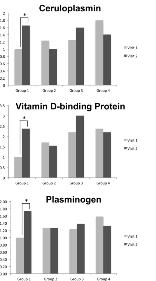
The primary objective of this study was to gain insights into changes in plasma proteome induced by METH in HIV-infected individuals. The study was designed based on participants who successfully stopped using drugs of abuse, therefore observed effects measured such change, rather than change induced directly by drug administration. Changes between visits in all four groups are summarized in Table 4. Group 1 measures changes in proteome in HIV-infected individuals when METH was still used, revealing a large number of changed proteins between visits. Groups 2 (i.e., reported stopping using METH over the course of the study for between two to 12 weeks) and Group 3 (i.e., reporting having at least three months of abstinence) who showed relatively few changes between visits. Similarly Group 4 (e.g., not HIV-infected and did not use METH) did not show many changes, thus in normal individuals the proteome is relatively stable as measured
by our methods. Fig. 1 shows differences between visits for three selected proteins. Statistical significance (increase) was observed only in Group 1.
The paucity of changes in the measured proteomes between visits in Groups 2 and 3 indicated that METH use had limited effect of these proteins at the time of the first visit, since METH abstinence in the context of recent use at the first visit (Group 2) or a history of METH use (Group 3) did not result in changes; as the amount of change in the proteome did not differ from that found in controls who were HIV negative and did not use METH (Group 4). However, many changes were found in Group 1, consisting of the significant increase of twenty one proteins, and a decrease of one protein. This effect of METH is very likely due to a recurring insult from taking METH. This can be either due to the continued use of METH over this period, and/or recent METH use; the positive urinary toxicology used to identify METH use indicates its use within the last three days.
Figure 2 demonstrates that a specific subset of proteins, designated by the arrows, was differentially perturbed in HIV-infected persistent METH users (HIV+/persistent METH+). The majority of these proteins were increased in expression in association with persistent METH use. A smaller subset of proteins showed reduced expression. HIV positive individuals who discontinued METH use (Groups 2 and 3) showed profiles more similar to those of HIV negative METH negative subjects.
Discussion
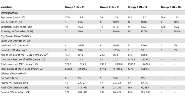
Use of METH is associated with multiple adverse health outcomes such as behavioral risks facilitating sexually transmitted diseases and mental health problems such as psychosis and depression [24,25]. A strong association between METH use and Table 2.Subject demographic, psychiatric, and clinical characteristics.
Variables Group 1 (N = 4) Group 2 (N = 8) Group 3 (N = 12) Group 4 (N = 10)
Demographics:
Age, years (mean, SD) 37.8 68.9 38.1 65.6 43.6 69.0 38.4 69.6
Sex, % male (N, %) 3 75% 8 100% 12 100% 7 70%
Education, years (mean, SD) 10 62.3 13 61.9 14 62.0 12.8 62.3
Ethnicity, % Caucasian, N, %) 2 50% 7 88.0% 10 83.0% 5 50.0%
Psychiatric Characteristics:
METH Use Disorder (N, %)a
Lifetime (.30 days ago) 4 100% 8 100% 12 100% 0 0%
Current (#30 days ago) 2 50% 3 37.5% 0 0% 0 0%
Age of 1st use of METH, years (mean, SD)b 15.7
64.0 24.3 67.5 31.1 630.0 – –
Days since last use of METH (mean, SD) 5.1 63.0 5.0 62.7 1174.2 63395.0 – –
Total days used METH (mean, SD) 527.0 6912.9 735.7 62080.8 1100.3 62624.7 – –
Total grams of METH used (mean, SD) 5569.5 69646.7 677.3 61915.6 477.5 6889.5 – –
Clinical Characteristicsc
:
On CART (N, %) 0 0% 5 63% 6 50% – –
Plasma VL (median, IQR) 4.3 2.4, 5.1 4.4 3.0, 5.2 1.7 1.7, 3.7 – –
Nadir CD4 (median, IQR) 422 119, 953 181 23, 282 168 76, 385 – –
Current CD4 (median, IQR) 579 209, 942 238 76, 427 416 323, 705 – –
Abbreviations: CART = combination antiretroviral therapy; SD = standard deviation; VL = viral load log10 copies/mL; IQR = interquartile range.
aMETH use disorder defined as meeting DSM-IV criteria for METH abuse or dependence. bThese variables are restricted to METH
+subjects.
increased risk of HIV has been substantiated by multiple studies [25,26,27]. Despite treatment, HIV infection is associated with a significant degree of neurocognitive impairment in one out of every seven subjects [28]. In SIV infected rhesus monkeys, METH treatment led to immune changes as well as increased virus in the brain [29]. Therefore, it can be expected that the combined effects of HIV infection and METH use will have not only additive or perhaps synergistic adverse effects on the brain, but also will have a detrimental impact on many physiological functions [30] including the immune system. We postulated that such effects would be reflected by changes in plasma proteome. Alterations in the plasma can affect the brain via communications between the periphery and the CNS, as well as through breach of blood-brain barrier (BBB) that can occur in HIV infection as well as drug abuse.
Interestingly the majority of significant changes were observed between visits in Group 1, those who did not discontinue using METH. Two major pathways were affected. First, was the complement cascade. This system is usually activated by HIV-1 infection alone; here continued METH use leads to further increase of C4A and C5 components. Such an increase likely represents non-specific response to an insult [31,32]. The second
activated system was blood coagulation and up-regulation of plasminogen, fibrinogen and kininogen. It has been shown that one toxic effect of METH use is disseminated intravascular coagulation (DIC, consumptive coagulopathy), which is a pathological activation of coagulation mechanisms [33]. As the small clots consume coagulation proteins and platelets, normal coagulation is disrupted; normal blood flow to organs is disrupted and this can lead to abnormal bleeding [34]. None of the subjects in this analysis had DIC but reported use of METH, at least in the setting of concurrent HIV infection, appears to result in less severe activation of coagulation pathways.
Increased expression of ceruloplasmin and hemopexin can represent a response to modulate increases in oxidative stress resulting from HIV infection and METH use. We expected that in response to METH exposure oxidative stress should be signifi-cantly enhanced and be correlated to increased expression of antioxidant enzymes, similar to our findings in SIV infected monkeys administered a chronic METH regimen [35]. Cerulo-plasmin is a copper-binding glycoprotein oxidizing Fe2+to Fe3+
ferroxidase without releasing radical oxygen species. Its main reported function is transport of iron across the cell membrane; however, several recent reports link this protein to an overall Table 3.Summary of differentially expressed proteins with statistical significance.
Protein names NCBI accession number
UniProtKB accession
number pI MW (kDa)
Alpha 2 macroglobulin variant gi|167017130 A8K2U0 5.8 161.1
Alpha-1-antichymotrypsin gi|1340142 P01011 5.5 49.8
Alpha-2-glycoprotein 1, zinc-binding gi|52790422 P25311 5.7 34.3
Alpha-2-HS-glycoprotein (Fragment) gi|7770227 P02765 6.4 22.7
Alpha2-HS glycoprotein gi|2521983 P022765 5.4 39.4
Antithrombin III gi|179161 P01008 6.3 52.6
Apolipoprotein B-100 precursor gi|28780 P04114 6.6 515.6
C4A variant protein gi|443671 P0C0L4 6.8 193.7
cDNA FLJ55673, highly similar to Complement factor B gi|194384366 B4E1Z4 6.8 140.9
cDNA FLJ56652, highly similar to Hemopexin gi|221044726 B7Z2Q4 6.4 15.7
Ceruloplasmin precursor gi|119599290 P00450 5.5 116.8
Coagulation factor XI precursor gi|4503627 P03951 8.5 70.1
Complement C5 preproprotein gi|38016947 P01031 6.1 188.3
Fibrinogen gamma chain gi|70906437 P02676 5.7 49.5
Group-specific component (vitamin D-binding protein) gi|34785355 P02774 5.3 52.9
Guanine nucleotide regulatory protein gi|404722 Q14344 8.1 44.1
Hemopexin precursor gi|386789 P02790 6.6 51.4
Immunoglobulin kappa light chain V gi|21669423 No UniProtKB number* 6.7 29.3
Inter-alpha globulin inhibitor H2 gi|70778918 P19823 6.4 106.5
Inter-alpha-trypsin inhibitor family heavy chain-related protein gi|221042206 B7Z544 6.2 98.3
Kininogen-1 isoform 2 gi|156231037 P01042 6.3 72
Leucine-rich alpha-2-glycoprotein 1 gi|21707947 P02750 6.5 38.2
Plasminogenisoform 1 precursor gi|4505881 P00747 7.0 90.6
PREDICTED: similar to immunoglobulin lambda-like polypeptide 1 gi|239752604 No UniProtKB number* 8.9 53.3
Prothrombin; coagulation factor II gi|1335344 P00734 5.5 69.3
Putative uncharacterized protein DKFZp686G11190 gi|34365282 Q6MZQ6 8.3 52.0
Recombinant IgG3 heavy chain gi|9857757 No UniProtKB number* 8.4 89.0
Vitamin D-binding protein/group specific component gi|455970 P02774 5.3 52.9
protective response of the host during increase of an oxidative stress. Ceruloplasmin is produced in the liver and our findings suggest that its expression increases during periods of METH use. These changes may remit following cessation of METH use, explaining why we did not observe changes in ceruloplasmin expression in patients after short or long term METH abstinence. In our previous plasma and cerebrospinal fluid (CSF) profiling experiments of HIV-infected individuals we also found cerulo-plasmin to be differentially expressed [23,36,37]. Interestingly, this protein was down regulated in CSF of subjects with HIV dementia whereas it was up-regulated in plasma, suggesting that the CSF-to-plasma ratio of ceruloplasmin may be an important correlate of HIV-associated neurocognitive impairment.
The observation that changes in the plasma proteome were largely limited to subjects who continued to use METH was also unexpected. Because of this, we could not confidently identify a signature of METH use versus METH abstinence. Continued use of METH is by its nature an unstable condition, and users ‘‘crash’’ after METH binges [38]. Similarly the plasma proteome changes we found in Group 1 were not consistent with adaptive long-term
changes, suggesting that METH use continues to lead to instability in normal physiology such as the complement and coagulation systems even during chronic use. It has to be noted that some of the subjects in Groups 2 and 3 were on treatment for HIV infection whereas none of those in Group 1 were treated for HIV. Our previous studies revealed that the proteome changes rapidly within the first two weeks of infection (acute phase) and comes back to background, especially if cART is implemented [22]. HIV-induced changes in proteome become obvious when viral infection is not well controlled and inflammation is on the rise. Therefore the relationship between METH use and HIV infection and the changes found in Group 1 is likely complex.
During the last decade of clinical and translational proteomics development much emphasis has been placed on technological development with expectations that more sensitive mass spec-trometers and multi-level pre-fractionations will lead us to discovery of multiple new diagnostic biomarkers [39,40]. Howev-er, the reality showed that although cataloging hundreds or even thousands of proteins in plasma and CSF has become more and more routine, quantitation and validation techniques are lagging Table 4.Changes in plasma protein expression levels between visits in all groups.1
Names Group 1 p-value Group 2 p-value Group 3 p-value Group 4 p-value
Alpha 2 macroglobulin variant 1.44 ,0.05 21.10 NS 1.35 NS 1.17 NS
Alpha-1-antichymotrypsin 2.09 ,0.001 21.01 NS 1.84 ,0.01 2.01 ,0.01
Alpha-2-glycoprotein 1, zinc-binding 21.12 NS 21.94 ,0.05 1.02 NS 21.82 ,0.05
Alpha-2-HS-glycoprotein (Fragment) 3.86 ,0.001 21.48 NS 1.09 NS 21.01 NS
Alpha2-HS glycoprotein 2.94 ,0.001 21.52 NS 1.14 NS 1.12 NS
Antithrombin III 2.25 ,0.001 1.02 NS 1.43 ,0.05 1.08 NS
Apolipoprotein B-100 precursor 1.20 NS 1.41 NS 1.60 ,0.05 1.36 NS
C4A variant protein 1.44 ,0.005 1.06 NS 21.04 NS 21.27 NS
cDNA FLJ55673, highly similar to Complement factor B 1.33 ,0.05 1.15 NS 1.44 ,0.005 21.32 NS
cDNA FLJ56652, highly similar to Hemopexin 2.21 ,0.001 21.13 NS 1.24 NS 21.15 NS
Ceruloplasmin precursor 1.65 ,0.001 21.19 NS 1.29 NS 21.29 NS
Coagulation factor XI precursor 1.89 ,0.05 21.31 NS 1.42 NS 1.01 NS
Complement C5 preproprotein 2.01 ,0.05 1.30 NS 1.28 NS 1.83 NS
Fibrinogen gamma chain 21.43 NS 21.31 NS 1.57 NS 21.61 NS
Group-specific component (vitamin D-binding protein) 3.05 ,0.001 21.13 NS 1.53 NS 21.03 NS
Guanine nucleotide regulatory protein 22.63 NS 1.27 NS 21.50 NS NS
Hemopexin precursor 1.85 ,0.001 21.13 NS 1.30 NS 21.06 NS
Immunoglobulin kappa light chain V 1.82 ,0.05 21.12 NS 1.06 NS 1.38 NS
Inter-alpha globulin inhibitor H2 1.55 ,0.05 1.11 NS 1.70 ,0.05 1.14 NS
Inter-alpha-trypsin inhibitor family heavy chain-related protein
1.99 ,0.01 21.03 NS 1.52 NS 21.16 NS
Kininogen-1 isoform 2 1.58 ,0.001 1.02 NS 1.07 NS 1.17 NS
Leucine-rich alpha-2-glycoprotein 1 1.58 ,0.05 21.11 NS 1.82 ,0.05 21.03 NS
Plasminogenisoform 1 precursor 1.74 ,0.005 1.00 NS 1.12 NS 21.19 NS
PREDICTED: similar to immunoglobulin lambda-like polypeptide 1
2.57 ,0.01 21.39 NS 1.21 NS 1.18 NS
Prothrombin; coagulation factor II 1.96 ,0.001 1.08 NS 1.20 NS 1.06 NS
Putative uncharacterized protein DKFZp686G11190 2.65 ,0.005 21.33 NS 1.32 NS 1.26 NS
Recombinant IgG3 heavy chain 2.59 ,0.05 21.28 NS 1.36 NS 1.21 NS
Vitamin D-binding protein/group specific component 2.37 ,0.001 21.09 NS 1.37 NS 21.07 NS
NS: p-value is not significant. 1
Figure 1. Quantitative changes in expression of ceruloplasmin, vitamin D-binding protein and plasminogen.Based on iTRAQ ratios, all three proteins showed significant increase from visit one to visit two in Group 1. No changes in other groups of subjects were found.
[15,41]. This makes it more difficult to draw conclusions about how to connect differentially expressed proteins to biology of disease and to judge whether a potential biomarker is specific and directly relevant to the pathology in question [42,43]. Another layer of complexity is correlating genomic and proteomic data into one, synthesizing within a comprehensive and biologically meaningful scheme [44,45]. Much progress has been made in this area during the last decade when genomic and proteomic data from cellular proteins are compared. In the case of secreted proteins, especially those expressed by multiple tissues, such connections are even harder to make [45,46]. Also our knowledge about molecular mechanisms governing CNS and periphery communication is lagging and will require further investigations of BBB function to better interpret changes in proteomes of these two compartments. Considering studies performed so far [ref], there is no clear answer whether we should or should not expect such correlations and what they are indicative of biologically.
Conclusions
Our study is the first report of systematic proteomic profiling of plasma samples, aiming to address the question of the effect of METH use or abstinence using well-defined groups of research subjects. Based on our results, we draw three conclusions for the effect METH use on the plasma proteome in the people living with HIV disease. First, changes in the hosts’ responses to METH use may be short lived or require continued METH use to reach
detectable levels. Second, the effect of METH is reflected by changes in plasma proteins that are linked to oxidative stress and inflammation. Third, METH use perturbs blood coagulation pathways, with upregulation of plasminogen, fibrinogen and kininogen. Consumption of clotting factors in a coagulation cascade may result in an imbalance between pro- and anti-coagulation. This could in turn increase the risk of both ischemic and hemorrhagic end-organ disease. Recent epidemiological studies demonstrated substantial increases in intracerebral hem-orrhage and myocardial infarction in young adults abusing METH [47], thus our work may provide insight into such complications of METH use, whether in the setting of HIV infection or in its absence.
Acknowledgments
We would like to thank Ms. Jayme Wiederin and Ms. Robin Taylor at the University of Nebraska Medical Center for help in preparation of this manuscript. We also thank Melinda Wojtkiewicz from the Mass Core Facility at the University of Nebraska Medical Center for providing assistance with mass spectrometry analyses.
Author Contributions
Conceived and designed the experiments: GP TJ FY SL RE NAD IG HEG HSF PC. Performed the experiments: GP TJ PC. Analyzed the data: GP FY NAD IG SL PC. Contributed reagents/materials/analysis tools: IG SL RE PC. Wrote the paper: GP HEG HSF FY NAD PC RE. Provided IRB protocols, clinical evaluations and collection of samples: IG SL RE. Figure 2. Distribution of the Log2fold changes in proteins’ expression in all groups.It is expected that proteins will be distributed around
log2 of 0 ( = 1, no change between visits). We observed two additional peaks in Group 1 indicated by arrowheads; larger peak with log2of 1
corresponding to 2-fold increase and smaller peak with log2of approximately -2 corresponding to 7-fold decrease.
References
1. Degenhardt L, Mathers B, Guarinieri M, Panda S, Phillips B, et al. (2010) Meth/amphetamine use and associated HIV: Implications for global policy and public health. Int J Drug Policy 21: 347–358.
2. Scott JC, Woods SP, Matt GE, Meyer RA, Heaton RK, et al. (2007) Neurocognitive effects of methamphetamine: a critical review and meta-analysis. Neuropsychol Rev 17: 275–297.
3. Letendre S, Paulino AD, Rockenstein E, Adame A, Crews L, et al. (2007) Pathogenesis of hepatitis C virus coinfection in the brains of patients infected with HIV. J Infect Dis 196: 361–370.
4. Krasnova IN, Cadet JL (2009) Methamphetamine toxicity and messengers of death. Brain Res Rev 60: 379–407.
5. Kiyatkin EA, Sharma HS (2009) Acute methamphetamine intoxication brain hyperthermia, blood-brain barrier, brain edema, and morphological cell abnormalities. Int Rev Neurobiol 88: 65–100.
6. Cadet JL, Brannock C, Krasnova IN, Ladenheim B, McCoy MT, et al. (2010) Methamphetamine-induced dopamine-independent alterations in striatal gene expression in the 6-hydroxydopamine hemiparkinsonian rats. PLoS One 5: e15643.
7. Thomas DM, Perez MA, Francescutti-Verbeem DM, Shah MM, Kuhn DM (2010) The role of endogenous serotonin in methamphetamine-induced neurotoxicity to dopamine nerve endings of the striatum. J Neurochem. 8. Saito M, Terada M, Kawata T, Ito H, Shigematsu N, et al. (2008) Effects of
single or repeated administrations of methamphetamine on immune response in mice. Exp Anim 57: 35–43.
9. Talloczy Z, Martinez J, Joset D, Ray Y, Gacser A, et al. (2008) Methamphetamine inhibits antigen processing, presentation, and phagocytosis. PLoS Pathog 4: e28.
10. Potula R, Hawkins BJ, Cenna JM, Fan S, Dykstra H, et al. (2010) Methamphetamine causes mitrochondrial oxidative damage in human T lymphocytes leading to functional impairment. J Immunol 185: 2867–2876. 11. Chana G, Everall IP, Crews L, Langford D, Adame A, et al. (2006) Cognitive
deficits and degeneration of interneurons in HIV+methamphetamine users. Neurology 67: 1486–1489.
12. Cadet JL, Krasnova IN (2007) Interactions of HIV and methamphetamine: cellular and molecular mechanisms of toxicity potentiation. Neurotox Res 12: 181–204.
13. Mahajan SD, Aalinkeel R, Sykes DE, Reynolds JL, Bindukumar B, et al. (2008) Methamphetamine alters blood brain barrier permeability via the modulation of tight junction expression: Implication for HIV-1 neuropathogenesis in the context of drug abuse. Brain Res 1203: 133–148.
14. Zetterberg H, Ruetschi U, Portelius E, Brinkmalm G, Andreasson U, et al. (2008) Clinical proteomics in neurodegenerative disorders. Acta Neurol Scand 118: 1–11.
15. Silberring J, Ciborowski P (2010) Biomarker discovery and clinical proteomics. Trends Analyt Chem 29: 128.
16. Haverland N, Pottiez G, Wiederin J, Ciborowski P (2011) Immunoreactivity of anti-gelsolin antibodies: implications for biomarker validation. J Transl Med 8: 137.
17. Rippeth JD, Heaton RK, Carey CL, Marcotte TD, Moore DJ, et al. (2004) Methamphetamine dependence increases risk of neuropsychological impairment in HIV infected persons. J Int Neuropsychol Soc 10: 1–14.
18. Wittchen HU, Robins LN, Cottler LB, Sartorius N, Burke JD, et al. (1991) Cross-cultural feasibility, reliability and sources of variance of the Composite International Diagnostic Interview (CIDI). The Multicentre WHO/ADAMHA Field Trials. Br J Psychiatry 159: 645–653, 658.
19. Wittchen HU (1994) Reliability and validity studies of the WHO–Composite International Diagnostic Interview (CIDI): a critical review. J Psychiatr Res 28: 57–84.
20. Kessler RC, McGonagle KA, Zhao S, Nelson CB, Hughes M, et al. (1994) Lifetime and 12-month prevalence of DSM-III-R psychiatric disorders in the United States. Results from the National Comorbidity Survey. Arch Gen Psychiatry 51: 8–19.
21. Robins LN, Regier DA (1991) Psychiatric Disorders in America: The Epidemiologic Catchment Area Study. New york: Free Press.
22. Wiederin JL, Donahoe RM, Anderson JR, Yu F, Fox HS, et al. (2010) Plasma proteomic analysis of simian immunodeficiency virus infection of rhesus macaques. J Proteome Res 9: 4721–4731.
23. Rozek W, Ricardo-Dukelow M, Holloway S, Gendelman HE, Wojna V, et al. (2007) Cerebrospinal fluid proteomic profiling of HIV-1-infected patients with cognitive impairment. J Proteome Res 6: 4189–4199.
24. Clark T, Marquez C, Hare CB, John MD, Klausner JD (2010) Methamphet-amine Use, Transmission Risk Behavior and Internet Use Among HIV-Infected Patients in Medical Care, San Francisco, 2008. AIDS Behav.
25. Marshall BD, Werb D (2010) Health outcomes associated with methamphet-amine use among young people: a systematic review. Addiction 105: 991–1002. 26. Peck JA, Shoptaw S, Rotheram-Fuller E, Reback CJ, Bierman B (2005) HIV-associated medical, behavioral, and psychiatric characteristics of treatment-seeking, methamphetamine-dependent men who have sex with men. J Addict Dis 24: 115–132.
27. Semple SJ, Zians J, Grant I, Patterson TL (2006) Sexual risk behavior of HIV-positive methamphetamine-using men who have sex with men: the role of partner serostatus and partner type. Arch Sex Behav 35: 461–471.
28. Heaton RK, Clifford DB, Franklin DR, Jr., Woods SP, Ake C, et al. (2010) HIV-associated neurocognitive disorders persist in the era of potent antiretroviral therapy: CHARTER Study. Neurology 75: 2087–2096.
29. Marcondes MC, Flynn C, Watry DD, Zandonatti M, Fox HS (2010) Methamphetamine increases brain viral load and activates natural killer cells in simian immunodeficiency virus-infected monkeys. Am J Pathol 177: 355–361. 30. Newsome SD, Johnson E, Pardo C, McArthur JC, Nath A (2011) Fulminant encephalopathy with basal ganglia hyperintensities in HIV-infected drug users. Neurology 76: 787–794.
31. Stoiber H, Kacani L, Speth C, Wurzner R, Dierich MP (2001) The supportive role of complement in HIV pathogenesis. Immunol Rev 180: 168–176. 32. Perricone R, Fontana L, de Carolis C, Carini C, Sirianni MC, et al. (1987)
Evidence for activation of complement in patients with AIDS related complex (ARC) and/or lymphoadenopathy syndrome (LAS). Clin Exp Immunol 70: 500–507.
33. Westover AN, Nakonezny PA, Haley RW (2008) Acute myocardial infarction in young adults who abuse amphetamines. Drug Alcohol Depend 96: 49–56. 34. White SR (2002) Amphetamine toxicity. Semin Respir Crit Care Med 23:
27–36.
35. Pendyala G, Trauger SA, Siuzdak G, Fox HS (2010) Quantitative plasma proteomic profiling identifies the vitamin E binding protein afamin as a potential pathogenic factor in SIV induced CNS disease. J Proteome Res 9: 352–358. 36. Wiederin J, Rozek W, Duan F, Ciborowski P (2009) Biomarkers of HIV-1
associated dementia: proteomic investigation of sera. Proteome Sci 7: 8. 37. Rozek W, Horning J, Anderson J, Ciborowski P (2008) Sera proteomic
biomarker profiling in HIV-1 infected subjects with cognitive impairment. Proteomics Clin Appl 2: 1498–1507.
38. Semple SJ, Patterson TL, Grant I (2003) Binge use of methamphetamine among HIV-positive men who have sex with men: pilot data and HIV prevention implications. AIDS Educ Prev 15: 133–147.
39. Imai K, Koshiyama A, Nakata K (2011) Towards clinical proteomics analysis. Biomed Chromatogr 25: 59–64.
40. Neilson KA, Ali NA, Muralidharan S, Mirzaei M, Mariani M, et al. (2011) Less label, more free: Approaches in label-free quantitative mass spectrometry. Proteomics 11: 535–553.
41. Ciborowski P (2009) Biomarkers of HIV-1-associated neurocognitive disorders: challenges of proteomic approaches. Biomark Med 3: 771–785.
42. Dowling P, Clynes M (2011) Conditioned media from cell lines: A complementary model to clinical specimens for the discovery of disease-specific biomarkers. Proteomics 11: 794–804.
43. Raimondo F, Morosi L, Chinello C, Magni F, Pitto M (2011) Advances in membranous vesicle and exosome proteomics improving biological understand-ing and biomarker discovery. Proteomics 11: 709–720.
44. Mertens I, Vandingenen A, Meeusen T, De Loof A, Schoofs L (2004) Postgenomic characterization of G-protein-coupled receptors. Pharmacoge-nomics 5: 657–672.
45. Dunn BK, Jegalian K, Greenwald P (2011) Biomarkers for early detection and as surrogate endpoints in cancer prevention trials: issues and opportunities. Recent Results Cancer Res 188: 21–47.
46. Fournier PE, Raoult D (2011) Prospects for the Future Using Genomics and Proteomics in Clinical Microbiology. Annu Rev Microbiol.