Regulated by GSK-3 in Quiescent Cells
John W. Tullai1, Silvia Tacheva1¤a, Laura J. Owens1, Julie R. Graham2¤b, Geoffrey M. Cooper1*
1Department of Biology, Boston University, Boston, Massachusetts, United States of America,2Program in Molecular Biology, Cell Biology and Biochemistry, Boston University, Boston, Massachusetts, United States of America
Abstract
Background:The protein kinase GSK-3 is constitutively active in quiescent cells in the absence of growth factor signaling. Previously, we identified a set of genes that required GSK-3 to maintain their repression during quiescence. Computational analysis of the upstream sequences of these genes predicted transcription factor binding sites for CREB, NFkB and AP-1. In our previous work, contributions of CREB and NFkB were examined. In the current study, the AP-1 component of the signaling network in quiescent cells was explored.
Methodology/Principal Findings: Using chromatin immunoprecipitation analysis, two AP-1 family members, c-Jun and JunD, bound to predicted upstream regulatory sequences in 8 of the 12 GSK-3-regulated genes. c-Jun was phosphorylated on threonine 239 by GSK-3 in quiescent cells, consistent with previous studies demonstrating inhibition of c-Jun by GSK-3. Inhibition of GSK-3 attenuated this phosphorylation, resulting in the stabilization of c-Jun. The association of c-Jun with its target sequences was increased by growth factor stimulation as well as by direct GSK-3 inhibition. The physiological role for c-Jun was also confirmed by siRNA inhibition of gene induction.
Conclusions/Significance:These results indicate that inhibition of c-Jun by GSK-3 contributes to the repression of growth factor-inducible genes in quiescent cells. Together, AP-1, CREB and NFkB form an integrated transcriptional network that is largely responsible for maintaining repression of target genes downstream of GSK-3 signaling.
Citation:Tullai JW, Tacheva S, Owens LJ, Graham JR, Cooper GM (2011) AP-1 Is a Component of the Transcriptional Network Regulated by GSK-3 in Quiescent Cells. PLoS ONE 6(5): e20150. doi:10.1371/journal.pone.0020150
Editor:Michael Polymenis, Texas A&M University, United States of America ReceivedMarch 16, 2011;AcceptedApril 19, 2011;PublishedMay 25, 2011
Copyright:ß2011 Tullai et al. This is an open-access article distributed under the terms of the Creative Commons Attribution License, which permits unrestricted use, distribution, and reproduction in any medium, provided the original author and source are credited.
Funding:This work was supported by the National Institutes of Health Grant R01 CA18689 (GMC) and by American Cancer Society Grant IRG-72-001-33-IRG (JWT). The funders had no role in study design, data collection and analysis, decision to publish, or preparation of the manuscript.
Competing Interests:The authors have declared that no competing interests exist. * E-mail: [email protected]
¤a Current address: Immune Disease Institute and Program in Cellular and Molecular Medicine, Children’s Hospital, Boston, Massachusetts, United States of America
¤b Current address: Pfizer BioTherapeutics Research, Cambridge, Massachusetts, United States of America
Introduction
The serine/threonine kinase glycogen synthase kinase-3 (GSK-3) is a master regulator of a variety of cellular processes. First characterized as the kinase responsible for phosphorylating and inactivating glycogen synthase, GSK-3 now has recognized roles in controlling cell proliferation, survival and differentiation. Abnormal GSK-3 regulation has been associated with many human diseases including diabetes, heart disease, cancer, Alzhei-mer’s disease and schizophrenia [1,2,3].
3 has two widely expressed mammalian isoforms, GSK-3aand GSK-3b, both of which are subject to regulation by the PI 3-kinase/Akt pathway [4]. GSK-3 has been shown to regulate cell survival and proliferation downstream of PI 3-kinase signaling through phosphorylation of cyclin D1 [5], Mcl-1 [6], and eukaryotic translation initiation factor 2B (eIF2B) [7,8], as well as a variety of transcription factors [1,3]. GSK-3 is also regulated through the Wnt pathway. Wnt signaling results in a decrease in the phosphorylation of b-catenin by GSK-3, causing a corre-sponding increase in the transcriptional activation of b-catenin/ TCF target genes [9].
Unlike most protein kinases, GSK-3 is constitutively active in quiescent cells, and undergoes an inhibitory phosphorylation by Akt (on serine 9 for GSK-3b, and on serine 21 for GSK-3a) in the presence of growth factors [4]. The activity of GSK-3 in quiescent cells suggests that it may actively maintain repression of growth factor-regulated genes in the absence of PI 3-kinase signaling. We have investigated the role of GSK-3 in quiescence by combining global expression profiling and computational analyses to examine gene expression downstream of PI 3-kinase/Akt/GSK-3 signaling [10]. These studies identified a set of twelve immediate early genes whose induction following growth factor stimulation of quiescent T98G human glioblastoma cells was dependent upon PI 3-kinase and which could also be induced by direct inhibition of GSK-3 without growth factor stimulation [10,11]. These genes mainly encoded growth factors and transcription factors involved in cell proliferation, so their repression by GSK-3 presumably contrib-uted to maintenance of the quiescent state of the cell.
mediated by common transcription factors, we examined the upstream sequences of the twelve GSK-3 repressed genes to identify statistically over-represented and evolutionarily conserved transcription factor binding sites. This computational analysis predicted AP-1, as well as CREB and NFkB transcription factors, as potential regulators of these genes downstream of GSK-3 [10,12].
In the present study, we have investigated the role of AP-1 family members in GSK-3 mediated transcriptional regulation. Two AP-1 family members, c-Jun and JunD, bound to predicted upstream regulatory sequences in 8 of the 12 GSK-3-regulated genes. Consistent with previous studies demonstrating inhibition of c-Jun by GSK-3 [13,14], c-Jun was phosphorylated by GSK-3 in quiescent cells. The association of c-Jun with its target sequences was increased by growth factor stimulation as well as by GSK-3 inhibition, and a physiological role for c-Jun was demonstrated by siRNA inhibition of gene induction. These results indicate that inhibition of c-Jun by GSK-3 contributes to the repression of growth factor-regulated genes during quiescence. Moreover, together with previous studies, these findings delineate an integrated transcriptional network in which AP-1, CREB and NFkB play major roles in GSK-3-mediated repression of target genes in quiescent cells.
Results and Discussion
Binding of c-Jun to GSK-3 Target Genes
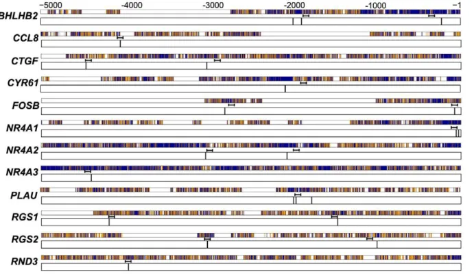
Our previous computational analysis identified a total of 22 conserved, putative AP-1 sites across all 12 of the genes that were induced by inhibition of GSK-3 (Figure 1). The AP-1 family of transcription factors is made up of three Jun family members (c-Jun, JunB, and JunD) and four Fos family members (c-Fos, FosB,
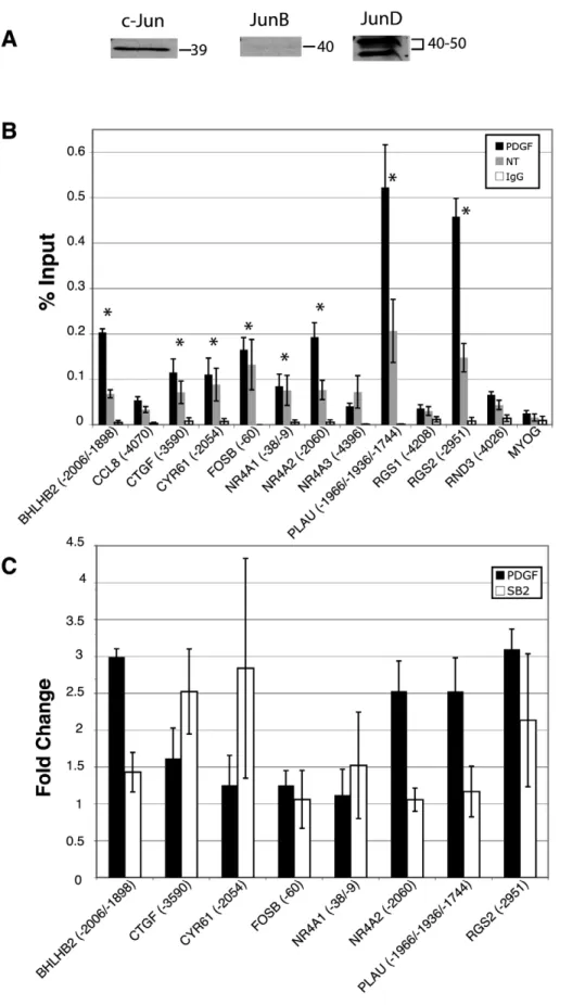
Fra-1 and Fra-2). AP-1 is a dimeric transcription complex that is formed from Jun-Jun family homodimers, or Jun-Fos family heterodimers [15,16]. It is the particular combination of the dimers that determines the transcriptional activity. FosB, c-Fos, and c-Jun are activators, whereas JunD, JunB, Fra-1 and Fra-2 have weaker transactivation domains and can act as repressors by competing for c-Fos, FosB and c-Jun binding. To determine which family members are present in quiescent T98G cells, immunoblots for all members were conducted. As expected, c-Fos, FosB, Fra-1 and Fra-2 were not detected in the quiescent cells, as they require growth factor signaling for their induction [17,18,19,20] (data not shown). c-Jun, JunD and JunB were all detected by immunoblots, although JunB was only weakly detected (Figure 2A). c-Jun, JunD and JunB therefore were pursued in subsequent chromatin immunoprecipitation (ChIP) experiments.
We sought to determine if c-Jun was bound to these predicted sites by ChIP analysis of quiescent T98G cells, as well as following stimulation with platelet-derived growth factor (PDGF). T98G cells were rendered quiescent as described in Materials and Methods, and then stimulated for 30 minutes with PDGF, which was the growth factor used in the initial studies of the PI 3-kinase/ Akt/GSK-3 regulated genes [11], from which the GSK-3 regulated subset was derived [10]. Eight of 12 genes (BHLHB2,
CTGF, CYR61, FOSB, NR4A1, NR4A2, PLAU, and RGS2) demonstrated occupancy/binding by c-Jun (greater than 3-fold as compared to the negative control promoter,MYOG) in both untreated and PDGF treated samples (Figure 2B). PDGF treatment resulted in a greater than 2-fold increase in binding of c-Jun at 4 sites in 4 different genes (BHLHB2,NR4A2,PLAUand
RGS2) as well as a moderate increase in binding (1.6 fold) for
CTGF (Figure 2B and C). We next tested for binding and recruitment of c-Jun following direct inhibition of GSK-3 with the
Figure 1. Conserved AP-1 sites predicted in the upstream regions of GSK-3 regulated genes.Predicted AP-1 binding sites are indicated by vertical black lines in the 5-kb aligned sequences of human and mouse genes [10]. Numeric positions are relative to the transcription start site. Positions of the ChIP PCR amplicons are indicated. Refer to Table S1 for precise positions of predicted sites and of primers. Alignments are indicated as dark blue, match; orange, mismatch; white, aligned to gap.
small molecule inhibitor SB-216763 in the absence of growth factor stimulation. Interestingly, inhibition of GSK-3 also resulted in a greater than 2-fold increase in binding of c-Jun to the upstream sites inCTGF,CYR61andRGS2(Figure 2C), indicating a direct effect of GSK-3 on c-Jun. Overall, the predicted upstream sequences of 8 out of 12 genes were bound by c-Jun and the occupancy of c-Jun at 6 of these genes was increased following either growth factor stimulation or direct inhibition of GSK-3.
Binding of JunD to GSK-3 Target Genes
JunD and JunB AP-1 family members have weaker activation domains and can potentially act as repressors that antagonize or inhibit the binding of activating family members [15,16]. We therefore hypothesized that gene induction through AP-1 may be the result of JunD and/or JunB being displaced by an activating
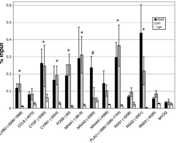
family member such as c-Jun. To test this, we conducted ChIP analysis for both JunB and JunD at the predicted AP-1 sites illustrated in Figure 1. JunB ChIP experiments did not show binding or recruitment greater than that of the negative control promoter, MYOG, or than that of the IgG control (data not shown). In contrast, ChIP analysis for JunD indicated that for many of the genes, JunD was bound to the upstream regions (Figure 3). In all, 8 out of 12 genes showed JunD binding greater than 3-fold as compared to the negative control promoter,MYOG. For seven of the genes showing JunD binding (BHLHB2,CTGF,
CYR61,FOSB,NR4A1,PLAU, andRGS2), the relative binding of the PDGF and untreated (NT) samples were not significantly different, but both higher than that of the negative control,MYOG, and of the control IgG sample. The eighth gene,NR4A2, showed increased binding of JunD upon PDGF stimulation. Binding of
Figure 3. Analysis of JunD binding to predicted AP-1 sites by chromatin immunoprecipitation.Quiescent T98G cells were treated with PDGF for 30 minutes or left untreated (NT), and then chromatin was immunoprecipitated with an anti-JunD antibody or normal rabbit IgG. Only the PDGF-stimulated samples are plotted for normal rabbit IgG. The numbers in parentheses refer to the 59-most position of the putative AP-1 binding site (see Figure 1) relative to the transcription start site. Data are presented as percent input averaged from 4 separate experiments6S.E.MYOG served as a negative control promoter. * indicates greater than 3-fold binding compared toMYOGin both untreated and PDGF treated samples;# indicates greater than 3-fold binding compared toMYOGin the PDGF treated sample only.
doi:10.1371/journal.pone.0020150.g003
transcription start site. When more than one position is listed, this refers to all possible sites that would be detected within the resolution limits of the ChIP PCR amplicon (approximately6250 nucleotides). For those genes with multiple putative AP-1 sites, only one representative site (if all were negative for a given gene) or that site which indicated binding are shown for clarity. For a complete list of all tested sites, see Table S1. Data are presented as percent input averaged from 4 separate experiments6S.E.MYOGserved as a negative control promoter. Asterisks indicate greater than 3-fold binding as compared toMYOGin both untreated and PDGF treated samples C. Recruitment of c-Jun upon stimulation with PDGF or direct inhibition of GSK-3 with SB-216763. Quiescent T98G cells were treated with PDGF for 30 minutes or left untreated, or treated with SB-216763 for 1 hour or with DMSO vehicle control. Only those genes that initially showed c-Jun binding (panel A) are shown. Data are presented as fold change over untreated (for PDGF) or fold change over DMSO vehicle control (for SB-216763), and are averaged from 4 separate experiments6S.E. No significant change was observed in the normal rabbit IgG orMYOGsamples (not shown).
JunD to these 8 genes corresponded to 100% overlap with those sites to which we demonstrated c-Jun binding (see Figure 2B).
These results suggested that JunD was occupying the same AP-1 sites as c-Jun, but with the exception ofNR4A2, was not changing in response to growth factor stimulation. This may reflect the presence of c-Jun-JunD heterodimers at some or all of these AP-1 binding sites. Genes targeted by AP-1 may therefore be activated by recruitment of the activating partner, c-Jun, or by posttrans-lational modifications of the complex to stimulate target gene expression.
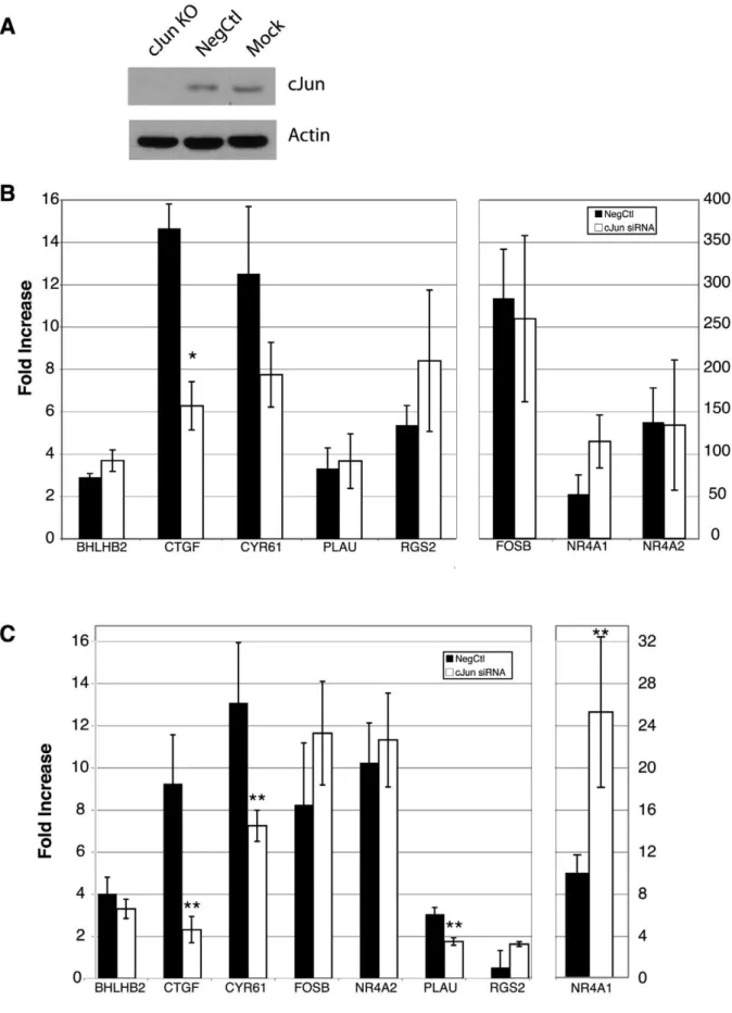
Inhibition of GSK-3 Leads to Dephosphorylation and Stabilization of c-Jun
Since the recruitment of c-Jun to its target sites was stimulated by inhibition of GSK-3, we investigated the effect of GSK-3 inhibition on c-Jun phosphorylation. It has been previously shown that GSK-3 phosphorylates c-Jun on threonine 239 [21]. This phosphorylation event has been shown to block c-Jun DNA binding activity [13] and transactivation [22], and target it for ubiquitination and proteasomal degradation [14]. We therefore assessed the phosphorylation status of c-Jun with the use of phospho-specific antibodies (Figure 4A). Quiescent T98G cells were treated for a time course up to 60 minutes with the GSK-3 inhibitor SB-216763, or the corresponding vehicle control, without growth factor stimulation. This was the treatment time corre-sponding to the gene inductions described previously [10]. As expected, phosphorylation of c-Jun threonine 239 was readily detectable in quiescent cells, consistent with the increased kinase activity of GSK-3 [10,12]. As compared to the untreated sample (NT), phosphorylation at threonine 239 decreased as rapidly as 15 minutes following addition of SB-216763, and declined for the duration of the time course. The 60-minute vehicle control was unchanged.
We next conducted a more extensive time course of GSK-3 inhibition to see if we could observe a stabilization of c-Jun (Figure 4B). Quiescent T98G cells were again treated with SB-216763 in the absence of growth factors. The levels of c-Jun
remained relatively unchanged until 2 hours, when a modest increase in signal was observed as compared to the vehicle control. The 4-hour time point showed a more marked increase in signal as compared to theb-actin loading control, suggesting that the amount of c-Jun protein had increased during the duration of the experiment. This apparent increase in the c-Jun signal upon dephosphorylation of threonine 239 would be consistent with a stabilization of the protein previously described [14], and with the corresponding gene activation observed in the present study. Taken together, GSK-3 inhibition caused the rapid dephosphorylation of c-Jun threonine 239 (allowing DNA binding and transactivation activity), and subsequent stabilization of the protein.
Effect of c-Jun siRNA on the Induction of GSK-3-regulated genes
To determine whether c-Jun is required for the induction of the GSK-3 regulated genes, RNA interference experiments were performed. Transfection of a specific c-Jun siRNA for 24 hours, followed by 48 hours of serum starvation resulted in a knockdown of greater than.90% in T98G cells (Figure 5A). At the end of the 48-hour serum starvation, the effect of the c-Jun knockdown was tested for both the PDGF and SB-216763 induction of the 8 genes for which we demonstrated c-Jun binding. Treatment with c-Jun siRNA decreased the induction ofCTGFby PDGF greater than two-fold (p,0.01) (Figure 5B). The induction CYR61 by PDGF was also decreased nearly two-fold in the presence of the c-Jun siRNA, but did not reach statistical significance (p= 0.11) (Figure 5B). RGS2, despite revealing the most dramatic c-Jun recruitment in the ChIP assays upon PDGF stimulation (Figure 2B), does not appear to require c-Jun for its PDGF-mediated induction. RNAi against c-Jun significantly blocked the induction of CTGF, CYR61 and PLAU (p,0.05) resulting from inhibition of GSK-3 with SB-216763 (Figure 5C). These experiments directly demonstrated that c-Jun is required for the induction of these three genes following GSK-3 inhibition.
Role of AP-1 in the Transcriptional Network Downstream of GSK-3
Previous studies showed that the GSK-3 regulated genes were also targeted by CREB [10] and NFkB [12]. We therefore sought to determine whether there were synergistic or antagonistic interactions between c-Jun, CREB and NFkB in the transcrip-tional response of these genes to inhibition of GSK-3. It is noteworthy in this regard that knockdown of c-Jun resulted in a greater than 2-fold increase in the induction of NR4A1 upon inhibition of GSK-3 with SB-216763 (Figure 5C). Thus, rather than activating transcription ofNR4A1, c-Jun appears to interfere with the induction of this gene, suggesting an inhibitory role for AP-1 upon NR4A1 transcription. Importantly, NR4A1 is also targeted by both CREB and NFkB, with siRNA knockdowns of either of these factors resulting in a significant inhibition ofNR4A1
induction [10,12]. It is thus noteworthy that AP-1 and CREB bind to similar sites [23], and that the AP-1 binding sites at29 and at
238 (relative to the transcription start site) were also found to be binding sites for CREB [10]. This suggests that c-Jun may suppress induction of NR4A1 by sterically interfering with the ability of CREB to bind at these sites and activate transcription.
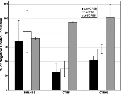
We investigated the possible synergistic roles of c-Jun, NFkB and CREB by performing double knockdown experiments with pairs of siRNAs for all 3 of these transcription factors. Overall, the pairwise knockout results were not significantly different from the effects of siRNA against the individual factors. Representative results are presented in Figure 6. For example, BHLHB2 has
Figure 4. Effect of GSK-3 inhibition on c-Jun phosphorylation and stability.T98G cells were rendered quiescent by serum starvation for 72 hours. Cells were then left untreated (NT), or treated with SB-216763 or vehicle control (DMSO) for the times indicated. Cell extracts were immunoblotted in parallel with anti-phospho threonine 239-c-Jun, pan-c-Jun and b-actin antibodies. Data shown are representative of three separate experiments. The left and right panels of the immunoblot in (B) were taken from the same autoradiography film, and therefore are identical exposure times.
SB-upstream binding sites for c-Jun and NFkB [12], but induction of
BHLB2 was not significantly inhibited by siRNAs against either NFkB [12] or c-Jun (see Figure 5) individually. Likewise, induction of BHLB2 was not significantly inhibited by any of the combinations of siRNAs against these factors (Figure 6).
CTGFandCYR61are examples of genes with c-Jun binding sites whose induction by inhibition of GSK-3 was inhibited by c-Jun siRNA (see Figure 5). Both of these genes have binding sites for NFkB and CYR61 also has a binding site for CREB, but the induction of these genes in response to inhibition of GSK-3 was not significantly inhibited by either NFkB or CREB siRNAs [10,12]. Consistent with the results of siRNAs against these individual factors, induction of these genes was inhibited only by combinations of siRNAs which included c-Jun siRNA, and the combinations had no greater effect than c-Jun siRNA alone (compare Figures 5 and 6). The triple knockdown of these factors likewise did not affect the SB-216763 induction of BHLHB2,
CTGForCYR61(data not shown) as compared to the knockdown of c-Jun alone.
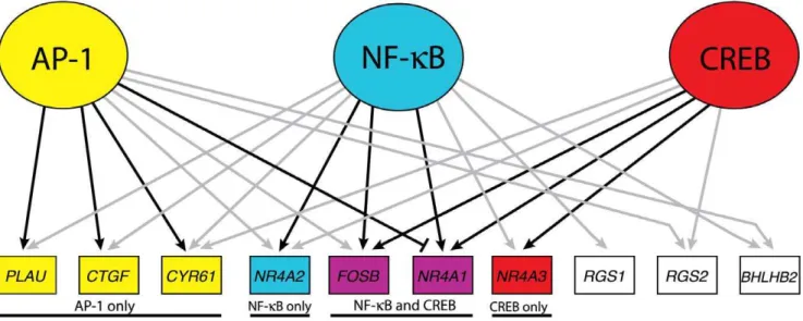
The results of gene regulation downstream of GSK-3 by AP-1 are integrated with the results of our previous studies on CREB [10] and NFkB [12] in Figure 7. These 3 factors comprise a transcriptional network that maintains repression of growth factor-inducible genes in quiescent cells. Of 12 genes that were factor-inducible by inhibition of GSK-3 in quiescent T98G cells, 10 were targeted by at least one of these three transcription factors. Moreover, 4 genes were targeted by all 3 transcription factors and 5 genes by 2 of the 3 factors.
The role of these transcription factors, as assessed by the effects of siRNA knockdowns, varied for the different target genes in the network. The induction of 3 of the 10 genes (RGS1, RGS2 and
BHLBH2) was not significantly affected by siRNAs against AP-1, CREB, or NFkB, either alone or in combination, suggesting that other transcription factors play a dominant role in regulation of these genes. Induction of 5 of the other 7 genes was significantly inhibited by siRNA against 1 of the transcription factors that bound to their upstream sequences, and induction of 2 genes (FOSBand
NR4A1) was inhibited by siRNAs against both CREB and NFkB. This suggests that one or two transcription factors play dominant roles in the induction of different target genes. Interestingly, induction ofNR4A1 was antagonized by AP-1, presumably as a result of competition for CREB binding as noted above.
AP-1 [15,16], NFkB [24,25] and CREB [26,27] are all known to play important roles in the induction of immediate-early genes in response to growth factor stimulation. Furthermore, previous studies have shown that AP-1 [13,22], NFkB [12,28,29,30] and CREB [10,31,32,33,34] all can be inhibited by GSK-3. Our results indicate that these 3 factors comprise a transcriptional network whose inhibition by GSK-3 plays an important role in maintaining repression of growth factor-inducible genes during quiescence.
Materials and Methods
Cell Culture and Treatments
T98G human glioblastoma cells were grown in Minimal Essential Medium (Invitrogen) containing 10% fetal bovine serum
216763 induction. Transfection and starvation were performed as above, and then cells were treated for 1 hour with SB-216763. Data are presented as fold change as compared to vehicle control (DMSO). Data are means for 4 separate experiments6S.E., **p,0.05.
doi:10.1371/journal.pone.0020150.g005
Figure 6. Effect of double siRNAs on gene induction by inhibition of GSK-3.Cells were rendered quiescent and transfected with the indicated combination of siRNAs. A total of 2.5 nM of each siRNA (5 nM total) was used as compared to 5 nM of negative control siRNA. Knockdown efficiencies of c-Jun, p65 and CREB were greater than 90% under these conditions. Data are presented as % of the SB-216763 induction as compared to the induction in the presence of the negative control. All data points are averages of a minimum of n = 3 (except for the p65/CREB combination, which is n = 2),6S.E.
(Hyclone) and penicillin-streptomycin (Invitrogen). For inhibitor and growth factor experiments, cells were switched to serum free media for 72 hours to place the cells in G0arrest [35]. Arrested
cells were then stimulated with 50 ng/mL human PDGF-BB (Peprotech) for 30 minutes (or left untreated), or 5mM SB-216763
(BioMol) or DMSO vehicle for 1 hour.
Chromatin Immunoprecipitation
Chromatin immunoprecipitations were performed as previously described [10] using 5mg of anti-c-Jun (Santa Cruz, sc-1696X ),
5mg of anti-JunD (Santa Cruz, sc-74), or 5mg of normal rabbit
IgG as a control (Santa Cruz, sc-2027). Salmon sperm DNA/ Protein A agarose bead slurry (50%) immunoprecipitates were successively washed in low salt, high salt, and lithium chloride buffers before being washed twice with 16TE (10 mM Tris-HCl,
1 mM EDTA pH 8.0). Immunoprecipitated chromatin was quantified with real-time PCR (see Figure 1 for mapped amplicons and Table S1 for precise locations of sites, primers and primer sequences).MYOGwas used as the negative control for all ChIPs.
Immunoblots
Whole cell lysates were electrophoresed in 10% polyacrylamide gels and transferred to either nitrocellulose or PVDF membranes. Membranes were then incubated with either anti-JunD (Santa Cruz, sc-74), anti-JunB (Santa Cruz, sc-73), anti-phospho threonine 239-c-Jun (Santa Cruz, sc-101720), anti-c-Jun (Santa Cruz, sc-1696) or anti-b actin (Sigma). Immunoblots were then incubated with a horseradish peroxidase secondary antibody (Bio-Rad) followed by visualization using chemiluminescence and exposure to autoradiography film.
Quantitative RT-PCR
Total RNA was isolated using Trizol (Invitrogen) as recom-mended by the manufacturer. RNA was used in quantitative real-time reverse transcription polymerase chain reactions (RT-PCR) as previously described [11], except using a minimum of 0.75mg
of total RNA in the reverse transcription reaction. PCR primers are as previously described [10].
RNA interference
Transfections were performed using pre-designed siRNAs against p65 (Ambion, S11915), CREB (Ambion, 109994), c-Jun (Ambion, s7658), or a non-specific negative control (Ambion, 4390843). Shortly before transfection, 105cells/ml were seeded on 60 mm plates in 4 ml of media containing 10% fetal bovine serum. Transfection reactions containing 5 nM of siRNA, 20ml of
HiPerfect (Qiagen) and 100ml of serum-free media were
incubated for 10 min at room temperature and added drop-wise onto the cells. For the double knockdowns, 2.5 nM of c-Jun siRNA, 2.5 nM of p65 siRNA and 2.5 nM of CREB siRNA were used. Cells were incubated at 37uC, 5% CO2for 24 hrs and then
serum starved for 48 hrs to induce quiescence prior to treatment with PDGF-BB for 30 minutes or SB-2167623 for 1 hour. Starvation times of 48 hrs were used in these experiments to minimize toxicity from the transfection; gene inductions were comparable after either 48 or 72 hrs of starvation [10]. Quiescent cells were then appropriately treated, after which RNA was extracted and analyzed by real-time RT-PCR.
Supporting Information
Table S1 Chromatin Immunoprecipitation Primers for Quantitative Real-Time PCR. Real-time PCR primer se-quences and predicted AP-1 binding site positions are shown. The ‘‘Sites’’ column denotes multiple predicted binding sites in one gene where applicable. The ‘‘Sites Covered’’ column, in turn, refers to those sites that are too close to be distinguished from one another (typically ,250 nucleotides away from each other, the approximate resolution of the ChIP experiment), and therefore both (or in some cases, three sites) are tested with one primer set. * = Experiments with these primer pairs were performed, but the data is not shown. Results were negative.
(XLS)
Figure 7. Model of the GSK-3 transcriptional network in quiescent cells.The AP-1 data has been combined with that of CREB [10] and NFkB [12]. Both grey and black arrows indicate ChIP binding by the given factor to the gene’s upstream sequence. Black arrows indicate that siRNA against the factor blocked induction of the gene in response to SB-216763 treatment greater than two-fold and statistically significant (p,0.05). The blunt-ended edge between AP-1 andNR4A1indicates a greater than two-fold inhibition (p,0.05) of induction in the presence of AP-1.RND3andCCL8have been excluded from the illustration, as no ChIP binding nor transcription factor knockdown data indicated any functional connections with AP-1, NFkB or CREB.
Acknowledgments
The authors wish to thank Sean Sepulveda for technical assistance.
Author Contributions
Conceived and designed the experiments: JWT GMC. Performed the experiments: JWT ST LJO JRG. Analyzed the data: JWT GMC. Wrote the paper: JWT GMC.
References
1. Doble BW, Woodgett JR (2003) GSK-3: tricks of the trade for a multi-tasking kinase. J Cell Sci 116: 1175–1186.
2. Frame S, Cohen P (2001) GSK3 takes centre stage more than 20 years after its discovery. Biochem J 359: 1–16.
3. Jope RS, Johnson GV (2004) The glamour and gloom of glycogen synthase kinase-3. Trends Biochem Sci 29: 95–102.
4. Cross DA, Alessi DR, Cohen P, Andjelkovich M, Hemmings BA (1995) Inhibition of glycogen synthase kinase-3 by insulin mediated by protein kinase B. Nature 378: 785–789.
5. Diehl JA, Cheng M, Roussel MF, Sherr CJ (1998) Glycogen synthase kinase-3b regulates cyclin D1 proteolysis and subcellular localization. Genes Dev 12: 3499–3511.
6. Maurer U, Charvet C, Wagman AS, Dejardin E, Green DR (2006) Glycogen synthase kinase-3 regulates mitochondrial outer membrane permeabilization and apoptosis by destabilization of MCL-1. Mol Cell 21: 749–760.
7. Hardt SE, Tomita H, Katus HA, Sadoshima J (2004) Phosphorylation of eukaryotic translation initiation factor 2B-eby glycogen synthase kinase-3b regulates beta-adrenergic cardiac myocyte hypertrophy. Circ Res 94: 926–935. 8. Pap M, Cooper GM (2002) Role of translation initiation factor 2B in control of cell survival by the phosphatidylinositol 3-kinase/Akt/glycogen synthase kinase 3bsignaling pathway. Mol Cell Biol 22: 578–586.
9. Logan CY, Nusse R (2004) The Wnt signaling pathway in development and disease. Annu Rev Cell Dev Biol 20: 781–810.
10. Tullai JW, Chen J, Schaffer ME, Kamenetsky E, Kasif S, et al. (2007) Glycogen synthase kinase-3 represses cyclic AMP response element-binding protein (CREB)-targeted immediate early genes in quiescent cells. J Biol Chem 282: 9482–9491.
11. Tullai JW, Schaffer ME, Mullenbrock S, Kasif S, Cooper GM (2004) Identification of transcription factor binding sites upstream of human genes regulated by the phosphatidylinositol 3-kinase and MEK/ERK signaling pathways. J Biol Chem 279: 20167–20177.
12. Graham JR, Tullai JW, Cooper GM (2010) GSK-3 represses growth factor-inducible genes by inhibiting NF-kB in quiescent cells. J Biol Chem 285: 4472–4480.
13. Boyle WJ, Smeal T, Defize LH, Angel P, Woodgett JR, et al. (1991) Activation of protein kinase C decreases phosphorylation of c-Jun at sites that negatively regulate its DNA-binding activity. Cell 64: 573–584.
14. Wei W, Jin J, Schlisio S, Harper JW, Kaelin WG, Jr. (2005) The v-Jun point mutation allows c-Jun to escape GSK3-dependent recognition and destruction by the Fbw7 ubiquitin ligase. Cancer Cell 8: 25–33.
15. Chinenov Y, Kerppola TK (2001) Close encounters of many kinds: Fos-Jun interactions that mediate transcription regulatory specificity. Oncogene 20: 2438–2452.
16. Eferl R, Wagner EF (2003) AP-1: a double-edged sword in tumorigenesis. Nat Rev Cancer 3: 859–868.
17. Cohen DR, Curran T (1988) fra-1: a serum-inducible, cellular immediate-early gene that encodes a fos-related antigen. Mol Cell Biol 8: 2063–2069.
18. Kovary K, Bravo R (1991) Expression of different Jun and Fos proteins during the G0-to-G1 transition in mouse fibroblasts: in vitro and in vivo associations. Mol Cell Biol 11: 2451–2459.
19. Muller R, Bravo R, Burckhardt J, Curran T (1984) Induction of c-fos gene and protein by growth factors precedes activation of c-myc. Nature 312: 716–720. 20. Nishina H, Sato H, Suzuki T, Sato M, Iba H (1990) Isolation and
characterization of fra-2, an additional member of the fos gene family. Proc Natl Acad Sci U S A 87: 3619–3623.
21. Morton S, Davis RJ, McLaren A, Cohen P (2003) A reinvestigation of the multisite phosphorylation of the transcription factor c-Jun. Embo J 22: 3876–3886.
22. Nikolakaki E, Coffer PJ, Hemelsoet R, Woodgett JR, Defize LH (1993) Glycogen synthase kinase 3 phosphorylates Jun family members in vitro and negatively regulates their transactivating potential in intact cells. Oncogene 8: 833–840.
23. van Dam H, Castellazzi M (2001) Distinct roles of Jun : Fos and Jun : ATF dimers in oncogenesis. Oncogene 20: 2453–2464.
24. Ghosh S, Karin M (2002) Missing pieces in the NF-kB puzzle. Cell 109 Suppl: S81–96.
25. Hayden MS, Ghosh S (2008) Shared principles in NF-kB signaling. Cell 132: 344–362.
26. Carlezon WA, Jr., Duman RS, Nestler EJ (2005) The many faces of CREB. Trends Neurosci 28: 436–445.
27. Mayr B, Montminy M (2001) Transcriptional regulation by the phosphoryla-tion-dependent factor CREB. Nat Rev Mol Cell Biol 2: 599–609.
28. Bachelder RE, Yoon SO, Franci C, de Herreros AG, Mercurio AM (2005) Glycogen synthase kinase-3 is an endogenous inhibitor of Snail transcription: implications for the epithelial-mesenchymal transition. J Cell Biol 168: 29–33. 29. Bournat JC, Brown AM, Soler AP (2000) Wnt-1 dependent activation of the
survival factor NF-kB in PC12 cells. J Neurosci Res 61: 21–32.
30. Sanchez JF, Sniderhan LF, Williamson AL, Fan S, Chakraborty-Sett S, et al. (2003) Glycogen synthase kinase 3b-mediated apoptosis of primary cortical astrocytes involves inhibition of nuclear factorkB signaling. Mol Cell Biol 23: 4649–4662.
31. Bullock BP, Habener JF (1998) Phosphorylation of the cAMP response element binding protein CREB by cAMP-dependent protein kinase A and glycogen synthase kinase-3 alters DNA-binding affinity, conformation, and increases net charge. Biochemistry 37: 3795–3809.
32. Grimes CA, Jope RS (2001) CREB DNA binding activity is inhibited by glycogen synthase kinase-3b and facilitated by lithium. J Neurochem 78: 1219–1232.
33. Liang MH, Chuang DM (2006) Differential roles of glycogen synthase kinase-3 isoforms in the regulation of transcriptional activation. J Biol Chem 281: 30479–30484.
34. Martin M, Rehani K, Jope RS, Michalek SM (2005) Toll-like receptor-mediated cytokine production is differentially regulated by glycogen synthase kinase 3. Nat Immunol 6: 777–784.