SPINDLE CELL CARCINOMA OF MAXILLA:CASE REPORT OF A
RARE ENTITY AND REVIEW OF LITERATURE
1
Rathy Ravindran Vishnu Mohan Ajish M. Saji
Department of Oral & Maxillofacial Pathology , Azeezia College of Dental Sciences & Research, Kollam, Kerala,
1
Department of Oral & Maxillofacial Surgery, Azeezia College of Dental Sciences & Research, Kollam, Kerala, India
Corresponding Author: Rathy Ravindran, Department of Oral & Maxillofacial Pathology , Azeezia College of Dental Sciences & Research, Kollam, Kerala, India, Ph:09447363459
Abstract
Spindle cell Carcinoma is a rare biphasic neoplasm consisting of epithelial and mesenchymal components and accounts for less than 1% of all tumours of oral region. It is a rare aggressive variant of squamous cell carcinoma which frequently recurs and metastasizes with poor prognosis compared to classical squamous cell carcinoma. The biologic behaviour is comparable to poorly differentiated Squamous Cell Carcinoma. The 5 year disease free survival rate is approximately 30% for all Oral Tumors. The variants of squamous cell carcinoma frequently arise in mucosa of upper aerodigestive tract. The most common site in head and neck region is in larynx and hypopharynx; the oral cavity being rarely affected. This biphasic malignant neoplasm often assumes a sarcomatous appearance and may present diagnostic difficulty. Hence careful histopathologic analysis is warranted. We report a rare case of spindle cell carcinoma in unusual location with immunohistochemical findings and review of the literature.
Key words: Spindle cell carcinoma, Maxilla, Pancytokeratin, Vimentin
Introduction carcinoma that frequently recurs &
metastasizes and hence the importance of
Spindle cell carcinoma is rare variant (1)
correct diagnosis & proper treatment . Only of squamous cell carcinoma. It is important
few cases reported in the medical literature to understand their microscopic peculiarities
hence the purpose of this case report; and a for correct diaganosis & proper treatment. It
(1) review of literature was carried out.
occurs mainly in upper aerodigestive tract .
They may be encountered frequently in Case Report
larynx, nasal cavity, hypopharynx, oral A female patient aged 60 years
cavities, esophagus, trachea, skin & breast. reported to dental OP with complaint of The tumour is designated by variety of terms swelling of gums and mobility of teeth in because of their microscopic pecularity and upper right posterior region since 4-5 months. the various terms include carcinosarcoma, Intra oral examination revealed exophytic pseudosarcoma, sarcomotoid squamous cell mass showing irregular to poorly defined carcinoma, pleomorphic carcinoma & margins with surface ulceration of size 4x3 p o l y p o i d c a r c i n o m a . T h e W H O cm (Fig. 1). Lesion extending anteriorly to classification of tumour has placed this entity upper right canine, Posteriorly to right under malignant epithelial tumours of maxillary tuberosity, medially to midpalatine squamous cell carcinoma and labeled it raphe. On palpation lesion was indurated
(2)
and tender. Routine lab investigation carried spindle cell carcinoma
out & found to be within normal limits. CT Spindle cell carcinoma is an unusual
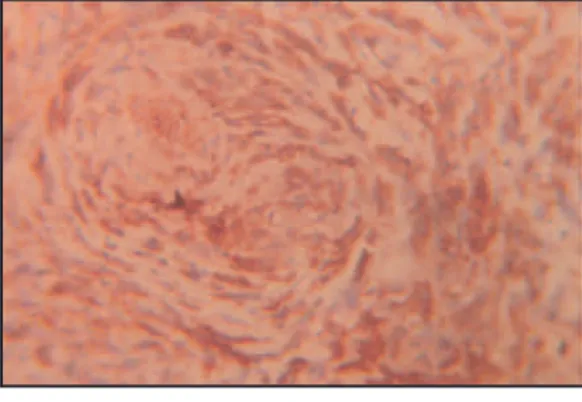
enhancing soft tissue lesion noted in right that spindle cells showed immunoreactivity maxilla and oral cavity. Superiorly it is eroding for pancytokeratin, vimentin (Figure 6 and 7), the right maxilla and floor of the right no immunoreactivity for desmin. Spindle maxillary sinus and invading into sinus cavity. cells showing positivity for pancytokeratin Posteriorly it is extending to lateral pterygoid suggested its epithelial origin and hence a plates & is closely related to right parotid diagnosis of spindle cell carcinoma was made. gland with clear planes of separation.
Anteriorly lesion is seen extending to anterior aspect of right maxilla till canine region. The lesion measures 39x25 mm with density difference of 40 HU causing destruction of right maxilla and alveolar process. Evidence of soft tissue density of fluid level noted in right maxillary sinus. Rest of frontal sinuses and maxillary sinus normal. There is evidence of multiple enlarged and small lymph nodes noted in right submandibular, cervical region along jugular vesels. Features suggestive of malignant process.
Incisional biopsy was taken from right upper molar region. Histopathological examination revealed dysplastic epithelium with islands of neoplastic epithelial cells invading underlying connective tissue & diagnosis of moderately differentiated squamous cell carcinoma was made.
The treatment undertaken was hemimaxillectomy and supraomohyoid l y m p h n o d e n e c k d i s s e c t i o n . O n histopathological examination the given tissue section consisted predominantly of spindle cell component which showed hypercellular and hypocellular area with variable collagen. On high power view anaplasitc pleomorphic cells showing frequent mitosis (Fig. 3 and 4). On serial section few cells with squamoid features seen which constituted a minor component of tumour section (Fig. 5). It presented with diagnostic difficult. On the basis of histopathology differential diagnosis sug gested was poorly differentiated squamous cell carcinoma, malignant mesenchymal neoplasm, spindle cell variant of melanoma.
The sarcomatoid like appearance predominated & hence to ascertain the true nature of spindle cell component I m mu n o h i s t o ch e m i c a l a n a l y s i s wa s undertaken. Immunohistochemistry revealed
Fig. 1 : Exophytic lesion with poorly defined margins showing surface
Fig. 2 : CT PNS & Neck suggestive of malignant process
Discussion
Spindle cell carcinoma account for less
(3)
than 1% of all tumours of oral regions . It is biphasic malignant neoplasm with confusion ever the basic nature of sarcomatoid element whether it is benign or malignant,and mesenchymal or epithelial in origin. The sarcomatoid cells thought to be derived from squamous cells and the epithelial nature of the sarcomatoid component of spindle cell carcinoma was revealed by combination of immuno histochemical staining for keratins and electron microscopic demonstration of tonofilament / or desmesome like
(2) structure .
The age at the time of diagnosis range from 47 to 88 yrs with mean age of 65.7 yrs (2) and shows predilection of male patient . The clinical presentation vary from exophytic ulcerated mass, polypoid nodule infiltrative
(4)
ulcerated lesion . The present case showed exophytic ulcerated mass. Four factors considered to be possibly predisposing for this disease - tobacco, alcohol, poor oral health, previous irradiation to area of tumour. Some author emphasized that radiation or
(6) trauma induces spindle cell carcinoma . The present case was female aged 60years with history of paan chewing.
The tumour shows the presence of two distinct, epithelial derived components and sarcomatoid or dysplastic spindle cell component. The spindle cell component may assume various histological patterns. The most common ones are pleomorphic (malignant histiocytoma like) and spindle cell
(2)
sarcoma (fibroscaroma like) . The spindle cell component may present diagnostic challenge especially when squamous cell component is not obvious. The spindle cell component may resemble lesions ranging from benign reactive lesion like radiation induced granulation tissue to malignant lesions like fibrosarcoma. Three different theories proposed to explain histogenetic nature of spindle cells. First theory states that spindle cells & epithelial cells arise from separate stem cells & hence the name collision tumour. Second theory explain spindle cell Fig. 4 : Anaplastic spindle cells with
hyperchromatism, pleomorphism, prominent mitosis (H &E stain 40 x)
Fig. 5 : High power view showing squamoid feature (H&E stain 40x)
Fig. 6 : Spindle cells positive for pancytokeratin
component as an atypical reactive squamous differentiation usually is seen, proliferation of the stroma & hence the term sometimes at the advancing front or within pseudosarcoma. Finally last theory states cells invaginations at surface where epithelium is
(13,16)
of both spindle & epithelial components have not ulcerated or denuded . In the present same monoclonal origin & dedifferentiation case we could see few squamous cells at the or transformation to spindle cells occurred. advancing front. The tumour may be Several studies revealed that spindle cells have hypocellular or hypercellular with obvious similar characteristic feature of squamous malignant pleomorphism with typical & cells in immunohistochemical, ultrastructural, atypical mitotic figures. The cellular
(5,11,16)
molecular & genetic aspects . The ar rangement may appear storifor m, carcinomatous portion comprise very minor herringbone, fasicular, loose or even
(16)
portion and hence the diagnostic difficulty hypocellular with dense collagen . The cells
encountered in the present case. were pleomorphic with large, oval-round,
vesicular nuclei. Bizarre cells, frequent mitotic Histological studies alone cannot
figures observed in sarcomatous component. explain the spindle cell components. Recent
The absence of keratin pearls, large number IHC studies attempted to explain histogenesis
of mitosis indicate that the clinical behaviour of the spindle cells within these tumours and
might be close to that of poorly differentiated the concept that spindle cell elements are
squamous cell carcinoma. epithelial in origin is now proven by positive
keratin immunostaining and demonstrating The importance of IHC undertaken
of desmosomes and tonofilaments in the in the present case to make a diagnosis (5)
cells strongly supported . Immuno because bulk of the tumour composed of histochemically most sensitive and reliable anaplastic cells and on repeated serial sections epithelial markers for demonstration of could make out only few cells with squamoid epithelial phenotype are keratin and epithelial features. Several studies regarding their membrane antigen which is useful in intermediate filament pattern has been differential diagnosis from other sarcomatous published. The spindle cells invariably lesions. The vimentin positivity reflects that express vimentin reactivity, keratin expression these bizarre fibroblast like cells are being more variable.
carcinoma cells with true mesenchymal Kudo et al found that spindle cell metaplasia. The results indicate that these squamous carcinoma cells expressed wnt-5a cells have acquired mesenchymcal properties and vimentin mRNA at high levels, but did both morpholgically and functionally through not express E-cadherin mRNA. This meteplastic changes and simply correlated to expression pattern was similar to that of the concept of a malignant epithelial cell fibroblast, not of oral squamous carcinoma undergoing alterations, resulting in loss of cells. Their findings suggest that nature of keratin and acquiring vimentin as cytoskeletal spindle cell squamous carcinoma cells may be protein. The double labeling with keratin and similar to mesenchymal cells & the positvity vimentin in spindle cells illustrating the for cytokeratin showed epithelial nature of
(1) versatility of the interemediate filament spindle cell squamous carcinoma cells phenotype. It has been suggested that
According to shibuya et al there was development of spindle cell phenotype
notable difference in immunostaining pattern involves functional loss of genes that control
between squamous cell carcinoma and spindle epithelial differentiation and that conversion
c e l l c a r c i n o m a . T h e p o s i t i v e to spindle morphology in a recessive entry.
immunoreactivity for cytokeratin, á-cat and â P63 has been reported as useful marker for
(2) cat shown everywhere in squamous cell
spindle cell carcinoma .
increases in spindle cell of spindle cell together with their polypoid configuration. (5)
Thus metastases usually contain squamous carcinoma . There was severely reduced
cell carcinoma or squamous cell carcinoma expression of E-cad & the heterogenous
and spindle cell component and rarely only expression of á cat or â cat are responsible for
just spindle cell component. morphologic shift from converntional
squamous cell carcinoma to sarcomatoid Genetically, the sarcomatoid & component in spindle cell carcinoma. epithelial components of spindle cell According to shibuya etal the expression of carcinoma harbor similar mutations and have cadherin- catenin complex was regarded as concordant ploidy. P53 overexpressed in both hall mark of epithelial cell. It is believed that components. The tumour shows LOH dysfunctional cadherin catenin complex frequencies similar to those of poorly causes cells to shift in morphology from differentiated squamous cell carcinoma. A squamoid to a more spindled type and permits specific marker on the short arm of a more infiltrative & diffuse pattern of chromosome 4 was shown to be more
(5)
growth . commonly lost in these tumours than in other
(14) squamous cell carcinoma variants The concept that sarcomatous
portion arises from transformation of Summary
squamous cells was proposed as early as 1900
Spindle cell carcinoma is biphasic (6)
by krompecher and was later supported by
malignant tumour & considered to be variant (11)
other light microscopists. Battifora has of squamous cell carcinoma. The interesting reported that sarcomatous portion of these aspect of this tumour is that it mimics other tumours represent actual mesenchmyal connective tissue sarcomas & spindle cell
(11)
metaplasia . The malignant epithelial cell malignancies at light microscopic level. In the undergoing alteration resulting in loss of past the nature of spindle cell & their keratin and acquiring vimentin as the histog enesis was strongly debated. cytoskeletal protein. I m m u n o h i s t o c h e m i c a l & e l e c t r o n
Spindle cell carcinoma in the oral microscopic studies have contributed to solve cavity and oropharynx is potentially the diagnostic difficulty & their epithelial aggressive and seems to recur easily and to origin. Spindle cell carcinoma is aggressive metastasize. It is difficult to predict biologic tumour & tend to recur easily & metastasize. behaviour in every case, patients whose The prognosis depends on depth of tumour tumour are deeply invasive tend to have poor invasion. It is difficult to predict the biologic prognosis, whereas those with early stage behaviour but tumours deeply invasive tend t u m o u r s u s u a l l y h a v e e x c e l l e n t to have poor prognosis.
(8)
prognosis .Unfortunately 5year disease free References (17)
survival rate is ~30% for all oral tumours.
1. Kudo Y, Ogawa I, Kitagawa M, Kitajima S, Some authors are of the opinion that Siriwardena BSMS, Aobara N et al. wide radical resection alone is the best mode Establishment and characterization of a spindle of treatment while some others are of the cell squamous carcinoma cell line. J oral pathol
opinion that surgery with radiotherapy med 2006;35:479-83.
required. Many authors are of the opinion
2. Parikh N, Desai N. Spindle cell carcinoma of radiotherapy &chemotherapy is ineffective.
the oral cavity: A case report of a rare entity and The treatment option of surgery followed by
review of literature. J academy Adv Dental radiotherapy was found to yield best long
Research 2011;2:31-36. term patient outcome similar to conventional
(13) 3. Jordan RCK, Regezi JA. Oral Spindle Cell
squamous cell carcinoma.
neoplasms: A review of 307 cases. Oral surg Distant metastases and depth of
oralmed Oral pathol Oral Radiol Endod 2003; tumour invasian into underlying structures
4. Romanach MJ, Azevedo RS, Carlos R, 11. Battifora H. Spindle cell carcinoma Almeida OP, Pires FR. Clinico pathological ultrastructural evidence of squamous origin and and immunohistochemical features of oral collagen production by tumour cells. Cancer spindle cell carcinoma. J oral pathol med 1976;37: 2275-82.
2010;39: 335-41. 12. Slootweg, Pieter J, Roholl PJM, Muller H,
5. Shibuya Y, Umeda M, Yokoo S, Komori T. Lubsen H. Spindle cell carcinoma of the oral Spindle cell squamous carcinoma of the maxilla: cavity and larynx. J Cranio- Mac- Fac.surg Report of a case with immunohistochemical 1989;17: 234-36.
Analysis. J oral Maxillofac surg 2000; 13. Thompson LDR, Squamous cell Carcinoma
58:1164-69. variants of the head and neck. Current
6. Munakata R, Cheng JMR, Nakajima T, Diagnostic Pathology 2003; 9:384-96.
Saku T. Spindle Cell carcinoma of the gingiva: 14. Stelow EB, Mills SE. Squamous cell carcinoma report of an autopsy case. J oral pathol med variants of upper aero digestive tracts. Am J
1998; 27:180-4. Clin Pathol 2005;124(suppl 1):S96-S109.
7. Lewis IS, Ritter JH, El-Mofty S. Alternative 15. Koseoglu RD, Sertcelik A, Ayva Y. A rare epithelial markers in sarcomatoid carcinomas of variant of squamous cell carcinoma of the head and neck, lung and bladder – P63, Moc – tongue; spindle cell carcinoma. J of Ankara 31 and TTF -1, Mod Pathol 2005;28:1471- University faculty of Medicine 2005;
58:11-81. 14.
8. Su HH, Chu ST, Hou YY, Chang KP, Chen 16. Oktay M, Kokenek-Unal TD, Ocal B, Saylam CJ. Spindle cell carcinoma of oral cavity and G, Korkmaz MH, Alper M. Spindle cell oropharynx; factors affecting outcome. J chin carcinoma of the tongue: A rare tumor in an
Med Assoc 2006;69:478-83. unusual location. Patholog y Research
9. Chaudhary S, Denny EC, Bastian TS, Singh International Volume 2011:1-6
A. Spindle cell carcinoma: A case report with 17. Randall G, Alonso WA, Ogura JH. Spindle review. Journal of Nepal Dental Association cell carcinoma (pseudosarcoma) of the larynx.
2009;10:68-71. Arch Otolaryngol 1975;101:63-66
10. Ellis GL, Corio RL. Spindle cell Carcinoma of oral cavity :a clinico pathologic assessment of fifty nine cases. Oral surg oral med oral pathol 1980;50:523-33.
Source of Support - Nil Conflict of Interest - None declared
How to cite this article: