U
NIVERSIDADE DE
L
ISBOA
F
ACULDADE DEC
IÊNCIASDEPARTAMENTO DE BIOLOGIA VEGETAL
Optimization of lipase production
in lipase-producing yeasts strains isolated from
olive mil wastewater
Vera Mónica Brito Salgado
Dissertação
Mestrado em Microbiologia Aplicada
U
NIVERSIDADE DE
L
ISBOA
F
ACULDADE DEC
IÊNCIASDEPARTAMENTO DE BIOLOGIA VEGETAL
Optimization of lipase production
in lipase-producing yeasts strains isolated from
olive mil wastewater
Vera Mónica Brito Salgado
Dissertação
Mestrado em Microbiologia Aplicada
Orientadores
Doutora Ana Eusébio
Professora Doutora Maria Manuela Carolino
Optimization of lipase production in
lipase-producing yeasts strains isolated
from olive mil wastewater
Vera Mónica Brito Salgado
2015
This thesis was fully performed at the Department of Bioenergy of Laboratório
Nacional de Energia e Geologia (LNEG) under the direct supervision of Dr. Ana
Eusébio in the scope of the Master in Applied Microbiology of the Faculty of
Sciences of the University of Lisbon.
Prof. Dr. Maria Manuela Carolino was the internal designated supervisor in the
scope of the Master in Applied Microbiology of the Faculty of Sciences of the
University of Lisbon
I like nonsense, it wakes up the brain cells. Fantasy is a necessary ingredient in living, it's a way of looking at life through the wrong end of a telescope. Which is what I do, and that enables you to laugh at life's realities.
Agradecimentos
Gostaria de agradecer a todas as pessoas que estiveram directamente ou indirectamente envolvidas na formação deste trabalho, cujo apoio foi essencial no decurso desta tese.
À Doutora Ana Eusébio, por ter aceitado orientar-me durante este último ano. Por estar sempre disponível a ajudar-me e a encaminhar-me na direção certa. O seu apoio e conselhos foram fundamentais no acompanhamento deste trabalho.
À Professora Manuela Carolino, por ter aceitado ser minha orientadora interna e ter estado sempre disponível ao auxiliar me.
À Doutora Lina Hall, por me ter guiado e ajudado durante os primeiros meses da minha tese e no isolamento dos vários microrganismos e na aplicação de técnicas de microbiologia básica.
Ao grupo de investigação e funcionários do Laboratório Nacional de Energia e Geologia (LNEG). Ao Doutor José Roseiro pela ajuda prestada em vários aspetos do meu trabalho e na análise de resultados do planeamento experimental. À Doutora Teresa Silva pelos ensinamentos e pela ajuda na análise de resultados relativamente à citometria de fluxo. Ao Doutor César Fonseca pela sua disponibilidade e auxílio na identificação dos microrganismos isolados. À Doutora Margarida, ao Engenheiro Carlos e à Dona Amélia pela vossa assistência e ensinamentos relativamente ao funcionamento do laboratório e na ajuda de vários problemas.
Aos meus colegas de laboratório do LNEG. À Engenheira Margarida Gonçalves, ao Doutor Nuno Faria pelo seus incentivos durante a escrita da tese, à Mestre Ekaterina “Kitty Kat” Vorobieva, ao mestre André Nascimento, às futuras Doutoras Patrícia e Camila e aos Engenheiros Francisco, Alicia, Diogo, Diana e Joana Ortigueira. Aos futuros Mestres Vera, Marta, Lara, Ana Sofia e Bruno. Ao Mestre Tiago, pela sua ajuda e conselhos na escrita da minha tese. Aos futuros e atuais mestres do “Edifício G”. À Catarina Marques, ao Guilherme, à Ana, à Margarida Nunes, à Joana Leonardo, ao Lucas, ao João e ao Luca. Quero agradecer vos a todos pelo companheirismo, pela boa disposição e pela vossa amizade. Pelas inúmeras festas, “coffee breaks” e bom ambiente que me proporcionaram. Obrigado por me terem aturado durante este tempo todo e pelos conselhos sábios que me deram. Vocês foram como uma “família” de laboratório e espero que a nossa camaradagem continue por muitos mais anos.
À minha mui querida Família. Aos meus avôs e avó, tios, tias e primas que sempre me apoiaram. O vosso amor e carinho foram essenciais na minha formação enquanto pessoa. À minha prima Joana pela tua cumplicidade, apoio e por seres como uma irmã para mim. Às minhas primas Anita e Filipa pelas vossas parvoíces e bom humor. Aos meus amigos e irmãos “fculianos”. À Catarina Santos, à Inês Range e à Aninhas. Aos “mestres-in-the-making” Raquel, Daniela, Lígia, Nuno, Margarida e Luísa. À Sara Rosa, Inês Pato e Sandra pelos lanches fenomenais que passámos e pela ajuda e conselhos que me deram. Ao Mestre Richard pela sua ajuda nos “ajustes” finais da tese. Ao futuro Doutor Davide pela sua companhia e “coffee breaks” durante a escrita. À Cássia, à Cristina, ao Aurélio e à Elisabete. À Sara Maria, Lucy Marisa e Inês pela vossa amizade resistente aos anos e pelos inúmeros cafés na vossa companhia. Obrigado a todos por partilharem comigo as aventuras mais incríveis, pela vossa companhia nas longas horas de trabalho, amizade e parvoíces ao longo de todos estes anos.
Ao Zé Jorge por teres aturado os meus desabafos, pelos conselhos, pelo apoio e carinho que me tens dado nestes últimos meses. E pelos pratos irresistíveis de choco frito. Aos meus pais João e M. Augusta. Por todo o amor e pelo apoio incondicional que me dão. Obrigado por me aturarem sempre e especialmente durante este ano. Obrigado por me terem financiado uma educação superior, quando poderiam ter gasto esse dinheiro em vários cruzeiros e viagens. Tudo o que sou devo-o a vocês. Obrigado.
Acknowledgement to the Project
This work was prepared in the framework of the project "Mediterranean Cooperation in the Treatment and Valorisation of Olive Mill Wastewater (MEDOLICO)" which is funded by the European Union under the “ENPI Cross-Border Cooperation Mediterranean Sea Basin Programme”. MEDOLICO total budget is 1.9 million Euro and it is co-financed through the European Neighbourhood and Partnership Instrument (90%) and national funds of the countries participating in the project (10%).
Resumo
Nas regiões da bacia mediterrânica, o processo de extração de azeite por três fases produz aproximadamente 7 a 30 milhões de metros cúbicos de efluente por ano. O descarte deste efluente, denominado por águas ruças devido à cor escura que apresenta, é um sério problema ambiental. Várias soluções têm sido apresentadas através de propostas para tecnologias de tratamento, no entanto nenhuma delas obteve uma eficácia completa. A maioria dos problemas associados com a poluição das águas ruças é atribuída à sua elevada fração orgânica, composta essencialmente por polissacáridos, açúcares simples, compostos fenólicos, taninos, poliálcoois, proteínas, ácidos orgânicos e lípidos. Parte da toxicidade das águas ruças provém dos seus compostos fenólicos, sendo estes responsáveis por sérios danos biológicos, incluindo a fitotoxicidade. Ragazzi e Veronese (1989) descrevem que os compostos fenólicos tirosol e hidroxitirosol são os principais contribuidores para a atividade antimicrobiana deste efluente. Estas águas têm sido usadas como fertilizante para o crescimento vegetal através de sistemas de espalhamento no solo, contundo o elevado teor em sais minerais e em matéria orgânica (ácidos gordos e polifenóis) é um fator desfavorável para a fertilidade do solo.
Apesar das características antimicrobianas apresentadas por este efluente, várias espécies de microrganismos são capazes de sobreviver neste meio, apresentando até uma microbiota moderadamente diversa. Esta microbiota apresenta alguma tolerância, e por vezes resistência, à toxicidade fenólica presente nas águas ruças. Certas espécies, tais como Yarrowia lipolytica, Candida rugosa e Candida cylindracea, conseguem crescer em meios de cultura que contém águas ruças e produzir compostos com interesse comercial como enzimas e ácidos orgânicos.
Recentemente, devido ao seu conteúdo lipídico, as águas ruças têm sido propostas como uma fonte alternativa de triacilgliceróis para reduzir o custo da produção de éster metílico de ácido gordo (biodiesel, designado pelo acrónimo inglês FAME - fatty acid methyl ester). Tem sido dedicada especial atenção ao uso de lipases como biocatalizadores no processo de produção de biodiesel, sendo considerado um método mais ecológico. As lipases (triacilglicerol hidrolases, EC 3.1.1.3) são um grupo importante de enzimas com aplicações nas indústrias alimentar, de lacticínios, dos detergentes e farmacêuticas. Podem ser produzidas por animais, plantas ou por microrganismos. As lipases microbianas apresentam certas características, tais como a estabilidade e a seletividade, que fazem com que sejam bastante procuradas e utilizadas pela área industrial. Os microrganismos que as produzem encontram-se
geralmente em ambientes lipidicos. As águas ruças demonstram potencialidade, não só como fonte de microrganismos produtores de lipases, mas também como meio complexo de crescimento indicado para a produção de lipases que, devido ao seu conteúdo residual lipídico, funciona como um indutor destas enzimas.
O objetivo deste estudo foi isolar, a partir de águas ruças, novas estirpes de leveduras com capacidade de produzir lipases extracelulares e avaliar a adequabilidade das águas ruças para a produção de lipases.
Para tal propósito, trinta e duas estirpes de leveduras foram isoladas de amostras de águas ruças, provenientes de lagares de azeite de Tavira, Portugal e da Jordânia, através dosmeios Yeast Malt Agar (YMA), Potato Dextrose Agar (PDA) e Cooke-Rose Bengal Agar (CRBA) e a sua capacidade de produção de lipases foi investigada. Através do cultivo em meios de Tributirina, Tween 20 e Vermelho de Fenol Agar, usando três métodos de deteção rápida em caixas de Petri. Os microrganismos Yarrowia lipolytica (LNEG 263F, NRRL Y-323) e Candida sp. (LNEG 356F, INETI), duas estirpes de leveduras lipolíticas, foram utilizados como controlos positivos. Após 72 a 96 horas de incubação a 30ºC, selecionaram-se seis estirpes de leveduras que conseguiram produzir halos lipolíticos em, pelo menos, 2 métodos de deteção. A atividade lipolítica extracelular foi determinada através do crescimento em meio líquido sintético: 0,5% (m/v) extrato de levedura, 0,1% (m/v) sulfato de magnésio, 0,1% (m/v) cloreto de potássio e 0,5% (v/v) azeite. Os seis isolados e o controlo Yarrowia lipolytica foram incubados a 30ºC com uma agitação de 180 rpm durante 144 horas e posteriormente avaliados pelo método Gomes et al. (2011). O melhor produtor de lipase extracelular foi o isolado designado por “JOR TR 5”, tendo-se obtido uma produção de 0,8 U mL-1 às 96h de incubação. Para a sua identificação, o ADN foi extraído e as regiões D1/D2 do rDNA 28S foram amplificadas por PCR, usando os primers NL1 e NL4. Os fragmentos genómicos amplificados foram enviados para sequenciação (StabVida®). O isolado foi identificado como Magnusiomyces capitatus (gene parcial 28S rRNA, estirpe Kw-230) com 99% de similaridade, usando o programa BLAST. M. capitatus é uma levedura filamentosa com uma distribuição cosmopolita. Tem uma maior incidência em zonas geográficas com elevada humidade e temperatura. Pode ser encontrada no solo, em frutas e em produtos lácteos (queijos, por exemplo). Também pode ser encontrada no trato digestivo e respiratório de animais e humanos.
O efeito da toxicidade dos compostos fenólicos sobre o crescimento de M.capitatus foi avaliado pelo crescimento desta levedura em diferentes diluições do efluente de águas ruças (10, 25, 50 e 100%). Ao meio de cultura natural foi adicionado extrato de levedura
com e sem azeite como suplementos nutricionais. Para avaliar o aumento da biomassa nas experiências com suplemento nutricional, foi elaborada uma reta de correlação entre os valores da densidade ótica a 640 nm que o meio de cultura apresentava e o peso seco da biomassa (g.L-1). Desta forma, foi possível converter nos restantes ensaios os valores de densidade ótica em unidades de peso seco. No ensaio da toxicidade, as várias diluições com réplicas foram incubadas a 30ºC com uma agitação de 180 rpm durante 36 horas. Foram retiradas amostras de 1 mL às 0, 12, 22horas e a todas as horas até às 36 horas. No ensaio de suplemento nutricional, os meios e as suas respetivas réplicas foram postos a incubar a 30ºC com uma agitação de 180 rpm durante 120 horas com recolha de amostras de 24 em 24 horas.
Relativamente à influência da toxicidade fenólica no crescimento da biomassa, concluiu-se que o crescimento de M. capitatus apreconcluiu-senta alguma inibição devido à toxicidade pelo teor em fenóis presente nas águas ruças. Não houve produção de lipase em nenhuma das diluições nem no efluente bruto. No entanto, com o suplemento nutricional, observou-se que 2 g.L-1 de extrato de levedura e 1 g.L-1 de azeite obteve os melhores resultados, tanto na produção de lipase (0,33 U.mL-1) como na quantidade da biomassa produzida (13,24 g.L-1).
Com o objetivo de melhorar a produção de lipase da estirpe M. capitatus, foi estudada a influência da variação de NH4Cl e da oxigenação. Com esse objetivo foi delineado um ensaio de acordo com um planeamento experimental que segue a distribuição de Doehlert. Sete ensaios com réplicas foram incubados a 30ºC com uma agitação de 200 rpm durante 48h. A concentração de NH4Cl variou entre 0,2 e 2,8 g.L-1 num meio de cultura constituído por águas ruças não diluídas e suplementadas com 2 g.L-1 de extrato de levedura e com 1 g.L-1 de azeite. A disponibilidade de oxigénio foi estudada através da variação do volume de meio (desde 100 até 400 mL) em erlenmeyers de 1L com anteparas. Estes valores de oxigenação foram convertidos para kLa (coeficiente volumétrico de transferência de oxigénio em min-1), variando desde 0,2 até 1,88 min-1. Em todos os ensaios testados, o crescimento da levedura atinge um valor médio de concentração de biomassa de 4,56 g.L-1. A integridade da membrana celular da levedura foi analisada por citometria de fluxo, e observou-se que, na sua grande maioria, em todos os testes, as membranas celulares estavam intactas, indicando que as células não sofreram stress fisiológico. A atividade máxima de lipase foi de 1,16 U.mL-1, com uma suplemento de NH4Cl de 1,5 g.L-1 e um valor de kLa de 1,880, em meio de água ruça não diluída.
Com a realização deste trabalho foi possível isolar uma estirpe de levedura com características lipolíticas a partir de um efluente recalcitrante. Esta estirpe foi identificada como Magnusiomyces capitatus e, através de um método de otimização das condições de cultura em água ruça não diluída e suplementada com NH4Cl e oxigénio, foi possível aumentar a produção de lipase por esta estirpe. Estes resultados vêm contribuir com uma nova estirpe produtora de lipases e reforçar o potencial da valorização das águas ruças como meio de fermentação para a indução da produção de lipases e fonte de isolamento de estirpes de leveduras produtoras de lipases.
Palavras-chave: Águas ruças; Lipases; Leveduras lipolíticas; Magnusiomyces capitatus; Valorização de efluentes.
Abstract
Olive mill wastewaters (OMW) are effluents originated from olive oil extraction. It consists of a great variety of compounds, including polysaccharides, sugars, phenolic compounds, tannins, polyalcohols, proteins, organic acids and lipids.Due to its olive oil residue (an inducer of lipase) OMW is a potential source of lipase-producing microorganisms and a complex medium potentially suitable for lipase production. The aim of the present study is to isolate lipolytic yeasts with the ability to produce extracellular lipases from OMW samples, test their phenolic resistance and optimize their lipase production.
For this purpose, thirty-two yeast strains were isolated from OMW samples. Screening for lipase activity using rapid plate detection methods allowed the selection of six isolates. Subsequently, extracellular lipolytic activity was determined in shake-flasks, and the best producer was the isolate “JOR TR 5” with an activity of 0.8 U.mL-1. This strain was identified as Magnusiomyces capitatus through DNA sequencing. The growth of M. capitatus with OMW as culture medium was assessed using several OMW dilutions and supplementation with yeast extract and olive oil. The culture growth did show some inhibition due to the phenol toxicity present in OMW. Nutrient supplementation improved both lipase production and biomass growth. The influence of ammonium chloride (NH4Cl) and oxygen availability was shown to improve lipase production, according to a statistical design following the Doehlert distribution. The highest lipase activity obtained was 1.16 U.mL-1, with 1.5 g.L-1 of NH4Cl supplementation and with Kla value of 1.880 min-1. The integrity of the yeast cell membrane was detected by flow cytometry and did not showed any signs of physiological stress.
With this work, it was possible to obtain one yeast strain with lipolytic activity isolated from OMW, tolerant to this type of recalcitrant medium and able to produce lipase in undiluted OMW with reduced nutrient supplementation. These results confirm the valorisation of OMW as fermentation medium to induce the production of lipases and as source for isolation of lipase-producing yeasts strains.
Key words: Olive Mill Wastewater; Lipases; Lipase-producing yeasts; Magnusiomyces capitatus; Effluent valorisation.
Table of Contents
1. Introduction ……….………… 1
1.1. Olive Mill Wastewater ……….……….….…. 1
1.2 Lipases ……….……….…….……..… 3
1.2.1 Microbial lipases ………....…….…….…… 5
1.3 Lipase-producing yeasts ………...……..……….... 6
1.3.1 Genus Candida ……….…………..………...………. 6
1.3.2 Yarrowia Lipolytica ……….…………...……...…… 7
1.4 Valorisation of olive mil wastewater ………….………..….…..…..………. 8
2. Materials and Methods ………..……… 9
2.1 Olive mill wastewater ……….……… 9
2.1.1 OMW origin and processing .……….……… 9
2.2.2 OMW characterization ………..…….………….. 10
2.2. Isolation and screening of lipase-producing yeasts strains ……...……….... 10
2.2.1 Screening of yeasts strains with lipolytic activity ……...…….………..……… 10
2.2.1.1 Tween 20 plate assay ……….………..…… 10
2.2.1.2 Phenol-red plate assay ………….………....……… 10
2.2.1.3 TBA plate assay ………..…..…… 11
2.2.3 List of yeast strains used for comparative tests ……….……..…….………... 11
2.3 Selection of yeasts for extracellular lipase production ………….….……..……… 11
2.3.1 Assay of lipase activity ………..……… 12
2.4 Yeast strain identification: DNA extraction and sequencing ………...…… 12
2.5 Evaluation of lipase production by Magnusiomyces capitatus in OMW ………… 13
2.5.2 Influence of yeast extract and olive oil supplementation on lipase activity ... 13
2.6 Optimization of lipase production by experimental design methodology ……….. 13
2.6.1 Statistical analysis ……….……… 14
2.7 Analytical methods ……….……….………....…………. 14
3. Results and Discussion ……….……….………...………. 15
3.1 Isolation of yeasts from OMW ………...………..… 15
3.2 Screening of yeasts strains with lipolytic activity ………...………….……..… 19
3.3 Production of extracellular lipase in shake-flask experiments ……… 22
3.4 Identification of yeast strains PT OMW 3 and JOR TR 5 by DNA sequencing … 26 3.5 Influence of phenol toxicity of OMW in Magnusiomyces capitatus growth …...… 27
3.6 Evaluation of lipase production by Magnusiomyces capitatus in OMW ... 31
3.7 Optimization of lipase production by M. capitatus on OMW medium ……..…….. 32
4. Conclusions ……….………. 40
5. Future works ……….……… 41
6. References ...………..……….………. 41
Abbreviation List
OMW – Olive Mill Wastewater FAME – Fatty Acid Methyl Ester COD – Chemical Oxygen Demand BOD – Biological Oxygen Demand TSS – Total Suspended Solids VSS – Volatile Suspended Solids TKN – Total Kjeldhal Nitrogen DCW – Dry Cell Weight
OD640 – Optical density at 640 nm
YMA – Yeast Malt Agar
CRBA – Cooke-Rose Bengal Agar PDA – Potato Dextrose Agar
1
1. Introduction
The use of fossil fuels to produce energy causes significant damage to the environment and human well-being. This general recognition has led to the research of other more “clean” energy sources. Energy demand is a key concern in this context. Our societies require energy to function and this need has tremendous consequences in our resource use and broader impacts on ecosystems. Renewable energy technologies offer an alternative to increase resource efficiency significantly — allowing society to satisfy its energy requirements at much lower environmental costs (www.eea.europa.eu).
Biodiesel (FAME - fatty acid methyl ester) is a biodegradable and non-toxic fuel that can be obtained from renewable sources, such as vegetable oils, grease or animal fats (second-generation biodiesel). In recent years, cooking oils have been used in biodiesel production. Using low cost feedstock, such as non-edible oils, is essential to reduce production costs and make biodiesel production competitive with petroleum diesel (www.hempcar.org).
Recently, due to their residual lipidic content, olive mill wastewater has been proposed to be used as an alternative source of triacylglycerols to reduce the cost of biodiesel production. Particular attention has been dedicated to the use of lipases as biocatalysts for esterification of vegetable oil wastes, being an environmentally friendly process to produce biodiesel (Yousuf et al., 2010; Gog et al., 2012).
1.1 Olive Mill Wastewater
Olive oil is a product obtained from the olive, the fruit of Olea europaea commonly known as olive tree, with an extensive range of therapeutic and culinary applications. Olive oil consumption has considerable health benefits due to its main components, oleic acid, squalene and phenolic compounds, and it is a key ingredient of the “Mediterranean diet” adopted in countries surrounding the Mediterranean Sea (Waterman and Lockwood, 2007).
In this context, around 97% of the total olive oil production worldwide derives from countries in the Mediterranean basin (Figure 1.1), of which 80-84% are originated from European Union countries. Average olive oil production in the EU in recent years has been 2.2 million tonnes, representing around 73% of world production. According to the data from 2013/2014, Spain, Italy and Greece account for about 97% of EU olive oil production, with Spain producing approximately 62% of this amount. Outside of this
2 major production area, olive trees are also grown in the USA, Argentina, the Middle East and Australia (http://ec.europa.eu/agriculture/olive-oil/economic-analysis_en.pdf).
Figure 1.1: Growth of olive trees around the world (http://www.internationaloliveoil.org).
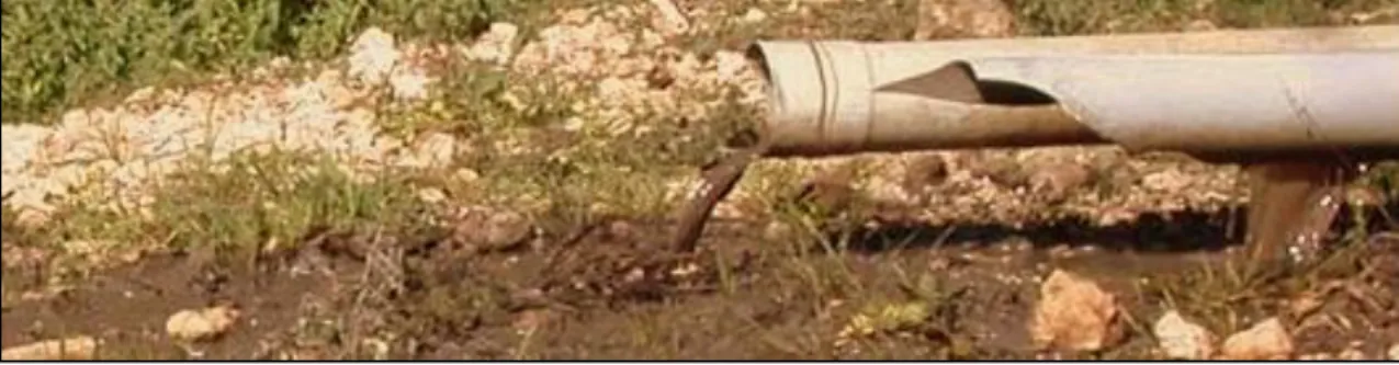
In the olive oil production, the wastewater generated by olive processing is one of the strongest industrial effluents, with COD values up to 220 g.L-1 and equivalent BOD values up to 100 g.L-1. The amount of wastewater originated from the milling process vary between 0.5-1.5 m3 per ton of olives, depending on the process that is used. This wastewater is usually acidic, with a black-brownish colour and is commonly denominated as olive mill wastewater (OMW) (Paixão et al., 1999; Rinaldi et al., 2003). An example of a discharge into the environment can be seen in Figure 1.2.
Figure 1.2: Dumping of wastewater into the Hasbani River by a local olive mill in Lebanon (Hashwa and
Mhanna, 2010).
The OMW composition varies according to several factors, such as: olive variety, climate conditions, cultivation practices, olive storage time and extraction process used. It is mainly composed by water (83-92%), organic acids, sugars and phenolic compounds. It
3 also has a high amount of recalcitrant compounds (lignins and tannins), which gives OMW its characteristic dark colour. Additionally, the phenolic compounds and the long-chain fatty acids existent in these wastewaters are very toxic to microorganisms and plants, making the disposal of OMW a very serious environmental issue (Niaounakis and Halvadakis, 2004). Up until now, there is no regulation of olive mill discharges in the European legislation and standards are defined by each individual country. OMW contain an incredibly high organic content, higher than the European legislation permits in effluents, which denies OMW a regular treatment in wastewater treatment plants. The practices that are currently applied involve land disposal, discharge into nearby lakes, rivers or seas and evaporation in lagoons. Soil contamination, underground leakages, water body pollution and foul odour are environmental problems that arise from those practices. (Paraskeva and Diamadopolus, 2006).
Despite their toxicity, several authors describe OMW as a resource for the simple and complex sugars that are required for fermentation processes (Federici et al., 1986; Montedoro et al., 1993; Crognale et al., 2003, Fenice et al., 2003) OMW also contains variable quantities of residual oil, depending on the extraction process efficiency. This contributes to the valorisation of OMW through microbial processes.
1.2 Lipases
Lipases (triacylglycerol acylhydrolases, E.C. 3.1.1.3) are ubiquitous enzymes of considerable physiological significance and industrial potential. They catalyse the hydrolysis of triglycerides to glycerol and free fatty acids. In contrast to esterases, lipases are activated only when adsorbed to an oil–water interface (Martinelle et al., 1995) and do not hydrolyse dissolved substrates in the bulk fluid. A true lipase will split emulsified esters of glycerine and long-chain fatty acids such as triolein and tripalmitin. Lipases are serine hydrolases, displaying little activity in aqueous solutions containing soluble substrates (Sharma et al., 2001).
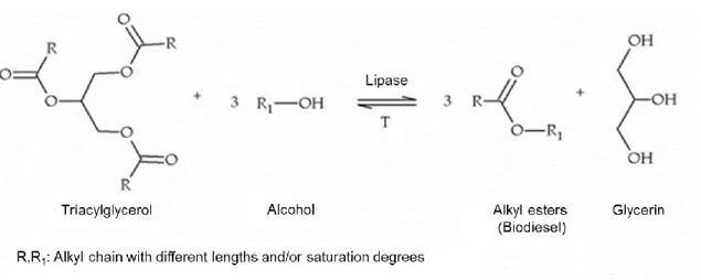
The chemical transesterification of vegetable oil for biodiesel production has several disadvantages, such as soap formation, due to the presence of free fatty oils and high reaction temperature. Therefore, enzymatic processes using lipases have been developed to overcome such problems (Figure 1.3). These enzymes require minimum amount of water and are very thermostable, even at high temperatures. Despite having low catalytic efficiency, processes such as immobilization, chemical modification and
4 protein engineering can be used to surpass the overall conversion process (Aires-Barros
et al., 1991; Tyagi and Gupta, 1998).
Figure 1.3: Enzymatic transesterification of fatty acid methyl esters, also known as biodiesel.
The ability to perform specific chemical transformations has made them progressively more popular in several industries, such as: food, detergent, cosmetic, organic synthesis and pharmaceutical (Ryu et al., 2006; Gupta et al., 2007; Franken et al., 2009). The use of lipases in organic chemical synthesis is becoming increasingly important. These enzymes catalyse the hydrolysis of water-immiscible triglycerides at water-liquid interface, which will be determined by the amount of water present. When there is little to none water, only esterification and transesterification are favoured. This aspect is very important in the context of enzymatic production of biodiesel (Klibanov et al., 1997). Lipases perform essential roles in the digestion, transport, and processing of dietary lipids (e.g., triglycerides, fats, and oils) in most living organisms. They are considered as a major group of biotechnologically valuable enzymes, mainly due to the versatility of their applied properties and easy mass production (Bornscheuer et al., 2002; Menoncin
et al., 2010).
1.2.1 Microbial lipases
Being more stable than animal or plant lipases, microbial lipases have earned special industrial attention due to their stability, selectivity and broad substrate specificity. The energy consumption required to conduct reactions at elevated temperature and pressure is eliminated as lipases are active under room temperature, reducing the denaturation of labile reactants and products (Dutra et al., 2008; Griebeler et al., 2011).
Lipase-producing microorganisms include actinomycetes and other bacteria, filamentous fungi and yeasts. They can be found in diverse habitats such as industrial wastes,
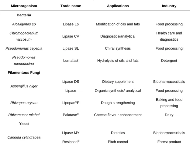
5 vegetable oil processing factories, dairies, soil contaminated with oil, oilseeds, and decaying food (Sztajer and Zboinska, 1988). Several methods have been developed to successfully identify these organisms and they usually involve agar plates with a lipidic substrate, in order to identify lipolytic activity. A simple agar medium with lipidic substrates has a turbid appearance and the detection of enzymes is is done by the observation of clear-through halos around the colonies (Sierra, 1957; Cardenas et al., 2001). Several substrates (basically, any oil or grease) can be used in this method. The use of chromogenic substrates, such as pH indicator Victoria blue and Phenol red, has also been described (Yeoh et al., 1986; Kouker et al., 1987, Wang et al., 1995, Hou, 1994). Chromogenic dyes can be used to assure a more visible detection, as they induce a change in the medium color around the enzyme-producing colonies. This color change occurs due to the formation of free fatty acids, which causes a change in the medium pH (Bornscheuer and Kazlauskas, 1999). There are other ways to detect lipase production without having to use chromogenic dyes. The precipitation of salts is also used for the detection of lipase-producing microorganisms. The fatty acids resulting from hydrolysis of tween binds with the calcium salts present in the media, forming insoluble crystals, visible around the colonies (Gopinath et al., 2005). Most of the industrial microbial lipases are derived from fungi and bacteria (Table 1.1).
Table 1.1: Examples of some commercially available microbial lipases, their most regular applications and
producing microorganisms (Jaeger and Reetz, 1998; Sharmaet al., 2001; Kademi et al., 2004).
Microorganism Trade name Applications Industry
Bacteria
Alcaligenes sp Lipase Lp Modification of oils and fats Food processing
Chromobacterium
viscosum Lipase CV Diagnostics/analytical
Health care and diagnostics
Pseudomonas cepacia Lipase SL Chiral synthesis Food processing
Pseudomonas
menodocina Lumafast Hydrolysis of oils and fats Detergent
Filamentous Fungi
Aspergillus niger
Lipase DS Lipase
Dietary supplement Organic synthesis/ analytical
Biopharmaceuticals Food processing
Rhizopus oryzae Lipopan©F Dough strengthening Baking and food
processing
Rhizomucor miehei Palatase© Cheese flavour enhancement Dairy
Yeast Candida cylindracea Lipase MY Resinase© Dietetics Pitch control Biopharmaceuticals Forest product
6
Pseudozyma antarctica Novozym© 435 Oil based specialities Food processing
Candida rugosa Lipase AY “Amano” 30 Organic synthesis Food processing
Geotrichum candidum Chirazyme© L-8 Lipolase© Oleochemistry Oleochemistry Food processing Food processing
These lipases are produced frequently by submerged cultures (Ito et al., 2001), although solid-state fermentation methods can also be used, namely for filamentous fungi (Chisti, 1999). For bacteria and yeasts, it is obligatory to use a submerged culture due to their minimum water requirement. Lipase production is influenced by both the nature and concentration of carbon and nitrogen sources, culture pH, dissolved oxygen concentration and growth temperature. It is reported that lipidic carbon sources appear to be largely crucial in order to obtain a high lipase yield (Elibol and Ozer, 2001).
1.3 Lipase-producing yeasts
In literature, several yeasts have been described to be capable of producing the enzyme lipase. Species belonging to the genus Candida are among the most used commercial producers (Larios et al., 2004).
1.3.1 Genus Candida
The genus Candida is extremely heterogenous and covers a wide diversity of yeasts of ascomycetous affinity with unknown sexual states (Figure 1.4).
7 Two well-known species that belong to this genus are Candida cylindracea and
Psedozyma Antarctica. Both species are extensively described in literature due to their
capability to produce valuable industrial biocatalysts, such as lipases. Because of their high production yield, they have been studied for the production of biodiesel using several lipidic substrates (Kurtzman et al., 2011). Additionally, lipase produced by
Candida rugosa is rapidly becoming one of the most used industrial enzymes due to its
high activity, both in hydrolysis as well in synthesis (Vakhlu and Kour, 2006). The referred lipases have several isoforms, which greatly contributes to their diverse use in biotechnology. This is due to the broad range specificity (substrate, positional, fatty acid and stereopreference), usually not found in other lipases (de María et al., 2006).
Over the past years, there has been a great demand for the use of non-conventional microorganisms, in this case, non-conventional yeasts (non-saccharomyces yeasts) that could be better suited for biotechnological purposes. One of these cases is Yarrowia
lipolytica, since its finding in 1980, has been made a model of study and research in
industrial microbiology (Spencer et al., 2002).
1.3.2 Yarrowia lipolytica
Yarrowia lipolytica is an aerobic, nonconventional ascomycetous yeast with multiple
biotechnological applications (Figure 1.5). Usually it can be found in hydrophobic substrates rich in fatty acids, such as dairy products (Spencer et al., 2002).
Figure 1.5: Microscopic image of Yarrowia lipolytica cells (Taken by Drº. Andreas Aurich, UFZ,
www.ufz.de).
It can be used for bioremediation, production of biosurfactants and other assorted substances such as citric and isocitric acids and enzymes (proteases, lipases, RNase) (Madzak et al., 2004). The most important of these metabolites is lipase due to its broad technological applications in several different areas. There are many studies related to lipase production by Y. lipolytica, in both wild and naturally occurring strains. Some
8 strains of this species are cultivable in effluents, making them a possible organism for a valorisation process in the case of OMW, for example (Federici et al., 2009; Dermeche
et al., 2013).
1.4 Valorisation of Olive Mill Wastewater
In recent years, increasing consideration has been dedicated to the possibility of valorising the olive oil extraction residues. The successful recovery of bioactive chemicals with commercial importance, as well the production of added-value products such as ethanol, organic acids and enzymes appear to be the new frontier in OMW valorisation (Dermeche et al., 2013).
Despite their toxicity, the high level of polyphenols present in OMW can act as a vantage point in their valorisation. Although to different extents, phenolic compounds are known to have antioxidant activity, a characteristic which is very sought out by cosmetic and pharmaceuticals industries. Hydrotyrosol, the main polyphenol in OMW, has the same antioxidant activity comparable to that one detected in other synthetic antioxidants has. Their synthesis is very costly, which means that the acquisition of hydrotyrosol from OMW could constitute an easier and inexpensive alternative for the acquirement of antioxidants (Federici et al., 2009).
The valorisation of OMW can also have a biotechnological approach, using this effluent in the production of added-value products, such as enzymes. As suggested in section 1.1, this effluent can be used as source for the simple and complex sugars required for fermentation processes.
Industrial enzymes can be produced by yeasts and filamentous fungi using OMW as a substrate. D’Annibale et al. (2006) assessed the suitability of OMW as growth medium for the production of microbial lipase using several yeasts and filamentous fungi strains from the NRRL collection (Culture Collections of the North Regional Research Laboratory, Peoria, Illinois). All strains were able to growth in undiluted OMW and produce extracellular lipase. The yeast Candida cylindracea (NRRL Y-17506) had the highest lipase production (9.23 U.mL-1), but only after supplementation with NH4Cl (2.4 g.L-1) and olive oil (3 g.L-1). Another case study was presented by Lanciotti et al. (2005), in which was used different strains of Yarrowia lipolytica to assess their ability to grow in undiluted OMW and compared it to the production of lipase in semi-synthtic medium. The majority of the strains tested did have a higher production of lipase on OMW than in the synthetic medium. They were also able to reduce COD values and polyphenols content.
9 Another approach to OMW valorisation was made by Yousuf et al. (2010), using the oleaginous yeast Lipomyces starkeyi. This author used undiluted OMW with the purpose of accumulating lipids as a feedstock for biodiesel production.
Based on the consulted literature, it is safe to assume that OMW has potential to be used in the growth of lipolytic strains and subsequent induction of the production of enzymes by those strains. OMW also has an associated microbiota, which are microorganisms that can live and growth in this effluent, withstanding its phenol toxicity. Isolation and selection of yeasts strains from OMW samples has already been described by Bleve et
al. (2011). The authors were able to isolate 300 yeasts isolates, showing that OMW has
a high quantity of yeasts in its microbiota, probably due to their resistance to high phenolic concentrations.
Due to the oil residue in OMW, it is very likely that most of the associated microbiota has lipolytic properties. Consequently this effluent can be used, not only as a growth medium (due to the presence of complex sugars and other carbon sources), but as well as a source of lipolytic yeasts. Taking these propositions into consideration, this thesis will focus on the isolation of lipolytic yeasts that are able to produce extracellular lipases from OMW samples, test their phenolic tolerance and optimize their lipase production.
2. Materials and Methods
2.1 Olive mill wastewater
2.1.1 OMW origin and processing
Raw OMW samples used in this work were collected from two different locations: a three-phase olive oil mill located in the south region of Portugal (Tavira, Algarve, during the last month of olive oil production campaign of 2013/2014) and several samples from Jordan (Irbid) with two origins: i) enriched OMW in shake-flask; ii) treated OMW in an aerobic bioreactor. All samples were kept at 4ºC until further use. Due to high suspended solids in the raw OMW content, a preliminary step of filtration and centrifugation (8600g, 15 min) for the removal of solid was performed before using as culture medium. This filtration is performed with normal gauze or a large strainer.
10
2.2.2 OMW characterization
The OMW characterization was performed in the subsequent 48 h after its collection. The effluent was characterized for pH, Chemical Oxygen Demand, Total Suspended Solids, Volatile Suspended Solids, Total Kjeldhal Nitrogen, total phenols, nitrates, total lipids and reducing sugars (see section 2.9 Analytical methods).
2.2. Isolation and screening of lipase-producing yeasts strains
The microbial characterization of used OMW samples was performed in Nutrient Agar in order to quantify total heterotrophic bacteria, filamentous fungi and yeasts. Serial dilutions (10-1 to 10-5) of the OMW samples were prepared and spread on agar plates, incubated at 30ºC during 48 hours, and quantification made in terms of Colony Forming Units (CFU). Yeast colonies were selected and isolated from the obtained CFU counts plates. Selective media, such as YMA (Difco©), CRBA (Difco©) and PDA (Difco©), containing final concentration of 35 µg/mL of chlortetracycline to prevent bacterial growth, were used. The plates were incubated at 30ºC during 48 hours. The isolates were first differentiated for their colony morphology and then analysed under optical microscopy (microscope Olympus BX51) in order to identify yeasts strains colonies.
2.2.1 Screening of yeasts strains with lipolytic activity
The yeast isolates were screened for lipolytic activity through rapid plate detection experiments. Three different media were used for that purpose: Tween 20, Phenol-red with lipidic substrates and tributyrin agar (TBA). The lipolytic activity was registered after 48 h of incubation at 30ºC.
2.2.1.1 Tween 20 plate assay
Tween 20 agar plates were prepared according to Gopinath et al. (2005). The culture media contained peptone (10 g.L-1), NaCl (5 g.L-1), CaCl2. 2H2O (0.1 g.L-1), agar (20 g.L -1) and tween 20 (10 mL, v/v). About 20 mL were distributed in Petri dishes and the isolates were inoculated. Lipolytic activity was indicated by the appearance of visible precipitate, as a result of deposition of calcium crystal salts formed by the fatty acid liberated by the extracellular lipase.
2.2.1.2 Phenol-red plate assay
Chromogenic substrate plates were prepared according to Singh et al. (2006) by using phenol red (0.01%) along with 1% olive oil, 0.1% CaCl2 and 2% agar. pH was adjusted to 7.3-7.4 by using NaOH 0.1 N. Cell suspensions were made using 1 mL of sterile distilled water, having a final concentration of approximately 107 CFU/mL (optical
11 turbidity, using a McFarland scale). A 3-5 mm diameter sterile paper discs were immersed in each cell suspension and placed in the phenol-red medium. The change in color of phenol from red to bright yellow, forming a halo, was used as an indicator of lipolytic activity.
2.2.1.3 TBA plate assay
The lipase production was examined in a yeast malt agar media supplemented with 0.5% (v/v) tributyrin, after autoclaving. Subcultures of each isolate were inoculated in the tributyrin plates and incubated at 30ºC during 72h. In the positive responses, a translucent halo around the colonies was formed.
2.2.2 List of yeast strains used for comparative tests
Three yeasts strains were obtained from Private Collection of Microorganisms of Bioenergy Unit of LNEG as described in Table 2.1. These strains were tested for lipolytic activity and compared with yeast strains isolated from OMW.
Table 2.1: List of yeasts cultures from Private Collection of Microorganisms of Bioenergy Unit of LNEG used
for comparative tests.
Microorganism/Isolate LNEG strain ID Origin and
reference Lipase production (Literature)
Yarrowia lipolytica 263F NRRL Y-323 Positive (Ogrydziak, 1988)
Candida sp. 356F INETI Positive (van Uden & H.R. Buckley,
1970)
11-T AR ---- INETI Unknown
NRRL – Recently changed to ARS (Agriculture Research Service) Culture collection; INETI – Instituto Nacional de
Engenharia, Tecnologia e Inovação.
2.3 Selection of yeasts for extracellular lipase production
Yeasts isolates were grown in 50 mL of YEPD medium (1% yeast extract, 2% peptone, 2% dextrose) overnight. 300 µL of freshly prepared cultures were used as inoculum. Experiments using 100 mL of control medium (1 g.L-1 yeast extract, 1 g.L-1 chloride potassium and 1 g.L-1 magnesium sulfate) with an inductor (0.5 g.L-1 olive oil) were performed in 500 mL-shake-flasks. Cultures were incubated at 30ºC under orbital shaking (180 rpm) during 120 hours. 1 mL samples were collected every 24 hours and absorbance was monitored at 600 nm. The samples were then centrifuged (7500 g, 15 min) at 4ºC and the supernatant was assayed for the enzyme activity.
12
2.3.1 Assay of lipase activity
The measurement of lipase activity was adapted from Gomes et al. (2011). A reaction mixture composed by 195 µL of substrate (2.63 mM ρ-nitrophenyl butyrate in sodium acetate buffer, 0.05 M, pH 6.8, with 4% (v/v) Triton X-100) and 5 µL of sample supernatant was incubated in a microplate at 37ºC for 15 min. In blanks, fresh medium was used. The absorbance was measured at 405 nm in a microplate reader (Multiskan™ GO Microplate Spectrophotometer, Thermo Fischer Scientific). One unit (U) of lipase activity was defined as the amount of enzyme that produces 1 µmol of ρ-nitrophenol per minute under assay conditions.
2.4 Yeast strain identification: DNA extraction and sequencing
DNA from selected yeast isolates was extracted according to a standard phenol-chloroform-extraction protocol (Cheng & Jiang, 2006): 1000 μL of cell suspension and a volume corresponding to 200 μL of sterile glass beads (∅ 0.43-0.6 mm) were centrifuged at 8600 g for 5 min and the supernatant was removed. Samples were frozen (-20ºC) for at least 60 min. After defrosting, 500 μL of lysis buffer (10 mM TrisHCl pH 8, 100 mM NaCl, 1 mM Na2-EDTA, 1 % SDS, 2 % (v/v) Triton x-100) and 500 μL of TE-phenol-chloroform were added to each sample, which was then vortexed (full speed, 20 min) and centrifuged, at 8600 g and for 25 min. 400 μL of the upper phase and 1000 μL of ethanol (100 %) were mixed and placed at -20ºC during 30 min, for precipitation purposes. The mix was then centrifuged at 8600 g for 15 min. The supernatant was removed and 50 μL of TE-RNase (pH 8.0; 50 μg.mL-1) were added. Samples were then placed in a thermoplate at 55 °C with occasional stirring for 15 min to solve the pellet. The DNA purity and quantification was measured via NanoDrop (NanoDrop 2000 Spectrophotometer, Thermo Scientific), in which the DNA absorption peak was observed at 260 nm.
The D1/D2 variable domains of the larger rDNA subunit (26S) were amplified by polymerase chain reaction (PCR) using NL1 (5’-GCA TAT CAA TAA GCG GAG GAA AAG) and NL4 (5’-GGT CCG TGT TTC AAG ACG G) primers (O’Donnell 1993). PCR was conducted using a 3 min initial denaturation at 94ºC, followed by 40 cycles of 30 s denaturation at 94ºC, 30 seconds primer annealing at 50ºC and 90 seconds extension at 72ºC, with a final 4 min extension at 72ºC. The amplified fragments were submitted to DNA sequencing (STAB Vida, Portugal). The sequences were analyzed through a BLAST search against the nonredundant nucleotide (nt) database (BLAST). Yeast species were identified on the basis of phylogenetic analysis.
13
2.5 Evaluation of lipase production by Magnusiomyces capitatus in OMW
Selected yeast strain was grown in 500-mL shaking flasks containing 100 mL centrifuged OMW (8600 g, 15 min) and the pH was adjusted to 6.8 before sterilization (121ºC for 20 min). The cell growth was monitored by analysis of the absorbance at 640 nm (Thermo Electron Corporation Spetrophotometer, model Genesys 6, Madison, USA).
The DCW values were obtained by filtrating 1 mL of every sample (three replicas of each sample were made) using membrane discs (0.45 µm, MicronSepTM membrane filter) and then dried overnight (18-24h) at 100ºC. Before use, the membrane discs were dried at the same conditions, to obtain their dry weight. The same samples were also used to measure the biomass by optical density (Thermo Electron Corporation Spetrophotometer, model Genesys 6, Madison, USA) at 640 nm. A correlation between the biomass dry weight values and absorbance at 640 nm was found.
2.5.1 Toxicity tests
The centrifuged OMW medium was diluted 10%, 25% and 50% using distilled water in order to assess the influence of the phenol concentration presented in the medium. The non-diluted medium was also tested (100% OMW). 300 µL of cell suspension, previously grown in YEPD (1% yeast extract, 2% peptone, 2% dextrose) medium overnight at 30ºC, served as inoculum. The flasks were placed in orbital incubator (180 rpm) at 30ºC during 36 hours. 1 mL samples were taken after 12 hours and every hour after 22 hours.
2.5.2 Influence of yeast extract and olive oil supplementation on lipase activity
Non-diluted OMW with adjusted pH (6.8) was supplemented with yeast extract (2.0 g.L -1) or olive oil (1.0 g.L-1) or both. 300 µL of cell suspension, previously grown in YEPD medium overnight at 30ºC, served as inoculum. The flasks were then incubated at 30ºC, 180 rpm, for 96 hours. Crude cell-free extract was obtained by recovering the supernatant after spinning down cell debris (7500 g, 15 min). This preparation was used to estimate lipase activity.
2.6 Optimization of lipase production by experimental design methodology
Experimental distribution for two factors, according to the Doehlert uniform design (Doehlert, 1970), was used to produce response analysis. Seven experiments (and respective replicates) were carried out within an experimental domain with 1000 mL-shake flasks containing working volume ranging from 100 to 400 mL, corresponding to a specific oxygen transfer coefficient kLa between 0.24 and 1.88 min-1. The nitrogen source tested was NH4Cl, according to D’Annibale et al. (2006), varying between 0.2 and
14 2.8 g.L-1. Cultures were incubated on undiluted OMW supplemented with yeast extract (2 g.L-1) and olive oil (1 g.L-1), at 30ºC and 180 rpm for 48 hours.
2.6.1 Statistical analysis
Coded representation of the factors and interaction terms was used for calculation purposes. The response studied in this design was the biomass and lipase activity. The model used to express the responses was a second order polynomial model provided by the Doehlert uniform design (equation 1):
Y=β0+β1X1+β2X2+β12X12+β11X12+β22X22 (Eq. 1)
where Y was the response from each experiment; β were the parameters of the polynomial model and X was the experimental factor level (coded units).
2.7 Analytical methods
COD (g.L-1), TSS (g.L-1), VSS (g.L-1), and TKN (g.L-1) were measured according to Standard Methods (APHA, 2001). Total phenols content was determined by a modified Folin-Ciocalteau colorimetric method (Singleton and Rossi, 1965) and expressed as caffeic acid (g.L-1). The nitrates determination (NO3-N, g.L-1), was performed by the method "Nitrate Cell Test 1.14542" (Spectroquant Merck) in a HACH DR/2010 spectrophotometer. Total lipids (g.L-1) content was determined gravimetrically after petroleum ether extraction. Reducing sugars were measured by the DNS method (Miller, 1959) and were expressed as glucose (g.L-1).
A flow cytometer (FACScalibur©, Becton-Dickinson, Franklin Lakes, NJ, USA) was used to evaluate the integrity of yeast cellular membrane of the cultured OMW after the experimental design methodology. The cytometer is equipped with a 488 nm argon laser and with photomultipliers FL1 (530 ± 30 nm), FL2 (585 ± 42 nm), FL3 (>670 nm) and FL4 (600 ± 16 nm). Cellular membrane integrity was assessed by fluorescence of stained cells using fluorochrome propidium iodide (IP, Invitrogen© EUA). The concentration of the IP stock solution was 1 mg.mL-1 (in filtered mili-Q water) and the final concentration in the cellular suspension was 0.5 µg.mL-1. IP fluorescence can be detected in channel FL3, due to this fluorochrome being excited at 536 nm and transmitting at 623 nm. Samples were pre-treated before being used in the flow cytometer, in order to remove the excess of particles normally presented in the OMW. Cells were centrifuged (5000 g, 10min). Supernatant was discarded and pellet was re-suspended in a buffered saline solution (PBS, pH 7.0, Oxoid© England). This procedure was repeated two more times. Before each analysis, the samples were sonicated during 10 seconds, in order to remove cellular aggregates and guarantee the analysis of single cells. This suspension was then
15 diluted using the same PBS in order to obtain a cellular concentration of 500 to 1000 events per second. 0.5 µL of IP was added to 499 µL of each sample and the mixture was analysed in the flow cytometer. The data obtained were treated in the program
Windows Multiple Document Interface flow cytometry (WinMDI 9.0).
3. Results and Discussion
3.1 Isolation of yeasts from OMW
According to several authors, OMW usually contain a diverse microbiota that is able to grow and colonize this effluent despite its high phenolic content. This microbiota consists in several species of yeasts, filamentous fungi and bacteria. Yeasts are especially well adapted to this particular environment, since they have a better tolerance to phenolic compounds when compared to bacteria (Rincón et al., 2006; Morillo et al., 2006; Ben Sassi et al., 2006). Samples of raw OMW of three different origins were analysed in terms of their physical-chemical properties (Table 3.1.1). The microbial content of these samples used in this study is shown in Table 3.1.2.
Table 3.1.1: Physical-chemical characterization of OMW samples.
Parameter Raw OMW (Portugal) Enriched OMW (Jordan) Treated OMW (Jordan) pH 4.99 8.44 7.90 COD (g.L-1) 55.20 13.00 10.45 Conductivity (mS.cm-1) 11.80 4.73 4.45 PO43- (g.L-1) 0.57 0.25 0.35 NO3- (g.L-1) 1.42 0.33 0.24 NH4 (g.L-1) 1.70 0.60 0.70 TSS (g.L-1) 21.9 1.12 4.83 VSS (g.L-1) 21.10 1.06 4.35 Total phenols (g.L-1) 3.97 0.07 0.05 Lipids (g.L-1) 3.20 n.d. n.d. n.d. – not determined
16
Table 3.1.2: Microbial counts in OMW.
OMW sample Microorganisms Counts (CFU.mL-1)
Enriched OMW, Jordan
Bacteria 2.32x10⁷
Yeasts 1.54x10⁷
Filamentous Fungi 1x10
Raw OMW, Portugal
Bacteria ---
Yeasts 3.14x10⁷
Filamentous Fungi ---
(---): absence of growth
The total number of viable yeasts was very high and similar in both samples. The counts of viable bacteria in the Jordanian samples show a clear dominance of this microbial group over remaining groups. This is not observed in the samples originated in Portugal. This is probably due to the Jordanian samples having an alkaline pH, which permits the existence of bacteria. The samples originated from Portugal show a more acidic pH, which is more favorable to the occurrence of fungi (yeasts and filamentous fungi) in general. Another explanation could arise from the phenol content in the samples, since most bacteria have a low tolerance to phenols, while yeasts and filamentous fungi can withstand a higher phenolic content (Ben Sassi et al., 2006). Figure 3.1.1 shows the morphological diversity of microorganisms obtained in Nutrient Agar (NA) plates. It was observed that colony morphology is very heterogeneous, with a clear dominance of bacteria.
Figure 3.1.1: Nutrient Agar Plates showing an example of the high microbial diversity observed in samples
of enriched OMW from Jordan [A], dilutions at 10-5 (on the left) and 10-4 (on the right). [B] and [C] are replicas
17 Yeast colonies were successfully replicated using selective media (PDA, CRBA-supplemented with 35 µg/mL of chlortetracycline and YMA), in order to obtain isolated pure strains. Colonies morphology was very similar in most isolates, forming circular white colonies. Under optical microscopy, the yeast cells were observed with a 40x ocular, making a total amplification of 400x (Figure 3.1.2). The 30 isolates obtained were divided into three major groups, based on their microscopic morphology.
In group A, the cells were small sized and round, very similar to the genus
Saccharomyces, sometimes forming aggregates. In group B, cells had a larger, more
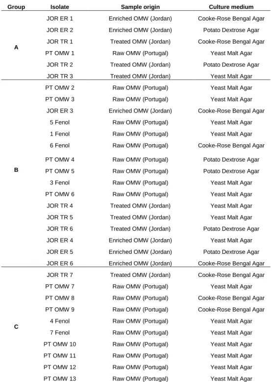
rectangular shape, with frequent formation of pseudo mycelia. Group C showed cells with an elliptical shape, without aggregates or filaments. Table 3.1.2 shows both the origin and the culture medium used to isolate the 32 yeasts strains from OMW samples in this work and the morphological groups in which they were divided.
18
Table 3.1.2: Yeast strains obtained from OMW with different origins.
Group Isolate Sample origin Culture medium
A
JOR ER 1 Enriched OMW (Jordan) Cooke-Rose Bengal Agar
JOR ER 2 Enriched OMW (Jordan) Potato Dextrose Agar
JOR TR 1 Treated OMW (Jordan) Cooke-Rose Bengal Agar
PT OMW 1 Raw OMW (Portugal) Yeast Malt Agar
JOR TR 2 Treated OMW (Jordan) Potato Dextrose Agar
JOR TR 3 Treated OMW (Jordan) Yeast Malt Agar
B
PT OMW 2 Raw OMW (Portugal) Yeast Malt Agar
PT OMW 3 Raw OMW (Portugal) Yeast Malt Agar
JOR ER 3 Enriched OMW (Jordan) Cooke-Rose Bengal Agar
5 Fenol Raw OMW (Portugal) Yeast Malt Agar
1 Fenol Raw OMW (Portugal) Yeast Malt Agar
6 Fenol Raw OMW (Portugal) Cooke-Rose Bengal Agar
PT OMW 4 Raw OMW (Portugal) Potato Dextrose Agar
PT OMW 5 Raw OMW (Portugal) Potato Dextrose Agar
3 Fenol Raw OMW (Portugal) Yeast Malt Agar
PT OMW 6 Raw OMW (Portugal) Yeast Malt Agar
JOR TR 4 Treated OMW (Jordan) Yeast Malt Agar
JOR TR 5 Treated OMW (Jordan) Yeast Malt Agar
JOR TR 6 Treated OMW (Jordan) Potato Dextrose Agar
JOR ER 4 Enriched OMW (Jordan) Yeast Malt Agar
JOR ER 5 Enriched OMW (Jordan) Potato Dextrose Agar
JOR ER 6 Enriched OMW (Jordan) Cooke-Rose Bengal Agar
C
JOR TR 7 Treated OMW (Jordan) Cooke-Rose Bengal Agar
PT OMW 7 Raw OMW (Portugal) Yeast Malt Agar
PT OMW 8 Raw OMW (Portugal) Cooke-Rose Bengal Agar
PT OMW 9 Raw OMW (Portugal) Cooke-Rose Bengal Agar
4 Fenol Raw OMW (Portugal) Yeast Malt Agar
7 Fenol Raw OMW (Portugal) Yeast Malt Agar
PT OMW 10 Raw OMW (Portugal) Yeast Malt Agar
PT OMW 11 Raw OMW (Portugal) Yeast Malt Agar
PT OMW 12 Raw OMW (Portugal) Yeast Malt Agar
19
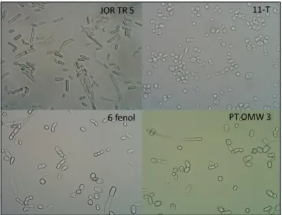
Figure 3.1.2: Isolate 11-T AR belongs to group A (amplification 1000x), while isolates JOR TR5, 6 fenol and
PT OMW 3 are part of group B (amplification 400x).
The content of filamentous fungi in the samples is evidently low, when compared to the findings of other authors (Millán et al., 2000). However, this could be due to a short incubation time of the plates (48h) as other authors describe an incubation time of 4-8 days. The bacteria and yeast counts in the Jordanian effluent are very similar, at relatively high amounts. Previous studies show that the microbial populations of olive mill wastewaters are formed by lactic acid bacteria and yeasts, which exist naturally at the surface of olives (Mouncif et al., 1993).
To compare the presumable lipase-producing ability of these isolates with the activity of other yeasts, three strains were used from cultures of the Private Collection of Microorganisms of Bioenergy Unit of LNEG. Namely, Yarrowia lipolytica and Candida
sp. are yeasts described in literature for being capable of producing lipase and were
used as positive controls. The isolate 11-T AR was also used due to being previously isolated from OMW, but its lipase production was unknown.
3.2 Screening of yeasts strains with lipolytic activity
Plate assays for rapid detection of hydrolytic enzymes have long been used in microbiology. The base of this method consists in the addition of substrates that can only be hydrolyzed by certain enzymes to the media. Chromogenic dyes, such as pH indicator dye Victoria blue, are specific for lipase detection. However, they are not very sensitive and require a long incubation time. Gupta et al. (2006) describes a highly sensitive and rapid pH-based protocol for detection of lipases which consists in using phenol-red as a
20 chromogenic substrate. When the lipases break the lipids present in the medium, this causes the pH to drop and induces a change of color from pink red to yellow (Gupta et
al., 2006).
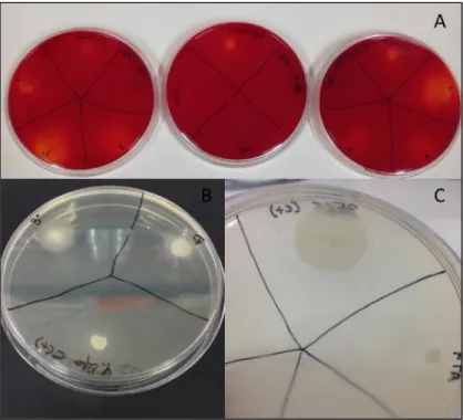
As previously stated, OMW usually contains oil residue, which allows the growth of yeasts able to produce lipases. The lipolytic activity of the 33 isolates was tested using three different detection media: Tween 20 agar; Phenol-Red agar and Trybutyrin agar. Yeasts that are positive for lipase production form a catalytic halo around the colonies (Figure 3.2.1). The medium Phenol-Red shows a change of color around the colonies (Figure 3.2.1-A), while the medium Tween 20 forms precipitates (Figure 3.2.1-B and
Figure 3.2.1-C).
Figure 3.2.1: Detection of lipase production in plate assays. Two strains, Yarrowia lipolytica and Candida
sp., were used as positive controls. [A] Several isolates in phenol-red agar plates supplemented with 1%
olive oil (substrate). [B] Isolates PT OMW3, JOR TR5 and Yarrowia lipolytica in a Tween 20 agar plate. [C] Close-up of a Yarrowia lipolytica with the precipitation halo in a Tween 20 agar plate.
After 72 to 96 hours at 30ºC, six isolates showed lipolytic activity (Table 3.2.1). These strains were referred as 1 Fenol, PT OMW3, JOR TR5, 6 Fenol, PT OMW6 and 11-T AR; showing similar results to the positive controls (Yarrowia lipolytica and Candida sp.).
21
Table 3.2.1: Screening of lipase activity in plate assays.
Isolate Tween 20 Tributyrin Phenol-red with 1% olive oil Y. lipolytica NRRL Y-323 + + + Candida sp. 356F + + + 11-T AR + + + JOR ER 1 - - n.d. JOR ER 2 - - n.d. JOR TR 1 - - n.d. PT OMW 1 + - n.d. JOR TR 2 - - n.d. JOR TR 3 - - n.d. PT OMW 2 - + n.d. 1 Fenol + - ++ JOR ER 3 - + n.d. PT OMW 3 + - ++ 5 Fenol - - n.d. 6 Fenol + - ++ PT OMW 4 - - n.d. PT OMW 5 + - n.d. 3 Fenol - - n.d. PT OMW 6 + + n.d. JOR TR 4 - - n.d. JOR TR 5 ++ + - JOR TR 6 - - n.d. JOR ER 4 - - n.d. JOR ER 5 - - n.d. JOR ER 6 - - n.d. JOR TR 7 - - n.d. PT OMW 7 - - n.d. PT OMW 8 - - n.d. PT OMW 9 - - n.d. 4 Fenol - - n.d. 7 Fenol - - n.d. PT OMW 10 - + n.d. PT OMW 11 - + n.d. PT OMW 12 + - n.d. PT OMW 13 + - n.d.
n.d: not defined; -: negative result; +: positive result; ++: positive result, but halo is visibly larger than the positive control halo.
22 The medium phenol-red showed poor performance in the evaluation of lipolytic activity of the isolates, most of it showing inconclusive results. However, this method is generally described in literature as a good lipase evaluator (Holmberg and Kielland, 1978).The remaining 27 isolates only revealed a positive result in one method and were not considered for further experiments. These tests are only a qualitative evaluation of lipase production, allowing the detection of producing isolates. However, the quantity of enzymatic production is not discerned. The next assays will evaluate extracellular lipase activity in liquid medium and optimize the lipase production of the selected strain in OMW as growth medium.
3.3 Production of extracellular lipase in shake-flask experiments
The strains 1 Fenol, PT OMW 3, 6 Fenol, PT OMW 6, 11-T and JOR TR 5 were tested for the production of extracellular lipases.
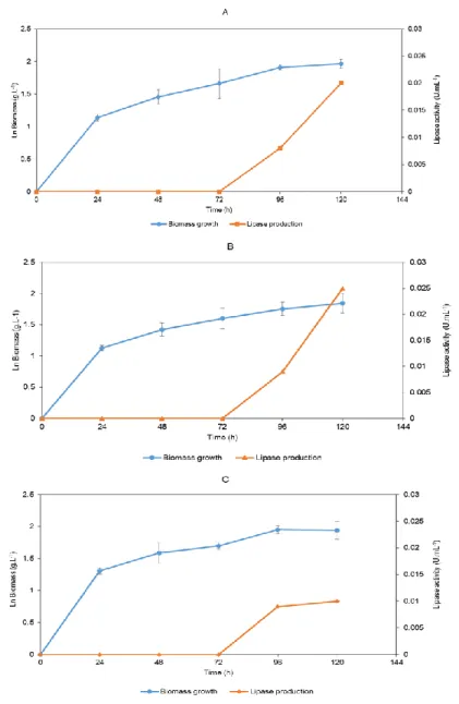
The cultures were grown during 120 hours in a synthetic medium with olive oil as an inductor. The strain Yarrowia lipolytica served as a control. It was grown in the same conditions as the remaining strains and had a maximum lipase activity of 0.12 U.mL-1 at 120 hours (Figure 3.3.1).
Figure 3.3.1: Time course of biomass growth and lipase production by Yarrowia lipolytica NRRL Y-323. All
results were obtained in triplicate. Standard desviation is indicated by the error bars.
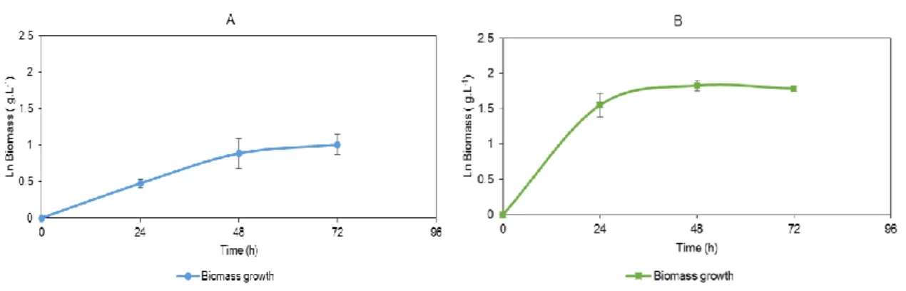
Figure 3.3.2 shows the biomass growth and enzymatic production of strains 1 Fenol, PT
23 (Figure 3.3.3). Figure 3.3.4 shows biomass growth and enzymatic production of strain JOR TR 5. Biomass growth of isolates 11-T, PT OMW 6 and JOR TR 5 was only determined during the first 72 hours, due to the occurrence of flocculation. The flocculation presented itself as small circular aggregates, floating in the medium (Figure
3.3.5). However, lipase production in all isolates was measured until 120 hours of culture
growth in order to obtain comparable data.
Figure 3.3.2: Biomass growth and lipase production of: A - 1 Fenol, B - PT OMW 3 and C – 6 Fenol, in
synthetic medium with 0.5 g.L-1 olive oil. All results were obtained in triplicate. Standard desviation is
24
Figure 3.3.3: Biomass growth of: A – 11-T and B – PT OMW 3, in synthetic medium with 0.5 g.L-1 olive oil.
All results were obtained in triplicate. Standard desviation is indicated by the error bars.
Figure 3.3.4: Time course of biomass growth and lipase production by strain JOR TR 5 in synthetic medium
with 0.5 g.L-1 olive oil. All results were obtained in triplicate. Standard desviation is indicated by the error
bars.
With these assays, strains 1 Fenol, PT OMW 3 and JOR TR 5 were selected as the most prominent lipase producers, with 0.02 U.mL-1, 0.03 U.mL-1 and 0.85 U.mL-1, respectively. Strain JOR TR 5 showed similar flocculation profile to the strains 11-T and PT OMW 6.

![Figure 3.1.1: Nutrient Agar Plates showing an example of the high microbial diversity observed in samples of enriched OMW from Jordan [A], dilutions at 10 -5 (on the left) and 10 -4 (on the right)](https://thumb-eu.123doks.com/thumbv2/123dok_br/15201981.1018238/30.892.234.660.788.1050/figure-nutrient-plates-microbial-diversity-observed-enriched-dilutions.webp)