U N IV E R S I D A D E D E L IS B O A FACULDADE DE MEDICINA VETERINÁRIA
PROTEIN DISULFIDE ISOMERASE AS A TARGET FOR BESNOITIOSIS THERAPY. MOLECULAR CHARACTERIZATION AND STUDIES OF ITS ROLE IN INFECTION AND
HOST IMMUNE RESPONSE.
EDUARDO MIGUEL BAPTISTA FERREIRA MARCELINO
Orientador(es): Doutor José Alexandre da Costa Perdigão e Cameira Leitão Doutor Carlos Manuel Mendes Novo
Tese especialmente elaborada para obtenção do grau de Doutor em Ciências Veterinárias na Especialidade de na especialidade de Sanidade Animal.
Júri:
Presidente: Doutor Rui Manuel de Vasconcelos e Horta Caldeira Vogais:
- Doutora Maria Helena Antunes Soares - Doutora Maria Luísa Santos Sousa Cyrne - Doutor José Augusto Farraia e Silva Meireles - Doutor Carlos Manuel Mendes Novo
- Doutor José Alexandre da Costa Perdigão e Cameira Leitão - Doutor Hélder Carola Espiguinha Cortes
i
Acknowledgments
This was a work of many! Many people, friends, colleagues and family, that shared my joys, my anger, my failures, my achievements but most of all that helped me in some way to make this work possible and accomplish this goal. For some of these many people a few words of gratitude are reserved.
First and foremost to my supervisors that guided my way throughout this journey and made it possible. To Dr. Alexandre Leitão I am very thankful for bringing me into the world of research and for letting me lean and learn on his knowledge. Thank you for the support, always present in good and bad times, for the motivation, patience and specially for your friendship. To Dr. Carlos Novo I am grateful for sharing his knowledge and expertise with me. Thank you for your patience, your continuous support and motivation and for having provided all my requests.
To Prof. Dr. Helder Cortes for introducing me to besnoitiosis, for sharing your knowledge, for your wised advises and, most of all, for your friendship.
To Prof. Dr. Andrew Hemphill, for welcoming me at the Institute of Parasitology, University of Bern and also for the teaching, the sharing of ideas and the wise and practical guidance you provided.
To Prof. Dr. Carlos Martins for embracing me in this journey and for the valuable assistance that the Infectious Diseases Laboratory at FMV provided me.
To the Laboratory of Parasitology at FMV for the good neighboring and for sharing their facilities. Special thanks to Prof. Dr. Isabel Fonseca, Prof. Dr. José Meireles, Prof. Dr. Luís Madeira de Carvalho and Lídia Gomes.
Also a special mention to Prof. Dr. Carlos Fontes and Prof. Dr. José Prates, for providing resources at their laboratories and the help that allowed me to faster and better accomplish my tasks, and to Dr. João Chagas e Silva, Prof. Dr. Armando Panhanha Serrão and Prof. Dr. Luis Costa for essential assistance in providing good care to a special bovine resident at FMV.
ii
I acknowledge Dr. Gereon Schares from the Federal Research Institute for Animal Health, Institute of Epidemiology (Germany), for kindly providing serum samples from a longitudinal study on bovine infected with B. besnoiti carried out in his laboratory.
Furthermore, I am truly thankful to the colleagues from the former UTPAM laboratory, INETI. You welcomed me with care and patience in my first steps in a laboratory. Special thanks for Tiago Martins and Angela Lopes for the learning and technical support you provided me.
And also to those special people from FMV, that in the very beginning of my work, helped me with patience and care, nurturing me through the basics of laboratory procedures. I would like to single out Raquel Portugal, Solange Gil, Clara Cartaxeiro and Maria de Jesus Silva.
To Abdelhak Lemsaddek, Afonso Basto, Dulce Santos and Sofia Nolasco for the knowledge and expertise they shared with me, for critical reviews and good advices. Most of all for being good friends, that were able to walk me through this journey with good doses of humor and motivation.
To my dear friends Alexandra Tavares, Helga Waap, Joana Morais, João Coelho, Margarida Simões, Rita Cardoso, Rodrigo Cunha, Sara Zúquete, Sílvia Almeida and Samuel Francisco a huge thank you, for making my days at the bench easier. Working with you was a privilege and already something that I miss.
To my friend Rui Vieira for the long conversations that helped me through the days and for the care and assistance in making sure all the basics were there.
Moreover I am sincerely appreciative to the institutions that made possible the development of the work required to produce this thesis:
Faculdade de Medicina Veterinaria (FMV), for embracing me and providing me with a healthy emotional, social and scientific environment that, in many ways, allowed me to mature as a scientist and as a person.
Centro de Investigação Interdisciplinar em Sanidade Animal (CIISA) in the persons of Prof. Dr. Luís Tavares and Prof. Dr. Luís Costa for granting me the essential support to conclude this journey.
iii
Institute of Parasitology, University of Bern, for welcoming me and providing with essential resources. A special mention to Prof. Dr. Bruno Gottstein, Prof. Dr. Norbert Muller, Prof. Dr. Joachim Muller and Dr. Karim Debache.
And also, the former Instituto Nacional de Engenharia, Tecnologia e Inovação (INETI), Instituto de Higiene e Medicina Tropical (IHMT) and the former Instituto de Investigação Científica Tropical (IICT).
Finally, to my family, that has supported me in many ways and gave me the strength, the tolerance, and the space to conclude this work, I cannot thank them enough! Specially Catarina, Maria and Joana that were so much deprived of my attention and to whom I did not had the right to do such. They stoically supported my absence, although sometimes physically present, and gave me the strength, the care and the love to pursue this goal.
iv
Financial support
This work was supported by:
Fundação para a Ciência e Tecnologia (FCT), through the fellowship SFRH/BD/ 31445/2006 and the project grant PTDC/CVT/65674/2006.
Centro de Investigação Interdisciplinar em Sanidade Animal (CIISA), Faculdade de Medicina Veterinária de Lisboa, Universidade de Lisboa (FMV-UL).
v
Abstract
Besnoitia besnoiti is an apicomplexan parasite responsible for bovine besnoitiosis, a
disease with a high prevalence in tropical and subtropical regions and re-emerging in Europe. Despite the great economical losses associated with besnoitiosis, this disease has been underestimated and poorly studied, and neither an effective therapy nor a vaccine to be used in Europe is available. Protein disulfide isomerase (PDI) is an essential enzyme for the acquisition of the correct three-dimensional structure of proteins. Current evidence suggests that in Neospora caninum and Toxoplasma gondii, which are closely related to B. besnoiti, PDI plays an important role in host cell invasion, is a relevant target for the host immune response, and represents a promising drug target and/or vaccine candidate. In this work, we presented the nucleotide sequence of the B. besnoiti PDI gene and a 3D theoretical model was built by comparative homology using Swiss-Model server. B. besnoiti expresses a PDI with 471 amino acids, structurally similar to human and yeast PDIs, with four thioredoxin-like domains a, b, b’, a’ and a C-terminal extension c. The a and a’ domains present the characteristic active site pattern CxxC, in this case CGHC and CGYC, respectively. Analysis of the phylogenetic tree for PDI within the phylum Apicomplexa reinforced the close relationship among B. besnoiti, N. caninum and T. gondii. Recombinant B. besnoiti PDI (recBbPDI) and truncated versions corresponding to domains a, b, b’ and a’c were expressed in a heterologous system. Mice were immunized with recBbPDI for the production of monoclonal antibodies (mAbs) by hybridoma technology and four mAbs were produced and characterized. RecBbPDI and domain a’c (recBb-a’c) were functionally active and exhibited a dose dependent cross-linking activity of insulin. In the presence of bacitracin, tocinoic acid, 5,5′-dithiobis(2-nitrobenzoic acid) (DTNB) and 4-chloromercuribenzoic acid (pCMBA) activity of both enzymes was inhibited, in a dose dependent manner. The same happened with recBbPDI in the presence of mAbs (with the exception of T8a), but not with recBb-a’c, whose activity was not sensitive to the presence of mAbs. In vitro proliferation of
B. besnoiti tachyzoites was diminished in the presence of PDI inhibitors and anti-PDI mAbs,
indicating that this enzyme seems to intervene in the process of host cell adhesion/invasion. In this way, considering the inhibitions obtained, both in the host cell invasion ability and in the enzyme catalytic activity, PDI can represent a potential target for addressing the treatment and/or prevention of besnoitiosis. The panel of monoclonal antibodies here developed represents an important tool for future studies.
Keywords: Besnoitia besnoiti, protein disulfide isomerase, PDI, monoclonal antibody, host cell invasion.
vii
Título:
A enzima isomerase de dissulfureto como alvo terapêutico contra a besnoitiose bovina. Caracterização molecular e estudo do seu papel na infeção e na resposta imunitária.Resumo
Besnoitia besnoiti, o agente etiológico da besnoitiose bovina, pertence ao filo
Apicomplexa, família Sarcocystidae, subfamília Toxoplasmatinae, estando filogeneticamente próximo dos géneros Neospora e Toxoplasma. A besnoitiose bovina é uma doença severa mas geralmente não fatal, endémica em vastas áreas tropicais e subtropicais de África e responsável por elevadas perdas económicas. Na Europa, após as primeiras descrições há mais de um século, em França e Portugal, a doença recebeu pouca atenção até finais do século XX, altura em que a sua incidência começou a aumentar com relatos em Portugal, Espanha,Itália e França. Mais recentemente o avanço geográfico da doença é relatado com casos na Grécia, Suíça, Alemanha, Hungria, Croácia e Irlanda. A fase aguda da doença é marcada por alterações respiratórias, febre, anasarca, diarreia e, por vezes, aborto. Na fase crónica, o animal apresenta grande espessamento da pele (elefantíase) com soluções de continuidade, muitas vezes complicadas por infecções oportunistas, má condição corporal e, nos machos, orquite necrosante e aspermia. Apesar de ser conhecida há mais de um século, assim como a sua etiologia parasitária, a besnoitiose bovina mantém muitos aspectos da sua epidemiologia desconhecidos, incluindo o ciclo de vida do seu agente etiológico. Outras espécies do género Besnoitia têm um ciclo heteroxeno mantido por uma relação presa-predador entre pequenos mamíferos e o gato. Acredita-se que o ciclo de vida de B. besnoiti seja semelhante, mas até à data ainda não foi identificado um hospedeiro definitivo. A transmissão homoxena de bradizoítos e taquizoítos de B. besnoiti foi comprovada experimentalmente, em bovinos e em diversos animais de laboratório, e uma maior incidência da doença nos meses mais quentes do ano, quando existe maior actividade de insetos hematófagos, sugere um papel destes na transmissão de B besnoiti. No entanto, o verdadeiro modo de transmissão deste parasita na natureza mantém-se desconhecido. A doença pode atingir uma elevada taxa de morbilidade, o que, associado à gravidade das manifestações clínicas e à inexistência de vacina segura ou tratamento eficaz, justifica a necessidade de aprofundar o conhecimento sobre a biologia deste parasita, tentando desenvolver novas abordagens à terapêutica e à formulação de vacinas.
A enzima isomerase de dissulfureto (PDI, do inglês protein disulfide isomerase), uma das mais abundantes do retículo endoplasmático, catalisa a formação de pontes dissulfureto e o rearranjo de emparelhamentos dissulfureto incorrectos, permitindo que as proteínas adquiram a sua correta conformação tridimensional. As suas funções (ou disfunções) têm
viii
sido implicadas em doenças neurodegenerativas como Alzheimer ou Parkinson ou na capacidade proliferativa de células neoplásicas. Sabe-se hoje que, para além da sua localização maioritariamente reticular, a PDI está presente à superfície das células, onde participa em diversas funções biológicas, como a ativação e agregação plaquetária, a adesão leucocitária ou a internalização de agentes patogénicos como alguns vírus e clamídias. Em diversos parasitas do filo Apicomplexa, incluindo Neospora caninum e
Toxoplasma gondii, foram já identificadas PDIs na superfície dos taquizoítos e foi
demonstrado que a inibição desta proteína, através de drogas ou anticorpos específicos, tem um efeito negativo sobre a capacidade de proliferação dos parasitas, evidenciando o papel que esta parece desempenhar no processo de adesão/invasão da célula hospedeira. Por outro lado, a PDI parece ter um papel relevante na resposta imunitária do hospedeiro, uma vez que foi identificada como uma proteína imunodominante e que foi demonstrada a presença de anticorpos anti-T. gondii-PDI e anti-N. caninum-PDI no repertório inato do homem e da vaca, respetivamente. Em conjunto, estas evidências sublinham a importância da PDI no processo de invasão celular e na resposta imunitária do hospedeiro.
Neste trabalho determinámos a sequência completa do gene da PDI em B. besnoiti, clonámos o respectivo cDNA e expressámos a proteína recombinante, assim como versões truncadas da mesma, num sistema heterólogo. A PDI de B. besnoiti (BbPDI) pertence à superfamília das tiorredoxinas (cluster 00388), estando incluída na família PDI_a (cluster defined cd02961) e subfamília PDI_a_PDI_a’_c (cd02995). Construímos um modelo tridimensional da BbPDI por homologia de comparação usando o software disponível no servidor Swiss-Model e tendo como modelo a estrutura cristalográfica da Tapasina-ERp57 (código do PDB: 3F8U chain C). Verificámos que B. besnoiti expressa uma PDI composta por 471 aminoácidos, com a organização típica descrita para a PDI humana e de levedura, ou seja os 4 domínios estruturalmente semelhantes à tiorredoxina, domínio a, b, b’, a’, com uma extensão C-terminal c. Os domínios activos a e a’ apresentam os característicos centros ativos CxxC, no caso de B. besnoiti com a sequência aminoacídica CGHC e CGYC, respectivamente. A análise filogenética para a PDI colocou a B. besnoiti no mesmo ramo que a N. caninum e T. gondii. Com a PDI recombinante de B. besnoiti (recBbPDI), imunizámos murganhos com o objetivo final de produzir anticorpos monoclonais (mAbs) anti-PDI, pela tecnologia de hibridomas. Obtivemos 4 mAbs, que foram caracterizados no reconhecimento das versões truncadas de PDI, correspondentes aos domínios a, b, b’ e a’c e na capacidade de reconhecerem cruzadamente a PDI de N. caninum e T. gondii. Todos os anticorpos produzidos, T8a, S4a, R60b e S16p, reconhecem a proteína recombinante e a natural de B. besnoiti, tanto em condições desnaturantes como em condições nativas. Dois mAbs (T8a e S4a) reconhecem o domínio a’c, um anticorpo reconhece o domínio b’ (R60b) e o outro (S16p) não reconhece nenhuma versão truncada da proteína. O anticorpo T8a é
ix
específico para a PDI de B. besnoiti, enquanto os restantes reagem cruzadamente com a PDI de N. caninum e T. gondii, quando testados por western blot e ELISA. Foi avaliada a actividade cinética da proteína recombinante pelo método da agregação da insulina. Este consiste na avaliação turbidimétrica da precipitação da insulina como resultado da formação de pontes dissulfureto, ação que é catalisada pela PDI. Foram avaliadas as versões truncadas dos domínios ativos (a e a’c) e a proteína integral, tendo-se verificado ausência de atividade para a versão truncada correspondente ao domínio a. Contudo, a versão truncada a’c (recBb-a’c) e recBbPDI mostraram-se funcionalmente ativas. Na presença de bacitracina, ácido tocinóico, ácido ditionitrobenzoico (DTNB) e ácido p-cloromercurobenzóico (pCMBA) a atividade destas enzimas foi, de uma forma geral, inibida de um modo dose-dependente. O mesmo aconteceu com a atividade catalítica da recBbPDI quando testada na presença dos mAbs produzidos (excepto mAb T8a), mas não na actividade de recBb-a’c, que não foi inibida por nenhum dos mAbs. Verificou-se a presença de PDI no produto de secreção/excreção de taquizoítos e, para avaliar o envolvimento desta no processo de invasão da célula hospedeira, foram colocados taquizoítos de B. besnoiti sobre um tapete confluente de células Vero na presença das drogas e dos mAbs acima mencionados. Após uma hora, removeram-se os inibidores e os taquizoítos em suspensão e as culturas foram incubadas por mais onze horas para permitir a fácil visualização dos vacúolos parasitóforos, por imunofluorescência indireta. A presença de inibidores da PDI e de mAbs anti-PDI permitiram diminuir a taxa de invasão de B. besnoiti, demonstrando a acção desta enzima no processo de adesão/invasão da célula hospedeira. Tendo em conta os resultados de inibição obtidos, quer na capacidade de invasão de célula hospedeira, quer na atividade cinética da enzima, a PDI pode de facto representar um alvo para o desenvolvimento de estratégias terapêuticas e/ou profilácticas contra a besnoitiose bovina. O painel de anticorpos monoclonais aqui desenvolvidos representa uma ferramenta importante para estudos futuros.
Palavras chave: Besnoitia besnoiti, enzima isomerase de dissulfureto, PDI, anticorpos monoclonais, invasão célula hospedeira.
xi
Index
Acknowledgments ... i Financial support ... iv Abstract ... v Resumo ... vi Index ... ixFigure Index ... xii
Table Index ... xiv
Abbreviations ... xv
Chapter 1 | Introduction ... 1
1.1 Bovine besnoitiosis ... 3
1.1.1 The genus Besnoitia and B. besnoiti ... 4
1.1.2 Epidemiology ... 16
1.1.3 Clinical signs and pathogenesis ... 19
1.1.4 Diagnosis ... 23
1.1.5 Treatment, vaccines and control measures ... 25
1.2 Protein disulfide isomerase ... 26
1.2.1 Protein disulfide isomerase in apicomplexan parasites ... 33
1.3 Aim of the work... 36
Chapter 2 | Besnoitia besnoiti protein disulfide isomerase (BbPDI): Molecular characterization, expression and in silico modelling. ... 37
2.1 Introduction ... 39
2.2 Materials and methods ... 41
2.2.1 Tissue culture and parasite purification ... 41
2.2.2 B. besnoiti PDI-gene sequence ... 41
2.2.3 Production of recombinant B. besnoiti PDI (recBbPDI) ... 42
2.2.4 Determination of the reductase activity of recBbPDI and its inhibition by bacitracin ... 43
xii
2.2.6 Western blotting ... 44
2.2.7 Immunofluorescence ... 44
2.2.8 Database search and biocomputing approaches ... 45
2.2.9 Comparative modelling and model validation ... 45
2.3 Results and discussion ... 46
2.3.1 Molecular characterization of the gene coding for BbPDI... 46
2.3.2 B. besnoiti PDI and its relationship to other members of the thioredoxin-like superfamily ... 48
2.3.3 Analysis of an in silico model of B. besnoiti PDI ... 52
2.3.4 Analysis of catalytic domains ... 52
2.3.5 RecBbPDI catalytic activity and bacitracin inhibition ... 54
2.3.6 Western blotting analysis and immunolocalization of PDI ... 56
2.4 Conclusion... 57
Chapter 3 | Monoclonal antibody production and characterization... 59
3.1 Introduction... 61
3.2 Materials and methods ... 66
3.2.1 Production of recombinant B. besnoiti PDI (recBbPDI) and truncated versions. ... 66
3.2.2 Production of monoclonal antibodies against B. besnoiti PDI ... 69
3.2.3 Epitope prediction ... 73
3.3 Results and discussion ... 73
3.4 Conclusion... 81
Chapter 4 | Kinetic characterization of B. besnoiti PDI and its involvement in host cell interaction ... 83
4.1 Introduction... 85
4.2 Materials and methods ... 90
4.2.1 Catalytic activity of recBbPDI and truncated versions, corresponding to the active domains a and a’c ... 90
4.2.2 PDI in the excretory-secretory compartment of B. besnoiti tachyzoites ... 90
xiii
4.2.4 Immunofluorescence ... 91
4.3 Results and discussion ... 92
4.3.1 Catalytic activity of recBbPDI and active domains. ... 92
4.3.2. Drug inhibition. ... 94
4.3.3 Monoclonal antibody inhibition. ... 103
4.3.4 Involvement of BbPDI in host cell interaction. ... 108
4.4 Conclusion ... 112
Chapter 5 | Concluding remarks and future perspectives ... 113
xv
Figure Index
Figure 1. Phylogenetic tree based on 18S rDNA sequences. ... 6
Figure 2. Phylogenetic tree of the ITS1 region of parasite isolates in the genus Besnoitia. ... 8
Figure 3. Phylogenetic tree based on ITS1 region sequences of Besnoitia neotomofelis and related organisms. ... 9
Figure 4. Schematic representation of host cell invasion. ... 14
Figure 5. Schematic representation of the ultrastructure of Besnoitia besnoiti. ... 15
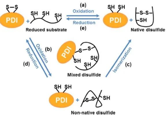
Figure 6. Schematic representation of thiol–disulfide interchange reactions catalyzed by PDI. ... 27
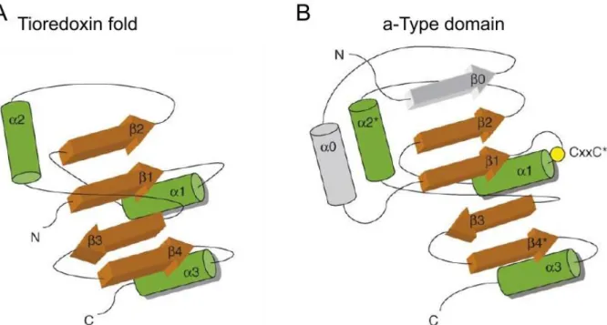
Figure 7. Domain organization of rat PDI. ... 29
Figure 8. Schematic representation of the organization of the thioredoxin fold and PDI domains. ... 31
Figure 9. The cDNA encoding BbPDI (GenBank accession number. DQ490130.1). ... 47
Figure 10. Multiple sequence alignment between the catalytic a and a' domains of Apicomplexa PDI family and predicted Secondary Structure Elements for the BbPDI model. ... 49
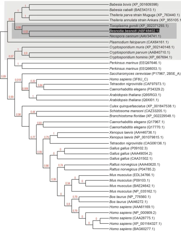
Figure 11. Mid-point rooting cladogram tree for PDI amino acid sequence. ... 51
Figure 12. B. besnoiti PDI model and binding pocket. ... 53
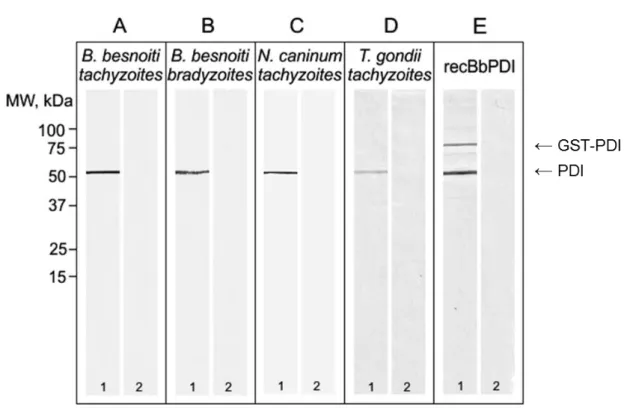
Figure 13. B. besnoiti PDI (BbPDI) enzymatic activity. ... 55
Figure 14. Western blot analysis using mouse serum. ... 56
Figure 15. Indirect immunolocalization of B. besnoiti PDI ... 57
Figure 16. Structure of an antibody molecule. ... 62
Figure 17. Proteolytic digestion of IgG by papain (left) and pepsin (right). ... 63
Figure 18. Schematic representation of the truncated version of recombinant BbPDI produced. ... 68
Figure 19. Coomassie stained SDS-PAGE (10%) of purified mAb (only elution fractions are shown). Under denaturing conditions the two light and heavy chains are separated. ... 76
Figure 20. Western blot analysis of monoclonal antibodies T8a, S4a, S16p and R60b over recBbPDI and truncated version produced, corresponding to domains a, a’c, b and b’. ... 77
Figure 21. Evaluation of cross reactivity of monoclonal antibodies T8a, S4a, R60b and S16p with PDI of N. caninum and T. gondii tachyzoites. ... 79
xvi
Figure 23. Reductase activity measured by the aggregation of insulin of (A) recombinant B. besnoiti PDI (recBbPDI) and (B) truncated version corresponding to domain a’c
(recBb-a’c). ... 92 Figure 24. Reductase activity, measured by the aggregation of insulin, of recombinant B.
besnoiti PDI (recBbPDI) in the presence of bacitracin. ... 95 Figure 25. Reductase activity, measured by the aggregation of insulin, of truncated
version corresponding to domain a’c (recBb-a’c) in the presence of bacitracin. ... 96 Figure 26. Reductase activity, measured by the aggregation of insulin, of recombinant B.
besnoiti PDI (recBbPDI) in the presence of tocinoic acid. ... 97 Figure 27. Reductase activity, measured by the aggregation of insulin, of truncated
version corresponding to domain a’c (recBb-a’c) in the presence of tocinoic acid. ... 98 Figure 28. Reductase activity, measured by the aggregation of insulin, of recombinant B.
besnoiti PDI (recBbPDI) in the presence of DTNB. ... 99 Figure 29. Reductase activity, measured by the aggregation of insulin, of truncated
version corresponding to domain a’c (recBb-a’c) in the presence of DTNB. ... 100 Figure 30. Reductase activity, measured by the aggregation of insulin, of recombinant B.
besnoiti PDI (recBbPDI) in the presence of pCMBA... 101 Figure 31. Reductase activity, measured by the aggregation of insulin, of truncated
version corresponding to domain a’c (recBb-a’c) in the presence of pCMBA. ... 102 Figure 32. Reductase activity, measured by the aggregation of insulin, of recombinant B.
besnoiti PDI (recBbPDI) in the presence of monoclonal antibody T8a (mAb T8a). ... 104 Figure 33. Reductase activity, measured by the aggregation of insulin, of recombinant B.
besnoiti PDI (recBbPDI) in the presence of monoclonal antibody S4a (mAb S4a). ... 105 Figure 34. Reductase activity, measured by the aggregation of insulin, of recombinant B.
besnoiti PDI (recBbPDI) in the presence of monoclonal antibody S16p (mAb S16p). ... 106 Figure 35. Reductase activity, measured by the aggregation of insulin, of recombinant B.
besnoiti PDI (recBbPDI) in the presence of monoclonal antibody R60b (mAb R60b). ... 107 Figure 36. Detection of B. besnoiti PDI in the excretory-secretory compartment. ... 108 Figure 37. In vitro invasion of Vero cells by B. besnoiti tachyzoites in the presence of
drug inhibitors. ... 110 Figure 38. In vitro invasion of Vero cells by B. besnoiti tachyzoites in the presence of
xvii
Table Index
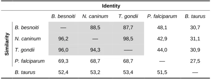
Table 1. List of primers used for PCR amplification in this chapter. ... 42 Table 2. Identity and similarity levels of PDI from B. besnoiti, N. caninum, T. gondii, P.
falciparum and B. Taurus. ... 46 Table 3. List of primers used for PCR amplification of the full length recBbPDI and the
truncated versions a, b, b’ and a’c. ... 67 Table 4. Recombinant proteins produced in this chapter. ... 69 Table 5. Resume of monoclonal antibody characterization. ... 78
xix
Abbreviations
ABS Absorvance
BbPDI Besnoitia besnoiti protein disulfide isomerase
BCR B-cell receptor
BPTI Bovine pancreatic trypsin inhibitor
cDNA Complementary DNA
DAPI 4',6-diamidino-2-phenylindole
DMEM Dulbeco’s Minimal Essential Medium DMSO Dimethyl sulfoxide
DNA Deoxyribonucleic acid
DsbA Disulfide bond formation protein A (thiol-disulfide oxidoreductase enzyme) DTNB 5,5′-dithiobis(2-nitrobenzoic acid)
DTT 1,4-Dithiothreitol
EFSA European Food Safety Authority ELISA Enzyme-linked immunosorbent assay
EM Electron microscopy
ER Endoplasmic Reticulum
FBS Fetal bovine serum
FITC Fluorescein isothiocyanate
GR Glutathione reductase
GSH Reduced glutathione
GSSG Oxidized glutathione GST Glutathione S-transferase
HAT Hypoxanthine, Aminopterin, Thymidine
HEPES 4-(2-hydroxyethyl)-1-piperazineethanesulfonic acid HGPRT Hypoxanthine guanine phosphoribosyl transferase hPDI Human protein disulfide isomerase
ICW Inner cyst wall
IFAT Immunofluorescence antibody test
Ig Immunoglobulin(s)
IgA Immunoglobulin, isotype A IgG Immunoglobulin, isotype G IgM Immunoglobulin, isotype M
IMC Inner membrane complex
xx
mAb Monoclonal antibody
NADPH Nicotinamide adenine dinucleotide phosphate
NTZ Nitazoxanide
OCW Outer cyst wall
ORF Open reading frame
pCMBA 4-chloromercuribenzoic acid, p-chloromercuribenzoic acid pCMBS p-chloromercuribenzenesulfonic acid
PCR Polymerase chain reaction
PDB Protein data base
PDI Protein disulfide isomerase PEG Polyethylene glycol
PMDB Protein model database PMDs Protein misfolding disorders p-NPP p- Nitrophenylphosphate
PV Parasitophorous vacuole
PVDF Polyvinylidene difluoride
rDNA Ribossomal DNA
recBbPDI Recombinant B. besnoiti protein disulfide isomerase
RMS Root mean square
RNA Ribonucleic acid
RNase Ribonuclease
RT-PCR Reverse transcription polymerase chain reaction
SDS-PAGE Sodium dodecyl sulphate-polyacrylamide gel electrophoresis SSU Small (ribosomal) subunit
TEM Transmission electron microscopy TRITC Tetramethylrhodamine
TRX Thioredoxin
UV Ultra violet
WB Western blot
1
Chapter 1 |
3
1.1 Bovine besnoitiosis
Besnoitiosis is a protozoal disease of cattle caused by the cyst-forming apicomplexan parasite Besnoitia besnoiti. It is a severe but usually non-fatal disease that leads to important economic losses due to significant reduction in productivity (Agosti, Belloli, Morini & Vacirca, 1994; Basso et al., 2013; Bigalke, 1968; Cortes et al., 2003; European Food Safety Authority [EFSA], 2010; Juste, Cuervo, Marco & Oregui, 1990; Olias et al., 2011; Pols, 1960). The first mention related to bovine besnoitiosis was published in 1884, when Cadéac described a skin disease in cattle from southern France, that he then, having no knowledge of the etiology, named bovine elephantiasis and anasarca («l’elephantiasis et de l’anasarque du boeuf») (Cadéac, 1884). The history of bovine besnoitiosis is, in fact, plenty of confusion and uncertainty concerning the identity of the etiological agent and the name of the disease, which conduced to different synonym based on the clinical aspects of the disease or the succeeding denominations for the parasite. With the investigations of Besnoit and Robin (1912), that revealed the presence of a large number of thick-walled cysts harboring numerous spores in the skin and subcutaneous tissues of affected cattle, the parasitic etiology of the disease was established. They realized that it was a new parasite, and suggested that it might be a Sarcocystis sp., designating the disease as (cutaneous) sarcosporidiosis. In the same year, Marotel (1912) pointed out that nothing similar had been found previously in cattle and, despite noting morphological characteristics that clearly differentiate this parasite from the known Sarcocystis spp. at the time, he proposed the species name of Sarcocystis besnoiti. The following year, Henry (1913), considering this morphological distinction, argued that the protozoa of Besnoit and Robin could not be placed in the genus Sarcocystis. He proposed the creation of the genus Besnoitia and that the parasite be referred as Besnoitia besnoiti, keeping the authorship of the species to Marotel. Meanwhile, in Portugal, Franco and Borges (1915, 1916) conducted a detailed description of the disease, in 67 animals they encountered in Lisbon abattoir from 1885 to 1914. In agreement with Henry (1913), they noticed the distinct characteristics that differentiated this parasite from the genus Sarcocystis and proposed also the name of Besnoitia besnoiti. However, in 1920 (Nöller), in a revision of the relevant literature up to the year of 1918 about the generic nomenclature of the genera Globidium, Gastrocystis, Ileocystis, Lymphocystis,
Haplogastrocystis and Besnoitia, regarded that the parasites with unknown complete life
cycle should be included in the genus Globidium, for the sake of simplicity. He then suggested the name Globidium besnoiti for the etiological agent of the Besnoit and Robin’s sarcosporidiosis. This suggestion was followed by other authors in subsequent publications (Pols, 1960), and lead to another designation of the disease: bovine globidiosis. In
4
subsequent years, the debate on how to classify this protozoan continued, but the consensus about the Besnoitia genus came in the mid twentieth century.
1.1.1 The genus Besnoitia and B. besnoiti
1.1.1.1 Life cycle and biological features
Besides B. besnoiti (type species), the genus Besnoitia comprises other three named species infecting large ungulates (Besnoitia caprae, Besnoitia bennetti and Besnoitia tarandi) and six infecting small mammals and lizards (Besnoitia akodoni, Besnoitia darlingi, Besnoitia
jellisoni, Besnoitia neotomofelis, Besnoitia oryctofelisi and Besnoitia wallacei) (Cortes, Leitao,
Gottstein & Hemphill, 2014; Olias et al., 2011). Presently, there are still questions regarding the differentiation of some of these species and their taxonomic status because only four have their life cycle clarified and morphological and molecular differences among the remaining species are poorly defined (Dubey, van Wilpe, Blignaut, Schares & Williams, 2013; EFSA, 2010; Olias et al., 2011).
It has been suggested that Besnoitia species, like other cyst-forming coccidian, have a facultative heteroxenous life cycle with a predator as a definite host and a prey as intermediate host (Basso, Schares, Gollnick, Rutten & Deplazes, 2011; Olias et al., 2011). The life cycle is well characterized only for four Besnoitia species found in small mammals from the American continent (B. darlingi, B. neotomofelis, B. oryctofelisi and B. wallacei), for which the cat was found to act as the definite host (Dubey & Lindsay, 2003; Dubey et al., 2002; Dubey et al., 2003b; Dubey & Yabsley, 2010; Frenkel, 1977; Smith & Frenkel, 1977, 1984; Wallace & Frenkel, 1975). Nevertheless, it is important to point that not all life forms (cysts/bradyzoites, tachyzoites or oocysts) of these parasites have been identified in nature. For B. darlingi, B. neotomofelis and B. oryctofelisi, the tissue cysts, containing bradyzoites, were identified in the intermediate host and the cat experimentally infected (Dubey & Lindsay, 2003; Dubey et al., 2002; Dubey et al., 2003b; Dubey & Yabsley, 2010; Smith & Frenkel, 1977, 1984), while for B. wallacei, the oocysts were isolated in the feces of one stray cat and susceptible intermediate hosts (Rattus norvegicus, R. exulans, and Mus
musculus) experimentally infected (Frenkel, 1977; Wallace & Frenkel, 1975). In this way,
while the life cycle of these species was experimentally maintained through a cat-prey-cat cycle, the natural intermediate host for B. wallacei remains unknown, as well as the cat real role in nature for the remaining species. No definite host for B. jellisoni and B. akodoni has been identified so far.
5
In regard to the Besnoitia species infecting large ungulates, although they have been studied for more than a century now (namely B. besnoiti, (Besnoit & Robin, 1912)), the definite host in the putative heteroxenous life cycle is still to be found (Alvarez-Garcia, Frey, Mora & Schares, 2013; Cortes et al., 2014; Olias et al., 2011). Homoxenous, direct transmission of bradyzoites and tachyzoites between intermediate hosts has been established experimentally in ungulates and several laboratory animals (Basso et al., 2011; Bigalke, 1968; Ng'ang'a & Kasigazi, 1994; Pols, 1960). Whether sexual reproduction really exists for all species of this genus is unknown, but since the closely related T. gondii, N.
caninum and Hammondia spp. use terrestrial carnivores to fulfill this process and the cat has
been identified has definite host in four Besnoitia species, it is very suggestive that large ungulate Besnoitia species also have a two-host life cycle (Olias et al., 2011). There have been several attempts to identify a definite host for these Besnoitia species, but only Peteshev, back in 1974, stated that both domestic cats and a wild cat (Felis Iybica), shed oocysts after ingestion of B. besnoiti cyst-containing tissues (Peteshev, Galuzo & Polomoshov, 1974). This observation, however, was never sustained by other authors. Diesing et al. (1988), fed domestic cats (Felis domestica), a dog (Canis familiaris), jungle cats (Felis chaus), caracals (Caracal caracal), small spotted genets (Genetta genetta), a lion (Panthera leo), leopards (Panthera pardus), cheetahs (Acinonys jubatus), banded mongooses (Mungos mungo), black-backed jackals (Canis mesomelas), a cape fox (Vulpes
chama), six species of snakes and white-backed vultures (Gyps africanus), with B. besnoiti
cystic material from chronically infected cattle and concluded that none of these species represents the definitive host of B. besnoiti, as no oocysts were found in their feces. Recently, these results were confirmed in experimental infections in domestic cats and beagle dogs (Basso et al., 2011). In the same way, experimental infections with B. tarandi failed in studies with domestic cats (Ayroud, Leighton & Tessaro, 1995), domestic cats and dogs (Dubey et al., 2004) and raccoon (Procyon lotor), domestic cat and arctic fox (Alopex
lagopus) (Glover, Swendrowski & Cawthorn, 1990), as well as experimental infections with B. caprae in cats (Ng'ang'a & Kasigazi, 1994). Evidences of a definitive host also fail in a
serological survey of B. besnoiti in free-living carnivores in Spain (Millan et al., 2012)
Apart from the search for the missing final host it should be considered that ungulates may in fact represent accidental hosts, playing a role as a reservoir of the parasite in a sylvatic life cycle (Basso et al., 2011; Diesing et al., 1988; EFSA, 2010; Kiehl et al., 2010; Olias et al., 2011). To the present date, there is no evidence of parasites of the genus
6
1.1.1.2 Taxonomy and phylogeny
The obligate intracellular protozoan parasites of the genus Besnoitia are classified in the sub-family Toxoplasmatinae of the family Sarcocystidae, within the phylum Apicomplexa (Ellis et al., 2000; Olias et al., 2011; Tenter et al., 2002). Phylogenetic analysis based on 18S rDNA sequences of B. besnoiti, B. jellisoni, B. caprae, Hammondia hammondi, Isospora spp., Frenkelia spp., Neospora caninum, Sarcocystis spp. and Toxoplasma gondii showed that Besnoitia species comprise a monophyletic and sister group to a clade containing other medically and veterinary important parasitic protozoa, such as Toxoplasma gondii, and
Neospora caninum (Figure 1) (Ellis et al., 2000; Olias et al., 2011). These results were
supported by Dubey et al. (2004) with analysis of both large and small subunit rDNA sequences.
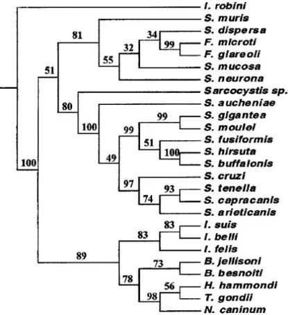
Figure 1. Phylogenetic tree based on 18S rDNA sequences.
Tree constructed from the parsimony analysis of the structure alignment of the SSU rDNA, representing the relationships amongst the cyst-forming coccidia sequences. Isospora robini was used as the outgroup. The numbers at the nodes represent the bootstrap values (% out of 500) (reprinted from Ellis et al., 2000, with permission from Elsevier)
7
Evolutionary relationships among Besnoitia species have been only scarcely investigated and are hindered by the deficit of gene sequences published for the different species and from different geographic regions. A striking example, is the fact that for B. wallacei, despite being known for forty years now (Wallace & Frenkel, 1975), there is no publically available sequence [GenBank search (http://www.ncbi.nlm.nih.gov/genbank/) for the term “Besnoitia
wallacei”; accessed on 18 February 2016]. The published phylogenetic analyses on the
genus Besnoitia have been based on the availability of ITS1 gene sequences. Although sequence information is not entirely complete for all species, it was possible to identify a
Besnoitia genus-specific cluster within the ITS1 region, which, by multiple alignments, was
demonstrated to be independent from other apicomplexans (Cortes et al., 2014). Phylogeny suggests that the Besnoitia genus is comprised of two distinct groups. One group includes the Besnoitia isolates from large ungulates (B. besnoiti, B. bennetti, B. caprae and B.
tarandi), while the other contains Besnoitia isolates from small mammals from the American
continent (B. akodoni, B. darlingi, B. jellisoni, B. oryctofelisi and B. neotomofelis) (Dubey & Yabsley, 2010; Kiehl et al., 2010; Olias et al., 2011). According to the Olias et al. (2011) analysis of available ITS1 sequences, that did not include the recently described B.
neotomofelis (Dubey & Yabsley, 2010), small mammal Besnoitia spp group show a genetic
divergence to large ungulate species group of 23.1% to 25.5%. Within the small mammal
Besnoitia species there is a higher degree of ITS1 sequence variation, between 1.6 to 5.9%,
while within large mammal Besnoitia species the sequences showed no variation (Figure 2). These differences might result from the contribution of the diploid phase, during the parasites differentiation and life cycle, where there is a bigger chance of recombination to occur. As previously discussed, in the case of B. besnoiti, the oral route of infection through oocysts shed by a definitive host has not been identified to date. However, it was demonstrated that transmission can occur by blood feeding insects such as horse flies and the stable fly,
Stomoxys calcitrans (Bigalke, 1968), therefore simply bypassing the sexual cycle and
promoting the parasite clonality (Cortes et al., 2014). This scenario is supported by comparison of the ITS1 sequences of B. besnoiti from different geographic regions, that show a lower degree of sequence variation for mechanically transmitted Besnoitia species. Olias et al. (2011) compared B. besnoiti ITS1 sequences from Israel, Portugal and South Africa and encountered no variation, while Kiehl et al. (2010) analyzed sequences from isolates of Israel, Portugal, Spain and Germany and found nearly identical sequences, with only a single nucleotide different in two of them.
8
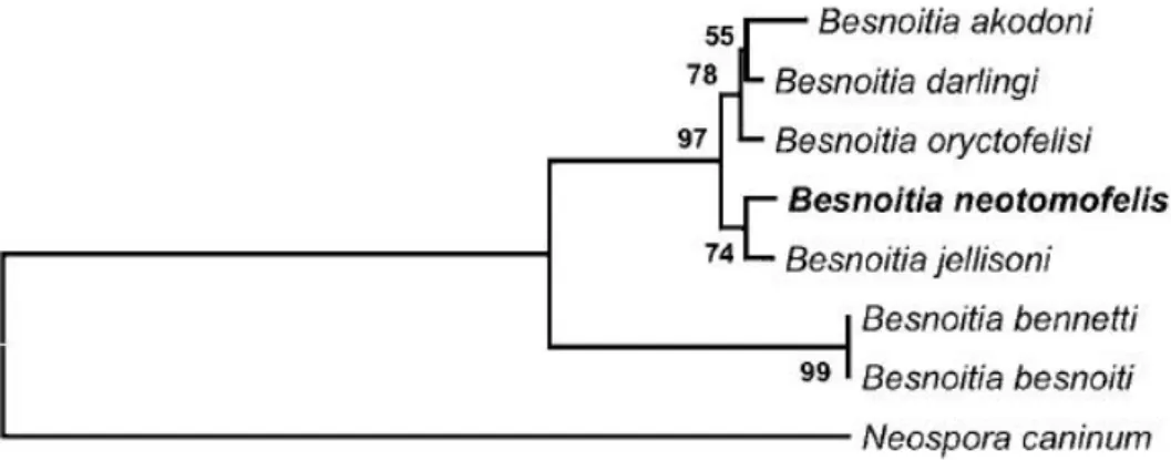
Phylogenetic analysis of the ITS1 sequence of B. neotomofelis, the latest identified
Besnoitia specie, placed it in a clade with B. jellisoni, which is a sister clade to one containing B. akodoni, B. darlingi, and B. oryctofelisi within the small mammal clade (Dubey & Yabsley,
2010) of the genus Besnoitia (Figure 3).
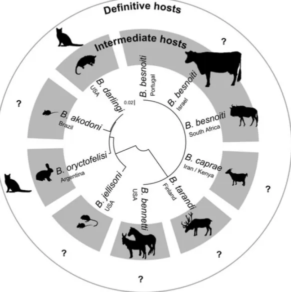
Figure 2. Phylogenetic tree of the ITS1 region of parasite isolates in the genus Besnoitia.
Tree constructed by unrooted neighbor-joining method. Eight out of 10 named Besnoitia species shown, as well as their known natural intermediate host (in gray). No natural definitive hosts are known for these species and the cat here depicted was established experimentally. Scale bar indicates genetic distance (reprinted from Olias, Schade & Mehlhorn, 2011, with permission from Elsevier).
9
Molecular analysis set in motion the debate about species differentiation that, especially in the case of coccidian, was traditionally based on phenotypic characters that include the morphology/ultrastructure of the available parasite stage and the host specificity. One problem with this approach is that in most cases only the oocyst stage and the ‘host’ by which it was shed were known when a new species was named. Such incomplete species descriptions are confounded by the fact that the ‘host’ described for the new species may not be its true natural host, because many animals may passage oocysts through their intestine without being a host for these parasites (Tenter et al., 2002). This may be further complicated in the case of heteroxenous or polyxenous parasites, where an incomplete understanding of the parasite life cycle can lead to different named species based on phenotypic characters such as the host specificity. An example of this situation is the well-known T. gondii, that, before the discovery of the oocyst and it’s heteroxenous life cycle (more than 60 years later from the description of the first stage of the parasite in the gondi (Ctenodactylus gundi)), several other species of Toxoplasma were named in accordance to the host species in which they were detected (Levine, 1977; Tenter et al., 2002). Besnoitia spp. may or may not be writing the same history than Toxoplasma and only further research, both at the molecular level and biological/epidemiological level, can elucidate this matter. Presently, we should deduce the status of a species from the analysis of molecular, morphological, biochemical, and/or ecological data all together (Kiehl et al., 2010). In the case of Besnoitia species, looking into the ITS1 sequences of large ungulates, we might be
Figure 3. Phylogenetic tree based on ITS1 region sequences of Besnoitia
neotomofelis and related organisms.
Percentages of 1000 bootstrap samplings that supported clades are shown on branches for Neighbor-joining analysis (reprinted from Dubey & Yabsley, 2010, with permission from Cambridge University Press).
10
drawn to conclude that presently there is no sufficient genetic evidence to consider B.
caprae, B. tarandi, B. bennetti and B. besnoiti separated species, since all published
sequences to date are identical or different with only one or two base insertions when compared to B. besnoiti (Ellis et al., 2000; Olias et al., 2011). Nevertheless, some points should be considered in this matter: first, as previously mentioned, genetic data available for these parasites from different isolates is limited and for sure represents a biased sampling of the biological diversity of Besnoitia spp. (Tenter et al., 2002); second, phylogenic definition of a species is almost impossible based in molecular data derived only from one gene, especially when there are no hints in which degree of diversity is needed to define a specie (Kiehl et al., 2010); Nevertheless, the size and sequence content of the ITS1 is recognized as a well-characterized species-specific marker amongst the coccidian such that parasite populations sharing the same, or highly similar, ITS1 sequences are likely to be derived from the same specie (Ellis et al., 2000); third, biological and ultrastructural details indicate substantial differences that, as stated, have to be weighed along with molecular data. Still, the Toxoplasma example shows us that we can have virulent and avirulent populations of T. gondii, which differ in their biological (e.g. spectrum of disease in the mouse) and genetic (e.g. SAG2 and RAPD PCR profiles) properties, possess identical ITS1 sequences (Ellis et al., 2000). When comparing B. besnoiti and B. caprae, we can found significant ultrastructural differences (Njenga, Bwangamoi, Kangethe, Mugera & Mutiga, 1995) and differences in host susceptibility: B. besnoiti is infectious for rabbits, mice, guinea pigs, hamsters, rats, sheep and cattle but B. caprae is not (Ng'ang'a & Kasigazi, 1994; Njenga, Bwangamoi, Mutiga, Kangethe & Mugera, 1993). Similarly, we can find differences in the ultrastructure and host susceptibility between B. besnoiti and B. tarandi (Dubey, Shkap, Pipano, Fish & Fritz, 2003a; Dubey et al., 2004), and between B. besnoiti and B. bennetti (Bigalke, 1970; Dubey et al., 2005; Pols, 1960). In this way, what we can presently sustain is that either B. caprae, B. tarandi and B. bennetti are different species with the same ITS1 sequence than B. besnoiti or that they represent distinct populations/strains of B. besnoiti. Clearly, additional information is required to resolve this controversy.
1.1.1.3 Morphological features
The rapidly dividing B. besnoiti tachyzoites represent the parasite proliferative stage responsible for the acute phase of the disease. With the onset of the host immune response and the presence of other physiological factors, tachyzoites differentiate into the slowly replicating encysted bradyzoites and a persistent tissue cyst infection is established along with the chronic phase of the disease (Alvarez-Garcia, Garcia-Lunar, Gutierrez-Exposito, Shkap & Ortega-Mora, 2014b; Buxton, McAllister & Dubey, 2002; Cortes et al., 2014;
11
Jacquiet, Lienard & Franc, 2010; Lyons, McLeod & Roberts, 2002). Tachyzoites and tissue cysts (containing bradyzoites) are the two tissue stages of B. besnoiti (and other Besnoitia species) found in intermediate hosts (Alvarez-Garcia et al., 2014b; Dubey et al., 2013).
B. besnoiti tachyzoites are typically crescent-shaped with a slightly pointed anterior
compared with the rounded posterior end (Langenmayer et al., 2015; Shkap, Yakobson & Pipano, 1988), measuring about 6-7.5 x 2.5-3.9 μm in cell culture (Reis et al., 2006). Similar to T. gondii and N. caninum, these tachyzoites can invade a wide range of host cells in vitro, with an asexual multiplication by endodyogeny (Cortes et al., 2014; Göbel, Widauer, Reimann & Munz, 1985), that takes place in the parasitophorus vacuole, a cytoplasmic compartment formed upon entry of the parasite into the host cell. By light microscopy tachyzoites of B. besnoiti, N. caninum or T. gondii cannot be distinguished. In vitro replication rates depend largely on the cell line used, going from 0.14 (on Vero cells) to 20 (on BHK21 cells) tachyzoites per hour per tachyzoite inoculated (Schares et al., 2009). Bradyzoites however, show a replication rate more than 100 times slower than that of cell cultures inoculated with tachyzoites (Schares et al., 2009). Morphologically they are very similar to tachyzoites, but reported measures fluctuate accordingly the author (Dubey et al., 2003a; Pols, 1960; Rostaher, Mueller, Majzoub, Schares & Gollnick, 2010; Sannusi, 1991), differing from 4.6 x 1.8 μm (Cortes et al., 2003) to 7-9 x 2.0 μm (Mehlhorn et al., 2009).
As with other apicomplexans, B. besnoiti bradyzoites persist for extended periods of time in the chronically infected animal, harbored in cysts in long living tissues, mainly the skin and aponevrosis (Cortes et al., 2014; Walker et al., 2014). The typical morphology of B. besnoiti tissue cysts is a strikingly large host cell containing a parasitophorous vacuole (PV) with large numbers of bradyzoites surrounded by a thick wall. They can grow up to 0.6 mm diameter (Pols, 1960) and are morphologically unusual because the host cell nuclei is incorporated inside the cyst and is hypertrophied (Dubey & Lindsay, 2003; Dubey et al., 2003b; Dubey et al., 2003c; Dubey et al., 2013; Dubey & Yabsley, 2010; Ernst, Chobotar, Oaksec & Hammond, 1968; Paperna & Lainson, 2001; Wobeser, 1976). This originates the characteristic double layer cyst-wall, that clearly distinguishes B. besnoiti cysts from its related apicomplexans (Cortes et al., 2014). Accurate morphological description of Besnoitia cysts is complicated by observed variation in cysts size and the thickness of the cyst wall, features that depend largely on the host affinity (natural or experimental host), tissue affinity and duration of infection (Dubey et al., 2003a). For B. besnoiti, although the existence of several studies that describe the cysts structure in naturally infected cattle, the nomenclature regarding the composition of the cysts varies significantly between authors. For example, Dubey et al. (2003a) and Cortes et al. (2006c) describe the cyst structures in a similar way (from in to out): intracellular cyst wall delineated by the parasitophorous vacuole membrane, host cell cytoplasm and membrane and outer cell cyst wall; Mehlhorn et al. (2009) talks of a
12
primary cyst wall and a secondary cyst wall; Later, Dubey et al. (2013) described B. besnoiti tissue cysts has having 3 layers or compartments: the outer layer with connective-like tissue, the middle layer incorporating host nuclei, and the cyst proper containing the bradyzoites. Recently, Langenmayer et al. (2014), in a study that followed cyst development on cattle through an 84-day trial post natural infection, proposed a new nomenclature for describing B.
besnoiti tissue cysts: 1) structures within the host cell should be called by their respective
name (bradyzoites, parasitophorous vacuole, membrane of the parasitophorous vacuole, host cell cytoplasm, nuclei, host cell membrane, etc.). 2) For the outermost acellular layer, they proposed the term outer cyst wall (OCW). The OCW is composed of interwoven collagenous material, is about 10-12 μm in thickness (Cortes et al., 2006c; Dubey et al., 2003a) and seems to be the result of the host physiological response (Cortes et al., 2014). 3) For the cyst wall of undeveloped cysts, which in developed cysts lies between the OCW and the membrane of the host cell, they proposed the name of inner cyst wall (ICW). This is a thick layer of up to 1 μm, also acellular, consisting of proteoglycan particles and fine filaments. Both OCW and the ICW seem to be the result of the host physiological response (Cortes et al., 2014). 4) The whole hypertrophied host cell, including the ICW and, if present, the OCW, is called a tissue cyst.
The parasitophorous vacuole is limited by a single cell membrane, frequently lined on the luminal side by a granular layer of approximately 0.2 μm thick (Dubey et al., 2003a; Langenmayer et al., 2015; Mehlhorn et al., 2009). No septation exists inside Besnoitia cysts as it is characteristic for Sarcocystis species (Mehlhorn et al., 2009). The host cell cytoplasm usually contains multiple large nuclei with occasional invaginations of the nuclear membrane. The organelles consisted mostly of dilated tubules of the ER, normal or swollen mitochondria and lysosomes. They are arranged in a circular manner around the PV, which occupies most of the host cell cytoplasm (Langenmayer et al., 2015; Mehlhorn et al., 2009).
1.1.1.4 Bradyzoite and tachyzoites ultrastructure features
The phylum Apicomplexa consists of intracellular parasites that have a characteristically polarized cell structure and a complex cytoskeletal and organellar arrangement at their apical end (Black & Boothroyd, 2000). Because the Apicomplexa are obligate intracellular parasites, survival depends on their ability to invade host cells (evading the host humoral immune response), avoid degradation by the host cell machinery (by blocking/modulating fusion of the vacuole with lysosomes, blocking vacuolar acidification and delaying premature cell death until parasite development is complete), and propagate intracellularly (recruiting cell resources and optimizing nutrient acquisition) (Binder & Kim, 2004; Nyalwidhe, Maier & Lingelbach, 2003; Sibley, 2011; Sinai & Joiner, 1997). The defining feature of Apicomplexa is
13
a complex assemblage of unique structural and secretory elements at the apical end of the parasite invasive life-cycle stages, named apical complex, that is intimately associated with these functions. (Binder & Kim, 2004; Katris et al., 2014; Kreier & Baker, 1987; Tenter et al., 2002).
The apical complex plays a key role in the mechanism of parasite motility, adhesion, invasion and formation of the parasitophorous vacuole, by providing both a semi-rigid framework to the parasite cell and a focal point for secretory organelles that release numerous invasion factors that mediate these functions. The apical complex is organized around an apical polar ring that serves as a microtubule organizing center that nucleates an array of subpellicular microtubules that descend toward the posterior of the cell (Katris et al., 2014; Morrissette & Sibley, 2002). These microtubules subtend flattened membrane sacs, or alveoli, that line most of the plasma membrane. A fibrous proteinaceous membrane skeleton supports the alveolar sacs against the microtubules. The alveoli and the proteinaceous skeleton form a structure called the inner membrane complex (IMC), which, together with the subpellicular microtubules, provides the shape and stability of the cell (Black & Boothroyd, 2000; Katris et al., 2014; Morrissette & Sibley, 2002). The plasma membrane is closely apposed over the IMC, forming together a structure called pellicle (Morrissette & Sibley, 2002). The apical polar ring marks the apical extremity of the IMC. A mobile conoid, consisting of tightly bent tubulin filaments fused to form a tapered hollow barrel, sits within the apical polar ring (Katris et al., 2014). The conoid can either be recessed in the cell, so that its tip is even with the apical polar ring, or, during invasion, be extruded from the apical polar ring to form an extended point to the cell. At the tip of the conoid are two preconoidal rings, and a pair of short microtubules sit eccentrically within the conoid. These preconoidal rings and interconoidal microtubules move together with the conoid during extrusion. The structural elements of the apical complex provide orientation to the cell, and the focal point for arrays of secretory organelles, micronemes and rhoptries and dense granules, that are organized towards the base of the conoid in readiness for a staged sequence of release (Katris et al., 2014). The electron-opaque dense granules are evenly distributed throughout the parasite cytoplasm, while the large club-shaped rhoptries and the small rod shaped micronemes are strictly confined to the anterior third of the cell (Carruthers, 1999). Several studies in other apicomplexans, especially T. gondii, have shown that microneme contents are secreted first, prior to invasion, and coat the parasite with proteins that facilitate host cell adhesion, gliding motility, and contribute to formation of an annular moving junction with the host plasma membrane through which the parasite enters the cell. During invasion rhoptries secrete further elements of the moving junction, as well as proteins that establish the properties of the parasitophorous vacuole in which the parasite will reside intracellularly (Katris et al., 2014; Sibley, 2011; Straub, Cheng, Sohn & Bradley, 2009). This junction can
14
be thought of as a “ring of contact” between the invading tachyzoite and the host cell membrane and is used by the parasite to forcibly enter the host cell. Once the parasite is completely inside the host cell, the moving junction disappears as the parasitophorous vacuole is sealed (Dubremetz, 2007; Walker et al., 2014). Finally, as the complete parasitophorous vacuole is detaching from the plasma membrane, dense granules penetrate the IMC within the apical region and liberate their content, by exocytosis, to the vacuole. Dense granule secretion coincides with the formation of a network of membrane tubules in the vacuole (named vacuolar network) and continues during the intracellular residence of the parasite (Figure 4) (Dubremetz, Achbarou, Bermudes & Joiner, 1993; Ngô et al., 2000).
Figure 4. Schematic representation of host cell invasion.
There is a sequential exocytosis of micronemes (green), rhoptries (red) and dense granules (blue) from different cellular locations. ER, host endoplasmic reticulum; Mi, host cell mitochondria; MJ, Moving junction PM, host cell plasma membrane; VM, vacuole membrane; VN, vacuole network; VP, vacuole pore. (adapted from Ngô, Hoppe & Joiner, 2000, with permission from Elsevier)
15
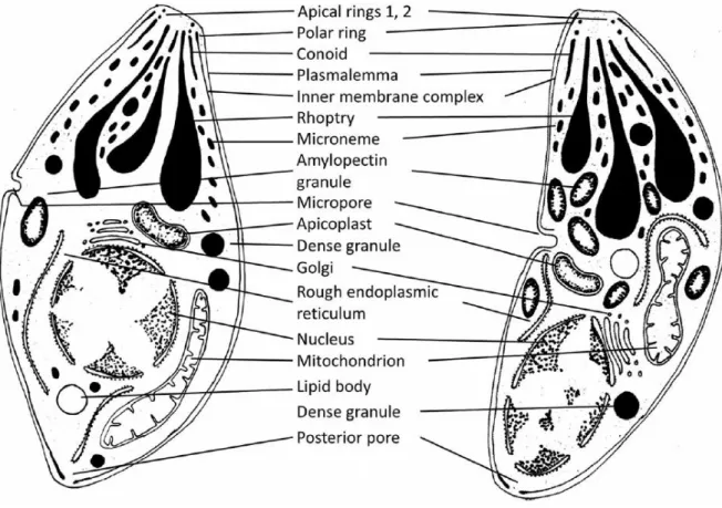
The elements of the apical complex are highly conserved throughout Apicomplexa (Katris et al., 2014), as it is the presence of the apicoplast, a plastid without photosynthetic ability (Arisue & Hashimoto, 2015). There are however some variations within the taxon and some species lack one or other feature of the apical complex (conoid in piroplasms, for example) or even lack the apicoplast (e.g. cryptosporidea) (Adl et al., 2012). B. besnoiti, being a coccidian, features the complete apical complex and the apicoplast and its ultrastructure is schematically presented in Figure 5. Here we can see the previously described elements, as well as other organelles and structures, including the nucleus, endoplasmic reticulum, Golgi complex, mitochondria, micropore, amylopectin granules and inclusion bodies (Dubey et al., 2013; Langenmayer et al., 2015; Shkap et al., 1988), similar to what is described for T. gondii (Dubey, Lindsay & Speer, 1998). The nucleus is usually situated in the middle or posterior third of the parasite (Langenmayer et al., 2015). In B. besnoiti zoites twenty two subpellicular microtubules radiate from the polar ring and extend far more than the 2/3 of the parasite length, sometimes reaching 4/5 of the cell body (Reis et al., 2006), as was defined for B.
jellisoni (D'Haese, Mehlhorn & Peters, 1977).
Figure 5. Schematic representation of the ultrastructure of Besnoitia besnoiti.
Bradyzoites (left) and Tachyzoites (right) (reprinted from Langenmayer et al., 2015, with permission of Springer)
16
1.1.2 Epidemiology
First reported cases of bovine besnoitiosis occurred more than a century ago in Europe, first in France (Besnoit & Robin, 1912; Cadéac, 1884) and after in Portugal (Franco & Borges, 1915, 1916). For years, besnoitiosis was considered an endemic sub-Saharan disease, because of the succeeding cases reported here and the lack of new cases reported in Europe. In Africa, the occurrence of bovine besnoitiosis was first discovered in 1945, by the South African state veterinary Christian Hofmeyr (Hofmeyr, 1945), with several other cases reported both in South Africa (Pols, 1960) and other countries, like Swaziland, Botswana, Namibia, Zimbabwe, Angola, Congo, Kenya, Tanzania, Uganda, Sudan, Cameroon and Nigeria (EFSA, 2010). In Asia the disease was reported in Israel (Goldman & Pipano, 1983; Neuman, 1972; Shkap, Reske, Pipano, Fish & Baszler, 2002; Shkap, Pipano & Greenblatt, 1987b), South Korea (Lee, Bak, Moon & Shin, 1970) and Russia (Krasov, Omarov Zh, Uvaliev & Khvan, 1975; Peteshev et al., 1974).
Bovine besnoitiosis remained silent in Europe until the last decade of the twentieth century, when new cases were described in northern Spain (Juste et al., 1990) and in Alentejo (Malta & Silva, 1991). But was until the beginnings of the twenty first century that the disease started to be a concern, having spread from southern France over western and central France (Alzieu, Dorchies, Schelcher & Gottstein, 2007; Bourdeau et al., 2004) and propagated within Portugal (Cortes et al., 2006c; Cortes et al., 2003; Cortes et al., 2005) and Spain (Castillo, Marcén, Ortega-Mora & Álvarez-García, 2009; Fernandez-Garcia et al., 2010; Fernandez-Garcia et al., 2009a; Fernandez-Garcia et al., 2009b; Irigoien et al., 2000).
B. besnoiti is considered endemic in large areas in Spain, Portugal and France, and recently
this status was also proposed for Italy (Gazzonis et al., 2014; Gentile et al., 2012a). Isolated outbreaks, described as resulting from the introduction of infected cattle from endemic areas, have been reported in Germany (Mehlhorn et al., 2009), Switzerland (Basso et al., 2013; Lesser et al., 2012), Italy (Agosti et al., 1994; Gentile et al., 2012b; Gollnick, Gentile & Schares, 2010; Mangili et al., 2012; Manuali et al., 2011; Mutinelli et al., 2011), Greece (Papadopoulos et al., 2014), Hungary (Hornok, Fedak, Baska, Hofmann-Lehmann & Basso, 2014), Croatia (Beck, Štoković & Pleadin, 2013) and Ireland (Ryan et al., 2016). The growing numbers of animals affected and the geographic expansion lead the European Food Safety Authority (EFSA) to consider bovine besnoitiosis as an emerging disease in Europe (EFSA, 2010).
There are still many aspects of the epidemiology of bovine besnoitiosis that are poorly understood, including prevalence and incidence data in endemic areas, routes of transmission and risk-factors associated to infection and disease. The complete life cycle of
17
of infected tissues, has not been identified yet, despite several studies with that purpose (Basso et al., 2011; Diesing et al., 1988; Millan et al., 2012). Hitherto, the only experimentally confirmed modes of transmission among cattle are mechanically, either through hematophagous insects, or iatrogenically through hypodermic needles (Bigalke, 1968; Pols, 1960). However, until now, no natural transmission of Besnoitia species has been proven and it is very likely that livestock trade of infected animals serves as a very important vehicle for long distance horizontal transmission of B. besnoiti (Frey et al., 2013; Olias et al., 2011), as it is discussed for recent outbreaks in France, Germany, Italy, Switzerland and Hungary (Agosti et al., 1994; Basso et al., 2013; Hornok et al., 2014; Mehlhorn et al., 2009; Mutinelli et al., 2011). After long-distance transport of single infected animals, the parasite might find adequate conditions to be transmitted locally to contact cattle, presumably by insect vectors and/or by direct contact between animals (Bigalke, 1968). Bigalke (1968), experimentally infected cattle orally with tachyzoites (bradyzoites were non-infective) and through the naso-pharyngeal route, with the inoculation of bradyzoites in the nostrils, but in nature, a feasible mechanism for the release of viable parasites (in infective quantities) from a chronically infected animal would have to be found. Skin cysts that might occasionally be exposed by trauma or spontaneously rupture to the surface are most unlikely to form a sufficiently reliable source of infection (Bigalke, 1968). The same should hold true when considering the venereal transmission of B. besnoiti, even though it has been suggested as possible (Frey et al., 2013; Gentile et al., 2012a; Gollnick, Scharr, Schares & Langenmayer, 2015). Consequently, the epidemiological significance of these modes of transmission is still unknown and further investigation is required. Moreover, it is not known if there are differences in the host specificity among B. besnoiti isolates from different parts of the world, or if other intermediate hosts than bovids are involved in the epidemiology of bovine besnoitiosis (Basso et al., 2011). In Europe, B. besnoiti natural infections were only confirmed in cattle, but in South Africa it was also described in wild ungulates (kudu (Tragelus strepsiceros), blue wildebeest (Connochaetes taurinus) and an impala (Aepyceros
melanpus) (McCully, Basson, Van Niekerk & Bigalke, 1966). For this reason, and also
because it can experimentally infect small mammals, the EFSA (2010) stated that wild ruminants and rodents should not be disregarded as reservoirs of the parasite. A serological survey in wild ungulates in Spain detected specific antibodies against Besnoitia spp. in a red deer (Cervus elaphus) and a roe deer (Capreolus capreolus) from the endemic region of the Pyrenees, suggesting a link between the sylvatic and domestic life cycles of Besnoitia spp. similar to what is observed in the lifecycles of other closely related apicomplexan parasites (Gutierrez-Exposito et al., 2013). In the same way, the relationship between B. besnoiti and other Besnoitia spp. isolated from ungulates (B. tarandi and B. caprae) should be investigated to assess the risk of infection for domesticated ruminants (EFSA, 2010).