Proteomic Identification of Oxidized Proteins
in
Entamoeba histolytica
by Resin-Assisted
Capture: Insights into the Role of Arginase in
Resistance to Oxidative Stress
Preeti Shahi1, Meirav Trebicz-Geffen1, Shruti Nagaraja1, Sharon Alterzon-Baumel1, Rivka Hertz1, Karen Methling2, Michael Lalk2, Serge Ankri1*
1Department of Molecular Microbiology, Bruce Rappaport Faculty of Medicine, Technion-Israel Institute of Technology, Haifa, Israel,2University of Greifswald, Institute of Biochemistry, Greifswald, Germany
*sankri@tx.technion.ac.il
Abstract
Entamoeba histolyticais an obligate protozoan parasite of humans, and amebiasis, an infectious disease which targets the intestine and/or liver, is the second most common cause of human death due to a protozoan after malaria. Although amebiasis is usually asymptomatic,E.histolyticahas potent pathogenic potential. During host infection, the par-asite is exposed to reactive oxygen species that are produced and released by cells of the innate immune system at the site of infection. The ability of the parasite to survive oxidative stress (OS) is essential for a successful invasion of the host. Although the effects of OS on the regulation of gene expression inE.histolyticaand the characterization of some proteins whose function in the parasite's defense against OS have been previously studied, our knowledge of oxidized proteins inE.histolyticais lacking. In order to fill this knowledge gap, we performed a large-scale identification and quantification of the oxidized proteins in oxida-tively stressedE.histolyticatrophozoites using resin-assisted capture coupled to mass spectrometry. We detected 154 oxidized proteins (OXs) and the functions of some of these proteins were associated with antioxidant activity, maintaining the parasite's cytoskeleton, translation, catalysis, and transport. We also found that oxidation of the Gal/GalNAc impairs its function and contributes to the inhibition ofE.histolyticaadherence to host cells. We also provide evidence that arginase, an enzyme which converts L-arginine into L-ornithine and urea, is involved in the protection of the parasite against OS. Collectively, these results emphasize the importance of OS as a critical regulator ofE.histolytica's functions and indi-cate a new role for arginase inE.histolytica's resistance to OS.
Author Summary
Reactive oxygen species are the most studied of environmental stresses generated by the host immune defense against pathogens. Although most of the studies that have
OPEN ACCESS
Citation:Shahi P, Trebicz-Geffen M, Nagaraja S, Alterzon-Baumel S, Hertz R, Methling K, et al. (2016) Proteomic Identification of Oxidized Proteins in Entamoeba histolyticaby Resin-Assisted Capture: Insights into the Role of Arginase in Resistance to Oxidative Stress. PLoS Negl Trop Dis 10(1): e0004340. doi:10.1371/journal.pntd.0004340
Editor:Alvaro Acosta-Serrano, Liverpool School of Tropical Medicine, UNITED KINGDOM
Received:August 18, 2015
Accepted:December 8, 2015
Published:January 6, 2016
Copyright:© 2016 Shahi et al. This is an open access article distributed under the terms of the Creative Commons Attribution License, which permits unrestricted use, distribution, and reproduction in any medium, provided the original author and source are credited.
Data Availability Statement:All relevant data are within the paper and its Supporting Information files.
investigated the effect of oxidative stress on an organism have focused on changes which occur at the protein level, only a few studies have investigated the oxidation status of these proteins. Infection withEntamoeba histolyticais known as amebiasis. This condition occurs worldwide, but is most associated with crowded living conditions and poor sanita-tion. The parasite is exposed inside the host to oxidative stress generated by cells of the host immune system. The nature of oxidized proteins in oxidatively stressedE.histolytica has never been studied. In this report, the authors present their quantitative results of a proteome-wide analysis of oxidized proteins in the oxidatively stressed parasite. They identified crucial redox-regulated proteins that are linked to the virulence of the parasite, such as the Gal/GalNAc lectin. They also discovered that arginase, a protein involved in ornithine synthesis, is also involved in the parasite's resistance to oxidative stress.
Introduction
Amebiasis is a parasitic infection of the intestines and is mainly caused by fecal contamination [1]. Although 90% of infected individuals are asymptomatic, amebic dysentery affects 50 mil-lion people in India, Southeast Asia, Africa, and Latin America and amebiasis is the cause of at least 100,000 deaths each year [2,3]. Following excystation within the small intestinal lumen, trophozoites colonize the large intestine and they usually reside in the colon as a non-patho-genic commensal in most infected individuals. Due to as yet unidentified causes, these tropho-zoites can cause amebic dysentery, become virulent and invasive, and migrate to the liver, via the portal veins, where they cause hepatocellular damage.
Following host invasion, invadingE.histolyticatrophozoites are challenged by oxidative stress (OS) and nitrosative stress (NS), which originate from fluctuations in ambient oxygen tension in the intestinal lumen and the generation of reactive oxygen species (ROS) and reac-tive nitrogen species (RNS) by cells of the immune system. Once formed, these reacreac-tive species can oxidatively damage proteins and change their structural conformation and functional activity [4], [5], [6]. The parasite's complex response to OS involves modulation of a large num-ber of genes which encode proteins that are associated with signaling/regulatory and repair/ metabolic pathways and proteins whose exact functions are still unknown [7]. It has been recently reported that the expression of these genes is regulated by a recently identified tran-scription factor that binds to a specific promoter motif of hydrogen peroxide (H2O2
)-respon-sive genes [8]. It has also been reported that those genes inE.histolyticawhich confer
resistance to OS also contribute to its virulence [9]. Since antioxidant enzymes, such as catalase, glutathione reductase, andγ-glutamyl transpeptidase, are missing fromE.histolytica's enzyme
resource [10], one of the functions of proteins, such as the 29-kDa peroxiredoxin [11] and the iron-containing peroxide dismutase [12], is to protect the parasite against OS. Since OS glycol-ysis is inhibited and metabolic flux is redirected towards glycerol production in oxidatively stressedE.histolyticatrophozoites, these findings suggest that the glycerol synthesis pathway is a component of the parasite's metabolic antioxidative defense system [13]. Despite these infor-mative data on the parasite's response to OS, our knowledge on the identity of oxidized pro-teins inE.histolyticais still incomplete. Here, we report the results of a study whose aim was to identify and to determine the biological relevance of oxidized proteins (OX) inE.histolytica using resin-assisted capture (RAC) coupled with mass spectrometry (MS) [14].
The results of this analysis revealed 154 OXs which include antioxidant proteins, cytoskele-ton proteins, protein involved in translation, and transport proteins. We also found that oxida-tion of cysteine residues in the carbohydrate recognioxida-tion domain (CRD) of the 260-kD
heterodimer and multifunctional virulence factor ofE.histolytica, Gal/GalNAc lectin (gl), impairs its ability to adhere to host cells. We also found that arginase, the enzyme which con-verts L-arginine into L-ornithine and urea, confers resistance to OS inE.histolytica.
Methods
Microorganisms
E.histolyticatrophozoites strain HM-1:IMSS were grown under axenic conditions in Dia-mond's TYI S-33 medium at 37°C. Trophozoites in the exponential phase of growth were used in all experiments.
DNA constructs
For the construction of the pJST4-arginase expression vector, arginase was amplified by poly-merase chain reaction (PCR) using the primers, Arginase KpnI and Arginase BglII (table 1). The PCR product was subcloned using the pGEM-T Easy vector system (Promega) and then digested with the restriction enzymes KpnI and Bgl II. The digested DNA insert was cloned into theE.histolyticaexpression vector pJST4 which had been previously linearized with KpnI and Bgl II. The pJST4 expression vector (pcontrol) contains a tandem affinity purification tag for use in protein purification and identification [15]. This CHH-tag contains the calmodulin binding protein, hemagglutinin (HA), and histidine (His) residues and its expression is driven by an actin promoter. This vector was used as control in our experiment in order to exclude the possibility that the CHH tag is responsible for the phenotypes of the arginase-overexpres-sing strain. A previously described protocol was used to transfectE.histolyticatrophozoites [16].
Viability assay
E.histolyticatrophozoites (1x10⁶) were exposed to 1 mM, 2.5 mM, 5 mM, 7 mM, or 10 mM H2O2for 60 minutes at 37°C. At the end of the exposure, a 10-μl aliquot of each culture was
stained with eosin (0.1% final concentration), and the number of living trophozoites was counted in a counting chamber under a light microscope. Resistance of the control, trophozo-ites overexpressing arginase and pcontrol trophozotrophozo-ites to OS was measured by calculating the median lethal dose (LD50) of hydrogen peroxide (H2O2) by linear regression analysis using
Microsoft Excel. The assay was repeated three times with two replicates in each assay.
Determination of intracellular ROS levels
Control and oxidatively stressedE.histolyticatrophozoites were incubated with 0.4 mM (final concentration) 2,7-dichlorofluorescein diacetate (H2DCFDA; Sigma) for 15 minutes in the dark. The cells were washed twice in phosphate buffered saline (PBS; pH 7.4) and immediately examined under a Zeiss Axio Scope.A1 fluorescence microscope. Intracellular ROS levels were determined by measuring fluorescence intensity using the ImageJ software [17].
Table 1. Oligonucleotides used in this study.
Primer Name Sequence Direction Restriction site-underline
Arginase Kpn1 GGTACCATGCAATTTGAAAAAGTTA Sense KpnI
Arginase Bgl11 AGATCTACACTTTATACCAAAAAGTG Antisense BglII
Detection of OXs by OX-RAC
E.histolyticatrophozoites (5x10⁷) were incubated with 2.5 mM H2O2for 60 minutes at 37°C.
At the end of the incubation, a total protein extract was prepared by lysing the oxidatively stressed trophozoites with 1% Igepal (Sigma) in PBS. OXs in the extract were detected by OX-RAC using a previously described protocol [14] with minor modifications. Briefly, the total protein extract (9 mg) was incubated in mixture of 50 mMN-ethylmaleimide and 2.5% sodium dodecyl sulfate (SDS) for one hour at 50°C with frequent vortexing in order to block the free thiols. The proteins were then precipitated with three volumes of cold 100% acetone and incubated at -20°C for 20 minutes. The mixture was centrifuged at 1820g for five minutes, and the pellet was then washed three times with 70% acetone (3 volumes) and then resus-pended in HENS buffer which contains 100 mM HEPES, 1 mM EDTA, 0.1 mM neocuproine, and 1% SDS). The resuspended samples were added to 80μl thiopropyl sepharose 6B resin (GE
Healthcare) in the presence or absence of dithiothreitol (DTT, final concentration 10 mM). DTT is a reducing agent, which enables the oxidized thiol group of cysteine to bind to the resin by forming disulfide bonds between the reduced thiol groups of the proteins and the thiol group of the resin. The samples were rotated in the dark at room temperature for 1–2 hours, and then overnight at 4°C. The resin was washed four times with 1 ml HENS buffer, and then twice with 1 ml HENS/10 buffer (1:10 HENS buffer). Captured proteins were eluted with 30μl
HENS/10 buffer which contained 100 mM 2-mercaptoethanol for 20 minutes at room temper-ature, and the proteins in each eluent were resolved on a 12.5% SDS-PAGE gel. Each gel was then stained with silver (Pierce Silver Stain) and each gel slice was independently analyzed by MS.
In-gel proteolysis for MS-based protein identification
The proteins in each gel slice were reduced with 2.8 mM DTT (60°C for 30 minutes), modified with 8.8 mM iodoacetamide in 100mM ammonium bicarbonate in the dark at room tempera-ture for 30 minutes, and digested overnight in 10% acetonitrile and 10 mM ammonium bicar-bonate with modified trypsin (Promega-Biological Industries, Israel) at 37°C. The resulting peptides were resolved by reverse-phase chromatography on 0.075 x 200-mm fused silica capillaries (J&W Scientific, Agilent Technologies, Israel) packed with Reprosil reversed phase material (Dr. Maisch GmbH, Germany). The peptides were eluted at flow rates of 0.25μl/min
on linear gradients of 7–40% acetonitrile in 0.1% formic acid for 95 minutes followed by eight minutes at 95% acetonitrile in 0.1% formic acid. MS was done by an ion-trap mass spectrome-ter (Orbitrap, Thermo) in a positive mode using a repetitively full MS scan followed by colli-sion-induced dissociation (CID) of the seven most dominant ions selected from the first MS scan. The MS data was analyzed using the Proteome Discoverer software version 1.3 which searches the Ameba section of the NCBI-NR database and the decoy databases (in order to determine the false discovery rate (FDR)) using the Sequest and the Mascot search engines.
Classification of OXs according to their protein class
The OXs were classified according to their protein class using the PANTHER software (Protein ANalysis THrough Evolutionary Relationships) Classification System (http://www.pantherdb. org/) [18].
Adhesion assay
H2O2for 20 minutes at 37°C, washed twice with Dulbecco's modified Eagle's medium
(DMEM) without serum, added to wells that contained fixed HeLa monolayers in 1 ml of DMEM without serum, and incubated for 30 minutes at 37°C. The number of adherent tropho-zoites was determined by counting the number of trophotropho-zoites that remained attached to the HeLa cells after gentle decanting (twice) of the non-adherent trophozoites with warm (37°C) DMEM under a light microscope.
Determination of
E
.
histolytica
motility
The Costar Transwell System (8-μm pore size polycarbonate membrane, 6.5-mm diameter,
Corning Inc, Corning, NY, USA) was used to determine trophozoite motility [20]. Briefly, 24-well culture plate was filled with serum-free Diamond’s TYI-S-33 medium (500-μl per
well). A transwell insert was then inserted into each well. Control and oxidatively stressed (2.5 mM and 1 mM for one hour at 37°C) trophozoites were washed three times in serum-free Dia-mond’s TYI-S-33 medium, and then suspended in serum-free Diamond’s TYI-S-33 medium. A 500-μl aliquot of the suspension (26x105trophozoites/ml) was then loaded into the transwell
inserts. The 24-well culture plate containing the transwell inserts was then placed in anaerobic bags (Mitsubishi Gas Chemical Company, Inc., Tokyo, Japan), and incubated for three hours at 37°C. At the end of the incubation, the inserts and culture medium were removed from the 24-well culture plate, and trophozoite migration was determined by counting the number of trophozoites that were attached to the bottom of the wells of the 24-well culture plate.
Purification of Gal/GalNAc lectin by affinity chromatography
Gal/GalNAc lectin was purified using a previously described protocol [21]Oxidation of purified Gal/GalNAc lectin
Aliquots (5μg) of purified Gal/GalNAc lectin were incubated with either 0.1 mM or 2.5 mM
H2O2for ten minutes at 37°C. The Gal/GalNAc lectin was then incubated with 10μl
D-galac-tose-coated agarose beads (Thermo Scientific-Pierce) overnight at 4°C. At the end of the incu-bation, the beads were washed in 20 volumes of PBS and then boiled in Laemmli sample buffer. The amount of Gal/GalNAc lectin that was released from the beads was determined using SDS-PAGE gel electrophoresis and silver staining (Pierce).
Detection of oxidized Gal/GalNac lectin
Aliquots (5μg) of purified Gal/GalNac lectin [21] were treated with 1 mM H2O2for ten
min-utes at room temperature in order to introduce carbonyl groups into protein side chains. Using the OxyBlot Protein Oxidation Detection Kit (Millipore, Israel) [22], the carbonyl groups are derivatized with 2,4-dinitrophenylhydrazine (DNPH). The DNPH-treated Gal/GalNac lectin was separated by SDS-PAGE, transferred onto a nitrocellulose membrane and then detected by a specific antibody against the dinitrophenyl (DNP) moiety of the OXs. The nitrocellulose membrane has been stripped and probed with a polyclonal Gal/GalNAc lectin antibody (a kind gift from of N. Guillen, Pasteur Institute, Paris, France) to confirm that equal amounts of puri-fied Gal/GalNac lectin were loaded on the gel.
Determination of protein synthesis by surface sensing of translation
(SUnSET)
trophozoites were incubated with 10μg/ml puromycin (Sigma), a structural analog of
tyro-syltRNA, for 20 minutes. For pretreatment of the trophozoites with cycloheximide (Sigma), the trophozoites were incubated with 100μg/ml cycloheximide for five minutes before adding
puromycin. The trophozoites were lysed using 1% Igepal (Sigma) in PBS. Puromycin was detected by immunoblotting using a monoclonal puromycin antibody (12D10 clone, Milli-pore). Protein quantification was measured by band intensity (densitometry) using ImageJ software [17].
Determination of arginase activity
Arginase activity inE.histolyticacrude lysate was spectrophotometrically measured by quanti-fying the amount of urea that is generated when L-arginine is hydrolyzed by arginase using a previously described protocol [24]. Briefly, 104trophozoites were dissolved in 100μl of 0.1%
Triton X-100 (Sigma) in the presence of 50μM
L-3-carboxy-2,3-trans-epoxypropionyl-leucyla-mido(4-guanidino)-butane (E-64) (Sigma), a cysteine protease inhibitor. The lysate (50μl) was
mixed with 50μl of Tris-HCl (50μM; pH 7.5) which contained 10 mM MnCl2, and then
acti-vated by heating for ten minutes at 55°C. The hydrolysis of L-arginine by arginase was initiated by adding 25μl L-arginine (0.5 M; pH 9.7) to a 25μl aliquot of activated lysate. After a
30-min-utes incubation at 37°C, the reaction was stopped by adding 400μl of an acid solution mixture
(H2SO4: H3PO4: H2O = 1: 3: 7). The urea concentration in the mixture was measured at 570
nm after addingα-isonitrosopropiophenone (25μl, 9% in absolute ethanol) to the mixture,
heating the mixture for 45 minutes at 100°C, and incubating the mixture in the dark for ten minutes at room temperature.
HPLC analysis of amino acids in culture supernatants and intracellular
amino acids
The amino acid levels in culture supernatants were analyzed by high-performance liquid chro-matography (HPLC) using a previously described protocol [24] in which the proteins in the culture supernatant are first precipitated with methanol, and the amino acids are derivertized using o-phthalaldehyde (OPA) in an alkaline medium. Briefly, 200μl of culture supernatants
are added to an 800-μl mixture of methanol and internal standard (homocysteic acid). After
centrifugation, the samples are loaded into the HPLC autosampler, which converts the samples to fluorescent derivatives (by mixing them with OPA) before their injection into the columns (C-18). A JASCO FP 1520 fluorescence detector at an excitation wavelength of 360 nm with emission detection at 455 nm was used to separate, detect, and quantify the fluorescent deriva-tives. For quantification of the intracellular amino acids, trophozoites (107) were lysed in 1 ml of trichloroacetic acid (TCA) 10% for 30 minutes at 4°C, and centrifuged, and the pH of the supernatants was adjusted to 12 using 10N NaOH. The amino acid concentration in the super-natants was then measured by HPLC on two biological replicates.
1
H-NMR spectroscopic analysis of putrescine in trophozoite lysates
The quantification of putrescine in the trophozoite lysates was performed using1H- nuclear magnetic resonance (NMR) spectroscopy as previously described [25,26]. Two biological repli-cates were used for each measure. Briefly, 400μl H2O was used to dissolve the lysate and mixedwith 200μl buffer solution containing the internal standard TSP (3-trimethylsilyl-[2,2,3,3-D4
software (Bruker Biospin GmbH) as previously described [25,26]. The quantification was per-formed using the software package CHENOMX (version 8.1).
Results
Characterization of oxidized proteins in oxidatively stressed
E
.
histolytica
trophozoites
WhenE.histolyticatrophozoites strain HM-1:IMSS were incubated with 1 mM, 2.5 mM, 5 mM, 7 mM, or 10 mM H2O2for 60 minutes at 37°C, the calculated LD50 of H2O2is 5.5 ± 0.1
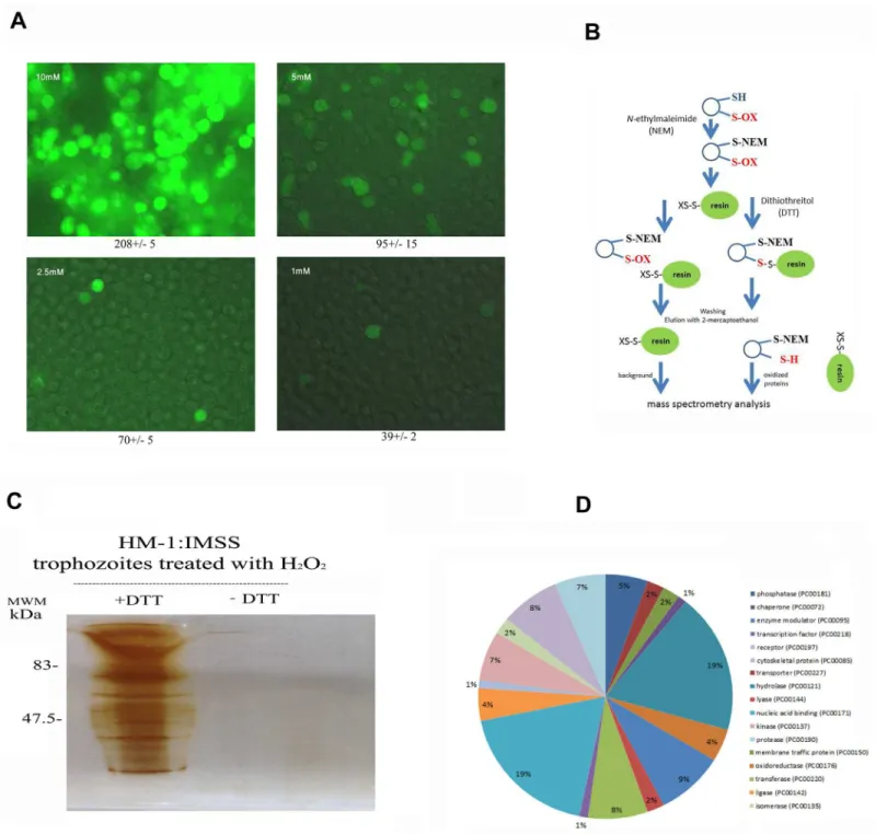
mM (Table 2) and the intracellular ROS levels in living trophozoites are high (Fig 1A). Based on these results, we selected 2.5 mM as the H2O2concentration to oxidatively stress
trophozo-ites in our various assays because this concentration is not lethal (85% of the trophozotrophozo-ites are viable;S1 Fig), the intracellular ROS levels are relatively low (Fig 1A), and OXs are formed (this work). We then used OX-RAC coupled to label-free quantification LC-MS for the detec-tion and quantificadetec-tion of OXs in the lysate of oxidatively stressed trophozoites (Fig 1B). A pro-tein was considered to be oxidized when its relative amount in the DTT-treated lysates was at least two times greater than that in the untreated lysates (Fig 1C). We identified 154 proteins that met this condition (S1–S3Tables). These 154 proteins were then classified (Fig 1D) using PANTHER sequence classification tool [27,28]. The protein classes were phosphatases (exem-plified by phosphoinositide phosphatase (EHI_141860); transporters (exem(exem-plified by plasma membrane calcium-transporting ATPase, EHI_030830); membrane traffic proteins (exempli-fied by putative vacuolar sorting protein, EHI_025270); chaperones (exempli(exempli-fied by Hsc70-in-teracting protein, EHI_158050); hydrolases (exemplified by arginase, EHI_152330);
oxidoreductases (exemplified by superoxide dismutase, EHI_159160); enzyme modulators (exemplified by Ras family GTPase, EHI_058090); lyases (exemplified by tRNA pseudouridine synthase, EHI_151650); transferases (exemplified by histone acetyltransferase, EHI_152010); nucleic acid binding proteins (exemplified by 13 kDa ribonucleoprotein-associated protein, EHI_104600); ligases (exemplified by ubiquitin-conjugating enzyme family protein, EHI_070750); kinases (exemplified by galactokinase, putative, EHI_094100); isomerases (exemplified by cysteine synthase A, EHI_024230); cytoskeletal proteins (exemplified by actin-binding protein, cofilin/tropomyosin family, EHI_168340) and proteases (exemplified by methionine aminopeptidase, EHI_126880). In order to evaluate the consistency of MS-based identification of OXs, the purified heavy subunit of Gal/GalNac lectin (Hgl) was exposed to 1 mM H2O2for ten minutes and its oxidation was confirmed independently by using the
Oxy-Blot kit. The presence of carbonyl groups was detected using a specific antibody which recog-nizes the DNP moiety in the purified lectin that has been exposed to H2O2and treated with
Table 2. LD50of H2O2inE.histolyticatrophozoites strain HM-1 IMSS, trophozoites overexpressing
arginase and pcontrol trophozoites.
Strain LD50of H2O2 (mM)
E.histolyticatrophozoites strain HM-1:IMSS 5.5±0.1
pcontrol trophozoites 5.1±0.1
arginase-overexpressingE.histolyticatrophozoites 6.2±0.08
Data are expressed as the mean and standard error of mean of three independent experiments that were performed in duplicate. The means of the different groups for three independent experiments were compared using an unpaired Student’s t test. The LD50of HM-1 IMSS or pcontrol trophozoites was
significantly different (p<0.05) from that of the arginase-overexpressing trophozoites.
Fig 1. Analysis of oxidized proteins inE.histolyticaafter resin-assisted capture.A. Detection of intracellular ROS level. Control and oxidatively stressed trophozoites were incubated with 0.4 mM (final concentration) 2,7-dichlorofluorescein diacetate for 15 minutes in the dark and then examined by fluorescence microscopy. The number in the top left side of each picture indicates the concentration of H2O2. The number in the bottom of each picture
indicates the mean fluorescence for 20 cells according to a measure performed by image J. B. Methodology of OX-RAC. The scheme has been adapted from [14]. C. Silver staining of oxidized proteins.E.histolyticatrophozoites strain HM-1:IMSS were treated with H2O2for 60 minutes at 37°C and a total protein
lysate was prepared by lysing the H2O2-treated trophozoites with 1% Igepal (Sigma) in phosphate buffered saline. The oxidized proteins in the cell lysates
were subjected to resin-assisted capture (RAC) in the presence of 10 mM DTT (+DTT) or the absence of DTT (–DTT). D. Functional categories of all oxidized proteins. Oxidized proteins inE.histolyticawere classified according to the protein class they encode using PANTHER sequence classification tool.
DNPH (Fig 2A). As expected, carbonyl groups were not detected in purified lectin that was not exposed to H2O2and treated with DNPH.
According to the PANTHER statistical overrepresentation test which compares classifica-tions of multiple clusters of lists to a reference list, very significant enrichment (fold
enrichment>5 and P>2.42x10⁻⁶) was found for proteins involved in the process of
transla-tion, such as the 60S ribosomal protein L9-like protein (EHI_193080) and the 13 kDa ribonu-cleoprotein-associated protein (EHI_104600).
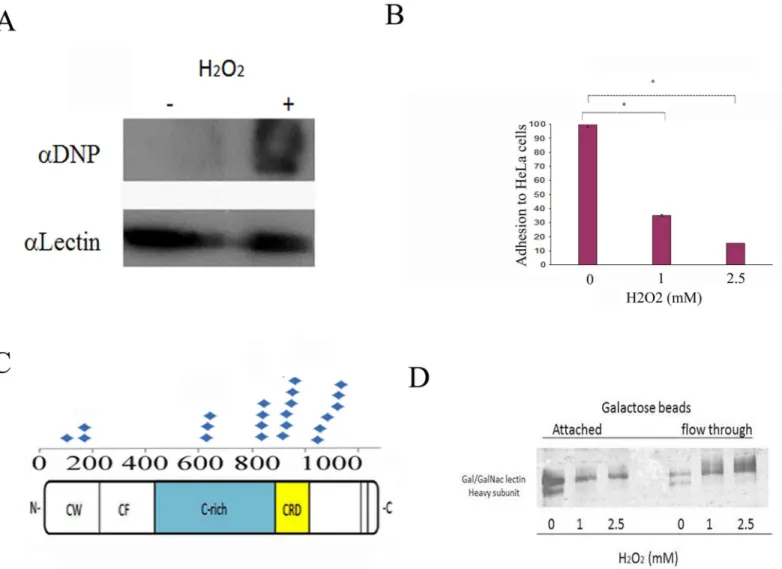
Fig 2. H2O2inhibits the adhesion ofE.histolyticato HeLa cells.A. Confirmation of OX-RAC data about the oxidation of the Hgl by using the OxyBlot
protein oxidation detection kit. This figure displays a representative result from two independent experiments. B.E.histolyticatrophozoites strain HM-1:IMSS were grown in Diamond’s TYI-S-33 medium and exposed to H2O2for 20 minutes before their incubation with a fixed HeLa cell monolayer for 30 minutes at
37°C. Data are expressed as the mean±standard deviation of three independent experiments that were performed in triplicate. The adhesion of the H2O2 -treated trophozoites was significantly different (p<0.05) from the control (100%) according to the results of an unpaired Student’s t-test in which statistical significance was set at 5%. C. Repartition of the carbamidomethylated cysteine residues in the Hgl as an indication of their oxidation status. These residues were mostly located in the cysteine-rich region (C-rich) in the carbohydrate recognition domain (CRD) and in the cysteine-tryptophan domain (CW) and were absent in the cysteine-free domain (CF). D. Dose-dependent inhibition of Gal/GalNAc lectin binding to D-galactose-coated agarose beads by H2O2. Gal/
GalNAc lectin was purified by D-galactose affinity chromatography and incubated with different concentrations of H2O2for 10 minutes at 37°C. The binding to
galactose-coated agarose beads was determined by SDS-PAGE gel electrophoresis and silver staining. This figure displays a representative result from two independent experiments.
The results of a previous study showed that (a) gene expression of oxidatively stressedE. his-tolyticatrophozoites triggers a stress response in which 185 genes are upregulated and 102 genes are downregulated and (b) these genes are involved in signaling/regulatory processes, metabolic/repair processes, energy metabolism, the stress response, and transport [7]. We found that some of the OXs in the oxidatively stressed trophozoites are involved in the stress response, transport, and metabolism. Of all the proteins that were found to be oxidized and all the genes whose expression had changed in the oxidatively stressed trophozoites, EHI_179080 (sulfate adenylyltransferase) was the only common gene.
Cysteine residues are the predominant targets of oxidation or S-nitrosylation in redox-sen-sitive proteins. We have recently identified 142 S-nitrosylated (SNO)-proteins inE.histolytica after its exposure to NO [21]. 21 proteins were shared in our OX-RAC and SNO-RAC analysis (Table 3). The shared proteins include rubrerythrin, protein disulfide isomerase and iron-con-taining superoxide dismutase which have been associated with resistance to OS [12,27–29] and the Gal/GalNac lectin, a cell surface protein which is involved in binding ofE.histolytica troohozoites to host cells [17,18].
Regulation of the Gal/GalNAc Lectin by OS
In order to gain information on the consequence of oxidation on the activity of some of the proteins that were identified in the OX-RAC analysis, we decided to focus our analysis on the function of the oxidized Gal/GalNac lectin. The Gal/GalNac lectin consists of Hgl (170 kDa) and a light subunit (Lgl) (35/ 31 kDa). Hgl mediatesE.histolyticaadherence, and indirect evi-dence suggests that Lgl plays a role inE.histolyticavirulence [13,19]. The occurrence of Hgl among the OXs (seeS2 Table) and SNO proteins ([21] andTable 3) suggests that OS and NS affect the parasite's adherence. In order to test this hypothesis, we compared the ability of
Table 3. Common proteins in the OX-RAC analysis (this study) and SNO-RAC analysis [21].
Accession Description
183232225 Plasma membrane calcium-transporting ATPase
67469645 Hsc70-interacting protein
67481145 Rab family GTPase
67474574 hypothetical protein
67462443 NA modification enzymes, MiaB-family
67472683 rubrerythrin
67469707 Rho family GTPase
67477041 Ras family GTPase
67465045 60S ribosomal protein L18a
5689218 serine acetyltransferase
67482289 peptidyl-prolyl cis-trans isomerase
67465747 60S ribosomal protein L7
67481543 40S ribosomal protein S13
183234048 alcohol dehydrogenase
67465285 Iron-containing superoxide dismutase
67464751 ARP2/3 complex 20 kDa subunit
67465767 Galactokinase
67481663 Hgl
183232524 adenylyl cyclase-associated protein
67478039 hypothetical protein
52421800 protein disulfide isomerase
untreated and oxidatively stressed trophozoites to adhere to HeLa cells [13]. We found that the binding of the oxidatively stressed trophozoites to the HeLa cell monolayer was significantly less than that of the untreated trophozoites (65% and 85%;Fig 2B). Previous research results show that the cysteine-rich region (CRR) of the Hgl and the CRD are important for the binding activity of the lectin [19,20]. Our MS analysis of OXs indicates that many carbamidomethy-lated cysteine residues, that possibly represent oxidized cysteines, are located in the CRR and in the CRD of Hgl (Fig 2C). When we investigated the binding ability of oxidized Gal/GalNAc lectin, we observed that H2O2prevents the binding of the Gal/GalNAc lectin to the galactose
beads (Fig 2D).
Effect of OS on
E
.
histolytica
motility
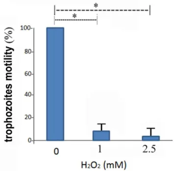
Motility is important forE.histolyticasurvival and pathogenicity, and requires a dynamic actin cytoskeleton [30]. The presence of cytoskeleton-associated proteins, such as the ARP2/complex 20kDa subunit, among the OXs (S2 Table) and the SNO- proteins ([21] andTable 3) suggests that the parasite's motility is redox-regulated. We tested this hypothesis by comparing the migration of control and oxidatively stressed trophozoites: the migration of the oxidatively stressed trophozoites was significantly less than that of the control trophozoites (Fig 3).
Effect of OS on protein synthesis
Inhibition of translation is a typical response of cells exposed to stress conditions [31,32], and such inhibition may avoid constant gene expression during error-prone environments. We recently reported that NS inhibits protein synthesis inE.histolytica[33]. The presence of the 60S ribosomal protein L7, the 60S ribosomal protein L18a and the 40S ribosomal protein
Fig 3. Effect of H2O2onE.histolyticamotility.The motility of control and oxidatively stressedE.histolytica
trophozoites was investigated using a transwell assay (seematerial and methodsfor details). The motility of control trophozoites that were not exposed to H2O2was set at 100%. Data are expressed as the
mean±standard deviation of three independent experiments that were repeated twice. The motility of the H2O2-treated trophozoites was significantly different (p<0.05) from that of the controls according to the results
of an unpaired Student’s t-test in which statistical significance was set at 5%.
S13 among the OXs (S2 Table) and SNO proteins ([21] andTable 3) suggest that oxidation is similar to S-nitrosylation [21], in that it regulates the translation of proteins in the parasite. In order to test this hypothesis, we used SUnSET [23][33,34] to determine protein synthesis. We found that protein synthesis is strongly inhibited in oxidatively stressed trophozoites and that the level of inhibition is comparable to that found in cyclohexamide-treated trophozoites (Fig 4) [35].
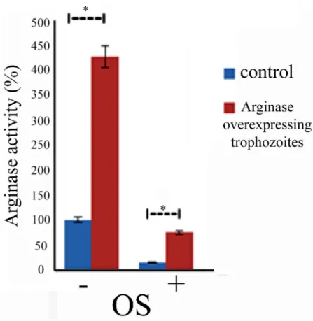
Overexpression of arginase confers resistance to OS
We previously reported that the enzymatic conversion of L-arginine to L-ornithine is an signif-icant source of L-ornithine forE.histolyticaand that arginase activity is essential for the resis-tance of the parasite to NS [24]. The presence of L-arginase among the OXs raises questions about the regulation of its activity by OS and its involvement in the resistance of the parasite to OS. In order to obtain information about the effect of OS on arginase activity, we measured arginase activity in control and oxidatively stressed trophozoites using a previously described assay [24]. We found that arginase activity is markedly inhibited (90% inhibition) in the oxida-tively stressed trophozoites (Fig 5).In order to establish whether arginase gene expression is
involved in the resistance of the parasite to OS, we decided to upregulate its expression (Fig 5). We detected modest arginase activity in arginase-overexpressing trophozoites and no arginase activity in control trophozoites that were exposed to 2.5 mM H2O2for 15 minutes (Fig 5). We
found that the arginase-overexpressing trophozoites are more resistant to OS (LD506.2±0.08
mM) than the control trophozoites (IC50LD505.1±0.1 mM) (table 2). We also found that the
intracellular arginine concentration in the arginase-overexpressing trophozoites (53 ± 5μM)
was significantly lower than that in the control trophozoites (136±9μM). No significant
differ-ence in the intracellular ornithine concentration in the arginase-overexpressing trophozoites (616±7μM) and the control trophozoites (577±15μM) was detected. In contrast, we found
that the intracellular concentration of putrescine in the arginase-overexpressing trophozoites (293±20 nmol/mg protein) was significantly higher than that of the control trophozoites (123 ±3 nmol/mg protein).
Fig 4. Effect of H2O2onE.histolyticaprotein synthesis measured using puromycin-labeled proteins.
Lane 1: Trophozoites were treated with 10μg/ml puromycin (lane 1), not treated with puromycin (lane 2),
treated with 2.5 mM H2O2for 15 minutes and then labeled with 10μg/ml puromycin for 20 minutes (lane 3)
and treated with cycloheximide (100μg/ml) before puromycin labeling (lane 4). The extracts were separated
by denaturing electrophoresis and analyzed by immunoblotting with a monoclonal puromycin antibody 12D10 clone. An actin immunoblot is shown as the loading control.
Discussion
The current understanding of the antiamebic effect of OS [36] has greatly beneficiated from previous omics studies [13] [7] on the response ofE.histolyticato OS. However, these studies did not address the nature of OXs in oxidatively stressedE.histolyticatrophozoites. We decided to use OX-RAC coupled to MS [14] to generate new data about OXs inE.histolytica. Some of the proteins that we identified in our OX-RAC analysis of OXs are of particular inter-est because (i) they have an important function in the parasite's physiology and/or virulence and (ii) their activity is regulated by OS in other organisms.
The first notable OX that we identified in our OX-RAC analysis is the cysteine-rich (C-rich) region (amino acids 356–1143) and a CRD (amino acids 895–998) of Hgl [20]. We recently showed that S-nitrosylation of cysteine residues in the CRD inhibits the galactose binding activity of the Gal/GalNAc lectin and contributes to the reduced binding of NO-treated tro-phozoites to their target cells [21]. In this study, we found that carbamidomethylated cysteine residues are also located in the CRR and the CRD of the lectin. This result suggests that oxida-tion of these cysteines is responsible for the loss of galactose-binding activity of the Gal/Gal-NAc lectin.
Fig 5. Overexpression of arginase confers resistance to OS.Arginase activity was measured in crude lysates that were prepared from control and arginase-overexpressingE.histolyticatrophozoites exposed or not to OS. Data are expressed as the mean±standard deviation of three independent experiments that were repeated twice. The arginase activity of the arginase-overexpressing trophozoites was significantly different (p<0.05) from the control (100%) according to the results of an unpaired Student’s t-test. The arginase activity of the oxidatively stressed arginase-overexpressing trophozoites was significantly different (p<0.05) from the oxidatively stressed control according to the results of an unpaired Student’s t-test. Arginase activity in the control trophozoites was 120μmol urea/min/mg proteins.
We found that cytoskeletal proteins are oxidized in oxidatively stressed trophozoites. For its invasion into the host's tissues,E.histolyticarelies on its dynamic actin cytoskeleton [37], [38]. Oxidation of the actin cytoskeleton can modulate its cellular functions in mammalian cells [39] [40] [41] and the results of proteomics studies in human peripheral blood mononuclear cells have previously identified actin as an oxidation target [42]. Actin oxidation inhibits its poly-merization [43] [44] and leads to cytoskeletal rearrangements [45]. According to previous reports, the cysteines in actin are some of the most susceptible targets of oxidation [45] and their oxidative modification is the likely cause of cytoskeletal rearrangements in oxidatively stressed mammalian cells [46] [47] [48]. Actin is a highly conserved protein among species [49]. The actin of rats andE.histolyticashare a number of oxidized amino acid residues (Met44, Met47) which are located in actin−actin contact regions [50] suggesting that actin
oxi-dation is part of the process that results in the inhibition of the transwell migration that we observed in the oxidatively stressed trophozoites.
We detected strong enrichment of OXs which are components of the parasite's translational machinery, such as ribosomal proteins and elongation factors. This finding is in agreement with the results of a recent study in yeast cells, in which it was reported increases in ribosomal proteins and elongation factors due to oxidative thiol modifications following a short-term exposure to H2O2[51]. At first glance, this finding is also in agreement with the inhibition of
protein synthesis in oxidatively stressedE.histolyticatrophozoites (this work) and mammalian cells [52]. However, a recent report suggests that the oxidation of components of the transla-tional machinery is not the direct cause of the inhibition of protein synthesis, but rather a global, enzymatic downregulation of almost all tRNA species in OS [53]. It will be interesting to determine whether the same enzymatic downregulation of tRNA species also occurs in oxi-datively stressedE.histolytica.
An additional group of OXs which we identified are the oxidoreductases, which includes the iron-containing superoxide dismutase. OS induces the expression of this protein [12] and the protein can form adducts with metronidazole metabolites [54]. InE.coli, iron-containing superoxide dismutase is inactivated by H2O2via a reaction of H2O2with the iron at the active
site that generates a potent oxidant which attacks tryptophan residues [55]. It has been recently showed forTrypanosomasuperoxide dismutase (Fe-SODB) that Cys(83) in Fe-SODB acts as an electron donor that repairs the tyrosyl radical (Tyr35-O•) via intramolecular electron trans-fer in order to prevent inactivation of Fe-SODB by peroxynitrite, which is produced by immu-nostimulated macrophages [56]. Interestingly, this Cys(83) is conserved inE.histolytica's iron-containing superoxide dismutase and Cys(83) was detected by MS as a carbamidomethylated cysteine residue which strongly suggest that it has been oxidized. Based on this data, it is tempt-ing to speculate that Cys(83) inE.histolytica's iron-containing superoxide dismutase is crucial for protecting the protein against inactivation by H2O2.
The phosphatases are another group of OXs which we identified in our OX- RAC analysis. This group includes the protein-tyrosine phosphatases (PTP). Two PTPs have been cloned fromE.histolytica, EhPTPA and EhPTPB. EhPTPA but not EhPTB is strongly up-regulated in trophozoites that have been recovered from amebic liver abscesses suggesting that EhPTPA is involved in the parasite’s virulence. [57].
histolyticaEhPTPA activity will be inhibited by OS and in the future, it will be interesting to study the consequence of this inhibition on the parasite’s virulence.
Another group of OXs which we identified in our OX- RAC analysis are the transport pro-teins and this group includes plasma membrane calcium-transporting ATPase. Ca2+-ATPases regulate intracellular calcium levels in eukaryotic cells and are thus essential to the correct functioning of the cell machinery. Calcium is also important in numerous cellular processes in E.histolytica, such as development and virulence [62,63]. Five putative Ca2+-ATPases, which could be important in the regulation of the cytoplasmic calcium concentration, have been recently identified inE.histolytica[64]. Ca2+-ATPases are very sensitive to OS and undergo functional and conformational changes when exposed to oxidants [65]. It is possible that the same observation applies to theE.histolyticaCa2+-ATPases identified in our Ox-RAC analysis of oxidized proteins.
Arginase (EHI_152330) is a hydrolase which we identified as being oxidized in our OX-RAC analysis. Whereas arginase activity has been associated with resistance to NS in vari-ous unicellular parasites includingE.histolytica[24,66,67], its involvement in OS resistance has never been investigated. In this study, we found that overexpression of arginase protectsE. histolyticaagainst OS. The inhibitory effect of OS on arginase activity has also been found in Helicobacter pillory[68]. Only three cysteine groups are present in Eharginase and their role in the enzyme's activity is unknown. A clue about their role may be deduced from the results of a previous study which found that arginase activity was inhibited in erythroleukemic K562 cells that were exposed to aurothiomalate, a gold analog that can specifically react with a protein sul-phydryl group to form a thiol-gold adduct [69]. Since this finding suggests that one or more of the cysteine residues in arginase are essential for its activity, we surmise that Eharginase activity is also dependent on the presence of cysteine residues.
A possible mechanism to explain why arginase overexpression protectsE.histolyticaagainst OS involves the strong reduction of intracellular arginine that we detected in the arginase over-expressing trophozoite. We presume that this reduction might be due to the conversion of argi-nine into ornithine by the excess of arginase and from ornithine into putrescine by ornithine decarboxylase (ODC) [70–72]. This presumption is supported by the higher intracellular con-centration of putrescine found in the arginase-overexpressing trophozoites compared with that of the control trophozoites. ODC is the only enzyme of polyamine biosynthetic pathway that has been reported to exist inE.histolytica, [73,74] [70]. Putrescine has been linked to OS resis-tance and one of the proposed mechanism of OS resisresis-tance is based on its polycationic nature that enables it to couple with nucleic acids and membrane phospholipids. Putrescine is also free radical scavenger and an antioxidant [75]. Putrescine probably plays the same antioxidant role inE.histolyticabut in absence of an efficient inhibitor ofE.histolytica's ODC (α
-difluoro-methylornithine, which is a potent irreversible ODC inhibitor in many organisms) is not effec-tive againstE.histolyticaODC [70]), this hypothesis cannot be directly tested.
heat-shock proteins or ubiquitin-conjugating enzymes because their expression is upregulated by OS [7] and oxidation inhibits their activity [77,78].
We identified 21 common OX and SNO proteins (Table 3). With the exception of the Gal/ GalNac lectin (this work), the effect of oxidation or S-nitrosylation on their activity/function has yet to be determined. This effect may be complex and often antagonist. For example, the activity of mammalian protein disulfide isomerase [79] and prokaryotic iron-containing super-oxide dismutase [80] is inhibited when they are S-nitrosylated. In contrast, S-nitrosylation of superoxide dismutase and protein phosphatase 1B prevents their inactivation by OS [81,82]. Thioredoxin must be S-nitrosylated for it to be an efficient antioxidant in plants [83]. It has been suggested that OXs and SNO proteins are mediators between stress pathways that are induced by OS and NS [84]. A good candidate for such mediator function is a member of the Ras-family GTPase, one of the common OX and SNO proteins which we identified in this study. Ras-family GTPases are involved in cell proliferation and their activity in mammalian is regulated by both S-nitrosylation [85] and oxidation [86].
To conclude, we inform on the presence of many novel OXs in oxidatively stressedE. histo-lyticatrophozoites. Of these oxidized proteins, we discovered that (a) protein oxidation can regulate the activity of an important virulence factor ofE.histolytica, namely Gal/GalNAc lec-tin, and (b) a protective role for arginase against OS. OX-RAC detects, enriches, and identifies OXsby detecting their oxidized cysteine residues, and it is possible that some of the OXs may not have been detected because of oxidation of other amino acid residues, such as methionine and tyrosine [87]. Finally, we envisage that these results will pave the way for further studies on the activity of OXs inE.histolytica.
Supporting Information
S1 Table. List of all OXs that were enriched by RAC in three independent experiments. (XLSX)
S2 Table. List of OXs that were enriched at least two times or more by RAC in three inde-pendent experiments.
(XLSX)
S3 Table. Description of the parameters that are given inS1andS2Tables. (DOCX)
S1 Fig. Viability ofE.histolyticatrophozoites which were exposed to different concentra-tions of H2O2for 60 minutes at 37°C.Data are expressed as the mean ± standard deviation of three independent experiments that were repeated twice.
(JPG)
Acknowledgments
We thank the Smoler Proteomics Center at the Technion for their help with the proteomic analysis of the data. We also would like to thank Dr. Kaplan and Dr. Dalit, Department of Lab-oratory Medicine, RAMBAM hospital, for their help with the HPLC analysis of amino acids. The authors would also like to acknowledge Dr. Arieh Bomzon, ConsulWrite (www. consulwrite.com) for his editorial assistance in preparing this manuscript.
Author Contributions
References
1. Schensnovich VB. [On the life cycle of Entamoeba histolytica]. Med Parazitol (Mosk). 1967; 36(6):712– 5. Epub 1967/11/01. PMID:4305047.
2. Amoebiasis. Wkly Epidemiol Rec. 1997; 72(14):97–9. Epub 1997/04/04. PMID:9100475.
3. Walsh JA. Problems in recognition and diagnosis of amebiasis: estimation of the global magnitude of morbidity and mortality. Rev Infect Dis. 1986; 8(2):228–38. Epub 1986/03/01. PMID:2871619.
4. Shacter E. Quantification and significance of protein oxidation in biological samples. Drug Metab Rev. 2000; 32(3–4):307–26. Epub 2001/01/04. doi:10.1081/DMR-100102336PMID:11139131.
5. Aiken CT, Kaake RM, Wang X, Huang L. Oxidative stress-mediated regulation of proteasome com-plexes. Mol Cell Proteomics. 2011; 10(5):R110 006924. Epub 2011/05/06. doi:10.1074/mcp.M110. 00692410/5/R110.006924 [pii]. PMID:21543789; PubMed Central PMCID: PMC3098605.
6. Wu WS, Tsai RK, Chang CH, Wang S, Wu JR, Chang YX. Reactive oxygen species mediated sus-tained activation of protein kinase C alpha and extracellular signal-regulated kinase for migration of human hepatoma cell Hepg2. Mol Cancer Res. 2006; 4(10):747–58. Epub 2006/10/20. 4/10/747 [pii] doi:10.1158/1541-7786.MCR-06-0096PMID:17050668.
7. Vicente JB, Ehrenkaufer GM, Saraiva LM, Teixeira M, Singh U. Entamoeba histolytica modulates a complex repertoire of novel genes in response to oxidative and nitrosative stresses: implications for amebic pathogenesis. Cell Microbiol. 2009; 11(1):51–69. Epub 2008/09/10. doi:10.1111/j.1462-5822. 2008.01236.xCMI1236 [pii]. PMID:18778413; PubMed Central PMCID: PMC3418052.
8. Pearson RJ, Morf L, Singh U. Regulation of H2O2 stress-responsive genes through a novel transcrip-tion factor in the protozoan pathogen Entamoeba histolytica. J Biol Chem. 2013; 288(6):4462–74. Epub 2012/12/20. doi:10.1074/jbc.M112.423467M112.423467 [pii]. PMID:23250742; PubMed Central PMCID: PMC3567695.
9. Rastew E, Vicente JB, Singh U. Oxidative stress resistance genes contribute to the pathogenic poten-tial of the anaerobic protozoan parasite, Entamoeba histolytica. Int J Parasitol. 2012; 42(11):1007–15. Epub 2012/09/27. doi:10.1016/j.ijpara.2012.08.006S0020-7519(12)00220-2 [pii]. PMID:23009748; PubMed Central PMCID: PMC3483436.
10. Tekwani BL, Mehlotra RK. Molecular basis of defence against oxidative stress in Entamoeba histolytica and Giardia lamblia. Microbes Infect. 1999; 1(5):385–94. Epub 1999/12/22. S1286-4579(99)80055-0 [pii]. PMID:10602671.
11. Sen A, Chatterjee NS, Akbar MA, Nandi N, Das P. The 29-kilodalton thiol-dependent peroxidase of Ent-amoeba histolytica is a factor involved in pathogenesis and survival of the parasite during oxidative stress. Eukaryot Cell. 2007; 6(4):664–73. Epub 2007/02/20. EC.00308-06 [pii]doi: 10.1128/EC.00308-06PMID:17307964; PubMed Central PMCID: PMC1865653.
12. Bruchhaus I, Tannich E. Induction of the iron-containing superoxide dismutase in Entamoeba histoly-tica by a superoxide anion-generating system or by iron chelation. Mol Biochem Parasitol. 1994; 67 (2):281–8. Epub 1994/10/01. PMID:7870132.
13. Husain A, Sato D, Jeelani G, Soga T, Nozaki T. Dramatic Increase in Glycerol Biosynthesis upon Oxi-dative Stress in the Anaerobic Protozoan Parasite Entamoeba histolytica. Plos Neglect Trop D. 2012; 6 (9). Artn E1831 doi:10.1371/Journal.Pntd.0001831ISI:000309528100031.
14. Kohr MJ, Sun J, Aponte A, Wang G, Gucek M, Murphy E, et al. Simultaneous measurement of protein oxidation and S-nitrosylation during preconditioning and ischemia/reperfusion injury with resin-assisted capture. Circ Res. 2011; 108(4):418–26. Epub 2011/01/05. doi:10.1161/CIRCRESAHA.110.
232173CIRCRESAHA.110.232173 [pii]. PMID:21193739; PubMed Central PMCID: PMC3042536.
15. Dastidar PG, Majumder S, Lohia A. Eh Klp5 is a divergent member of the kinesin 5 family that regulates genome content and microtubular assembly in Entamoeba histolytica. Cell Microbiol. 2007; 9(2):316– 28. Epub 2006/08/24. CMI788 [pii]doi:10.1111/j.1462-5822.2006.00788.xPMID:16925786.
16. Fisher O, Siman-Tov R, Ankri S. Pleiotropic phenotype in Entamoeba histolytica overexpressing DNA methyltransferase (Ehmeth). Mol Biochem Parasitol. 2006; 147(1):48–54. PMID:16497397.
17. Schneider CA, Rasband WS, Eliceiri KW. NIH Image to ImageJ: 25 years of image analysis. Nat Meth-ods. 2012; 9(7):671–5. Epub 2012/08/30. PMID:22930834.
18. Rist C, Johnson TR, Becker A, Leber AW, Huber A, Busch S, et al. [Dual-source cardiac CT imaging with improved temporal resolution: Impact on image quality and analysis of left ventricular function]. Radiologe. 2007; 47(4):287–90, 92–4. Epub 2007/02/08. doi:10.1007/s00117-007-1479-7PMID: 17285272.
20. Gilchrist CA, Baba DJ, Zhang Y, Crasta O, Evans C, Caler E, et al. Targets of the Entamoeba histolytica transcription factor URE3-BP. PLoS Negl Trop Dis. 2008; 2(8):e282. Epub 2008/10/11. doi:10.1371/ journal.pntd.0000282PMID:18846235; PubMed Central PMCID: PMC2565699.
21. Hertz R, Ben Lulu S, Shahi P, Trebicz-Geffen M, Benhar M, Ankri S. Proteomic identification of S-nitro-sylated proteins in the parasite Entamoeba histolytica by resin-assisted capture: insights into the regu-lation of the Gal/GalNAc lectin by nitric oxide. PLoS One. 2014; 9(3):e91518. Epub 2014/03/15. doi:10. 1371/journal.pone.0091518PONE-D-13-51231 [pii]. PMID:24626316; PubMed Central PMCID: PMC3953491.
22. Aksenov MY, Aksenova MV, Butterfield DA, Geddes JW, Markesbery WR. Protein oxidation in the brain in Alzheimer's disease. Neuroscience. 2001; 103(2):373–83. Epub 2001/03/14. S0306-4522(00) 00580-7 [pii]. PMID:11246152.
23. Hertz R, Tovy A, Kirschenbaum M, Geffen M, Nozaki T, Adir N, et al. The Entamoeba histolytica Dnmt2 homolog (Ehmeth) confers resistance to nitrosative stress. Eukaryot Cell. 2014; 13(4):494–503. Epub 2014/02/25. doi:10.1128/EC.00031-14EC.00031-14 [pii]. PMID:24562908; PubMed Central PMCID: PMC4000097.
24. Elnekave K, Siman-Tov R, Ankri S. Consumption of arginine mediated by Entamoeba histolytica L-arginase (EhArg) inhibits amoebicidal activity and nitric oxide production by activated macrophages. Parasite Immunol. 2003; 25(11–12):597–608. Epub 2004/04/01. doi:10.1111/j.0141-9838.2004. 00669.xPIM669 [pii]. PMID:15053781.
25. Dorries K, Lalk M. Metabolic footprint analysis uncovers strain specific overflow metabolism and D-iso-leucine production of Staphylococcus aureus COL and HG001. PLoS One. 2013; 8(12):e81500. Epub 2013/12/07. doi:10.1371/journal.pone.0081500PONE-D-13-28725 [pii]. PMID:24312553; PubMed Central PMCID: PMC3849228.
26. Dorries K, Schlueter R, Lalk M. Impact of Antibiotics with Various Target Sites on the Metabolome of Staphylococcus aureus. Antimicrob Agents Ch. 2014; 58(12):7151–63. doi:10.1128/Aac.03104-14 ISI:000345221000015.
27. Cabeza MS, Guerrero SA, Iglesias AA, Arias DG. New enzymatic pathways for the reduction of reactive oxygen species in Entamoeba histolytica. Biochim Biophys Acta. 2015; 1850(6):1233–44. Epub 2015/ 03/01. doi:10.1016/j.bbagen.2015.02.010S0304-4165(15)00072-0 [pii]. PMID:25725270.
28. Mares RE, Minchaca AZ, Villagrana S, Melendez-Lopez SG, Ramos MA. Analysis of the isomerase and chaperone-like activities of an amebic PDI (EhPDI). Biomed Res Int. 2015; 2015:286972. Epub 2015/02/20. doi:10.1155/2015/286972PMID:25695056; PubMed Central PMCID: PMC4324885.
29. Wassmann C, Hellberg A, Tannich E, Bruchhaus I. Metronidazole resistance in the protozoan parasite Entamoeba histolytica is associated with increased expression of iron-containing superoxide dismut-ase and peroxiredoxin and decredismut-ased expression of ferredoxin 1 and flavin reductdismut-ase. J Biol Chem. 1999; 274(37):26051–6. Epub 1999/09/03. PMID:10473552.
30. Kumar N, Somlata, Mazumder M, Dutta P, Maiti S, Gourinath S. EhCoactosin stabilizes actin filaments in the protist parasite Entamoeba histolytica. PLoS Pathog. 2014; 10(9):e1004362. Epub 2014/09/12. doi:10.1371/journal.ppat.1004362PPATHOGENS-D-14-00504 [pii]. PMID:25210743; PubMed Cen-tral PMCID: PMC4161475.
31. Clemens MJ. Initiation factor eIF2 alpha phosphorylation in stress responses and apoptosis. Prog Mol Subcell Biol. 2001; 27:57–89. Epub 2001/09/29. PMID:11575161.
32. Proud CG. eIF2 and the control of cell physiology. Semin Cell Dev Biol. 2005; 16(1):3–12. Epub 2005/ 01/22. S1084-9521(04)00106-5 [pii]doi:10.1016/j.semcdb.2004.11.004PMID:15659334.
33. Venkataramaiah NR, Reinaerts HH, Van Raalte JA, Shaba KJ. Pseudomalignant cutaneous amoebia-sis. Trop Doct. 1982; 12(4 Pt 1):162–3. Epub 1982/10/01. PMID:7179440.
34. Schmidt EK, Clavarino G, Ceppi M, Pierre P. SUnSET, a nonradioactive method to monitor protein syn-thesis. Nat Methods. 2009; 6(4):275–7. Epub 2009/03/24. doi:10.1038/nmeth.1314nmeth.1314 [pii]. PMID:19305406.
35. Korner A. Effect of cycloheximide on protein biosynthesis in rat liver. Biochem J. 1966; 101(3):627–31. Epub 1966/12/01. PMID:16742435; PubMed Central PMCID: PMC1270163.
36. Ghosh AS, Dutta S, Raha S. Hydrogen peroxide-induced apoptosis-like cell death in Entamoeba histo-lytica. Parasitol Int. 2010; 59(2):166–72. Epub 2010/01/19. doi: 10.1016/j.parint.2010.01.001S1383-5769(10)00002-4 [pii]. PMID:20079879.
38. Labruyere E, Guillen N. Host tissue invasion by Entamoeba histolytica is powered by motility and phagocytosis. Arch Med Res. 2006; 37(2):253–8. Epub 2005/12/29. S0188-4409(05)00343-7 [pii]doi: 10.1016/j.arcmed.2005.10.005PMID:16380326.
39. Goldschmidt-Clermont PJ, Moldovan L. Stress, superoxide, and signal transduction. Gene Expr. 1999; 7(4–6):255–60. Epub 1999/08/10. PMID:10440226.
40. Clempus RE, Griendling KK. Reactive oxygen species signaling in vascular smooth muscle cells. Car-diovasc Res. 2006; 71(2):216–25. Epub 2006/04/18. S0008-6363(06)00114-3 [pii]doi:10.1016/j. cardiores.2006.02.033PMID:16616906; PubMed Central PMCID: PMC1934427.
41. Hertelendi Z, Toth A, Borbely A, Galajda Z, van der Velden J, Stienen GJ, et al. Oxidation of myofila-ment protein sulfhydryl groups reduces the contractile force and its Ca2+ sensitivity in human cardio-myocytes. Antioxid Redox Signal. 2008; 10(7):1175–84. Epub 2008/03/12. doi:10.1089/ars.2007.2014 PMID:18331201.
42. Laragione T, Bonetto V, Casoni F, Massignan T, Bianchi G, Gianazza E, et al. Redox regulation of sur-face protein thiols: identification of integrin alpha-4 as a molecular target by using redox proteomics. Proc Natl Acad Sci U S A. 2003; 100(25):14737–41. Epub 2003/12/06. doi:10.1073/pnas. 24345161002434516100 [pii]. PMID:14657342; PubMed Central PMCID: PMC299788.
43. DalleDonne I, Milzani A, Colombo R. H2O2-treated actin: assembly and polymer interactions with cross-linking proteins. Biophys J. 1995; 69(6):2710–9. Epub 1995/12/01. S0006-3495(95)80142-6 [pii] doi:10.1016/S0006-3495(95)80142-6PMID:8599677; PubMed Central PMCID: PMC1236508.
44. Milzani A, DalleDonne I, Colombo R. Prolonged oxidative stress on actin. Arch Biochem Biophys. 1997; 339(2):267–74. Epub 1997/03/15. S0003-9861(96)99847-1 [pii]doi:10.1006/abbi.1996.9847 PMID:9056258.
45. Dalle-Donne I, Rossi R, Milzani A, Di Simplicio P, Colombo R. The actin cytoskeleton response to oxi-dants: from small heat shock protein phosphorylation to changes in the redox state of actin itself. Free Radic Biol Med. 2001; 31(12):1624–32. Epub 2001/12/18. S0891584901007493 [pii]. PMID: 11744337.
46. Hinshaw DB, Burger JM, Beals TF, Armstrong BC, Hyslop PA. Actin polymerization in cellular oxidant injury. Arch Biochem Biophys. 1991; 288(2):311–6. Epub 1991/08/01. PMID:1898028.
47. Omann GM, Harter JM, Burger JM, Hinshaw DB. H2O2-induced increases in cellular F-actin occur with-out increases in actin nucleation activity. Arch Biochem Biophys. 1994; 308(2):407–12. Epub 1994/02/ 01. S0003-9861(84)71057-5 [pii]doi:10.1006/abbi.1994.1057PMID:8109969.
48. Mocali A, Caldini R, Chevanne M, Paoletti F. Induction, effects, and quantification of sublethal oxidative stress by hydrogen peroxide on cultured human fibroblasts. Exp Cell Res. 1995; 216(2):388–95. Epub 1995/02/01. S0014-4827(85)71049-X [pii]doi:10.1006/excr.1995.1049PMID:7843283.
49. Hightower RC, Meagher RB. The molecular evolution of actin. Genetics. 1986; 114(1):315–32. Epub 1986/09/01. PMID:3770469; PubMed Central PMCID: PMC1202938.
50. Fedorova M, Kuleva N, Hoffmann R. Identification of cysteine, methionine and tryptophan residues of actin oxidized in vivo during oxidative stress. J Proteome Res. 2010; 9(3):1598–609. Epub 2010/01/13. doi:10.1021/pr901099ePMID:20063901.
51. Brandes N, Reichmann D, Tienson H, Leichert LI, Jakob U. Using quantitative redox proteomics to dis-sect the yeast redoxome. J Biol Chem. 2011; 286(48):41893–903. Epub 2011/10/07. doi:10.1074/jbc. M111.296236M111.296236 [pii]. PMID:21976664; PubMed Central PMCID: PMC3308895.
52. Patel J, McLeod LE, Vries RG, Flynn A, Wang X, Proud CG. Cellular stresses profoundly inhibit protein synthesis and modulate the states of phosphorylation of multiple translation factors. Eur J Biochem. 2002; 269(12):3076–85. Epub 2002/06/20. 2992 [pii]. PMID:12071973.
53. Zhong J, Xiao C, Gu W, Du G, Sun X, He QY, et al. Transfer RNAs Mediate the Rapid Adaptation of Escherichia coli to Oxidative Stress. PLoS Genet. 2015; 11(6):e1005302. Epub 2015/06/20. doi:10. 1371/journal.pgen.1005302PGENETICS-D-14-03287 [pii]. PMID:26090660.
54. Leitsch D, Kolarich D, Wilson IB, Altmann F, Duchene M. Nitroimidazole action in Entamoeba histoly-tica: a central role for thioredoxin reductase. PLoS Biol. 2007; 5(8):e211. Epub 2007/08/07. 07-PLBI-RA-0204 [pii]doi:10.1371/journal.pbio.0050211PMID:17676992; PubMed Central PMCID: PMC1933457.
55. Beyer WF Jr., Fridovich I. Effect of hydrogen peroxide on the iron-containing superoxide dismutase of Escherichia coli. Biochemistry. 1987; 26(5):1251–7. Epub 1987/03/10. PMID:3552043.
57. Herrera-Rodriguez SE, Baylon-Pacheco L, Talamas-Rohana P, Rosales-Encina JL. Cloning and partial characterization of Entamoeba histolytica PTPases. Biochem Biophys Res Commun. 2006; 342 (4):1014–21. Epub 2006/03/04. S0006-291X(06)00350-0 [pii]doi:10.1016/j.bbrc.2006.02.055PMID: 16513090.
58. Leslie NR, Lindsay Y, Ross SH, Downes CP. Redox regulation of phosphatase function. Biochem Soc Trans. 2004; 32(Pt 6):1018–20. Epub 2004/10/28. BST0321018 [pii]doi:10.1042/BST0321018PMID: 15506952.
59. Wright VP, Reiser PJ, Clanton TL. Redox modulation of global phosphatase activity and protein phos-phorylation in intact skeletal muscle. J Physiol. 2009; 587(Pt 23):5767–81. Epub 2009/10/21. doi:10. 1113/jphysiol.2009.178285jphysiol.2009.178285 [pii]. PMID:19841000; PubMed Central PMCID: PMC2805384.
60. Ostman A, Frijhoff J, Sandin A, Bohmer FD. Regulation of protein tyrosine phosphatases by reversible oxidation. J Biochem. 2011; 150(4):345–56. Epub 2011/08/23. doi:10.1093/jb/mvr104mvr104 [pii]. PMID:21856739.
61. van Montfort RL, Congreve M, Tisi D, Carr R, Jhoti H. Oxidation state of the active-site cysteine in pro-tein tyrosine phosphatase 1B. Nature. 2003; 423(6941):773–7. Epub 2003/06/13. doi:10.1038/ nature01681nature01681 [pii]. PMID:12802339.
62. Bhattacharya A, Padhan N, Jain R, Bhattacharya S. Calcium-binding proteins of Entamoeba histolytica. Arch Med Res. 2006; 37(2):221–5. Epub 2005/12/29. S0188-4409(05)00335-8 [pii]doi:10.1016/j. arcmed.2005.10.002PMID:16380322.
63. Makioka A, Kumagai M, Ohtomo H, Kobayashi S, Takeuchi T. Effect of calcium antagonists, calcium channel blockers and calmodulin inhibitors on the growth and encystation of Entamoeba histolytica and E. invadens. Parasitol Res. 2001; 87(10):833–7. Epub 2001/11/02. PMID:11688889.
64. Martinez-Higuera A, Salas-Casas A, Calixto-Galvez M, Chavez-Munguia B, Perez-Ishiwara DG, Xime-nez C, et al. Identification of calcium-transporting ATPases of Entamoeba histolytica and cellular locali-zation of the putative SERCA. Exp Parasitol. 2013; 135(1):79–86. Epub 2013/06/27. doi:10.1016/j. exppara.2013.06.004S0014-4894(13)00162-8 [pii]. PMID:23800535.
65. Zaidi A. Plasma membrane Ca-ATPases: Targets of oxidative stress in brain aging and neurodegen-eration. World J Biol Chem. 2010; 1(9):271–80. Epub 2011/05/04. doi:10.4331/wjbc.v1.i9.271PMID: 21537484; PubMed Central PMCID: PMC3083975.
66. De Muylder G, Daulouede S, Lecordier L, Uzureau P, Morias Y, Van Den Abbeele J, et al. A Trypano-soma brucei kinesin heavy chain promotes parasite growth by triggering host arginase activity. PLoS Pathog. 2013; 9(10):e1003731. Epub 2013/11/10. doi: 10.1371/journal.ppat.1003731PPATHOGENS-D-13-00249 [pii]. PMID:24204274; PubMed Central PMCID: PMC3814429.
67. da Silva MF, Floeter-Winter LM. Arginase in Leishmania. Subcell Biochem. 2014; 74:103–17. Epub 2013/11/23. doi:10.1007/978-94-007-7305-9_4PMID:24264242.
68. McGee DJ, Kumar S, Viator RJ, Bolland JR, Ruiz J, Spadafora D, et al. Helicobacter pylori thioredoxin is an arginase chaperone and guardian against oxidative and nitrosative stresses. J Biol Chem. 2006; 281(6):3290–6. Epub 2005/12/16. M506139200 [pii]doi:10.1074/jbc.M506139200PMID:16354674.
69. Iyamu EW, Perdew HA, Woods GM. Oxidant-mediated modification of the cellular thiols is sufficient for arginase activation in cultured cells. Mol Cell Biochem. 2012; 360(1–2):159–68. Epub 2011/09/16. doi: 10.1007/s11010-011-1053-5PMID:21918827.
70. Jhingran A, Padmanabhan PK, Singh S, Anamika K, Bakre AA, Bhattacharya S, et al. Characterization of the Entamoeba histolytica ornithine decarboxylase-like enzyme. PLoS Negl Trop Dis. 2008; 2(1): e115. Epub 2008/02/01. doi:10.1371/journal.pntd.0000115PMID:18235846; PubMed Central PMCID: PMC2217671.
71. Arteaga-Nieto P, Lopez-Romero E, Teran-Figueroa Y, Cano-Canchola C, Luna Arias JP, Flores-Car-reon A, et al. Entamoeba histolytica: purification and characterization of ornithine decarboxylase. Exp Parasitol. 2002; 101(4):215–22. Epub 2003/02/22. S0014489402001376 [pii]. PMID:12594962.
72. Preeti, Tapas S, Kumar P, Madhubala R, Tomar S. Biochemical, mutational and in silico structural evi-dence for a functional dimeric form of the ornithine decarboxylase from Entamoeba histolytica. PLoS Negl Trop Dis. 2012; 6(2):e1559. Epub 2012/03/06. doi: 10.1371/journal.pntd.0001559PNTD-D-11-00951 [pii]. PMID:22389745; PubMed Central PMCID: PMC3289617.
73. Anderson IJ, Loftus BJ. Entamoeba histolytica: observations on metabolism based on the genome sequence. Exp Parasitol. 2005; 110(3):173–7. Epub 2005/06/16. S0014-4894(05)00085-8 [pii]doi:10. 1016/j.exppara.2005.03.010PMID:15955308.
75. Groppa MD, Benavides MP. Polyamines and abiotic stress: recent advances. Amino Acids. 2008; 34 (1):35–45. Epub 2007/03/16. doi:10.1007/s00726-007-0501-8PMID:17356805.
76. Mares RE, Magana PD, Melendez-Lopez SG, Licea AF, Cornejo-Bravo JM, Ramos MA. Oxidative fold-ing and reductive activities of EhPDI, a protein disulfide isomerase from Entamoeba histolytica. Parasi-tol Int. 2009; 58(3):311–3. Epub 2009/04/14. doi: 10.1016/j.parint.2009.04.001S1383-5769(09)00037-3 [pii]. PMID:19361571.
77. Obin M, Shang F, Gong X, Handelman G, Blumberg J, Taylor A. Redox regulation of ubiquitin-conjugat-ing enzymes: mechanistic insights usubiquitin-conjugat-ing the thiol-specific oxidant diamide. FASEB J. 1998; 12(7):561– 9. Epub 1998/05/12. PMID:9576483.
78. Mollapour M, Neckers L. Post-translational modifications of Hsp90 and their contributions to chaperone regulation. Biochim Biophys Acta. 2012; 1823(3):648–55. Epub 2011/08/23. doi:10.1016/j.bbamcr. 2011.07.018S0167-4889(11)00217-5 [pii]. PMID:21856339; PubMed Central PMCID: PMC3226900.
79. Uehara T, Nakamura T, Yao D, Shi ZQ, Gu Z, Ma Y, et al. S-nitrosylated protein-disulphide isomerase links protein misfolding to neurodegeneration. Nature. 2006; 441(7092):513–7. Epub 2006/05/26. nature04782 [pii]doi:10.1038/nature04782PMID:16724068.
80. Misra HP. Inhibition of superoxide dismutase by nitroprusside and electron spin resonance observa-tions on the formation of a superoxide-mediated nitroprusside nitroxyl free radical. J Biol Chem. 1984; 259(20):12678–84. Epub 1984/10/25. PMID:6092342.
81. Chen YY, Chu HM, Pan KT, Teng CH, Wang DL, Wang AH, et al. Cysteine S-nitrosylation protects pro-tein-tyrosine phosphatase 1B against oxidation-induced permanent inactivation. J Biol Chem. 2008; 283(50):35265–72. Epub 2008/10/09. doi:10.1074/jbc.M805287200M805287200 [pii]. PMID: 18840608; PubMed Central PMCID: PMC3259880.
82. Sun J, Steenbergen C, Murphy E. S-nitrosylation: NO-related redox signaling to protect against oxida-tive stress. Antioxid Redox Signal. 2006; 8(9–10):1693–705. Epub 2006/09/22. doi:10.1089/ars.2006. 8.1693PMID:16987022; PubMed Central PMCID: PMC2443861.
83. Haendeler J, Hoffmann J, Tischler V, Berk BC, Zeiher AM, Dimmeler S. Redox regulatory and anti-apo-ptotic functions of thioredoxin depend on S-nitrosylation at cysteine 69. Nat Cell Biol. 2002; 4(10):743– 9. Epub 2002/09/24. doi:10.1038/ncb851ncb851 [pii]. PMID:12244325.
84. Lounifi I, Arc E, Molassiotis A, Job D, Rajjou L, Tanou G. Interplay between protein carbonylation and nitrosylation in plants. Proteomics. 2013; 13(3–4):568–78. Epub 2012/10/05. doi:10.1002/pmic. 201200304PMID:23034931.
85. Raines KW, Bonini MG, Campbell SL. Nitric oxide cell signaling: S-nitrosation of Ras superfamily GTPases. Cardiovasc Res. 2007; 75(2):229–39. Epub 2007/06/15. S0008-6363(07)00194-0 [pii]doi: 10.1016/j.cardiores.2007.04.013PMID:17559822.
86. Ferro E, Goitre L, Retta SF, Trabalzini L. The Interplay between ROS and Ras GTPases: Physiological and Pathological Implications. J Signal Transduct. 2012; 2012:365769. Epub 2011/12/17. doi:10.1155/ 2012/365769PMID:22175014; PubMed Central PMCID: PMC3235814.


![Table 3. Common proteins in the OX-RAC analysis (this study) and SNO-RAC analysis [21].](https://thumb-eu.123doks.com/thumbv2/123dok_br/16407053.194098/10.918.301.864.627.1049/table-common-proteins-rac-analysis-study-sno-analysis.webp)