Contextual game design: from interface
development to human activity
recognition
Helena Carolina Teixeira Lopes
Mestrado Integrado em Engenharia Eletrotécnica e de Computadores
Supervisor: Hélder Filipe Pinto de Oliveira (PhD) Co-Supervisor: João Pedro Monteiro (MSc)
O cancro de mama é considerado um problema de saúde pública pela Organização Mundial de Saúde, sendo também o tipo de cancro mais recorrente em mulheres, em todo o mundo.
Apesar dos avanços significativos em diversas modalidades de tratamento (e.g. Tratamento Conservador do Cancro da Mama, radioterapia, remoção dos nódulos linfáticos, ...), e da existên-cia de programas de rastreio mamográfico, tratamento e acompanhamento no cancro da mama, as sobreviventes de cancro da mama estão ainda em risco de experienciar várias debilidades que afectam directamente o seu dia-a-dia. De entre os efeitos pós tratamento do cancro da mama mais comuns, é possível verificar uma maior prevalência da restrição de mobilidade do braço/ombro, as-sim como inchaço do braço, que deterioram a função corporal superior, que pode levar a linfedema crónico.
Considerando esta perspectiva, que tem sido identificada com bastante frequência, é imper-ativo que as mulheres mantenham uma actividade física específica e constante, de modo a obter mobilidade nos membros superiores do corpo considerada normal. Para isso, actividades a realizar em casa, de acordo com a avaliação clínica do estado da paciente, estão a ser apresentadas como a melhor opção de forma a garantir a melhor recuperação possível.
Atualmente, o emergente aparecimento de novas tecnologias levou a drásticas mudanças na sociedade. Um exemplo é a tecnologia associada a videojogos, que tem vindo a ser adaptada como parte de tratamentos médicos, com particular enfâse na fase de recuperação, quer para avaliação clínica como quer para maior motivação da paciente.
Neste trabalho pretende-se comparar um conjunto de interfaces contextualizadas com o pro-lema de cancro de mama, que usam dados proveniente de um sensor Microsoft Kinect para moni-torizar a paciente. Estas interfaces foram desenhadas com base nos fundamentos de jogos sérios, com o objetivo de exemplificar de forma interativa um conjunto de exercícios escolhidos pela co-munidade médica. O funcionamento de cada interface proposta foi avaliada num setup clínico com um grupo de controlo composto por sobreviventes de cancro da mama. Por conseguinte, os resultados obtidos levaram a uma exploração do reconhecimento de atividade humana, de modo a melhorar a deteção de exercícios corretamente realizados pelo utilizador.
Em resumo, os objetivos deste trabalho são dar suporte e melhorar o período de reabilitação da paciente e avaliar as restrições funcionais causadas pelo tratamento do cancro, permitindo ao médico um seguimento do estado físico da paciente, com o intuito de melhorar significativamente a qualidade de vida da paciente.
Keywords: Cancro de mama; Tratamentos médicos; Função do Corpo Superior; Período de reabilitação; Jogos sérios; Sensor Microsoft Kinect.
Breast cancer is not only considered a public health problem by the World Health Organization (WHO), but also reckoned as the most recurrent invasive cancer affecting women worldwide.
Despite significant advances on different treatment modalities (e.g. Breast Cancer Conserva-tive Treatment (BCCT), radiation therapy, surgical removal of the lymphatic nodes, ...), and the existence of cancer treatment programs, breast cancer survivors are still at risk of experience sev-eral functional impairments with direct impact on theirs daily life. Among the most common breast cancer treatment-associated effects, it is possible to recognize a high prevalence of arm/shoulder restricted mobility and arm swelling, that deteriorate the Upper-Body Function (UBF) and may lead to chronic lymphedema.
Within such perspective, and as it has been recurrently identified, it is imperative for woman to sustain a constant and specific physical activity, in order to regain a normal UBF. For that, home-based programs, to be developed accordingly with the clinical assessment of the patient state, are being presented as a desirable path to follow for the best possible recovery.
Concurrently, a recent emergence of new technologies has led to major changes in society. For instance, technology from video games has being used as part of medical treatments, with emphasis in the recovery stage, to evaluate and motivate the patient.
The present work aims to compare a set of contextual interfaces, that uses data acquired with a Microsoft Kinect sensor to monitor the user. Furthermore, fundamental design guidelines from serious games are explored within the context of developing a system aid for physical follow up care in the form of a set of exercises selected by the medical community. The proposed interfaces were evaluate in a clinical set-up with a control group of breast cancer survivors. This assessment raised the need to tackle problems related with recognition of human activity. The later observation led to the study of human action recognition methods based on histograms of 3D joint locations in order to improve the interfaces detection of correctly exercises performed by the user.
In resume, the purpose of the subsequent work is to support and improve the patient rehabili-tation period and assess the functional restrictions caused by cancer treatment, in order to evaluate the quality of procedures and to avoid further complications, while also allowing the clinician to follow-up the patient performance and ultimately improve significantly the patient Quality of Life (QOL).
Keywords: Breast Cancer; Medical treatments; Upper-Body Function; Rehabilitation period; Serious games; Microsoft Kinect sensor.
First of all, I would like to express my deepest gratitude to my advisor Hélder Oliveira, for giving me the chance to work in this engaging area. I sincerely appreciate his advice and guidance, his continuous support, his encouragement and his trust in me. I feel very lucky that he granted me the opportunity to join his group.
I would like to express my sincere thanks to João Monteiro for the time, the guidance, the brainstorming sessions and his encouragement. From whom I learned not only the foundation knowledge and state-of-the-art research, but also his dedication, patience and passion towards research. He gave me many helpful and insightful advice/contributions on my work.
To the Breast Research Group and INESC TEC a special thanks.
Also, I am very grateful to Dr. André Magalhães and Dr. Isabel Lopes of Hospital de S. João for the medical support in the data acquisition process.
A huge thank you to all the patients who put time and effort to test the interfaces created. Without them it would not have been possible to complete this work.
I feel lucky to have incredibly supportive friends. The courage and inspiration they gave enabled me to walk through all the difficulties.
To Francisco thank you for all the moments, distractions and encouragement.
Finally, I owe my thanks to my parents and my brother. Without their continuous support, encourage and love, this would not have been possible. I attribute all my success to them.
Carolina Lopes
William Blake, Proverbs of Hell
Resumo i Abstract iii 1 Introduction 1 1.1 Context . . . 1 1.2 Motivation . . . 1 1.3 Objectives . . . 2 1.4 Contributions . . . 2 1.5 Dissertation structure . . . 3 2 Medical overview 5 2.1 Breast Anatomy and Physiology . . . 5
2.2 Cancer . . . 6
2.2.1 Breast carcinoma . . . 7
2.2.2 Breast Cancer treatments . . . 8
2.2.3 Lymph Node Dissection . . . 9
2.3 Impairments caused by breast cancer treatments . . . 10
2.3.1 Post Mastectomy Syndrome . . . 11
2.3.2 Lymphedema . . . 11
2.4 Summary . . . 14
3 Technological overview 17 3.1 Movement recognition . . . 17
3.1.1 Non-visual tracking systems . . . 17
3.1.2 Visual based tracking systems . . . 18
3.1.3 Robot-aided tracking systems . . . 20
3.1.4 Summary . . . 21
3.2 RGB-D sensors . . . 21
3.2.1 Kinect v1 sensor . . . 23
3.2.2 Kinect v2 sensor . . . 26
3.3 Methods for UBF Evaluation . . . 28
3.3.1 Subjective methods for UBF assessment . . . 28
3.3.2 Objective methods for UBF assessment . . . 28
3.3.3 Key Timing of Measures . . . 28
3.4 Game design . . . 30
3.4.1 Game engines overview . . . 30
3.5 Summary . . . 32
4 Human-Computer Interaction 33
4.1 Serious Games . . . 34
4.1.1 Serious Games for Rehabilitation . . . 35
4.1.2 Taxonomy for Rehabilitation Serious Games . . . 36
4.1.3 Examples of Rehabilitation Serious Games . . . 37
4.2 Summary . . . 39
5 Interfaces for Breast Cancer Rehabilitation 41 5.1 Methodology . . . 42
5.1.1 Technology Selection . . . 42
5.1.2 Exercise Selection . . . 44
5.1.3 Interfaces for medical data acquisition . . . 45
5.1.4 Questionnaire . . . 56
5.2 Data Collection . . . 58
5.3 Results and Discussion . . . 61
5.3.1 Demographic information . . . 61
5.3.2 Characterization of population health related with breast cancer . . . 62
5.3.3 Relation with new technologies . . . 63
5.3.4 Evaluation of the proposed interfaces . . . 63
5.3.5 Relationship with the virtual environment . . . 69
5.3.6 Considerations / feelings after demonstration of interfaces . . . 72
5.3.7 Suggestions about the interfaces presented . . . 72
5.4 Additional work . . . 72
5.5 Summary . . . 74
6 Goniometer Tool Simulation 77 6.1 Interface for goniometer tool simulation . . . 78
6.1.1 Angles computation . . . 79
6.2 Protocol used for acquisition . . . 80
6.2.1 Methods . . . 80 6.2.2 Participants . . . 81 6.3 Experimental results . . . 82 6.3.1 Shoulder abduction . . . 82 6.3.2 Elbow rotation . . . 84 6.3.3 Shoulder flexion . . . 85 6.4 Summary . . . 87
7 Recognizing Human Activity Using RGB-D Data 89 7.1 Machine learning overview . . . 90
7.1.1 k-Nearest Neighbour . . . 92
7.1.2 Support Vector Machines . . . 93
7.1.3 Summary: Advantages and Disadvantages of available classifiers . . . . 96
7.1.4 Dimension Reduction . . . 97 7.1.5 Confusion Matrix . . . 100 7.2 Activity Classification . . . 101 7.3 Methodology . . . 102 7.3.1 Feature Extraction . . . 102 7.3.2 Dataset Description . . . 103 7.4 Experimental Results . . . 105
7.4.1 Experiment number one . . . 105
7.4.2 Experiment number two . . . 108
7.5 Summary . . . 110
8 Conclusion and Future Work 113 8.1 Conclusion . . . 113
8.2 Future Work . . . 115
A Interfaces Protocol 117 A.1 Protocol for interfaces: Single-player . . . 117
A.2 Protocol for interfaces: Multi-player . . . 118
A.3 Acquisition Environment . . . 118
B Interfaces Requirements 119 B.1 Kinect and Unity system . . . 119
B.1.1 Hardware requirements . . . 119
B.1.2 Limits . . . 119
B.1.3 Skeleton Joints . . . 120
B.1.4 Position of the Sensor . . . 120
B.1.5 Room environment . . . 120
B.2 Saving Data . . . 120
B.2.1 Files Organization . . . 120
C Personalized Avatar 123 C.1 Software used for acquisition . . . 123
C.2 Protocol used for acquisition . . . 123
D Informed consent 125 E Questionnaire 127 E.1 Questionnaire for patients with breast cancer . . . 127
F Questionnaire answers 143 F.1 Characterization of the study population . . . 143
F.2 Relation with new technologies . . . 144
F.3 Considerations / feelings after demonstration of interfaces . . . 145
G Goniometer tool simulation 147 G.1 Saving Data . . . 147
G.1.1 Files Organization . . . 147
G.2 Protocol used for acquisition . . . 147
H Recognizing Human Activity Using RGBD Data 149 H.1 Confusion Matrices for tests conduct in UTD-MHAD dataset . . . 149
2.1 Breast anatomy scheme . . . 6
2.2 Incidence of cancers in women in the United States between 1975-2009 . . . 7
2.3 Invasive Lobular Carcinoma . . . 8
2.4 Common mastectomies procedures in breast cancer patients . . . 9
2.5 Axillary Lymph Node Dissection . . . 10
2.6 Sentinel lymph node biopsy of the breast . . . 10
2.7 Normal lymph flow in skin, towards the lymph nodes . . . 11
2.8 Specific Exercises for Upper Limb Lymphedema . . . 13
2.9 Exercises for Upper Limb Lymphedema where mobility is limited . . . 13
2.10 Pneumatic compression pump . . . 14
2.11 Phases of complete decongestive therapy . . . 14
3.1 Classification of human motion tracking using sensor technologies . . . 18
3.2 Markers and camera position in a example system . . . 19
3.3 2-D model of the human body . . . 20
3.4 Concept of reconstruction of the shape of the object . . . 20
3.5 RGB image and depth information capture by an RGB-D camera . . . 21
3.6 Kinect key components . . . 23
3.7 An illustration of the skeleton space . . . 24
3.8 Typical steps of motion tracking and analysis with Kinect . . . 25
3.9 External view of the Microsoft Kinect v2 . . . 26
3.10 Evaluation of depth values over time . . . 27
3.11 Unity3D screenshot: Interface development . . . 30
4.1 Definition of serious games . . . 34
4.2 Number of “Serious Games” released each year up until 2009 . . . 35
4.3 Market repartition of "Serious games" released before and after 2002 . . . 35
5.1 Autodesk Maya screenshot: Avatar exercise implementation . . . 43
5.2 Exercises selection . . . 45
5.3 Interfaces usage scheme for single-player . . . 46
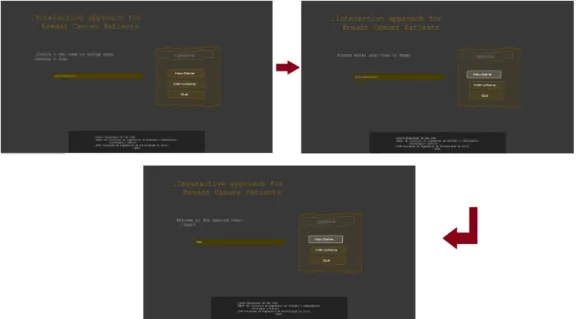
5.4 Interface components: Main menu . . . 47
5.5 Interface components: Instructions . . . 48
5.6 Description of the interfaces common components during game play . . . 49
5.7 Illustration of the medical avatar and its states . . . 49
5.8 Interface components: Statistics . . . 50
5.9 Exercise counter measures . . . 50
5.10 Decision tree for type of visual feedback . . . 51
5.11 Interface with no visual feedback . . . 51
5.12 Interface with mirror-based feedback . . . 52
5.13 Block diagram system for animation of the player-avatar . . . 52
5.14 Examples of the available avatars with different levels of abstraction . . . 53
5.15 Interface components: Player selection . . . 53
5.16 Interfaces screenshots for avatar-based feedback . . . 54
5.17 Interface screenshot for avatar based feedback, with sparse activity between the medic-avatar and the player-avatar . . . 55
5.18 Interface avatar-based visual feedback: User packs and respective subsystem use cases . . . 56
5.19 Diagram for questionnaire conception . . . 57
5.20 Questionnaire information: 5-point scales to three semantic differential pairs of adjectives . . . 58
5.21 Number of sessions performed per day . . . 60
5.22 Acquisition environment . . . 60
5.23 Data acquisition scheme . . . 60
5.24 Questionnaire information: Using the favourite interface at home . . . 63
5.25 Questionnaire information: Interface preference . . . 64
5.26 Questionnaire information: Interface evaluation . . . 65
5.27 Questionnaire information: Representation of the movement by the avatar . . . . 66
5.28 Questionnaire information: Perception of movement . . . 66
5.29 Questionnaire information: Choice of interface according to patients who did or did not physiotherapy . . . 67
5.30 Questionnaire information: Choice of interface according to patient age range . . 67
5.31 Questionnaire information: Choice of interface according to the academic level of patients . . . 68
5.32 The boxplot shows the median evaluation for immersion in the virtual environment per rating on the avatar representation of the user . . . 71
5.33 The boxplot shows the median evaluation for immersion in the virtual environment per frequency in playing games . . . 71
5.34 Interface with avatar-based feedback for multi-player . . . 73
5.35 Multi-player interface with avatar-based visual feedback: User packs and respec-tive subsystem use cases . . . 73
6.1 Goniometer used in this study . . . 77
6.2 Goniometer simulation tool: Main menu . . . 78
6.3 Goniometer simulation tool: Interface of the acquisition environment . . . 79
6.4 Goniometer simulation tool: Body segments and angle calculation of shoulder motions . . . 81
6.5 Goniometer simulation tool: Subject ID=8, right shoulder sequence acquired . . 82
6.6 Goniometer simulation tool: Results for shoulder flexion in front pose . . . 83
6.7 Goniometer simulation tool: Results for shoulder abduction to 90◦in front pose . 83 6.8 Goniometer simulation tool: Results for elbow rotation . . . 84
6.9 Goniometer simulation tool: Results for shoulder flexion in sagittal pose . . . 85
6.10 Goniometer simulation tool: Results for shoulder flexion to 90◦in sagittal pose . 86 7.1 Conventional pattern recognition system . . . 90
7.2 A simple example of 3-Nearest Neighbour Classification . . . 92
7.3 Linear separating hyperplanes for the separable case . . . 93
7.5 SVM: Feature space . . . 95
7.6 Two types of multi-class SVM classifier . . . 96
7.7 Cosine similarity between objects described by 5 and 20 features . . . 99
7.8 A sample format of a confusion matrix with n-classes . . . 100
7.9 Overview of the implemented method for human action recognition . . . 102
7.10 Modified spherical coordinate system for joint location binning . . . 103
7.11 Comparison between the accuracy of the proposed method with classifier SVM and classifier kNN . . . 106
7.12 Confusion matrix of kNN: Test Two, AS1 . . . 109
7.13 Confusion matrix of kNN: Test Two, AS2 . . . 109
7.14 Confusion matrix of kNN: Test Two, AS3 . . . 110
7.15 Process for further improvement of the accuracy in the proposed method . . . 111
C.1 Acquisition process for the personalized avatar . . . 123
E.1 Questionnaire: Section 1 - Authorization for the use of information provided . . . 127
E.2 Questionnaire: Section 2, part 1 - Characterization of the population . . . 128
E.3 Questionnaire: Section 2, part 2 - Characterization of the population . . . 129
E.4 Questionnaire: Section 2, part 3 - Characterization of the population . . . 130
E.5 Questionnaire: Section 3, part 1 - Personal information . . . 131
E.6 Questionnaire: Section 3, part 2 - Personal information . . . 132
E.7 Questionnaire: Section 4, part 1 - Relation with new technologies . . . 133
E.8 Questionnaire: Section 4, part 2 - Relation with new technologies . . . 134
E.9 Questionnaire: Section 5, part 1 - Evaluation of proposals interfaces . . . 135
E.10 Questionnaire: Section 5, part 2 - Evaluation of proposals interfaces . . . 136
E.11 Questionnaire: Section 5, part 3 - Evaluation of proposals interfaces . . . 137
E.12 Questionnaire: Section 6, part 1 - Relationship with the virtual environment . . . 138
E.13 Questionnaire: Section 6, part 2 - Relationship with the virtual environment . . . 139
E.14 Questionnaire: Section 7, part 1 - Considerations/feelings about the proposals in-terfaces . . . 140
E.15 Questionnaire: Section 7, part 2 - Considerations/feelings about the proposals in-terfaces . . . 141
E.16 Questionnaire: Section 8 - Suggestions . . . 142
F.1 Questionnaire information: Technological resources at home . . . 144
H.1 Confusion matrix of 27 UTD-MHAD human actions with a SVM classifier . . . 150
2.1 Comparison of the most significant methods for Lymphedema assessment . . . . 12
2.2 Brief description for Lymphedema assessment methods . . . 12
3.1 Performance assessment of different motion tracking systems . . . 21
3.2 Available RGB-D sensors characteristics . . . 22
3.3 Description of Kinect central components . . . 23
3.4 Comparison of main features of the two versions of the Kinect sensor . . . 27
3.5 Main self-reports to UBF Evaluation . . . 29
4.1 The Games for Health Taxonomy developed by the Games for Health Project . . 36
4.2 Classification and Comparison of Serious Games for Rehabilitation . . . 38
5.1 Properties of the chosen game engine . . . 43
5.2 Main requirements for the interfaces . . . 46
5.3 Nomenclature used to explain the development of interfaces . . . 47
5.4 Type of visual and temporal representation in the developed interfaces . . . 55
5.5 Summary of the Self-Presence Framework . . . 58
5.6 Strengths and Limitations of Existing Gaming Scales . . . 59
5.7 Questionnaire information: Brief summary of the sample tested . . . 61
5.8 Questionnaire information: Age ranges of the study population . . . 61
5.9 Questionnaire information: Academic status . . . 61
5.10 Questionnaire information: Type of surgery performed during treatment . . . 62
5.11 Questionnaire information: Chemotherapy, Radiotherapy and Physiotherapy . . . 62
5.12 Questionnaire information: Complications during treatment . . . 62
5.13 Questionnaire information:Frequency on game play . . . 63
5.14 Questionnaire information:Free time evaluation . . . 63
5.15 Questionnaire information: Use at home according to patients with or without physical therapy . . . 64
5.16 Questionnaire information: Speed of the exercises . . . 65
5.17 Questionnaire information: Type of visual feedback according to patients who use or not technological resources . . . 68
5.18 Questionnaire information: When playing the game/using the virtual environment, how much do you feel like your avatar is an extension of your body within the game/virtual environment? . . . 70
5.19 Questionnaire information: When playing the game/using the virtual environment, to what extent do you feel inside of the game/virtual environment? . . . 70
5.20 Questionnaire information: When something happens to your avatar’s body, to what extent does it feel like it is happening to any part of your body? . . . 70
5.21 Questionnaire information: When playing the game/using the virtual environment, how much do you feel your avatar is a part of your body? . . . 70
5.22 Questionnaire information: Suggestions choices . . . 72
5.23 Classification of the implemented interfaces using the Taxonomy of Serious Games for Rehabilitation . . . 76
6.1 Goniometer simulation tool: Postures for acquisition data . . . 80
6.2 Goniometer simulation tool: Brief summary of the test samples . . . 81
6.3 Goniometer simulation tool: Results for shoulder flexion in front pose . . . 82
6.4 Goniometer simulation tool: Results for shoulder abduction to 90◦in front pose . 84
6.5 Goniometer simulation tool: Results for elbow rotation . . . 85
6.6 Goniometer simulation tool: Results for shoulder flexion in sagittal pose . . . 86
6.7 Goniometer simulation tool: Results for shoulder flexion to 90◦in sagittal pose . 86
6.8 Goniometer simulation tool: Overall results . . . 87
7.1 The three components of learning algorithms . . . 91
7.2 Comparing learning algorithms (**** stars represent the best and * star the worst performance) . . . 98
7.3 UTD-MHAD dataset actions . . . 104
7.4 The three subsets of actions used in the experiments . . . 105
7.5 Human action recognition accuracy comparison for UTD-MHAD dataset . . . . 106
7.6 Statistics extracted from the confusion matrices of the methods with classifier SVM and kNN . . . 107
7.7 Recognition accuracies of different tests . . . 108
F.1 Questionnaire information: Weight . . . 143
F.2 Questionnaire information: Height . . . 143
F.3 Questionnaire information: Civil status . . . 143
F.4 Questionnaire information: Serious game . . . 144
F.5 Questionnaire information: Technological resources . . . 144
F.6 Questionnaire information: Considerations / feelings after demonstration of inter-faces . . . 145
Abbreviations
2-D Two-dimensional
3-D Three-dimensional
ALND Axillary lymph node dissection ANN Artificial Neural Networks
API Application programming interface BCCT Breast cancer conservative treatment BCRL Breast cancer-related lymphedema CDT Complete decongestive therapy CGI Computer-generated imagery DCIS Ductal carcinoma in situ DBN Dynamic Bayesian Networks DNA Deoxyribonucleic acid
fs Sampling rate
FoV Field of View
HCI Human - computer interaction HMM Hidden Markov Model HOJ3D Histograms of 3D Joints IDC Invasive ductal carcinoma ILC Invasive lobular carcinoma
IR Infra–red
kNN k-Nearest Neighbors
M Mean
ML Machine learning
NUI Natural user interface
NN Nearest Neighbors
OMS World Health Organization
P2P Peer-to-peer
PPMP Persistent post mastectomy pain QOL Quality-of-life
ROM Range of motion SD Standart deviation
SDK Software development kits SLND Sentinel lymph node dissection SPQ Self-Presence Questionnaire SVM Support Vector Machine UBF Sentinel lymph node dissection UTD-MHAD UTD Multimodal Human Dataset VE Virtual environment
VR Virtual reality
Introduction
1.1
Context
Breast cancer is a matter of public health, with approximately 60% of incidence, being the one that affects females the most, having overall a tremendous impact on the woman body image and self-esteem given the aesthetic appearance of the breast [1]. Nearly 25% of the women diagnosed with breast cancer have malignant cells in the axillary lymph node system. Which implicates, besides the tumor removal, additional treatments have to be applied to remove the axillary lymph nodes [2]. Though, this type of procedures are usually accompanied with several upper-limb problems, comprising shoulder mobility, arm/shoulder pain and lymphedema. The former is a chronic disease that manifest itself with swelling of the hand, arm, chest, torso leading to a physical strain, and psychosocial functioning in patients [3].
Breast cancer survivors usually have to deal with long-term effects that will be a constant everyday which can also be accompanied with emotional distress, directly associated with the subsequent body changes results and its impairments [1]. Of all the complications of treating breast cancer, lymphedema of the upper limb is one of the most troubling and unpleasant for the patient, being also very frustrating to the surgeon [4]. The next five years after surgery are marked with arm/shoulder pain in the scale of 30-40%, of lymphedema 10-15% and conditioned arm/shoulder mobility is 15-30% [5]. These upper-body impairments are highly correlated with the QOL of breast cancer patient.
1.2
Motivation
An immediate study of the patient situation is imperative in order to put into action an appropriated and reliable rehabilitation program to prevent/attenuate these impairments in order to provide the patient a better QOL [6]. Therefore, it is the utmost necessity to detect as early as possible the development of side effects in order to identify which procedures have the best results to battle its adverse symptoms [7].
There are many ways to manage symptoms resulting of breast cancer such as exercises for Upper Limb Lymphedema (having in mind there are different levels of impairment), surgery, med-ication, manual lymph drainage, pneumatic compression, wearables for example. Although, most of this techniques have been shown as insufficient at some sort of level, the main reason being most of this procedures just treat the member in question without dealing with the movement of the confined lymphatic fluid long-term [8].
Resort to computational techniques such as Virtual Reality (VR) can be a source of motivation throw-out the recovery process of the patient. So the use of serious games in VR can became a great ally to the pre-existent procedures.
1.3
Objectives
In order to be possible to the patient regain full or at least a satisfactory level of mobility on the upper limbs, is imperative in the recovery stage a constant level of physical activity. So a list of exercises is prescribed, but sometimes the exercises are not done as they should and/or as frequent. The main purpose of the following work is to solve this problem with a new rehabilitation tool. The basic premise is to show the patient how to do the exercises properly in the comfort of their own home, and at the same time, doing an evaluation of its overall performance concealed as a serious game using as resource some type of sensor.
The exercises will have been main focus in the affected upper body members in order to help regain as much as possible arm/shoulder mobility and prevent the risk of developing arm/shoulder impairments. The work aims to implement a series of interfaces for rehabilitation of breast cancer patients with partnership of researchers from S. João hospital, the data recover from its test subjects will be used to evaluate the research done.
In resume, this work focus on developing and testing of various interfaces, subsequently im-prove and test particular aspects such as, Microsoft Kinect joint location accuracy and action recognition.
1.4
Contributions
This work had the following main contributions:
1. Eight different interfaces were develop using the Microsoft sensor and Unity game engine, with six of them tested in breast cancer patients;
2. An already seizable database with collected data, was composed by diverse exercises and movements typically used for breast cancer patients, which can be further used in other works in the area like movement recognition, for example. This dataset composed by skeleton positions and color images is expected to continue growing for a year, approximately;
3. An application to simulate a goniometer was implemented, tested and validated; 4. A study and implementation of a simple framework for human action recognition.
1.5
Dissertation structure
After this section, this document is composed by seven more chapters. Chapter 2 presents a biolog-ical overview of the breast and breast cancer condition and in Chapter 3 a technologbiolog-ical overview is done from subjective methods UBF assessment and human tracking systems are analysed. Also, some insights of RGB-D cameras are provided focusing in the Microsoft Kinect. Lastly, a anal-yses of several tools/programs to create a game environment and its components. In Chapter 4, a literature review of Human-Computer interaction and analysis of serious games in healthcare. Chapter 5, describes the application conception and results of the developed interfaces. In Chapter 6 which describes the implementation, methods and results for an application which simulates a goniometer. A study and a explanation of the framework created. And, in Chapter 7 shows the study and development of a framework for action recognition. Lastly, Chapter 8 is composed by a conclusion to the presented research and possible future work.
Medical overview
2.1
Breast Anatomy and Physiology
The breast (see Figure 2.1) refers to the front of the chest or, more specifically, to the mammary gland responsible for producing milk being composed mostly of fat with a shape resembling a teardrop. Within this organ a complex network of branching ducts that exit from sac-like structures called lobules, which can produce milk in females. The ducts exit the breast at the nipple [9].
The lobules and ducts are supported in the breast by surrounding fatty tissue and ligaments. In the breast there are blood vessels and lymphatics, thin channels similar to blood vessels; they do not carry blood but collect and carry tissue fluid which ultimately reenters the blood stream. Breast tissue fluid drains through the lymphatics into the lymph nodes located in the underarm (axilla) and behind the breast bone (sternum) [10].
Although the primary biologic function of the breast is to produce milk to feed a baby, the breast has for many centuries been a symbol of femininity and beauty. The appearance of the nor-mal fenor-male breast differs greatly between individuals and at different times during a woman’s life before, during and after adolescence, during pregnancy and menstrual cycle, and after menopause [9].
The lymphatic system is part of the circulatory system and a vital part of the immune system, comprising a network of lymph nodes distributed along the lymphatic vessels that carry a clear fluid called lymph that contains tissue fluid and waste products, as well as immune system cells. Also, helps to maintain fluid balance in tissues and to absorb fat from the digestive tract. In the upper-limbs, all the lymph vessels drain into the lymph nodes in the axilla [12]. Furthermore, axillary nodes receive fluid from the upper back and shoulder, the lower neck, the chest, and the upper anterolateral abdominal wall. Approximately, 75% of the drainage of lymph fluid of the mammary gland is performed via lymphatic vessels into axillary nodes [13].
Figure 2.1: Breast Anatomy: (a) Nonlactating breast, only with the duct system (b) Lactating breast, with alveoli at the ends of the ducts, produce milk, (From [11])
2.2
Cancer
Cancer is defined by an abnormal and unregulated growth of cells, which may appear into sur-rounding tissues. It can occur in almost every part of the human body. Usually, human cells grow and divide to form new cells (replacing the dying ones) to match the needs of the human body, this cell cycle is regulated by Deoxyribonucleic acid (DNA) [12]. This regulation is done by two gene groups: oncogenes, responsible to promote cell growth and reproduction and suppressor genes, which constrains cell division and survival. However, this orderly process breaks down in the presence of cancer. The regulatory mechanisms become unable to prevent the perpetuation of this error, the cell starts to replicate without control, old and/or damaged cells survive when they should die, and new cells form when they are not needed, this may result in growths called tumors [14].
The regulation of those phases is mainly performed by two gene categories: oncogenes, which promote cell growth and reproduction, and tumour suppressor genes, which inhibits cell division and survival. If an over-expression of an oncogeneor an under-expression of tumour suppressor genes occur, and the regulatory mechanisms do not prevent the perpetuation of thiserror, the cell starts to replicate without control [15].
Cancerous tumors are considered malignant, once they are able to spread into, or invade, nearby tissues. In addition, some cancer cells can break off and travel to distant places in the body through the blood or the lymph system and form new tumors far from the original tumor. On other hand, benign tumors do not spread into, or invade, nearby tissues. When removed, they usually do
not grow back, whereas malignant tumors sometimes reappear [14].
A malignant tumor forming from the cells of the breast is commonly known as breast cancer. These cells have a tightly regulated cell cycle that controls their growth, maturity, division and death. A cancer cell appears when a normal cell undergoes damage to the DNA that it is not repaired and the cell does not die, as it should. Instead, the cell undertakes division and the damage is propagated by the out-of-control growth of abnormal cells [13]. A malignant tumor forming from the cells of the breast is commonly known as breast cancer. The most common breast cancer frequently either develops in the cells of the lobules or the lactiferous ducts. The rarest, breast cancer takes is starting point in the stromal tissues, which include the fatty and fibrous connective tissues of the breast [16]. A study made in 2013, was found to be the most frequent in woman with 29% occurrence (see Figure 2.2) and the second deadliest with 14% [17].
2.2.1 Breast carcinoma
Figure 2.2: Incidence of cancers in women in the United States between 1975-2009, (From [13])
Breast cancer is frequently divided into non-invasive and invasive. Non-invasive breast cancer, also known as carcinoma in situ, is when the cancer remains within the place of origin and does not grow or spreads beyond the breast. Ductal carcinoma in situ (DCIS), one type of non-invasive cancer, is considered a pre-cancerous lesion. Which means that, although the abnormal cells have not spread out, they can eventually develop into invasive breast cancer. In invasive breast cancer, the abnormal cells spread outside the membrane that lines a duct or lobule, attacking the surrounding tissues. The spread of this cells can be made through the bloodstream or the lymphatic system to other parts of the body such as the bones, liver or lungs, creating metastasis. The most common types of invasive breast cancer are invasive ductal carcinoma (IDC) and invasive lobular carcinoma (ILC) (see Figure 2.3) [12].
Figure 2.3: Invasive Lobular Carcinoma. The breast cancer cells originate in the breast lobules, (From [18])
2.2.2 Breast Cancer treatments
The clinicians approach to treat breast cancer as many factors to take in consideration, for instance women’s general health and age, position and size of the cancer and how far it has spread.Also aims to reduce the risk of recurrence and/or its spreading (metastases), and obtain the best possible aesthetic outcome, relief of symptoms and restoring the QOL prior to diagnosis [18]. The treat-ments options normally include chemotherapy, radiotherapy and surgery. The treatment elected may be the surgical removal of the tumor by a mastectomy or a more conservative approach by Breast Cancer Conservative Treatment (BCCT) [19]. Non-surgical treatments, chemotherapy and radiotherapy, have an important role in order to prevent reemergence. It might be used before surgery to help shrink the tumor or after surgery [18].
Mastectomy is the surgical removal of the entire breast. These are the following types [20]: • Simple mastectomy procedure: the entire breast tissue, the nipple, areola and, in certain cases, the sentinel lymph node (first node of axillary lymph node) are removed (see Figure 2.4(a)); • Radical mastectomy procedure: this method is performed only in severe cases as metastases in to the pectoralis muscles is the one that causes the more damage. Encompasses the removing of the entire breast, the axillary lymph nodes and pectoralis muscles;
• Modified radical mastectomy procedure: the entire breast tissue and all the axillary content is removed (see Figure 2.4(b));
• Skinsparing mastectomy: this type of surgery consists in removing breast tissue through an incisionmade around the areola. This type of procedure allows the reconstruction of the breast; • Nipplesparing/subcutaneous mastectomy: breast tissue is removed but the nipple-areola complex is preserved. This procedure is recently used for tumors outside the subnipple-areolar position.
For most women in initial stages of breast cancer, BCCT is as effective as mastectomy since the survival rates of women treated with both approaches are similar [21]. This procedure preserves
Figure 2.4: Common mastectomies procedures in breast cancer patients, (From [20])
the breast without compromising the survival of the patient. In breast conserving surgery it is only removed the tumor and some of the normal tissue that surrounds it, while preserving the natural shape and appearance of the breast. BCCT also includes a phase of radiation therapy after the surgical removal of the tumor in order to eradicate any residual cancer cells. However, side effects of radiation therapy, caused by interference on lymphatic drainage, include swelling and heaviness of the arm that will affect its mobility [22] .
2.2.3 Lymph Node Dissection
The axillary lymph node status represents one of the most important prognostic factors in breast cancer patients and determines among others subsequent treatment. The percentage of node pos-itive patients who benefit from routine axillary lymph node dissection (ALND) is constantly de-creasing as breast cancer is inde-creasingly detected at an early stage. About 40% of women di-agnosed with breast cancer have cancer cells in their axillary lymph nodes [23]. As a result, in addition to the removal of breast cancer through one of the methods mention prior, often is neces-sary remove of one or more axillary lymph nodes to discover if the cancer has spread beyond the breast, so it is necessary to perform a lymph node biopsy to determine the following treatment [24] (Figure 2.5). The patients which will undergoing ALND there is a high probability of developing severe impairments in upper-extremity function since the removal of lymph nodes will affect the drainage of the limbs. So, significant impairments in UBF are associated with ALND, such as restricted arm and/or shoulder motion and arm edema [22].
An alternative to ALND is sentinel lymph node biopsy (SLNB), which can spare the patients a more invasive surgery and side effects, the procedure involves the identification and removal of sentinel lymph node, subsequently tested to determine whether cancer cells are present. Although, it has its own limitations and drawbacks. For context, sentinel lymph node (SLN) is defined as the first lymph node to which cancer cells are most likely to spread from a primary tumor. Sometimes, there can be more than one sentinel lymph node. In figure 2.6 – first panel, a radioactive substance
Figure 2.5: Axillary Lymph Node Dissection (ALND), (From [25]])
and/or blue dye is injected near the tumor. In the second panel, the injected material is located visually and/or with a device that detects radioactivity. The last panel, sentinel node is removed and checked for carcinogenic cells [26].
Figure 2.6: Sentinel lymph node biopsy of the breast, (From [25])
2.3
Impairments caused by breast cancer treatments
Persistent postsurgical pain is an increasingly documented problem, negatively impacting QOL and comprising approximately 20% of new chronic pain patients. The reported incidence of per-sistent post mastectomy pain (PPMP) ranges from 25-60%. In addition, developments in breast cancer detection and treatment have dramatically reduced mortality, around 2.5 million survivors in the United States. Among breast cancer patients, PPMP is rated as the most troubling symptom, leading to disability and psychological distress, and is notably resistant to management. While surgical factors, including more extensive surgery (total or partial mastectomy), axillary lymph node dissection and reconstruction have been suggested to serve as important risk factors for chronic pain, although several studies do not support this association [27]. Adjuvant treatment, such as radiation, chemotherapy, and hormone therapy, has also been associated with persistent
pain. Among demographic factors, younger age correlates with increased persistent pain inci-dence in some studies but not others. Preexisting pain is also more frequent in those who go on to develop PPMP [28].
2.3.1 Post Mastectomy Syndrome
The causes of post mastectomy pain syndrome are due to several factors althout they remain un-certain. The most commonly cited theory is the removal of the intercostobrachial nerves that run through the axillary region into the arm which provoques chronic postoperative pain in breast can-cer patients, estimated that up to 50% [29]. And in addiction, chemotherapy and radiation therapy provoque neuropathies. This symthom provoques massive imparments in performing physical and recreational activities, increased body image distress, and decreased sexual interest and function-ing. Injury to the nerves will result in neuromas due to ineffective regeneration and swelling within the nerves. Neuralgic quality pain (burning, shooting) associated with hypersensitivity, numbness, tingling, and muscle weakness depending on whether the nerve involved is purely sensory, purely motor, or mixed sensory and motor. The treatment involves physical therapy, topical agents, anti-convulsants, antidepressants, antiarrhythmic, nerve block and scar desensitization injections with dilute local anesthesia and steroids [29,30].
2.3.2 Lymphedema
Lymphedema is a chronic disease which refers to swelling that generally occurs in one of your arms or legs sometimes either arms or legs swell. Lymphedema is most commonly caused by the removal of or damage to your lymph nodes as a part of cancer treatment. It results from a blockage in your lymphatic system (see Figure 2.7), which is part of your immune system. The blockage prevents lymph fluid from draining well, and the fluid buildup leads to swelling. However, it can be managed with early diagnosis and diligent care of your affected limb [31].
2.3.2.1 Lymphedema incidence in breast cancer patients
Patients undergoing axillary surgery and/or axillary radiation therapy for breast cancer are at higher risk for developing lymphedema of the arm, such treatments previous mention such as axillary node removal, sentinel node biopsy and local radiation [33]. Breast cancer-related lym-phedema (BCRL) is a progressive, debilitating condition affecting millions of breast cancer sur-vivors with a significant negative impact on QOL, employment and health, and it is not limited to arm swelling alone. Many survivors have complex symptoms which include breast and truncal swelling [34]. The amount of breast cancer-related arm edema can range from mild to severe, but once the condition starts, there is the possibility of progression to more severe lymphedema [31]. Even in early stage breast cancer, one study showed that when lymphedema progressed, lym-phedema therapy could not completely reverse it.
2.3.2.2 Methods for Lymphedema Assessment
Lymphedema detection is normally assessed comparing the limb volume with the unaffected limb. The Table 2.1 shows the most common approaches, with a brief summary of each procedure in Table 2.2.
Table 2.1: Comparison of the most significant methods for Lymphedema assessment, (Adapted from [33])
Method Time consumed Applied at home Accuracy Cost Complex
Water Displacement Low Yes Low Low Medium
Circumferential Measurements High Yes Low Low Low
Perometer R Medium No High Medium High
CT Low No High High High
DEXA Low No High High High
BIS Medium No Medium High Medium
Table 2.2: Brief description for Lymphedema assessment methods, (Adapted from [33])
Method Procedure summary
Water Displacement In a container with water the limb is immersed into and the amount of the displaced water represents the volume of the limb.
Circumferential Measurements The volume is estimated assuming cylindrical/conic volumes between several measures taken along the limb.
Perometer R The device scans the limb with infra-red (IR) light and assess limb
volume at small intervals.
CT The device scans the limb with IR light and assess limb volume at small intervals.
DEXA Uses a tissue-specific mode with attenuation of X-ray dependent on the thickness, density, and chemical structure of the tissue examined. BIS Measures volume of the limb by comparing impedance values of both
2.3.2.3 Lymphedema treatments
Lymphedema treatment focuses on reducing the swelling and managing the pain. For instance, some of the treatments are the following:
• Physical exercises: in which the movement of the affected limb may encourage lymph fluid drainage and help improve everyday tasks (see Figure 2.8 and Figure 2.9), the exercises should not be strenuous or tiresome but needs to focus on gentle contraction of the muscles in the affected limb [35].
Figure 2.8: Specific Exercises for Upper Limb Lymphedema, (From [36])
Figure 2.9: Exercises for Upper Limb Lymphedema where mobility is limited, (From [36])
• Manual lymph drainage: a specific massage technique called manual lymph drainage may encourage the flow of lymph fluid out of your arm or leg. This procedure is not recommending for every patient, should be avoid by whom displays symptoms such as skin infection, active cancer, blood clots or congestive heart failure. Also, the areas that were exposed to radiation therapy [37].
• Pneumatic compression: a sleeve worn over your affected arm or leg connects to a pump that intermittently inflates the sleeve, putting pressure on your limb and moving lymph fluid away from your fingers or toes (see Figure 2.10). Compression garments, long sleeves or stockings made to compress your arm or leg encourage the flow of the lymph fluid out of your affected limb [38].
Figure 2.10: Pneumatic compression pump, (From [36])
• Complete decongestive therapy (CDT): which involves combining therapies with lifestyle changes. Generally, CDT is not recommended for people who have high blood pressure, diabetes, paralysis, heart failure, blood clots or acute infections [39].
Figure 2.11: Phases of complete decongestive therapy, (Adapted from [40])
2.4
Summary
Nowadays, a more conservative approach in the treatment of breast cancer is a constant prac-tice of the clinics. In the past years, the treatments haven been suffered constant improvements, which contributed greatly for a less invasive approach. For instance, surgical procedures such as the radical mastectomy and ALND were replaced by BCCT and SLND, respectively. The last one manages to reduce the number of avoidable lymph node dissections. Even with these im-provements which outcome is less extensive procedures, there are still a considerable level of morbidity in several patients [12]. The most recurrent impairment is in a restricted upper-body function, which include reduced motion of the arm/shoulder, strength and flexibility, arm/shoulder pain and/or arm edema. That were caused by additional radiation therapy of the axilla or/and the lymph node removal. Both procedures interfere with the axillary lymphatic system [41]. Several methods of volume assessment of the upper limb are available, with water displacement as the gold
standard. In summation, restricted UBF as a direct correlation with the QOL of the patient, impact and sometimes dictate the lifestyle completely, having implications at a physical and emotional level such as chronic pain and depression [1].
Technological overview
In this chapter, its done a technological review of state of the art available systems, assessment methods and accessible software’s currently or possible applied in the rehabilitation of breast cancer patients. It begins with an overview of movement recognition systems, a key aspect to analyse change in the human body’s pose, which requires a well-developed motion-sensor, leading to the followed section composed by an introduction of RGB-D cameras and an assessment of different sensors. Next the attention falls in several methods (subjective and objective) for UBF assessment. Lastly, it is contemplate some of the most relevant game engines available.
3.1
Movement recognition
The way humans interact with computers is constantly evolving, with the general purpose being to increase the efficiency and effectiveness by which interactive tasks are completed. Real-time, static and dynamic hand gesture recognition affords users the ability to interact with computers in more natural and intuitive ways [42]. Motion tracking (see Figure 3.1) is an essential build-ing block of many advanced applications in diverse areas, for instance HCI (Human Computer Interaction) [43].
3.1.1 Non-visual tracking systems
Sensors available within these systems are placed in the human body with objective of collecting movement data. Being commonly classified as mechanical, inertial or microwave and magnetic based. In the general scheme of things, each type of sensor has its own advantages and limitations being particularly affected when exposed to different environments [44].
3.1.1.1 Magnetic sensor based systems
Magnetic motion tracking systems have various advantages, such as size, high sampling rate (fs) and shortage of blocking. Which make it largely used for tracking user movements in VR. How-ever, also present some serious disadvantages, for instance latency (because of its asynchronous
Figure 3.1: Classification of human motion tracking using sensor technologies, (Adapted from [44])
nature), jitter (appears in the existence of electronic or ferrous devices in the surroundings), and noise in the measurements. To tackle these problems, numerous research projects have been done, having its basis in predictive filtering methods [44].
3.1.1.2 Inertial sensor based systems
Inertial sensors are based on inertia, this sensors are easy to use and cost efficient way for full-body human motion detection. For instance, gyroscopes and accelerometers are often used in navigation and augmented reality modelling. The motion data can be transmitted wireless to a work base for further process or visualization. Inertial sensors can be of high sensitivity and large capture areas. For example, a project developed with this technology: Displacement Estimation in Micro-Sensor Motion Capture, with the following main objectives [45]:
- Multi-modal sensor data in order to attain high accuracy and low drift; - Combination of the motion models to obtain full body motion model; - Build a power effective and low error rate motion model.
3.1.2 Visual based tracking systems
Optical sensors (e.g. cameras) are routinely applied to improve precision in position assessment. Having as a precondition on whether the indicators need to be attached to body parts, tracking systems can be classied as either visual marker or marker-free [44].
3.1.2.1 Visual marker based tracking systems
Visual marker based tracking systems consists in using cameras to track human movements, with the visual markers positioned on the human body. Subsequently, an image processing program combines the 2D data and calculates the 3D position. The human skeleton is a vastly articulated structure and its different members have a random and intricate motion trajectory, which might cause an inconsistent and defective motion estimation. Other factors, may also introduce errors in the real position of the marker, such as mixt lighting or cluttered scenes [44].
Moving Light Display from Johansson in 1973, was the first work in this field, consisting in tracking trajectories due to reflective markers positioned in human joints. Nowadays, the purchase of these systems is a quite common thing and it is a growing sector. In controlled conditions this method is very accurate, being a reason to knowledgeable experienced investments from fields as animation movies and computer-generated imagery (CGI).
One example, is shown in Figure 3.2 which demonstrates a procedure for unconstrained wrist and elbow motions developed by Schmidt et al. [46]. However, the sensors need to performed some corrections in the results due to the influence of skin-movement on join angles. Besides, for patients with complications in upper limb it can become troublesome to setting the sensors the system requires, and a lot of calibration and professional intervention are needed.
Figure 3.2: Markers and camera position in Schmidt et al. system. (a) Disposition of the markers (b) 5 cameras are used to pick the localization of the markers, (From [46])
3.1.2.2 Marker-free visual based tracking systems
2-D approaches
A 2-D approach is a framework frequently used. This approach produces object models (see Figure 3.3) with acquired image data from arbitrary human movements, it merely concerns human movement in an image plane; due to its dimensions adapts in a simple and quick way. However,
there are natural restrictions, due to their viewing angle. To improve a tracker in an unpredicted environment, 2-D motion tracking does not necessary needs an explicit shape model. Normally, this approach appears in rehabilitation environments [44].
Figure 3.3: 2-D model of the human body, (From [47])
3-D approaches
3-D approaches for tracking human movements allows the tracking problem to be minimized. The future movements of a human body can be foreseen regardless of occlusion or self-collision. With these approach it is possible to constructed a 3-D model of an object by using various cameras(see Figure 3.4). This approach has been promoted as an alternative to 2-D mod-elling techniques [44].
Figure 3.4: Concept of reconstruction of the shape of the object, (From [47])
3.1.3 Robot-aided tracking systems
Robot-aided tracking systems, is an established approach in rehabilitation systems that are driven by electromechanical or electromagnetic tracking approaches and, integrate individual sensor technologies to conduct "sense-measure-feedback" strategies. For instance, to delivering neuro-rehabilitation for human limbs following stroke [44].
3.1.4 Summary
People detection and tracking is an important and fundamental component for many robots, inter-active systems and intelligent vehicles. Popular sensors for this task are cameras and range finders (see Table 3.1). Whereas both sensing modalities have advantages and drawbacks, their distinction may become obsolete with the availability of affordable and increasingly reliable RGB-D sensors that provide both image and range data [48].
Table 3.1: Performance assessment of different motion tracking systems according to Figure 3.1, (Adapted from [43])
Sensor Accuracy Compactness Computation Cost Drawbacks
Inertial High High Efficient Low Drifts
Magnetic Medium High Efficient Low Ferromagnetic materials
Combinatorial High Low Inefficient High Multidisciplinary
Marker High High Inefficient Medium Occlusion
Marker-free High High Inefficient Low Occlusion
3.2
RGB-D sensors
In recent years, a wide range of low-cost RGB-D sensors (see Table 3.2) have become available, consequence of a growth in the development of applications for home entertainment (e.g video games) using RGB-D sensors. Low-cost range sensors are an attractive alternative to other expen-sive scanners in application areas such as indoor mapping, surveillance, robotics and forensics. At the moment, acquire 3-D images using RGB-D cameras can be a quite simple task. This sensors capture RGB color images and depth data for each pixel. In order to produce depth estimates tech-niques as time-of-flight imaging and structured light stereo are commonly used. As an example, a frame obtain by using Microsoft Kinect sensor which provides dense depth estimates (see Figure 3.5) [49].
Figure 3.5: RGB image (left) and depth information capture (right) by an RGB-D camera. For the most part the white pixels in the right image means no depth value, due to occlusion, maximum distance, relative surface angle or material, (From [49])
T able 3.2: A v ailable RGB-D sensors characteristics, (Adapted from [ 50 ] ) Sensor T ype Depth Range 3D Resolution RGB Resolution Frame Rate Latency FO V Interface ASUS R XtionPr oTM Li v e Structured light 0.8 to 3.5 m 640 x 480 1280 x 1024 30 fps 1.5 frames 58 ◦ H, 45 ◦ V USB 2.0 Micr osoft R Kinect TM 1.0 Structured light 0.5 to 8m 640 x 480 41280x102 30 fps -57 ◦ H, 43 ◦ V USB 2.0 Micr osoft R Kinect TM 2.0 T ime of flight 0.5 to 4.5 m 512 x 424 1920 x 1080 30 fps 1 frame 70 ◦ H, 60 ◦ V USB 2.0 Intel RealSense R200 Stereo with pattern projector 0.6 – 3.5 m 640 x 480 1920 x 1080 60 fps (3D) 30 fps (RGB) 1 frame 59 ◦ H, 46 ◦ V USB 3.0 Ster eolabs R ZED TM Embedded stereo 1.5 to 20 m 2208 x 1242 max 2208 x 1242 max 15 fps at max 1 frame 96 ◦ H, 54 ◦ V USB 3.0 Car negie Robotics R MultiSense TM S7 Embedded stereo 0.4 m to infinity 2048 x 1088 2048 x 1088 max 15 fps at 2048 x 544 1 frame 80 ◦ H, 45 ◦ V Ethernet
The analyse of RGB sensors focus on the Kinect sensor (which has gone through two versions), the reasons for this specificity are the following:
- The sensor is less expensive compare with the ones in the market [51];
- Numerous applications are develop in several sectors, with emphasis in the healthcare sector;
- There are a enormous amount of information compared to other sensors; - It is integrated in numerous game engines (e.g. Unity, see Table 3.4).
3.2.1 Kinect v1 sensor
In November of 2010, Microsoft released a new Natural User Interface (NUI), the first version of the Kinect sensor (Kinect v1) as XBOX 360 gaming platform. Similarly, as other RGB-D sensors, the Kinect provides color information and the estimated depth for each pixel. The Kinect is an order of magnitude cheaper than similar sensors that had existed before it. This has dramatically strengthened interest in RGB-D sensors and their applications in areas such as NUI, reconstruction and virtual reality or 3D mapping [52]. The key hardware components of this sensor are illustrated in Figure 3.6 and a brief description is performed in Table 3.3.
Figure 3.6: Kinect key components, (Adapted from [51])
Table 3.3: Description of Kinect central components, (Adapted from [51])
Components Brief Description 1 Multi-array microphone
An array of four microphones able to separate the voices of the user from the background noise. The voice source can be placed as a result of comparing the delay in each microphone. 2 IR(Infra–Red) laser emitter
Dynamically emitting infrared spectrum, which are distorted by uneven surfaces and then randomly formed as reflected speckles that can be received by infrared camera.
3 IR camera Capture infra-red signal which can potentially be converted into depth map.
3.2.1.1 Microsoft Kinect for Windows SDK
The Kinect for Windows SDK started by its very first beta version being a preview version with a temporary Application Programming Interface (API) and allowed users to work with depth and color data and also supported an advanced Skeletal. The first major update came along with the 1.5 version that included a Face Tracking library and Kinect Studio, a tool for recording and replaying sequences captured by the sensor. The Kinect for Windows SDK version 1.7 was released in March 2013 and included advanced libraries such as Kinect Fusion, a library for 3D scanning and reconstruction, and a library for hand grip detection which has opened doors for more natural way of interaction. The API of the Kinect for Windows SDK provides sensors depth, color and skeleton data in a form of data streams. Each of these streams can produce actual data frame by polling or by using an event that is raised every time a new frame is available [53]. The Kinect sensor, together with the Microsoft Kinect SDK provides the user with several streams of information such as:
• A stream of 2D color image frames;
• A stream of 3D depth image frames, the depth information is provided by the depth stream, which is represented as a frame of pixels that contains the distance in millimetres from the camera to the nearest objected. The depth data are represented as 16–bit unsigned integer value: the first 3 bits are reserved for the player segmentation data and the rest 13 bits for the distance, so the most distance stored in the depth data is about 8 meters. The depth frame is available in different resolutions (30 frames per seconds, for all), for instance its maximum resolution is 640x480 pixels [54];
• A stream of 3D skeletal frames, one of the most significant functionalities of the Kinect for Windows SDK is the Skeletal Tracking. In its field of view can identify up to 8 individuals and monitor for 2 users their actions. The skeleton which illustrates the skeleton space consists of 20 joints that represent the locations in X, Y, Z coordinates in meters of the key parts of the human body (see Figure 3.7). The tracking algorithm is designed to recognize users facing the sensor (the standing or sitting pose) with no particular pose or calibration action necessary for a user to be tracked [54].
Figure 3.7: (a) An illustration of the skeleton space (b) Tracked skeleton joints overview, (From [54])
The process of developing a Kinect application was greatly expedited by availability of the skeletal joint 3D positions cause it freed developers from dealing with the complicated task of human pose estimation. Nonetheless, the accessibility to RGB and 3D depth frames enabled re-searchers and software developers to perform their own pose estimation instead of using the built-in skeletal frames provided by the SDK [55]. As an example, in a broad sense, the steps typically needed to develop a Kinect application involving human motion recognition are the following (see Figure 3.8):
• Human skeleton estimation: In order to achieve real-time human skeleton estimation, a model is developed to represent a human skeleton. Then, the model is trained with extensive labelled data. Finally, the trained model is incorporated in the SDK for real-time skeleton tracking. The main steps for human skeleton estimation include:
– Acquire the depth frames stream (needs to contain at least one human subject);
– Execute detection and foreground extraction of the subject (i.e., background subtraction); – To estimate the current pose, the extracted human subject is match with the trained model;
– Once the current pose is estimated infer the skeleton joint positions.
• Motion recognition: For this part of the application, is recognised the gesture formed by the motion.
• Finally, feedback is provide to users and/or actions are triggered.
3.2.2 Kinect v2 sensor
The second version of Kinect (referred to as Kinect v2) was officially released in summer 2014. Kinect v2 (see Figure 3.9) uses a completely different depth sensing technology and offers much improved depth sensing accuracy as well as color image resolution. Compared with Kinect v1 it provides overall superior precision, responsiveness, and intuitive capabilities allowing a faster development of applications that respond to movement, gesture, and voice. The sensor’s colour camera is enhanced with full 1080p video that can be displayed in the same resolution as the viewing screen [56]. The higher depth fidelity makes it significantly easier to image objects more clearly in three dimensions (3D).
Figure 3.9: External view of the Microsoft Kinect v2, (From [56])
The Kinect v2 has better overall performance, although its necessary a more recent software (see Table 3.4) and a Kinect v2 has a strong correlation of depth accuracy and temperature, re-sulting in the recommendation to turn on the device at least 25 min in order to achieve reliable results. The Kinect v1 captures reliable images already after a few initial images (see Figure 3.10). Another important disadvantages, it is the absence of a tilt motor in a running application. It is an important aspect in scenarios were testes are being perform in a sample population where the height vary from subject to subject.
Figure 3.10: Evaluation of depth values over time, the sensor are pointed to a wall while the camera heats up. For the Kinect v1 the depth values slightly decrease but almost constant over time. For the Kinect v2 the depth values strongly correlate to the device temperature. The red line depicts the ground truth distance, (From [57])
Table 3.4: Comparison of main features of the two versions of the Kinect sensor, (Adapted from [55])
Feature Kinect v1 Kinect v2
Depth Sensing Technology Triangulation with structured light Time of ight Color Image Resolution 640x480 30fps
1280x960 12fps
1920x1080 30fps (12fps low light) IR Image Resolution 640x480 30fps 512x424 30fps Depth Sensing Resolution
640x480 30fps 320x240 30fps 80x60 30fps 512x424 30fps Field of View 43 ◦vertical 57◦horizontal 60◦vertical 70◦horizontal
Tilt motor Yes No
Skeleton Tracking (with full skeleton)
Up to 2 subjects 20 joints per skeleton
Up to 6 subjects 25 joints per skeleton Built-in Gestures None Hand state (open, close, lasso)
Hand pointer controls; lean
Unity Support Yes Yes
Face APIs Basic Extended massively
Runtime Design
Can run multiple Kinect sensors per computer; One app per Kinect
At most one Kinect per computer; Multiple apps share,same Kinect Windows Store Cannot publish to Yes
3.3
Methods for UBF Evaluation
In this section, several methods for UBF evaluation are analysed. In Chapter 2, it was discussed the main causes and consequences of the breast cancer treatments regarding the UBF. It was stated the importance of a functional evaluation of the upper-body motion, in order to identify the procedures that present better results and to have a timely diagnosis in order to prevent further complications and, thereby, improve overall women’s QOL. The approaches to evaluate UBF can be divided in subjective and objective. The most commonly used is the subjective method because of its low cost with encouraging results however [58].
3.3.1 Subjective methods for UBF assessment
Normally, UBF evaluation relies on subjective measurements of patients’ experiences and function limitation. Thus, several generic self-report questionnaires have been developed to capture the effects of injury on the upper-body function (see Table 3.5) [59].
3.3.2 Objective methods for UBF assessment
Even though subjective methods have good results in UBF evaluation, it can be somewhat mis-leading. Thus, objective methods have been studied, including tests of flexibility, strength and endurance. The most common method is the goniometry, used to assess ROM in all planes [58]. After that, comparisons between limbs are performed, allowing the evaluation of abduction, flex-ion, extensflex-ion, internal and external rotation. Endurance tests are usually performed using isomet-ric and maximal performance of a set of tasks/exercises using the repetition maximum method, with each stage taking one minute and in each iteration exists an increment on speed of movement or weight held. However, objective methods are not usually used, since they have limited use to specify function, unlike subjective methods.
3.3.3 Key Timing of Measures
Measuring ROM function should be administered at key intervals along the cancer treatment and, the rehabilitation period that follows. The chosen time points should consider the natural course and expected rate of recovery from breast cancer, and the levels of change in the methods being applied (ROM assessment with goniometer, for instance). Measures should also be considered as a way to facilitate discussion among health care providers, employers, breast cancer survivors, third-party payers, and policy makers. Also, accurate documentation of symptoms and physical limitations may provide justification for funding and resource allocation at both a patient and program level [44].
![Figure 2.2: Incidence of cancers in women in the United States between 1975-2009, (From [13])](https://thumb-eu.123doks.com/thumbv2/123dok_br/15459987.1030615/31.892.317.609.491.814/figure-incidence-cancers-women-united-states.webp)
![Table 2.1: Comparison of the most significant methods for Lymphedema assessment, (Adapted from [33])](https://thumb-eu.123doks.com/thumbv2/123dok_br/15459987.1030615/36.892.116.738.637.779/table-comparison-significant-methods-lymphedema-assessment-adapted.webp)
![Figure 3.2: Markers and camera position in Schmidt et al. system. (a) Disposition of the markers (b) 5 cameras are used to pick the localization of the markers, (From [46])](https://thumb-eu.123doks.com/thumbv2/123dok_br/15459987.1030615/43.892.226.701.598.861/figure-markers-position-schmidt-disposition-markers-cameras-localization.webp)
![Table 3.4: Comparison of main features of the two versions of the Kinect sensor, (Adapted from [55])](https://thumb-eu.123doks.com/thumbv2/123dok_br/15459987.1030615/51.892.177.752.640.1095/table-comparison-main-features-versions-kinect-sensor-adapted.webp)
![Figure 4.3: Market repartition of "Serious games" released before and after 2002 up until 2009, (Adapted from [68])](https://thumb-eu.123doks.com/thumbv2/123dok_br/15459987.1030615/59.892.164.777.563.733/figure-market-repartition-games-released-adapted.webp)
![Table 4.1: The Games for Health Taxonomy developed by the Games for Health Project, (Adapted from [69])](https://thumb-eu.123doks.com/thumbv2/123dok_br/15459987.1030615/60.892.123.733.492.779/table-games-health-taxonomy-developed-health-project-adapted.webp)
![Table 5.1: Properties of the chosen game engine, (Adapted from [62]) Engine Asset Pipelining Programming](https://thumb-eu.123doks.com/thumbv2/123dok_br/15459987.1030615/67.892.166.778.180.420/table-properties-chosen-engine-adapted-engine-pipelining-programming.webp)