CHANGES IN GAIT OVER TIME IN RESPONSE TO
EXERCISE
Academic dissertation submitted with the purpose of obtaining a doctoral degree in sport sciences according to the Degree-Law nº. 74/2006 March 24th.
Supervisor
Prof. Dr. João Paulo Vilas-Boas
Camila Fonseca de Oliveira
Porto, 2016
UNIVERSITY OF PORTO
Faculty of Sport
Center of Research, Education,
Innovation, and Intervention in Sport
Oliveira, C.F. (2017). Changes in gait over time in response to exercise. (Ph.D.). Center of Research, Education, Innovation, and Intervention in Sport, Faculty of Sport, University of Porto.
I Funding
This Doctoral thesis was supported by the Coordination for the Improvement of Higher Education Personnel (CAPES), Grant 1637-13-4.
II Statement of Originality
I hereby certify that all the work described in this thesis is the original work of the author. Any published (or unpublished) ideas, techniques, or both from the work of others are fully acknowledged by the standard referencing practices.
Camila Oliveira February 2017
III Ethical Disclaimer
Ethical approval for the studies mentioned in this thesis has been granted by the Ethics Committee of Faculty of Sport, University of Porto (Process number: CEFADE 10/2014).
All subjects who participated in the studies of this thesis were free from any physical impairment and signed a consent form. All participants were fully informed about the nature and objectives of the studies.
V Acknowledgements
The completion of this thesis was accomplished through the support of many people, to who I wish to express my gratitude.
To my advisor, Prof. Dr. João Paulo Vilas-Boas for the assistance and suggestions throughout my project. Without your guidance, this work would not have been possible. To Prof. Dr. Leandro Machado and Prof. Dr. Filipa Sousa for the support and guidance. I am greatly indebted to Denise Soares for her contributions to all aspects of my work on this thesis, as well as my life in Porto. She was the most enthusiastic and generous researcher and co-author.
To the staff of the Faculty of Sports, thank you.
To all those that were directly, and indirectly, involved in the data collection, namely graduate students, friends, professors, and faculty staff.
To all colleagues and friends for the help and support throughout this process, in special to Michel Bertani, Tania Soares, and Federico Pizzuto.
To all my friends for helping me survive all the stress from these years and not letting me give up. They all kept me going, and this work would not have been possible without them.
My heartfelt thanks to Maria de Lurdes, my dear friend, for her understanding, friendship, enthusiasm, and encouragement.
Profuse thanks must go to my mom and sister, Luiza, for all the love, tolerance and support.
Last, but not least, to Justin Grant for his support and encouragement, without mentioning his incredible patience and support towards the end of this journey.
I thank to all who assisted in bringing this thesis to fruition. I am eternally grateful.
“Gratitude is not only the greatest of virtues, but the parent of all the others”. (Cicero)
VII List of publications
This Doctoral Thesis is based on the following scientific papers, which are referred in the text by their Arabic numbers:
1. Oliveira, C.F., Vieira E., Machado, L.J., Sousa, F., Vilas-Boas, J.P. Kinematics changes during fast walking in old and young adults. Manuscript submitted for publication.
2. Oliveira C.F., Soares D.P., Bertani M.C., Rodrigues Machado L.J., Vilas-Boas J.P. Effects of Fast-Walking on Muscle Activation in Young Adults and Elderly Persons. Journal of Novel Physiotherapy and Rehabilitation. 2017; 1: 012-019.
3. Oliveira, C.F., Vieira, E., Sousa F., Machado, L.J., Vilas-Boas, J.P. Age-related changes in kinematic and muscle activation variability during fast-walking. Manuscript submitted for publication.
4. Oliveira, C.F., Soares D.P., Sousa, F., Machado, L.J., Vilas-Boas, J.P. The influence of fast-walking on the variability of slip and trip-related kinematics. Manuscript submitted for publication.
5. Oliveira, C.F., Vieira, E., Machado, L.J., Sousa, F., Vilas-Boas, J.P. Older adults make use of different strategies to control toe clearance during fast-walking. Manuscript submitted for publication.
VIII
IX Table of Contents
ACKNOWLEDGEMENTS ... V LIST OF PUBLICATIONS ... VII INDEX OF TABLES ... XI INDEX OF FIGURES ... XIII INDEX OF EQUATIONS ... XVII RESUMO ... XIX ABSTRACT ... XXI LIST OF ABBREVIATIONS AND SYMBOLS ... XXIII
CHAPTER 1 GENERAL INTRODUCTION ... 25
CHAPTER 2 LITERATURE REVIEW ... 33
2.1 Falls in elderly population ... 33
2.2 Fatigue in older adults ... 34
2.3 Fatigue effects on gait and its repercussion on risk of falls ... 37
2.4 Effects of fast speed on gait ... 40
CHAPTER 3 ... 47
Kinematics changes during fast walking in old and young adults ... 47
3.1 Introduction ... 49 3.2 Methods ... 50 3.3 Results ... 52 3.4 Discussion ... 56 CHAPTER 4 ... 63 4.1 Introduction ... 65 4.2 Methods ... 66 4.3 Results ... 69 4.4 Discussion ... 71 CHAPTER 5 ... 77
X
Age-related changes in kinematic and muscle activation variability during
fast-walking ... 77 5.1 Introduction ... 79 5.2 Methods ... 79 5.3 Results ... 82 5.4 Discussion ... 86 CHAPTER 6 ... 93
The influence of fast-walking on the variability of slip and trip-related kinematics 93 6.1 Introduction ... 95
6.2 Methods ... 96
6.3 Results ... 100
6.4 Discussion ... 101
CHAPTER 7 ... 107
Older adults make use of different strategies to control toe clearance during fast-walking ... 107
7.1 Introduction ... 109
7.2 Methods ... 110
7.3 Results ... 113
7.4 Discussion ... 115
CHAPTER 8 GENERAL DISCUSSION ... 121
1. Impact of fast-walking on spatial and temporal characteristics of gait. ... 123
2. Impact of fast-walking on the variability of spatial and temporal characteristics of gait ... 123
3. Impact of fast-walking activity on lower-limb angular kinematics ... 125
4. Effects of fast-walking on slip and trip-related kinematics ... 131
5. Effects of fast-walking on muscular activation and coactivation ... 133
6. Brief considerations regarding motor variability ... 136
CHAPTER 9 CONCLUSIONS AND SUGGESTIONS FOR FUTURE RESEARCH ... 143
XI Index of Tables
CHAPTER 1
Table 1: Study design description. ... 28 CHAPTER 3
Table 1: Characteristics of study’s participants. ... 52 Table 2: Means and standard deviations for gait characteristics measures across all the time-points during fast-walking activity. ... 55 CHAPTER 4
Table 1: Subject characteristics. ... 66 CHAPTER 5
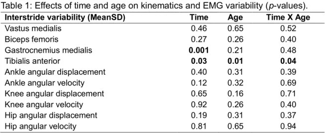
Table 1: Effects of time and age on kinematics and EMG variability (p-values).... 82 CHAPTER 6
Table 1: Pearson correlation analysis results. ... 101 CHAPTER 7
XIII Index of Figures
CHAPTER 1
Figure 1: Workflow of the fast-walking protocol ... 29 CHAPTER 3
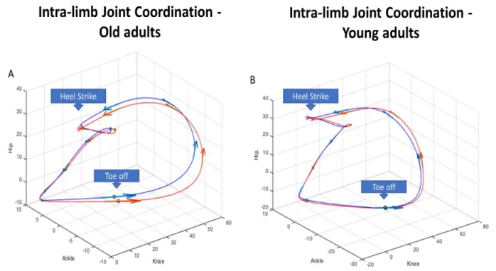
Figure 1: Spatial temporal parameters during fast-walking activity at the top. The mean and standard deviation for each subject were subsequently averaged across subjects within the age-group. At the bottom, differences in the coefficient of variation revealed the differences in spatial temporal variability along the task. * indicates statistically significant time effect. ... 53 Figure 2: Angular displacements for the hip, knee and ankle at heel strike and toe off in young and old adults during the five time points along the fast-walking activity. * indicates statistically significant time effect. The mean and standard deviation for each subject were subsequently averaged across subjects within the age-group.54 Figure 3: Illustration of Hip angle - Ankle angle - Knee angle intra-limb coordination patterns for the Old adults (A) and the Young adults (B). Blue line represents the mean values of the first stage, red line represents the mean values of the 5th / last stage. These two stages are displayed to highlight the differences induced by the activity. Arrows indicate the direction of the gait. ... 54 CHAPTER 4
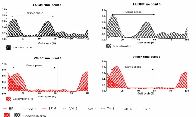
Figure 1: Illustration of coactivation area. Lines represent the mean linear envelope of muscle 1 activation. Dashed lines represent the mean linear envelope of muscle 2 activation. Crosshatched areas represent regions of coactivation. ... 69 Figure 2: The changes of muscle activities in Vastus Medialis (VM), Biceps Femoris (BF), Tibialis Anterior (TA), and Gastrocnemius Medialis (MG) for young and old adults over time. * represents significant changes over time; ** represents significant differences between groups. ... 70 Figure 3: Changes during the activity of the coactivation Indices between older (blue line) and young adults (green line). * represents significant differences over time. ... 71 CHAPTER 5
Figure 1: Mean ensemble curves for the sagittal plane angular displacement at the ankle, knee and hip during the gait cycle. The color scale was used to illustrate the intra-cycle region with higher variability among the subjects. The gradient of colors
XIV
represents the percentage of the sample that had values higher than their respective MeanSD at that particular time-point within the gait-cycle. ... 83 Figure 2: Mean ensemble curves for the sagittal plane angular velocity at the ankle, knee and hip during the gait cycle. The color scale was used to illustrate the intra-cycle region with higher variability among the subjects. The gradient of colors represents the percentage of the sample that had values higher than their respective MeanSD at that particular time-point within the gait-cycle. ... 84 Figure 3: Ensemble averaged electromyographic activities for TA, GM, VM, and BF during the gait cycle. The color scale was used to illustrate the intra-cycle region with higher variability among the subjects. The gradient of colors represents the percentage of the sample that had values higher than their respective MeanSD at that particular time-point within the gait-cycle. ... 85 CHAPTER 6
Figure 1: Mean ensemble curves for the heel horizontal velocity, and the toe vertical displacement normalized for the gait cycle (%). The color scale represents the percentage of the sample higher that had values higher than their respective MeanSD at that particular time-point within the gait-cycle. Dashed green circle represents the minimum toe clearance. Dashed grey circle represents the heel contact velocity. ... 100 CHAPTER 7
Figure 1: MTC and Lower limb angles between groups over time ... 114 CHAPTER 8
Figure 1: Sagittal angle displacement for the hip, knee, ankle within a gait cycle. Arrows are illustrating the three discrete events at which sagittal angle position were analyzed. HS = heel strike; TO = toe off; MTC = minimum toe clearance. ... 126 Figure 2: Illustration of Hip angle - Ankle angle - Knee angle intra-limb coordination patterns for the elderly subjects. Blue line represents the mean values of the first stage, red line represents the mean values of the 5th (last stage). These two stages
are displayed to highlight the differences induced by the activity. Small arrows indicate the direction of the gait. ... 128 Figure 3: Mean ensemble curve for the ankle angular displacement of an individual. The grey cloud around the mean line represents the standard deviation in its averaged value (MeanSD). The red cloud represents the original values of standard
XV
deviation for every percentage of the gait. The region where the orginal standard deviation is higher than the MeanSD was considered regions with a higher variability. ... 130 Figure 4: By overlapping the individuals’ plots (A), we obtained a gradient color indicating the percentage of the sample with higher variability for each ith% of the gait cycle. This information is then displayed over the mean ensemble curve for the variable analyzed (B). The vertical line indicates the average time of the toe off for all the subjects. ... 130 Figure 5: Mean ensemble curves for the toe vertical displacement normalized for the gait cycle (%). The gradient area represents the portion of the gait cycle were the variability is higher among the subjects. Color scale represents the percentage of the sample with values with great variability. ... 131
XVII Index of Equations CHAPTER 3 𝑪𝑽 =𝑴𝒆𝒂𝒏𝑺𝑫 × 𝟏𝟎𝟎% ... 51 CHAPTER 4 𝑪𝒐𝑰 = ∫ 𝑬𝑴𝑮𝑨𝑵𝑻 ∫ 𝑬𝑴𝑮𝑨𝑵𝑻+𝑬𝑴𝑮𝑨𝑮×𝟏𝟎𝟎% ... 68 CHAPTER 5 𝑴𝒆𝒂𝒏𝑺𝑫 = ⟨𝑺𝑫(𝒊)〉 𝒊, 𝒊 𝝐 {𝟎– 𝟏𝟎𝟎 % 𝒈𝒂𝒊𝒕 𝒄𝒚𝒄𝒍𝒆} ... 81 CHAPTER 6 𝐙 = 𝐔𝐭𝐗 . ... 98 𝐒𝐔𝐧 = 𝛌𝐔𝐧 . ... 98
XIX Resumo
Os efeitos da caminhada rápida nos parâmetros da marcha parecem destacar potenciais ameaças durante a execução desta atividade. Assim, o objetivo geral deste estudo foi investigar as alterações da marcha durante um exercício esporadicamente executado pelos idosos, a caminhada rápida. Durante esta atividade, dados cinemáticos e eletromiográficos foram avaliados. Os idosos apresentaram alterações nas articulações do quadril e do tornozelo em instantes discretos do ciclo de marcha sugerindo uma mudança na estratégia de produção de energia para impulsionar o corpo. Em geral, as alterações cinemáticas encontradas foram acompanhadas por mudanças no padrão de ativação muscular do membro inferior. Durante a tarefa, a atividade do músculo tibial anterior aumentou durante a fase de absorção e reduziu durante a sua fase propulsiva. Ao longo do protocolo, os índices de co-activação mudaram significativamente. Padrões de ativação muscular do tibial anterior e do gastrocnémio medial, reduziram e as regiões de maior variabilidade dentro do ciclo de marcha mudaram. Estas alterações neuromusculares foram semelhantes às encontradas na cinemática angular. Além disso, as reduções observadas na atividade muscular, níveis de coativação e variabilidade no padrão de ativação muscular, sugerem uma tentativa de redução do consumo metabólico durante o exercício. A presente pesquisa contribui para o aumento do conhecimento nesta área, fornecendo informações adicionais sobre a presença de variabilidade dentro do ciclo de marcha. Adicionalmente, encontramos mudanças nessas regiões de maior variabilidade dentro do ciclo de marcha durante a caminhada rápida. Estas alterações sugerem que adaptações a perturbações externas são dependente da fase do ciclo de marcha e podem ser consideradas como uma tentativa para manter o padrão motor dentro dos limites de segurança. Além disso, dada a associação encontrada entre a cinemática angular e o controle do movimento da trajetória vertical do pé e da velocidade horizontal do calcanhar, estratégias para prevenir quedas por tropeçar ou escorregar devem priorizar a estabilidade articular do tornozelo e do joelho. No entanto, apesar de melhorar o controle do movimento em eventos críticos do ciclo de marcha, os idosos parecem ter aumentado o risco de queda enquanto caminhavam em ritmos mais acelerados.
PALAVRAS-CHAVE: CINEMÁTICA, FADIGA, CAMINHADA, TROPEÇO, QUEDAS EM IDOSOS
XXI Abstract
Understanding the ongoing effects of fast-walking activity on gait parameters may highlight potential threats that can arise while walking at a faster pace. Therefore, our general purpose was to investigate the alterations to gait during an activity that can be sporadically performed by the elderly such as fast-walking. During the activity, kinematics and electromyography data have been assessed. Old adults showed alterations at the hip and ankle at discrete moments of the gait cycle suggestive of a change in the power generation strategy for propelling the body forward. Moreover, the alterations found on gait kinematics due to fast-walking were followed by changes in muscular activation pattern of the lower limb. During the task, the activity of the tibialis anterior increased during the absorptive phase and reduced during the propulsive phase. Throughout the protocol, coactivation indexes significantly changed over time. Measures of interstride variability of muscle activation for tibialis anterior and gastrocnemius medialis reduced, and regions of intra-stride variability changed during fast-walking. These neuromuscular alterations were similar to those of the angular kinematics. The reductions observed in muscular activity, levels of coactivation and variability in muscular activation pattern could be interpreted as an attempt to reduce metabolic consumption during the activity. The research presented here contributes to the body of literature by providing additional information regarding the presence of intra-cycle variability. Additionally, we have also found changes in these regions of higher variability within the gait cycle during fast-walking. These changes suggest that adaptations to a perturbation are phase-dependent and can be considered as an attempt to maintain the motor pattern within safety boundaries. Moreover, given the association found between the angular kinematics and the movement control of the toe vertical trajectory and heel horizontal velocity, strategies to prevent falls by tripping or slipping should address ankle joint kinematics, also as the knee joint kinematics. Despite improving movement control at critical events of the gait cycle, the elderly may have increased the risk of fall by tripping or slipping while walking at faster pace.
XXIII List of Abbreviations and symbols
ADL Activity of daily living
ANOVA Analysis of variance
BF Biceps femoris
CAPES Coordination for the Improvement of Higher Education Personnel
CoI Coactivation index
COM Center of mass
EMG Electromyography
GM Gastrocnemius medialis
HR Heart rate
HCV Heel contact velocity
HHV Heel horizontal velocity
IBGE Brazilian Institute of Geography and Statistics INE National Statistical Institute of Portugal
IQR Interquartile range
MTC Minimum toe clearance
n Number of subjects
OA Old adults
PC1, PC2, PCn First, second, nth principal component (PCA)
PCA Principal component analysis
ROM Range of motion
RMS Root mean square
SD Standard deviation
SPSS Statistical package for the social sciences
TA Tibialis anterior
TVD Toe vertical displacement
U Eigenvectors of the covariance matrix (PCA)
VM Vastus medialis
WHO World Health Organization
YA Young adults
β Coefficient of regression
XXIV
λ Eigenvalues (PCA)
25 Chapter 1 General Introduction
Currently, society is facing a marked ageing of the world population. Approximately 14% of the European population are over 65 years and it is expected that these numbers will double by 2050 (WHO, 2011). In Portugal, between 2001 and 2011, the percentage of the elderly population increased from 16.6% to 19% (INE, 2011). In Brazil, currently, about 10% of the population are elderly, i.e., approximately 20 million people (IBGE, 2010). With more than a third of elderly experiencing at least one fall per year worldwide, the consequences of falls represent a major problem for the health systems of many countries with huge financial costs associated (WHO, 2011).
Falls are a result of a complex interaction between extrinsic (environment) and intrinsic (related to the individual) factors. In a systematic review regarding the association of biomechanical aspects of gait in elderly fallers and non-fallers, Kirkwood et al. (2006) observed that 55% of falls were related to gait alterations, 32% to balance alterations, and the remaining to extrinsic factors. Among the extrinsic factors found were: inappropriate shoes, uneven surfaces, poor lighting, etc. The intrinsic factors included poor health, lack of static and dynamic postural control (i.e. remain standing and walking), insufficient muscle strength and power of the lower limb muscles, visual difficulties, history of falls, and fatigue (Maki, 1997; WHO, 2011). There are currently over than 400 known risk factors for falls (Masud & Morris, 2001). Besides the extrinsic (environmental) and intrinsic (individual) factors, aspects related to the task are among those that can bring an individual to fall. Factors such as speed, task complexity or fatigue during the task are considered to increase the probability of a fall incident (Callisaya et al., 2011; Tinetti & Speechley, 1989).
Healthy, active seniors are more likely to fall in outdoor activities where walking faster can be sporadically performed by this population (Li et al., 2006). Research supports the premise that age-related differences on gait observed in elderly are primarily due to reduced muscle strength and lower limb joint range of motion (ROM) (Kang & Dingwell, 2008), which in turn, is attributable to physiological and neuromuscular changes displayed by older adults (Faulkner et
26
al., 2007; Prince et al., 1997). Therefore, the extent of these changes may reduce the capacity of this population to adapt their gait pattern when demanded. Given that walking biomechanics are known to be influenced by speed, some studies have investigated the effects of the walking speed as well as the underlying factors that may drive these alterations. Significant associations have been found between gait speed and joint kinematics (Hanlon & Anderson, 2006; Monaco et al., 2009), joint kinetics (Burnfield et al., 2000; Chung & Wang, 2010; Silder et al., 2008), muscle activity (Schmitz et al., 2009) and gait stability (England & Granata, 2007; Kang & Dingwell, 2008). Irrespective of the factors underlying the alterations caused by walking at a faster pace, some authors have associated fast-walking with increased risk of falls (Callisaya et al., 2011; Faulkner et al., 2009; Nagano et al., 2014; Pavol et al., 1999).
A successful locomotion needs the integration of different physiological systems. According to England and Granata (2007) fast-walking velocity may influence the dynamic stability by a combination of several mechanisms, as the ability to control movement could be disrupted by the effects of fast-walking over gait kinematics and other clinical correlates of stable walking. When walking, the ability to adjust the speed requires different levels of muscular activities for appropriate adaptations to changes in the task demands. Nevertheless, increasing walking speed is associated with an increase in muscle stress, particularly of the plantar-flexors and dorsi-flexors (Neptune, 2004). Indeed, the ability of the ankle plantar flexors to produce force as walking speed increased was greatly impaired (Neptune et al., 2008), which could potentially compromise the trajectory of the swing foot, increasing risks of tripping and slipping (Lockhart & Kim, 2006; Winter, 1992). In consonance with this, Nagano et al. (2014) demonstrated that after a short period of fast-walking, older adults were more susceptible to falls by tripping, the authors attributed this to fatigue. In addition, older adults increased coactivation at the knee and ankle during mid-stance when walking faster than the preferred speed. Whilst coactivation is believed to be used to stiffen the joint and enhance stability (Hortobágyi & DeVita, 2006), a potential side-effect would be higher metabolic costs (Mian et al., 2006). Furthermore, increased joint stiffness decreases the capacity to produce force during toe off
27
(Watelain et al., 2000), as well as increases horizontal heel velocity at foot landing (Lockhart & Kim, 2006). Therefore, the broad effects of increased coactivation in older adults can increase the risk of falling by tripping or slipping.
The great variety of fall-risk’s factors reflect the diversity of health determinants that directly or indirectly affect the individual's well-being. Increased speed and walking duration may magnify the limitations of old adult’s gait characteristics, changes may occur over time, and such assessments may highlight other factors that may be associated with risk of falls. Studies involving analyses of biomechanical parameters and muscle activity in the elderly during an activity that can be a sporadic practice among this population, such as fast walking, are crucial for understanding the strategies that may be involved in falls’ prevention. Thus, there is a growing need to prevent falls in the elderly; to present relevant information regarding the abnormal gait patterns induced by fast-walking in the elderly; to determine mechanisms of falls allowing the development of a feedback mechanism in order to prevent the occurrence of falls and reduce such alarming numbers of morbidity associated with this part of the population; to give a differential treatment of the information obtained, considering data under different levels as well as the possible interactions between them; and to provide further information to public health systems to allow the planning of strategies addressed to the current reality, taking into account the context of the increase in life expectancy in most countries.
Objectives
Based on these assumptions, we observed a lack of information regarding the emerging changes to gait in an ecological context that may determine the predictive behavior of falls in the elderly population. The overall aim of this thesis is to evaluate the effects of fast-walking on gait, with a special focus on evaluating parameters previously linked to risk of falls in older adults. The specific aims were as follows: (1) to describe alterations on gait kinematics during fast-walking in old and young adults; (2) to investigate the influence of fast-walking on muscle activity of lower limb muscles of young and older adults; (3) to determine how the variability of gait and muscle activity change during prolonged time walking at faster speed; (4) to determine whether the variability of EMG and kinematic
28
patterns during fast-walking were reflected in the variability of kinematics associated with risk of slipping and tripping; (5) to verify alterations on slip and trip-related parameters during the activity; (6) to determine the influence of the swing and stance lower limb joints on altering the swing toe trajectory.
The work presented here aims to determine the biomechanical gait changes induced by fast-walking in elderly, with the experimental accomplishments presented in Chapters 3 to 7. In Chapter 2 we provide an overlook of the scientific literature pertaining to the topic. Then, we describe in Chapter 3 kinematic alterations over time in both age groups. Chapter 4 explores neuromuscular adaptations during fast-walking in old and young adults. Chapter 5 explores the variability of muscle and kinematic pattern during the task. In chapter 6 we sought to investigate whether the variability of trip and slip-related signals were associated with those from muscle and kinematic pattern described in chapter 5. Chapter 7 reveals the risk of falls by tripping along the fast-walking protocol. Chapter 8 includes a general discussion in the context of the findings from the experimental studies. Chapter 9 presents the main conclusions and offers recommendations for future research. Finally, the bibliography references are presented in Chapter 10. An overview of study designs is presented in Table 1.
Table 1: Study design description.
Study Design Population n included
I
Descriptive analysis of gait kynematics alterations over time
Healthy young and older adults 50
II Muscle activation study Healthy young and older adults 23
III Variability of EMG and
kinematic patterns Healthy young and older adults 23
IV
Gait variability influence on slip and trip-related
Association
Healthy older adults 08
V Multilevel Regression
Analysis Healthy young and older adults 51
29 Study delimitation
The study was delimited to healthy old and young adults above 65 and 21 years old, respectively. All participants were free of any orthopaedical, neurological, visual, vestibular or cardiovascular conditions that would prevent the subject from performing all the proposed activities. Old participants were recruited from the local community and the young participants from among the university’s students. The walking-based fatigue protocol consisted of walking at 70% of their maximum heart rate for twenty minutes or until their voluntary exhaustion. The flow of this protocol is illustrated in Figure 1. Further explanations regarding data acquisition and analysis are described in the experimental papers throughout the thesis.
30 References
Burnfield, J. M., Josephson, K. R., Powers, C. M., & Rubenstein, L. Z. (2000). The influence of lower extremity joint torque on gait characteristics in elderly men. Archives of Physical Medicine and Rehabilitation, 81(9), 1153-1157.
Callisaya, M. L., Blizzard, L., Schmidt, M. D., Martin, K. L., McGinley, J. L., Sanders, L. M., & Srikanth, V. K. (2011). Gait, gait variability and the risk of multiple incident falls in older people: a population-based study. Age and Ageing (Vol. 40, pp. 481-487).
Chung, M.-J., & Wang, M.-J. J. (2010). The change of gait parameters during walking at different percentage of preferred walking speed for healthy adults aged 20–60 years. Gait & Posture, 31(1), 131-135.
England, S. A., & Granata, K. P. (2007). The influence of gait speed on local dynamic stability of walking. Gait & Posture, 25(2), 172-178.
Faulkner, J. A., Larkin, L. M., Claflin, D. R., & Brooks, S. V. (2007). Age‐related changes in the structure and function of skeletal muscles. Clinical and Experimental Pharmacology and Physiology, 34(11), 1091-1096.
Faulkner, K. A., Cauley, J. A., Studenski, S. A., Landsittel, D. P., Cummings, S. R., Ensrud, K. E., Donaldson, M. G., & Nevitt, M. C. (2009). Lifestyle predicts falls independent of physical risk factors. Osteoporosis International, 20(12), 2025-2034.
Hanlon, M., & Anderson, R. (2006). Prediction methods to account for the effect of gait speed on lower limb angular kinematics. Gait & Posture, 24(3), 280-287.
Hortobágyi, T., & DeVita, P. (2006). Mechanisms responsible for the age-associated increase in coactivation of antagonist muscles. Exercise and Sport Sciences Reviews, 34(1), 29-35.
IBGE. (2010). Síntese de Indicadores Sociais. Uma análise das condições de vida da população brasileira. Estudos & pesquisas. Informação
demográfica e socioeconômica. Available in
http://www.ibge.gov.br/home/estatistica/populacao/condicaodevida/indica doresminimos/sinteseindicsociais2010/SIS_2010.pdf [Accessed 12 October 2016]
INE. (2011). XV recrutamento geral da população. Censos 2011. V recrutamento
geral da habitação. Available in
http://censos.ine.pt/xportal/xmain?xpid=CENSOS&xpgid=censos2011_a presentacao. [Accessed 25 November 2016]
Kang, H. G., & Dingwell, J. B. (2008). Effects of walking speed, strength and range of motion on gait stability in healthy older adults. Journal of Biomechanics, 41(14), 2899-2905.
31
Kirkwood, R. N., Araújo, P. A., & Dias, C. S. (2006). Biomecânica da marcha em idosos caidores e não caidores: uma revisão da literatura. Revista Brasileira de Cineantropometria e Movimento, 4(14), 103-110.
Li, W., Keegan, T. H., Sternfeld, B., Sidney, S., Quesenberry Jr, C. P., & Kelsey, J. L. (2006). Outdoor falls among middle-aged and older adults: a neglected public health problem. American Journal of Public Health, 96(7), 1192-1200.
Lockhart, T. E., & Kim, S. (2006). Relationship between hamstring activation rate and heel contact velocity: factors influencing age-related slip-induced falls. Gait & Posture, 24(1), 23-34.
Maki, B. E. (1997). Gait changes in older adults: predictors of falls or indicators of fear? Journal of the American Geriatrics Society, 45(3), 313-320. Masud, T., & Morris, R. O. (2001). Epidemiology, of falls. Age and Ageing, 30,
3-7.
Mian, O. S., Thom, J. M., Ardigo, L. P., Narici, M. V., & Minetti, A. E. (2006). Metabolic cost, mechanical work, and efficiency during walking in young and older men. Acta Physiologica (Oxford), 186(2), 127-139.
Monaco, V., Rinaldi, L. A., Macrì, G., & Micera, S. (2009). During walking elders increase efforts at proximal joints and keep low kinetics at the ankle. Clinical Biomechanics, 24(6), 493-498.
Nagano, H., James, L., Sparrow, W. A., & Begg, R. K. (2014). Effects of walking-induced fatigue on gait function and tripping risks in older adults. Journal of NeuroEngineering and Rehabilitation, 11, 155.
Neptune, R. R., Sasaki, K., & Kautz, S. A. (2008). The effect of walking speed on muscle function and mechanical energetics. Gait & Posture, 28(1), 135-143.
Pavol, M. J., Owings, T. M., Foley, K. T., & Grabiner, M. D. (1999). Gait characteristics as risk factors for falling from trips induced in older adults. Journals of Gerontology. Series A, Biological Sciences and Medical Sciences, 54.
Prince, F., Corriveau, H., Hébert, R., & Winter, D. A. (1997). Gait in the elderly. Gait & Posture, 5(2), 128-135.
Schmitz, A., Silder, A., Heiderscheit, B., Mahoney, J., & Thelen, D. G. (2009). Differences in lower-extremity muscular activation during walking between healthy older and young adults. Journal of Electromyography and Kinesiology, 19(6), 1085-1091.
Silder, A., Heiderscheit, B., & Thelen, D. G. (2008). Active and passive contributions to joint kinetics during walking in older adults. Journal of Biomechanics, 41(7), 1520-1527.
Tinetti, M. E., & Speechley, M. (1989). Prevention of falls among the elderly. New England Journal of Medicine 320(16), 1055-1059.
32
Watelain, E., Barbier, F., Allard, P., Thevenon, A., & Angué, J.-C. (2000). Gait pattern classification of healthy elderly men based on biomechanical data. Archives of Physical Medicine and Rehabilitation, 81(5), 579-586.
World Health Organization. (2011). Global Health and Ageing. World Health Organization.
Winter, D. A. (1992). Foot trajectory in human gait: a precise and multifactorial motor control task. Physical Therapy, 72(1), 45-53; discussion 54-46.
33 Chapter 2 Literature Review
This chapter reviews the scientific literature pertaining the topic being investigated in four major sections: incidence and aetiology of risk of falls in the elderly population; a comprehensive review of fatigue in older adults; and a summary about fatigue and increased speed effects on gait parameters.
2.1 Falls in elderly population
Ageing of the population and its arising implications led the construction of a new epidemiological profile, characterized by a higher prevalence of chronic diseases, bringing new challenges in health care (Williams & Manfredi, 2004). According to WHO (2011) more than a third of elderly experienced at least one fall per year. A study carried out in Catalonia (Spain) revealed that 17.9% of people over 65 have experienced at least one fall in the 12 months preceding the interview and the falls frequency increases with age (Séculi et al., 2004). In Portugal, after direct consultation with the National Statistical Institute (INE) and the Central Administration of the Health System (ACSS), no official data were found regarding the prevalence of falls in the elderly. In Brazil, the falls and their consequences in the elderly have assumed epidemic dimension, with huge financial costs associated with (IBGE, 2010).
The combination between high incidence with a high susceptibility to injury makes falls in the elderly population particularly dangerous, because of the high prevalence of clinical diseases (e.g. osteoporosis) and age-related physiological changes (e.g. slower reflexes) in this population (Rubenstein, 2006). In addition, the rate of falls increases with age, and the fear of falling down-regulates activity, which in turn, further contributes to subsequent falls (Tinetti & Powell, 1993).
Despite being the leading cause of non-fatal injuries worldwide, and therefore motivating several research studies focusing on fall prevention, there remains a lack of detailed national estimates of falls incidence, and fall-related injuries by age and gender. Data on the older Portuguese population relative to this matter is scarce, however Moniz-Pereira et al. (2013) in a prospective study reported a first known faller risk profile in the Portuguese elderly population. Their results showed that being a woman, having fear of falling and lower functional
34
fitness levels were determinant factors for both episodic and recurrent falls. Given the high incidence of falls and the associated morbidity, mortality and other costs, combined with the ageing of the population, the incidence of falls in the elderly and the associated costs are expected to rise unless effective preventive techniques are implemented. If preventive measures are not taken in the immediate future, the numbers of injuries caused by falls are projected to be 100% higher in the 2030 (Kannus et al., 2007).
Falls occur as a result of a complex interaction between numerous environmental hazards and individual factors. Among the extrinsic factors, we found inappropriate shoes, uneven surfaces, poor lighting, etc. The intrinsic factors included poor health, lack of static and dynamic postural control (i.e. remain standing and walking), insufficient muscle strength and power of the lower limb muscles, visual difficulties, and history of falls (WHO, 2008). The presence of environmental hazards creates the opportunity for a fall, particularly for individuals already impaired by a combination of intrinsic factors (Rubenstein, 2006). Gait or balance deficits emerged as the most consistent independent predictors from a systematic review regarding falls risk factors (Ganz et al., 2007). Ageing brings about profound changes in human locomotion. Older adults seem to adopt a more conservative walking pattern characterised by a slower walking velocity, shorter steps, greater base of support and prolonged double support phase (Lord & Dayhew, 2001; Maki, 1997). It is generally assumed that these changes lead to an increased stability during walking (Lord & Dayhew, 2001). One critical factor closely related with the task of walking itself is fatigue, which may discourage physical activity and further compromise safe progression. Nevertheless, several studies have identified fatigue as an important risk of falling, due to the deleterious influence in several aspects of gait modified by fatigue (Helbostad et al., 2007; Parijat & Lockhart, 2008b).
2.2 Fatigue in older adults
Given the ageing of our population and the improvements in medicine that have extended the life expectancy of human beings over the last century, the investigation about whether and how skeletal muscles of the elderly resist fatigue has become an important area of research. In an intact organism, several factors
35
can influence the development of muscle fatigue. Because the ability to maintain force is a critical aspect of neuromuscular function, understanding fatigue is essential to understand the biology of senescence (Kent-Braun, 2009). Some changes in the motor system related to age have an impact on the resistance to muscle fatigue. Examples of changes are:
- Loss and atrophy of muscle fibers, with a great decrease in the number and size of type II muscle fibers (Campbell et al., 1973);
- Progressive decline in the number of motoneurons with subsequent re-innervation and expansion of the re-innervation area of the surviving motoneurons (Manini et al., 2013);
- Substantial loss of upper motoneurons in the central nervous system (Eisen et al., 1991).
The functional consequences of these changes include substantial loss of strength and decreased contractile characteristics in most muscles assessed. Further studies suggest that type I muscle fibres contribute proportionally more to the strength production in elderly (≥70 years) than in young people (Roos et al., 1997). However, although it seems reasonable to suggest that age-related changes in muscle morphology and motor unit remodelling, as well as associated loss of strength and contractile properties, may improve resistance to neuromuscular fatigue in elderly, the results highlighted and discussed in a literature review performed by Allman and Rice (2002) suggest that this generalization cannot be made. The lack of agreement between the studies is due, in part, to the differences by which fatigue was induced or assessed. These inconsistencies may result in the quite varied methodologies used to quantify fatigue, as well as the different muscle groups that have been tested (i.e., elbow flexors, knee extensors, thumb adductor, ankle dorsiflexors), the exercise modality selected (i.e., isometric, isokinetic, dynamic, with electrical stimulation fatigue), the particularities of the study populations, and the relatively small number of individuals studied (Katsiaras et al., 2005).
The variability of fatigue susceptibility in the elderly relies heavily on the task used to induce fatigue. The fatigability in the elderly depends on the performance of different types of exercises (voluntary vs. electric, isometric vs.
36
dynamic, sustained versus intermittent, maximal vs submaximal) and yet, on the performance of the same exercise by muscles with different contractile properties (Bigland-Ritchie et al., 1995). According to Chan et al. (2000), muscle fatigue can result from the failure of any component along the motor pathway, from the volitional effort to the contractile ability of the muscle. This approach resulted in the distinction between central and peripheral fatigue, or central nervous system and muscle fatigue. Thus, the concept of fatigue begins to consider its multifactorial nature involving issues related to the depletion of energy systems, the accumulation of catabolism products, the involvement of the nervous system and the failure of the contractile mechanism of skeletal muscle fibre. The central fatigue was also considered as the failure to activate all motor units at optimal discharge rates and it can be assessed by comparing the reduction in force during a maximal voluntary contraction, which requires full functioning of the entire muscle force production pathway, to the decreased force induced by electrical stimulation, which evaluates changes distal to the stimulation point (Kent-Braun, 2009). For clinical use, the central fatigue is defined as the difficulty of initiation, or the ability to maintain volunteer activities (Chaudhuri & Behan, 2000). Finally, the ability to resist fatigue, sometimes expressed as ‘muscle endurance’, was defined as the time to failure to maintain target tension (Hicks et al., 2001).
In contrast to the concept of muscle fatigue suggested by Enoka and Stuart (1992), as an acute impairment of performance that includes both an increase in the perceived effort necessary to exert a desired force and an eventual inability to produce this force, a new review carried out after 15 years by Barry and Enoka (2007) defines muscle fatigue as an exercise-induced reduction in the ability of the muscle to generate force or power, even if the task can still be sustained. Therefore, fatigue often begins soon after the onset of sustained activity, even though an individual can continue performing the task. Although the impairments that contribute to fatigue will eventually limit the ability of the individual to continue that task, fatigue and task failure should be distinguished (Bigland-Ritchie et al., 1995).
Considering aspects of different systems, some studies observed that in the elderly population, when compared to young adults, presented greater
37
resistance to fatigue. By incorporating information from multiple systems, the fatigue resistance observed by the elderly seems to lie on a neuro-energetic basis centered on the largest metabolic economy in the neuromuscular system (Lanza et al., 2005). Regarding fatigue in the elderly, age-related changes that occur within the neuromuscular system may result in some sites that are more prone to fatigue. The changes may increase or decrease their susceptibility to failure under specific task conditions. The effect of age on the various central and peripheral sites more prone to fatigue is discussed considering their relative contributions during the different fatigue-induced tasks, without neglecting the impact of possible confounding effects on fatigability related to subjects' habits, physical activity status and gender (Allman & Rice, 2002). The assumption obtained after the results of more recent studies, presented in a review performed by Kent-Braun (2009), indicate that energy-producing pathways in skeletal muscle may combine with changes in motor unit behaviour and in contract properties of muscle with the objective to provide a unique environment to resist the muscle fatigue in healthy men and women over the age of 65.
2.3 Fatigue effects on gait and its repercussion on risk of falls
Fatigue can have a widespread impact on biological functions, altering the capacity of most systems to operate at the desired level (Enoka & Stuart, 1992; Gandevia, 1998). The effects of fatigue on gait in elderly people have been a crucial focus of some recent studies.
The impact of fatigue is not restricted to a decline in force producing capacity of the system, but also it leads to an inability to adequately control specific movement dynamics to produce a controlled movement (Cortes et al., 2014). A certain level of variability associated with many biomechanical measures contains structure in the form of long range correlations. This means that a repeated movement at any point in the time series is related to or dependent upon previous cycles. This scaling behaviour in human gait is of interest because it indicates the overall adaptability of the motor system. Increased variability may bring the dynamic state to their limits of stability, where error corrections are less effective. In this context, increased variability has been found to be associated with fall risk (Kressig et al., 2008). In an exemple, Helbostad et al. (2007) reported
38
that a repeated sit-to-stand task affected gait control in older person in terms of an increased variability in step width and length.
Stable walking kinematics requires generating appropriate motor patterns (Lee & Kerrigan, 1999). The movement pattern presumably reflects that of the muscles. However, Granacher et al. (2010) reported that ankle fatigue resulted in a decrease in functional reflex activity of tibialis anterior, and increases in coactivity and mean angular velocity in the ankle joint complex. It has been suggested that increased coactivation reduces the capacity to produce force in a short period of time, which could lead to lower capacity to react to motor disturbances (Bellew, 2002). A later study demonstrated coactivation as an important risk factor for falling (Pereira & Gonçalves, 2011), since it negatively affects torque production and increases energy expenditure during gait in elderly. The authors suggested that these results may impair an individual’s ability to compensate for gait perturbations. Appropriate temporal separation between agonist and antagonist activation of muscles has been observed for well-controlled voluntary movements (Frey-Law & Avin, 2013). When fatigated, however, this separation is attenuated, muscle coactivation is increased reducing knee extensor force (Pereira & Goncalves, 2011). Furthermore, higher coactivation increases joint stiffness decreasing the capacity to produce force during toe off (Watelain et al., 2000), as well as also increasing horizontal heel velocity at foot landing (Lockhart & Kim, 2006). Therefore, since loss of dorsiflexor strength at toe-off can be critical to the ability of overcome an obstacle (Nagano et al., 2011), and higher horizontal heel velocity at landing increases chances of slips (Lockhart & Kim, 2006), the broad effects of increased coactivation in older adults can increase risk of falling by tripping or slipping.
As previously mentioned, fatigue can lead to a loss of muscle strength, which may compromise obstacle clearance. The safe trajectory of the foot during swing is essential to the successful obstacle avoidance. The swing toe describes its forward trajectory reaching its lowest vertical point, represented by the minimum toe clearance (MTC). This biomechanical event occurs at the highest forward velocity of the swing foot, representing a highly-controlled movement to maintain the clearance around a safety value and prevent tripping (Winter, 1992).
39
Previous work has explored fatigue influence on the MTC and reported significant reductions in toe clearance, which in turn increases risk of falls by tripping. Along with tripping, slipping have also been shown to contribute to high rates of falls. The knee joint musculature is considered important in producing large flexion and extension moments when recovering from a slip. Fatigue of the knee extensors is associated with decreases in stabilization time (Parijat & Lockhart, 2008b).
As successful locomotion needs the integration of different physiological systems, therefore many factors have influence on gait performance. The inevitable consequences of fatigue can alter neuromuscular processes both centrally and peripherally. Since walking requires contracting muscles to move an imposed load through an adequate joint ROM for successful task completion, fatigue can affect the movement performance for a given individual. Previous studies indicated that slower walking speed in elderly subjects may be associated with the physiological or neuromuscular limitations in older adults (Anderson & Madigan, 2014; McGibbon, 2003). Regarding fatigue effects on joint angular kinematics, previous studies reported reduction in ankle joint ROM, greater knee flexion at heel contact, less knee extension during terminal stance, and less dorsiflexion at heel contact (Cheng & Rice, 2012; Parijat & Lockhart, 2008b). The reduction in the ROM of one distal joint, which has been reported as a consequence of fatigue, can lead the adjacent joints to adapt to a greater extent, producing different kinematic combination. The resulting coordination pattern of the whole limb, therefore, might have to adapt in a non-conventional way to accomplish the motor task required. However, the studies have been limited to a description of the fatigue effects on singular joints, without considering the intersegmental interactions.
In summary, fatigue can affect sensorimotor function and walking performance in many ways. Ultimately, fatigue has been shown to increase risk of falls by increasing gait variability, reducing minimum toe clearance, and increasing slip risk. Furthermore, when exposed to fatigue, the sensorimotor system seems to adjust motor planning and execution at the expense of the angular kinematics pattern to ensure the success of a motor task (Cheng & Rice, 2012; Parijat & Lockhart, 2008a). However, most of the known effects of fatigue
40
on gait parameters were assessed after using protocols that are not commonly reproducible in daily life. Recently, walking-based fatigue has been used to induce fatigue, although most of the studies have been limited to preferred walking speed. The only known exception regarding the association between fatigue and risk of falls is the study of Nagano et al. (2014), which have demonstrated that after a short period of fast-walking, older adults were more susceptible to falls by tripping.
2.4 Effects of fast speed on gait
In general, healthy older adults are more susceptible to fall in outdoors activities (Li et al., 2006), where walking at a faster pace can be a sporadic practice among this population. Previous experiments have demonstrated that walking at a faster speed significantly affect gait biomechanics. In a systematic review by Figueiredo et al (2011), the review showed the influence of speed in biomechanical parameters that characterize the gait action. Significant associations have been found between gait speed and joint kinematics (Hanlon & Anderson, 2006; Monaco et al., 2009), joint kinetics (Burnfield et al., 2000; Chung & Wang, 2010; Silder et al., 2008), muscle activity (Schmitz et al., 2009) and gait stability (England & Granata, 2007; Kang & Dingwell, 2008).
Previous studies have reported that a higher walking speed caused a greater joint motion in hip and knee joints (Hanlon & Anderson, 2006; Lelas et al., 2003; Monaco et al., 2009). In Hanlon and Anderson (2006), it is brought to our attention that the degree to which speed affected each angle depends not only on the angle itself but also on the time within the gait cycle. This is important for addressing the underlying reasons that drive these changes within the gait cycle. Some of these alterations, as the increased peak knee flexion during pre-swing, have been attributable to the need for greater shock absorption at higher speeds (Lelas et al., 2003). In addition, due to the increasing speed, older adults could exhibit a significant increase of the kinetics at the proximal joints (Burnfield et al., 2000). Previous studies have reported that older adults exhibit a proximal joint power strategy to propel the body forward (DeVita & Hortobagyi, 2000; Neptune et al., 2004). This distal to proximal shift in power production seems to be attributable to the plantar-flexor weakness (Silder et al., 2008). Assessment of
41
joint kinetics provides a better understanding of normal motor patterns. Thus, the estimation of the relative contribution of each joint to the total energy generated and absorbed during gait at fast-walking is a useful approach to understand the extent, and degree of compensation across joints and to address potential risks due to fast-walking.
Furthermore, when walking at a faster speed, older adults increase gait and neuromuscular variability (Almarwani et al., 2016; Kang & Dingwell, 2008; Raffalt et al., 2017), and greater joint kinematic variability has been consistently associated with risk of falls (Kobayashi et al., 2014; Mills et al., 2008). In addition, a significant association have been found between gait speed and gait stability (England & Granata, 2007; Kang & Dingwell, 2008). According to England and Granata (2007) fast-walking velocity may influence the dynamic stability by a combination of several mechanisms, as the ability to control movement could be disrupted by the effects of fast-walking over gait kinematics and other clinical correlates of stable walking.
When walking, the ability to adjust the speed requires different levels of muscular activity for appropriate adaptations to changes in the task demands. In general, muscle activity tends to increase at faster walking speeds, resulting in a larger muscular force output (Neptune et al., 2008). According to Schmitz et al. (2009), the changes in muscle activity observed when walking at a faster pace enhances the distal-to-proximal shift in power production exhibited in old adults. In addition, old adults increased coactivation at the knee and ankle during mid-stance. Whilst coactivation is believed to be used to stiffen the joint and enhance stability (Hortobágyi & DeVita, 2006), a potential side-effect would be at a higher metabolic cost (Mian et al., 2006). Nevertheless, increasing walking speed is associated with an increase in muscle stress, particularly of the plantar-flexors and dorsi-flexors (Neptune, 2004). Indeed, the ability of the ankle plantar flexors to produce force as walking speed increased was greatly impaired (Neptune et al., 2008), which could potentially compromise the trajectory of the swing foot, increasing risks of tripping (Winter, 1992). In consonance with this, as mentioned earlier in this chapter, Nagano et al. (2014) demonstrated that after a short period
42
of fast-walking, older adults were more susceptible to falls by tripping, to which the authors attributed to fatigue.
Therefore, increments in gait speed have been shown to have a significant effect on gait in older adults. However, most of these studies are limited to describe the changes that occur when walking speed is modified. Understanding the effects of fast-walking activity over time on gait parameters may highlight potential threats that may arise while walking at a faster pace.
References
Allman, B. L., & Rice, C. L. (2002). Neuromuscular fatigue and ageing: Central and peripheral factors. Muscle and Nerve, 25(6), 785-796.
Almarwani, M., VanSwearingen, J. M., Perera, S., Sparto, P. J., & Brach, J. S. (2016). Challenging the motor control of walking: Gait variability during slower and faster pace walking conditions in younger and older adults. Archives of Gerontology and Geriatrics, 66, 54-61.
Anderson, D. E., & Madigan, M. L. (2014). Healthy older adults have insufficient hip range of motion and plantar flexor strength to walk like healthy young adults. Journal of Biomechanics, 47(5), 1104-1109.
Barry, B. K., & Enoka, R. M. (2007). The neurobiology of muscle fatigue: 15 years later. Integrative and Comparative Biology, 47(4), 465-473.
Bellew, J. W. (2002). A Correlation Analysis Between Rate of Force Development of the Quadriceps and Postural Sway in Healthy Older Adults. Journal of Geriatric Physical Therapy, 25(1), 11-15.
Bigland-Ritchie, B., Rice, C. L., Garland, S. J., & Walsh, M. L. (1995). Task-dependent factors in fatigue of human voluntary contractions. Advances In Experimental Medicine And Biology, 384, 361-380.
Burnfield, J. M., Josephson, K. R., Powers, C. M., & Rubenstein, L. Z. (2000). The influence of lower extremity joint torque on gait characteristics in elderly men. Archives of Physical Medicine and Rehabilitation, 81(9), 1153-1157.
Campbell, M. J., McComas, A. J., & Petito, F. (1973). Physiological changes in ageing muscles. Journal of Neurology Neurosurgery and Psychiatry, 36(2), 174-182.
Chan, K. M., Raja, A. J., Strohschein, F. J., & Lechelt, K. (2000). Age-related changes in muscle fatigue resistance in humans. Canadian Journal of Neurology Science, 27(3), 220-228.
Chaudhuri, A., & Behan, P. O. (2000). Fatigue and basal ganglia. Journal of the Neurological Sciences, 179(1–2), 34-42.
43
Cheng, A. J., & Rice, C. L. (2012). Factors contributing to the fatigue-related reduction in active dorsiflexion joint range of motion. Applied Physiology, Nutrition, and Metabolism, 38(5), 490-497.
Chung, M.-J., & Wang, M.-J. J. (2010). The change of gait parameters during walking at different percentage of preferred walking speed for healthy adults aged 20–60 years. Gait & Posture, 31(1), 131-135.
Cortes, N., Onate, J., & Morrison, S. (2014). Differential effects of fatigue on movement variability. Gait & Posture, 39(3), 888-893.
DeVita, P., & Hortobagyi, T. (2000). Age causes a redistribution of joint torques and powers during gait. Journal of Applied Physiology, 88(5), 1804-1811. Eisen, A., Siejka, S., Schulzer, M., & Calne, D. (1991). Age-dependent decline in
motor evoked potential (MEP) amplitude: With a comment on changes in Parkinson's disease. Electroencephalography and Clinical Neurophysiology - Electromyography and Motor Control, 81(3), 209-215. England, S. A., & Granata, K. P. (2007). The influence of gait speed on local
dynamic stability of walking. Gait & Posture, 25(2), 172-178.
Enoka, R. M., & Stuart, D. G. (1992). Neurobiology of muscle fatigue. Journal of Applied Physiology, 72(5), 1631-1648.
Figueiredo, M. C., Abreu, S., Castro, M.P., Vilas-boas, J. P. (2011). The influence of ambulatory speed on gait biomechanical parameters. Revista Portuguesa de Ciências do Desporto, 11(3), 64-87.
Frey-Law, L. A., & Avin, K. G. (2013). Muscle coactivation: a generalized or localized motor control strategy? Muscle and Nerve, 48(4), 578-585. Gandevia, S. C. (1998). Neural control in human muscle fatigue: Changes in
muscle afferents, moto neurones and moto cortical drive. Acta Physiologica Scandinavica, 162(3), 275-283.
Ganz, D. A., Bao, Y., Shekelle, P. G., & Rubenstein, L. Z. (2007). Will my patient fall? Journal of the American Medical Association, 297(1), 77-86.
Granacher, U., Gruber, M., Forderer, D., Strass, D., & Gollhofer, A. (2010). Effects of ankle fatigue on functional reflex activity during gait perturbations in young and elderly men. Gait & Posture, 32(1), 107-112.
Hanlon, M., & Anderson, R. (2006). Prediction methods to account for the effect of gait speed on lower limb angular kinematics. Gait & Posture, 24(3), 280-287.
Helbostad, J. L., Leirfall, S., Moe-Nilssen, R., & Sletvold, O. (2007). Physical fatigue affects gait characteristics in older persons. In Journals of Gerontology. Series A, Biological Sciences and Medical Sciences (Vol. 62, pp. 1010-1015).
Hicks, A. L., Kent-Braun, J., & Ditor, D. S. (2001). Sex differences in human skeletal muscle fatigue. Exercise and Sport Sciences Reviews, 29(3), 109-112.
44
Hortobágyi, T., & DeVita, P. (2006). Mechanisms responsible for the age-associated increase in coactivation of antagonist muscles. Exercise and Sport Sciences Reviews, 34(1), 29-35.
IBGE. (2010). Síntese de Indicadores Sociais. Uma análise das condições de vida da população brasileira. Estudos & pesquisas. Informação
demográfica e socioeconômica. Available in
http://www.ibge.gov.br/home/estatistica/populacao/condicaodevida/indica doresminimos/sinteseindicsociais2010/SIS_2010.pdf. [Accessed 25 November 2016]
INE. (2011). XV recrutamento geral da população. Censos 2011. V recrutamento
geral da habitação. Available in
http://censos.ine.pt/xportal/xmain?xpid=CENSOS&xpgid=censos2011_ap resentacao [Accessed 12 October 2016]
Kang, H. G., & Dingwell, J. B. (2008). Effects of walking speed, strength and range of motion on gait stability in healthy older adults. Journal of Biomechanics, 41(14), 2899-2905.
Kannus, P., Palvanen, M., Niemi, S., & Parkkari, J. (2007). Alarming rise in the number and incidence of fall-induced cervical spine injuries among older adults. Journals of Gerontology. Series A, Biological Sciences and Medical Sciences, 62(2), 180-183.
Katsiaras, A., Newman, A. B., Kriska, A., Brach, J., Krishnaswami, S., Feingold, E., Kritchevsky, S. B., Li, R., Harris, T. B., Schwartz, A., & Goodpaster, B. H. (2005). Skeletal muscle fatigue, strength, and quality in the elderly: The Health ABC Study. Journal of Applied Physiology, 99(1), 210-216.
Kent-Braun, J. A. (2009). Skeletal Muscle Fatigue in Old Age: Whose Advantage? Exercise & Sport Sciences Reviews, 37(1), 3-9.
Kobayashi, Y., Hobara, H., Matsushita, S., & Mochimaru, M. (2014). Key joint kinematic characteristics of the gait of fallers identified by principal component analysis. Journal of Biomechanics, 47(10), 2424-2429.
Kressig, R. W., Herrmann, F. R., Grandjean, R., Michel, J.-P., & Beauchet, O. (2008). Gait variability while dual-tasking: fall predictor in older inpatients? Ageing Clinical and Experimental Research, 20(2), 123-130.
Lanza, I. R., Befroy, D. E., & Kent-Braun, J. A. (2005). Age-related changes in ATP-producing pathways in human skeletal muscle in vivo. Journal of Applied Physiology, 99(5), 1736-1744.
Lee, L. W., & Kerrigan, D. C. (1999). Identification of Kinetic Differences Between Fallers and Nonfallers in the Elderly1. American Journal of Physical Medicine & Rehabilitation, 78(3), 242-246.
Lelas, J. L., Merriman, G. J., Riley, P. O., & Kerrigan, D. C. (2003). Predicting peak kinematic and kinetic parameters from gait speed. Gait & Posture, 17(2), 106-112.
45
Li, W., Keegan, T. H., Sternfeld, B., Sidney, S., Quesenberry Jr, C. P., & Kelsey, J. L. (2006). Outdoor falls among middle-aged and older adults: a neglected public health problem. American Journal of Public Health, 96(7), 1192-1200.
Lockhart, T. E., & Kim, S. (2006). Relationship between hamstring activation rate and heel contact velocity: factors influencing age-related slip-induced falls. Gait & Posture, 24(1), 23-34.
Lord, S. R., & Dayhew, J. (2001). Visual risk factors for falls in older people. Journal of the American Geriatrics Society, 49.
Maki, B. E. (1997). Gait changes in older adults: predictors of falls or indicators of fear? Journal of the American Geriatrics Society, 45(3), 313-320. Manini, T. M., Hong, S. L., & Clark, B. C. (2013). Ageing and muscle: A neuron's
perspective. Current Opinion in Clinical Nutrition and Metabolic Care, 16(1), 21-26.
McGibbon, C. A. (2003). Toward a better understanding of gait changes with age and disablement: neuromuscular adaptation. Exercise and Sport Sciences Reviews, 31(2), 102-108.
Mian, O. S., Thom, J. M., Ardigo, L. P., Narici, M. V., & Minetti, A. E. (2006). Metabolic cost, mechanical work, and efficiency during walking in young and older men. Acta Physiologica (Oxford), 186(2), 127-139.
Mills, P. M., Barrett, R. S., & Morrison, S. (2008). Toe clearance variability during walking in young and elderly men. Gait & Posture, 28(1), 101-107.
Monaco, V., Rinaldi, L. A., Macrì, G., & Micera, S. (2009). During walking elders increase efforts at proximal joints and keep low kinetics at the ankle. Clinical Biomechanics, 24(6), 493-498.
Moniz-Pereira, V., Carnide, F., Ramalho, F., Andre, H., Machado, M., Santos-Rocha, R., & Veloso, A. P. (2013). Using a multifactorial approach to determine fall risk profiles in portuguese older adults. Acta Reumatologica Portuguesa, 38(4), 263-272.
Nagano, H., Begg, R. K., Sparrow, W. A., & Taylor, S. (2011). Ageing and limb dominance effects on foot-ground clearance during treadmill and overground walking. Clinical Biomechanics, 26(9), 962-968.
Nagano, H., James, L., Sparrow, W. A., & Begg, R. K. (2014). Effects of walking-induced fatigue on gait function and tripping risks in older adults. Journal of NeuroEngineering and Rehabilitation, 11, 155.
Neptune, R. R., Sasaki, K., & Kautz, S. A. (2008). The effect of walking speed on muscle function and mechanical energetics. Gait & Posture, 28(1), 135-143.
Neptune, R. R., Zajac, F. E., & Kautz, S. A. (2004). Muscle mechanical work requirements during normal walking: the energetic cost of raising the body's center-of-mass is significant. Journal of Biomechanics, 37(6), 817-825.