Co -circulatio n o f ge no type s IA and IB o f
he patitis A virus in No rthe ast Brazil
1Departamento de Virologia, Instituto O swaldo Cruz, FIO CRUZ, Rio de Janeiro, RJ, Brasil
2Laboratório Central de Saúde Pública do Estado de Pernambuco, Recife, PE, Brasil
L.M. Villar1, L.M. Morais1, R. Aloise1, M.M.M. Melo2, I.A. Calado2, E. Lampe1 and A.M.C. Gaspar1
Abstract
The Northeast region is the location of most cases of acute hepatitis A virus (HAV) in Brazil. In the present study, the genotypes of HAV strains from Pernambuco State, one of most populous states in the Northeast region, were characterized. Blood samples positive for anti-HAV IgM from 145 individuals (mean age = 29.1 years), collected during 2002 and 2003, were submitted to nested RT-PCR for amplifi-cation of the 5’non-translated region (5’NTR) and VP1/2A regions of the HAV genome. The VP1/2A and 5’NTR regions were amplified in 39 and 21% of the samples, respectively. Nucleotide sequencing was carried out in 46% of VP1/2A and in 53% of 5’NTR isolates. The identity in nucleotide sequence of the VP1/2A region ranged from 93.6 to 100.0%. Phylogenetic analysis of the VP1/2A sequences showed that 65% belong to sub-genotype IA and 35% to sub-genotype IB. Co-circulation of both sub-genotypes was observed in the two years studied. Distinct clusters of highly related sequences were observed in both sub-genotypes, suggesting endemic circulation of HAV strains in this area. In the 5’NTR isolates, 92.7-99.2% identity was observed andtwo isolates presented one deletion at position 413. Phylogenetic analysis showed that genotype IA strains cluster in the tree in the same way as genotype IB strains, but one IIIA isolate from Spain clusters with genotype IB strains. These results do not allow us to state that 5’NTR could be used to genotype HAV sequences. This is the first report of co-circulation of sub-genotypes IA and IB in this region, providing additional information about the molecular epide-miology of HAV strains in Brazil.
Co rre spo nde nce L.M. Villar
Departamento de Virologia, FIO CRUZ Av. Brasil, 4365
21045-900 Rio de Janeiro, RJ Brasil
Fax: + 55-21-2270-6397 E-mail: lvillar@ ioc.fiocruz.br
Research supported by CNPq, CAPES, FACEP, and CGLAB.
Received June 28, 2005 Accepted March 20, 2006
Ke y words
•Hepatitis A virus
•Molecular epidemiology
•Sub-genotype IA
•Sub-genotype IB
Intro ductio n
Hepatitis A virus (HAV) is an RNA virus belonging to the hepatovirus genus in the Picornaviridae family (1). HAV isolates from different parts of the world have been classi-fied into six genotypes based on a
168-nucleo-tide sequence at the virion protein 1 and 2A genes (VP1/2A junction) (2,3). The most frequent human genotype is genotype I (more than 80%), which has been divided into two sub-genotypes: IA and IB.
food or by person-to-person contact in schools and in day-care centers (4-7). The epidemiology of HAV infection in Brazil is characterized by a heterogeneous pattern, with low endemicity observed in the South and Southeast regions, with an anti-HAV prevalence rate of 55.7%, whereas the high-est anti-HAV rates, 92.8 and 76.5%, are observed in the North and Northeast re-gions, respectively (8). This situation dem-onstrates the importance of carrying out epi-demiological studies in the North and North-east regions of Brazil.
In previous studies by our group on the molecular epidemiology of HAV in Brazil (7,9,10), sub-genotypes IA and IB were de-tected among acute HAV cases and among HCV-co-infected patients in the Rio de Ja-neiro population, whereas in other 8 states of Brazil only sub-genotype IA was found. More detailed studies are necessary to deter-mine the real distribution of HAV genotypes in our country, and for this reason we pres-ently evaluated the circulation of HAV sub-genotypes in Pernambuco State, one of the most populous states of the Northeast re-gion.
Recently, it was demonstrated that the central part of the 5' non-translated region (5’NTR) of HAV might be correlated with the severity of HAV infection (11,12). In order to study the variability of the nucleo-tide sequence in both regions of the HAV genome and to identify the genotypes circu-lating in the Northeast region of Brazil, di-rect nucleotide sequencing and phylogenetic analysis were performed on HAV strains obtained from patients reported to the Public Health Laboratory of Pernambuco State from October 2002 to September 2003.
Patie nts, Mate rial, and Me thods
Se rum sample s
A total of 145 anti-HAV IgM cases of hepatitis from the Central Public Health
Lab-oratory of Pernambuco State (C.P.H.L.P.S.) were detected during 2002-2003. Samples were collected by venipuncture, transported in an ice box to the laboratory and kept at -20ºC until assayed. Pernambuco is a mixed urban and rural area of Brazil that receives a large number of travelers and visitors. It is located in the northeast region of Brazil and has approximately 8,000,000 inhabitants dis-tributed among 185 counties.
Se rological te sts
Anti-HAV IgM antibodies were detected in serum samples with commercially avail-able ELISA kits (Organon Teknika, Boxtel, The Netherlands) according to manufacturer instructions. To exclude other etiologies of hepatitis, serum samples were also tested for hepatitis B surface antigen (HBsAg) and for IgM antibodies directed against the HBV core protein (anti-HBc IgM) using Hepanos-tika HBsAg Uni-form II and HBc IgM kits (Organon Teknika), respectively. The pres-ence of anti-HCV antibodies was tested by enzyme immunoassay (UBI HCV EIA 4.0 kit, Organon).
HAV RNA amplification and se que ncing
Viral RNA was extracted from 100 µL of serum by a modified version of the TRIzol®
method (Life Technologies, Rockville, MD, USA) as described previously (7). Reverse transcription was carried out at 37ºC for 1 h with a random primer (Life Technologies) and using Moloney murine leukemia virus reverse transcriptase (Life Technologies). One half of the cDNA was used in a PCR assay that amplified parts of VP1/2A junc-tion. After 4-min denaturation at 94ºC, DNA was amplified for 30 cycles at 94ºC for 30 s, 40ºC for 30 s, 72ºC for 1 min, and an addi-tional 7 min at 72oC in the last cycle, in a
previ-ously described sense +2897 and antisense -3288 primers (13), degenerated as follows to be able to amplify all HAV genotypes (‘universal primers’): 5' CTATTCAGATTG CAAATTAYAAT 3' (sense) and 5' AAYTT CATYATTTCATGCTCCT 3' (antisense), where Y represents C or T. Nested PCR was carried out with 1 µL of the first round PCR product for 30 cycles under the same condi-tions (except that the annealing temperature was increased to 48oC). Internal primers were
+2949 and -3192 (2) modified as follows: 5' TATTTGTCTGTYACAGAACAATCAG 3' (sense) and 5' AGGRGGTGGAAGYACTT CATTTGA 3' (antisense), where R repre-sents A or G.
For nested RT-PCR for 5’NTR, we used a protocol described by Pina et al. (14). A 10-µL sample of cDNA was used for ampli-fication that was carried out in a 50-µL reac-tion mixture with 25 pmol of each primer: HAV1 (TTGGAACGTCACCTTGCAGTG) and HAV2 (CTGAGTACCTCAGAGG CAAAC) and Taq polymerase (Life Tech-nologies). The first denaturation cycle was carried out for 3 min at 95ºC, followed by 30 cycles of denaturation at 95ºC for 60 s, an-nealing at 55ºC for 60 s, and extension at 72ºC for 60 s. The final extension step was done at 72ºC for 5 min. Then, 1 µL (1/50) of the reaction mixture was added to a new batch of a 50-µL PCR mixture containing 25 pmol of each nested primer: neHAV1 (ATCTCTTTGATCTTCCACAAG) and neHAV2 (GAACAGTCCAGCTGTCAA TGG) for a new PCR amplification cycle following the same procedure.
Amplicons of expected size (244 bp for VP1/2A and 290 bp for 5’NTR) were puri-fied using the QIAquick Gel extraction kit (Qiagen, Valencia, Spain) according to manu-facturer instructions. The direct nucleotide sequencing reaction was carried out in both directions with the Big Dye Terminator kit
(Applied Biosystems, Foster City, CA, USA) and an automatic DNA sequencer model ABI Prism 310 (Applied Biosystems). The
sequences reported in this paper have been deposited in the GenBank sequence data-base under the following accession num-bers: AY995696 to AY995711 for the 5’NTR sequences and AY994302-AY994310; DQ002551-DQ002567 for the VP1/2A se-quences.
To avoid cross-contamination between samples, standard precautions were applied in all manipulations. Separate areas were used for reagents, for samples and for ma-nipulation of the amplified products. All samples were analyzed twice in independent experiments and samples from healthy per-sons were always used as negative control for each reaction.
Se que nce analysis
Algorithms within the GCG package (Wisconsin Sequence Analysis Package; Genetic Computer Group, Madison, WI, USA, version 10.1) were used for alignment of nucleotide and deduced amino acid se-quences. Initial multiple alignments were performed with the PILEUP program; fur-ther adjustments of the alignments were per-formed manually using visual correction based on sequence comparison generated with the PRETTY program in GCG. Phylo-genetic trees were created by the neighbor-joining method and the Kimura two-param-eter model (15) using the computer software MEGA 2.1 (16) and their reliability was assessed by bootstrap resampling (1000 pseudo-replicates).
Statistical analysis
Data are reported as means ± SD. Fre-quencies were compared using the χ2 test for
Re sults
Of the 145 anti-HAV IgM samples col-lected from patients (sporadic cases) who were referred to C.P.H.L.P.S., none was posi-tive for anti-HBc IgM, HBsAg or anti-HCV. Of 145 acute hepatitis A cases, 51.03% were male, the mean age was 29.1 years (range, 0-58 years), and 45% were older than 10 years. Fifty-six percent of the individuals reported signs and symptoms of acute hepatitis such as jaundice, nausea, and dark urine and 18% were asymptomatic. The VP1/2A sequence was amplified in 39% samples whereas the 5’NTR region was detected in 21%. In addi-tion, 17% of the individuals were positive for both regions. HAV positivity did not correlate with age, sex, city of residence, or presence of symptoms (Table 1).
For nucleotide sequencing, only positive nested RT-PCR samples presenting suffi-cient DNA quantities (20 ng/µL) were used; thus, 46% of the VP1/2A regions and 53% of the 5’NTR regions were sequenced. Com-parison of 218 nucleotide sequences from the VP1/2A region of Pernambuco HAV
isolates with different reference strains showed that all sequences belonged to geno-type I, 17 of them as sub-genogeno-type IA and 9 as sub-genotype IB. The identity in nucleo-tide sequence ranged from 93.6 to 100.0% among genotype IA strains and from 95.0 to 100% among genotype IB isolates. HAV sequences from the VP1/2A region studied are displayed in the phylogenetic tree con-structed by the neighbor-joining method (Fig-ure 1) that includes reference sequences from genotypes IA, IB, IIIA, and IIB. Co-circula-tion of sub-genotypes IA and IB was ob-served in the two years of the study (2002 to 2003), but in the last year most of the se-quences belonged to sub-genotype IB (6/ 11). Separate clusters of related HAV strains can be observed among isolates from Per-nambuco of both genotypes.
Analysis of 248 nucleotides of the 290-bp nested PCR amplicons from the 5’NTR sequences showed 92.7-99.2% identity among them. None of these sequences were identical to each other; however, two iso-lates (PE-19920 and PE-19921) shared one deletion at position 413. These samples
be-Table 1. Characteristics and risk factors for hepatitis A virus infection in the population studied. Characteristic VP1/2A region 5’NTR region
Positive Negative Positive Negative N = 56 (38.62%) N = 89 (61.37%) N = 30 (20.68%) N = 115 (79.31%) Age (years)a
≤10 39 (45%) 48 (55%) 17 (20%) 70 (80%) >10 14 (31%) 31 (69%) 8 (18%) 37 (82%) Sex
Male 29 (39%) 45 (61%) 18 (24%) 56 (76%) Female 27 (38%) 44 (62%) 12 (17%) 59 (83%) City of residence
Recife 17 (47%) 19 (53%) 7 (19%) 29 (81%) Other counties 39 (36%) 70 (64%) 23 (21%) 86 (79%) Presence of symptoms
Yes 50 (42%) 69 (58%) 26 (22%) 93 (78%) No 6 (23%) 20 (77%) 4 (15%) 22 (85%) Risk factors were represented by age, city of residence and presence of symptoms. Data are reported as number of subjects with percent in parentheses. aTotals do not add up to 145 because of missing data.There
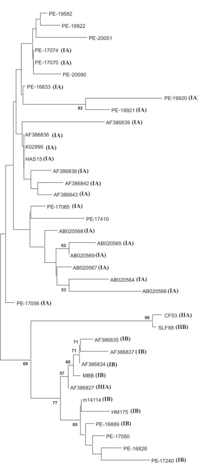
longed to genotype IA as determined by sequence analysis in the VP1/2A region and were isolated from two patients living in different counties during the same month (July 2003). Phylogenetic analysis was per-formed among 5’NTR sequences and se-quences from genotype IA were found to cluster in the tree in the same way as geno-type IB strains (Figure 2). However, one sample identical to one previously isolated in Spain (AF386827) belongs to genotype IIIA and clusters together with other strains from genotype IB. Furthermore, not all of 5’NTR sequences of this study were ampli-fied in the VP1/2A region, and therefore the genotype of these samples was not deter-mined. These results do not allow us to state that 5’NTR can be used to genotype HAV sequences.
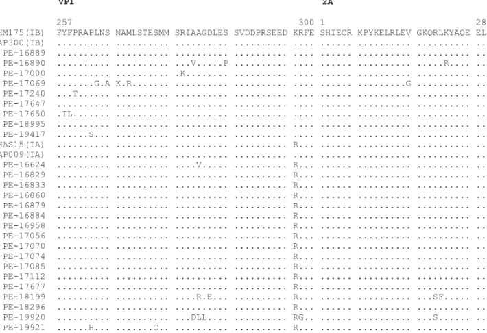
A comparison of the predicted amino acid sequences of the VP1/2A region from Pernambuco, of two isolates representative of genotypes IA and IB in the world and two more sequences from South America is shown in Figure 3. Identity in amino acid sequence ranged from 93.1 to 100% among
Figure 1. Phylogenetic tree analysis of the VP1-2A region in hepatitis A virus (HAV) isolates from Pernam-buco (PE). A 218-bp segment of the HAV VP1/2A junction was analyzed using the two-parameter model of Kimura. The numbers on the branches show boot-strap percentages obtained after 1000 replicates of bootstrapping sampling. The length of bars shows the distances. The numbers in parentheses show the month and year of strain isolation. Genotype and sub-genotype are indicated for each branch. Reference sequences from Genbank included: genotype IA strains (HAS15 x15464), genotype IB strains (HM175 m14707), genotype IIB strain (SLF88 ay644670), geno-type III strain (NOR21 aj299464), BA303 (ay 323017), BA351 (ay 323018), RJ104 (ay 323022), RJ178 (ay 323023), MA367 (ay 323036), MA347 (ay 323039) MA378 (ay 323037), BA251 (ay 323033), RJ12 (ay 323034), MA374 (ay 323040), RN243 (ay 323005), MG287 (ay 323007), RJ193 (af 410388), HAP009 (af 538723), RJPMC1 (af410386), HAP108 (DQ198361), Nor13 (af 050234), HAP231 (DQ198360), RJNSG2 (af 410383), HAP279 (DQ198362), HAP343 (DQ198363), RJ004 (af410380), RJ005 (af 410381), RJ049 (ay 322851), HAP300 (af 538727).
BA303 BA351 RJ104
RJ178
MA367 MA347 BA251 MA378 RJ12 MA374
PE-17677 (02/02)
PE-17056 (12/02)
PE-17112 (01/03)
PE-16879 (12/02)
PE-16884 (12/02)
RN243
PE-16958 (12/02)
MG287 PE-16829 (12/02)
RJ193
PE-18199 (04/03)
HAP108 RJPMC1 HAP009 Nor13
PE-19921 (07/03)
PE-19920 (07/03)
PE-16833 (12/02)
PE-18296 (04/03)
PE-16860 (12/02)
PE-17070 (12/02)
PE-16624 (11/02)
PE-17074 (12/02)
HAS15 PE-17085 (12/02)
PE-19417 (06/03)
HAP231 RJNSG2 HAP279 HAP343 RJ004 RJ005 PE-16889 (12/02)
PE-18995 (06/03)
HM175 RJ049 PE-17647 (02/03)
HAP300 PE-17240 (01/03)
PE-17000 (01/03)
PE-17650 (02/03)
PE-17069 (12/02)
PE-16890 (12/02)
SLF88
Nor21
61 52
85 99 93
88 71
50 65
52
87 69
55 58 64
51
0.05
Genotype I
IA
IB
IA strains and from 90.3 to 100% among IB strains. In sub-genotype IB, mutation in nucleotide sequence produces proportion-ally more changes than in IA strains, where most substitutions are in the third position. Therefore, 13 sub-genotype IA strains showed identical amino acid sequences whereas only three IB sub-genotype isolates showed the same amino acid sequence. How-ever, the difference in the number of amino acid changes among isolates from two sub-genotypes was not statistically significant (P = 0.82; χ² for independence = 0.05).
D iscussio n
Brazil exhibits a very heterogeneous pat-tern of endemicity for HAV infection, vary-ing accordvary-ing to geographical region and socioeconomic groups. In the past, Brazil was considered to be an area of high ende-micity with nearly all people being infected in early childhood (4). However, recent epi-demiological studies have demonstrated that, except for the North region, there has been a shift from high to medium endemicity of HAV infection, which may result in more clinical cases as the consequence of an in-creased number of individuals susceptible to infection, mainly adolescents and adults
(5-Figure 2. Phylogenetic tree analysis of the 5’NTR in hepatitis A virus (HAV) isolates from Pernambuco (PE). A 290-bp segment of the HAV 5’NTR was analyzed using the two-parameter model of Kimura (see Ref. 15). The numbers on the branches show bootstrap percentages obtained after 1000 replicates of boot-strapping sampling. The length of bars shows dis-tances. The numbers in parentheses indicate the geno-type of this isolate according to the VP1/2A region. Note that not all of the 5’NTR sequences were ampli-fied in the VP1/2A region. Isolate AF386827, displayed in the box, belongs to genotype IIIA and clusters with IB strains. Reference sequences from Genbank included genotype IA strains (HAS15 x15464), genotype IB strains (HM175 m14707) (MBB m20273), genotype IIB strain (SLF88 ay644670), genotype IIA strain (CF53 aj299464), and other sequences from acute cases of hepatitis A virus that appear as the Genbank number.
PE-19582
PE-19922
PE-20051
PE-17074
PE-17070
PE-20090
PE-16833
PE-19920
PE-19921
AF386839
AF386836
K02990
HAS15
AF386838
AF386842
AF386843
PE-17085
PE-17410
AB020568
AB020565
AB020569
AB020567
AB020564
AB020566
PE-17056
CF53
SLF88
AF386835
AF386837
AF386834
MBB
AF386827
m14114
HM175
PE-16889
PE-17050
PE-16828
PE-17240 99 92
85 71
71
60
57
77 69
53 62
0.01
(IB) (IA)
(IB) (IA)
(IA) (IA)
(IA) (IA)
(IA)
(IA)
(IIA)
(IIB)
(IB)
(IB) (IA)
(IA)
(IA)
(IA)
(IA)
(IA)
(IA)
(IA)
(IA)
(IA)
(IA)
(IA)
(IB)
( IB)
(IB)
7,17,18). In the present study, we observed that 45% of acute cases were identified among individuals older than 10 years, a situation normally found in regions of inter-mediate endemicity, confirming a change from high to medium endemicity in this region. Furthermore, the prevalence of HAV infection increased proportionally to age in the other 55% of individuals, since 16% of the individuals aged less than 5 years and 39% of the individuals aged 5 to 10 years were acutely infected. These findings agree with the results of several seroepidemiolog-ical studies that have shown a marked change in the epidemiological pattern of hepatitis A in many parts of the world due to improve-ments in living standards (19-21).
Phylogenetic analysis of the nucleotide sequence of the VP1/2A region of the HAV
genome classified all isolates as genotype I, confirming former epidemiological data demonstrating that this is the main genotype circulating in Brazil (7,10,22) as well the most widespread genotype in the world (2,23,24). Additionally, we identified the simultaneous presence of sub-genotypes IA and IB in 65 and 35% of cases of HAV infection, respectively. The presence of both sub-genotypes has not been detected previ-ously in the Northeastern region. de Paula et al. (10) observed that all HAV sequences belonged to sub-genotype 1A in other States of the Northeastern region. Only in the South-eastern region was the co-circulation of sub-genotype 1A and 1B detected before (7,22). HAV isolates from Pernambuco State formed distinct clusters of highly related sequences and this is consistent with
previ-Figure 3. Comparison of the predicted amino acid sequence of the VP1/2A junction of the hepatitis A virus strains studied.
VP1 2A
257 300 1 28
ous speculation about low rates of accumu-lating mutations among HAV strains and the development of specific ecological niches (23). Thus, these data suggest that there is a circulation of endemic HAV strains in this region, as demonstrated in other regions of Brazil (7,10). Moreover, the presence of the same strains sharing the same nucleotide sequence might indicate the presence of un-identified epidemic foci, since these isolates were identified in cases that occurred during a short period of time (most of them in December 2002).
The Pernambuco genotype IB isolates differ in predicted amino acid sequences amongst themselves as compared to geno-type IA strains. However, the difference in the number of amino acid changes is not significant. Furthermore no one has shown that these differences alter infectivity. Fuji-wara et al. (25) demonstrated that disease severity was not associated with the nucleo-tide sequence of the HAV genotype-deter-mining region.
In the 5’NTR, few nucleotide substitu-tions were observed among our isolates when compared to VP1/2A sequences, confirming the conserved nature of this region. How-ever, the amplification of 5’NTR was less efficient than one would have expected in this conserved region. Phylogenetic analy-sis based on the 5’NTR region produced an apparent separation between IA and IB sub-genotypes. However, taking into consider-ation the misclassificconsider-ation of the IIIA strain into the IB group, we may assume that the 5’NTR region, probably due to the highly conserved nature of the sequences, does not contain sub-genotype-specific motifs which faithfully reflect the diversity of the VP1/2A region and therefore is not appropriate for use for sub-genomic classification.
Interestingly, two isolates shared one deletion at position 413 in the 5’NTR region. Recent studies have demonstrated that alter-ations in the central part of the 5’NTR region of HAV could result in significant func-tional changes of the virus. Brown et al. (26) showed that deletion of base 447 slightly decreased translation, while deletion of base 533 almost completely abolished it. Further-more, they demonstrated that G to U muta-tion at nucleotide 646 localized in the termi-nal part of 5’NTR could alter the secondary structure of the HAV genome. Schultz et al. (27) reported that mutations within the 5’NTR region of cell-adapted HAV enhance cap-independent translation directed by HAV IRES in a cell type-specific fashion. The deletion found in the present study had never been identified in HAV isolates and there-fore we could not predict an alteration in secondary structure or HAV translation. Fujiwara et al. (12) reported a possible asso-ciation between the severity of type A hepa-titis and nucleotide substitutions in the middle part of 5’NTR. However, in our study the presence or absence of symptoms was not associated with the mutations in this part of genome.
Re fe re nce s
1. Minor P. Classification and nomenclature of viruses (Arch. Virol. Suppl. 2). In: Francki RIB, Fauquet CM, Knudson DL, Brown F (Editors), Picornaviridae. Wien: Springer-Verlag; 1991. p 320-326. 2. Robertson BH, Jansen RW, Khanna B, Totsuka A, Nainan OV, Siegl
G, et al. Genetic relatedness of hepatitis A virus strains recovered from different geographical regions. J Gen Virol 1992; 73 (Pt 6): 1365-1377.
3. Lu L, Ching KZ, de Paula VS, Nakano T, Siegl G, Weitz M, et al. Characterization of the complete genomic sequence of genotype II hepatitis A virus (CF53/Berne isolate). J Gen Virol 2004; 85: 2943-2952.
4. Vitral CL, Yoshida CFT, Lemos ERS, Teixeira CS, Gaspar AMC. Age-specific prevalence of antibodies to hepatitis A in children and adolescents from Rio de Janeiro, Brazil, 1978 and 1995. Relation-ship of prevalence to environmental factors. Mem Inst Oswaldo Cruz 1998; 93: 1-5.
5. Villar LM, da Costa MCE, de Paula VS, Gaspar AMC. Hepatitis A outbreak in a public school in Rio de Janeiro, Brazil. Mem Inst Oswaldo Cruz 2002; 97: 301-305.
6. Santos DC, Souto FJ, Santos DR, Vitral CL, Gaspar AM. Seroepide-miological markers of enterically transmitted viral hepatitis A and E in individuals living in a community located in the North Area of Rio de Janeiro, RJ, Brazil. Mem Inst Oswaldo Cruz 2002; 97: 637-640. 7. Villar LM, Lampe E, Meyer A, Gaspar AM. Genetic variability of
hepatitis A virus isolates in Rio de Janeiro: implications for the vaccination of school children. Braz J Med Biol Res 2004; 37: 1779-1787.
8. Clemens SA, da Fonseca JC, Azevedo T, Cavalcanti A, Silveira TR, Castilho MC, et al. Hepatitis A and hepatitis B seroprevalence in 4 centers in Brazil. Rev Soc Bras Med Trop 2000; 33: 1-10.
9. Devalle S, de Paula VS, de Oliveira JM, Niel C, Gaspar AM. Hepati-tis A virus infection in hepatiHepati-tis C Brazilian patients. J Infect 2003; 47: 125-128.
10. de Paula VS, Lu L, Niel C, Gaspar AM, Robertson BH. Genetic analysis of hepatitis A virus isolates from Brazil. J Med Virol 2004; 73: 378-383.
11. Fujiwara K, Yokosuka O, Ehata T, Imazeki F, Saisho H. PCR-SSCP analysis of 5'-nontranslated region of hepatitis A viral RNA: com-parison with clinicopathological features of hepatitis A. Dig Dis Sci
2000; 45: 2422-2427.
12. Fujiwara K, Yokosuka O, Ehata T, Saisho H, Saotome N, Suzuki K, et al. Association between severity of type A hepatitis and nucleo-tide variations in the 5' non-translated region of hepatitis A virus RNA: strains from fulminant hepatitis have fewer nucleotide substi-tutions. Gut 2002; 51: 82-88.
13. Bower WA, Nainan OV, Han X, Margolis HS. Duration of viremia in hepatitis A virus infection. J Infect Dis 2000; 182: 12-17.
14. Pina S, Buti M, Jardi R, Clemente-Casares P, Jofre J, Girones R. Genetic analysis of hepatitis A virus strains recovered from the environment and from patients with acute hepatitis. J Gen Virol
2001; 82: 2955-2963.
15. Felsenstein J. Phylogenetic inference package, version 3.5. Seattle: Department of Genetics, University of Washington; 1993.
16. Kumar S, Tamura K, Jakobsen IB, Nei M. MEGA2: molecular evolu-tionary genetics analysis software. Bioinformatics 2001; 17: 1244-1245.
17. Tanaka J. Hepatitis A shifting epidemiology in Latin America. Vac-cine 2000; 18 (Suppl 1): S57-S60.
18. de Almeida LM, Amaku M, Azevedo RS, Cairncross S, Massad E. The intensity of transmission of hepatitis A and heterogeneities in socio-environmental risk factors in Rio de Janeiro, Brazil. Trans R Soc Trop Med Hyg 2002; 96: 605-610.
19. Lagos R, Potin M, Munoz A, Abrego P, San Martin OS, Ureta AM, et al. Serum antibodies against hepatitis A virus among subjects of middle and low socioeconomic levels in urban area of Santiago, Chile. Rev Med Chil 1999; 127: 429-436.
20. Lopez H, Zitto T, Bare P, Vidal G, Vukasovic J, Gomez R. Preva-lence of anti-hepatitis A antibodies in an urban middle class area of Argentina: some associated factors. Int J Infect Dis 2000; 4: 34-37. 21. Fix AD, Martin OS, Gallicchio L, Vial PA, Lagos R. Age-specific
prevalence of antibodies to hepatitis A in Santiago, Chile: risk fac-tors and shift in age of infection among children and young adults.
Am J Trop Med Hyg 2002; 66: 628-632.
22. de Paula VS, Baptista ML, Lampe E, Niel C, Gaspar AM. Character-ization of hepatitis A virus isolates from subgenotypes IA and IB in Rio de Janeiro, Brazil. J Med Virol 2002; 66: 22-27.
23. Diaz BI, Sariol CA, Normann A, Rodriguez L, Flehmig B. Genetic relatedness of Cuban HAV wild-type isolates. J Med Virol 2001; 64: 96-103.
24. Mbayed VA, Sookoian S, Alfonso V, Campos RH. Genetic charac-terization of hepatitis A virus isolates from Buenos Aires, Argentina.
J Med Virol 2002; 68: 168-174.
25. Fujiwara K, Yokosuka O, Imazeki F, Saisho H, Saotome N, Suzuki K, et al. Analysis of the genotype-determining region of hepatitis A viral RNA in relation to disease severities. Hepatol Res 2003; 25: 124-134.
26. Brown EA, Day SP, Jansen RW, Lemon SM. The 5' nontranslated region of hepatitis A virus RNA: secondary structure and elements required for translation in vitro. J Virol 1991; 65: 5828-5838. 27. Schultz DE, Honda M, Whetter LE, McKnight KL, Lemon SM.