Skin morphology of the mutant
hairless USP mouse
Departamentos de 1Patologia, Faculdade de Medicina Veterinária e Zootecnia, and 2Imunologia, Instituto de Ciências Biomédicas, Universidade de São Paulo,
São Paulo, SP, Brasil
3Laboratório de Biologia do Reconhecer, Centro de Biotecnologia,
Universidade Estadual do Norte Fluminense, Campos dos Goytacazes, RJ, Brasil S.M.G. Massironi2,
M.R. Giacóia1,
P.C. Maiorka1,
T.L. Kipnis3 and
M.L.Z. Dagli1
Abstract
The morphology of the skin of the mutant hairless USP mouse was studied by histological, histochemical and immunohistochemical meth-ods and compared to the skin of BALB/c mice. Representative sec-tions of the dorsal skin from mice of both strains aged 18 days, and 1, 3, 6, and 8 months were studied. Sections stained with hematoxylin and eosin showed cystic formations called utricles and dermal cysts in the dermis that increased in size and number during growth. Skin thickness increased significantly at 8 months. Sections stained with picrosirius and examined with polarized light, displayed different colors, suggesting different thicknesses of dermal collagen fibers (probably types I and III). Weigert, Verhoeff and resorcin-fuchsin stains revealed fibers of the elastic system. The PAS and Alcian blue methods revealed neutral and acid glycosaminoglycans in the skin ground substance of both mouse strains. Immunohistochemical stain-ing for fibronectin and laminin did not show differences between the mutant and BALB/c mice. Mast cells stained by the Gomori method and macrophages positive for HAM 56 antibodies were observed in both mouse strains. Except for the presence of enlarged cysts inthe hairless strain, no qualitative differences were found during develop-ment of the skin of BALB/c and the mutant hairless mice.
Correspondence M.L.Z. Dagli
Departamento de Patologia Faculdade de Medicina Veterinária e Zootecnia, USP
Av. Prof. Dr. Orlando M. Paiva, 87 05508-900 São Paulo, SP Brasil
Fax: +55-11-3091-7829 E-mail: mlzdagli@usp.br
Part of a Master’s thesis presented by M.R. Giacóia to the Experimental and Comparative Program, Department of Pathology, Faculty of Veterinary Medicine, University of São Paulo, São Paulo, SP, Brazil.
M.R. Giacóia was the recipient of a FAPESP fellowship (No. 95/1737-7).
Received July 16, 2004 Accepted October 15, 2004
Key words •Mutant mice •hrUSP mutants •Hairless mice •BALB/c mice •Ethyl-nitrosourea
Introduction
The hairless and rhino mutants are the most intensively studied models of hair cycle defects. Affected mice lose their hair pro-gressively from the head to the tail at the end of the first hair cycle because the second hair cycle is defective (1). The rhino mutant pre-sents severe lesions at the skin, which be-comes progressively loose and redundant, forming folds, flaps and ridges (2).
Two of the authors (SMGM and TK) developed an ethyl-nitrosourea (ENU)-in-duced mutant phenotypically similar to the rhino mutant which was called hairless USP (hrUSP). Differently from the hairless mice
of the same gene, heterozygous mice of both strains were mated. Fifty percent of hrUSP/+
and rhino/+ offspring showed the hairless phenotype, indicating that the two mutations do not complement each other in vivo, being alleles of a single gene.
The skin of hrUSP mice displays a thin
epidermis formed by a stratified epithelium covering a dermis rich in cyst-like struc-tures. These cysts are surrounded by scarce connective tissue and are apparently con-nected to sebaceous glands (3). In this re-port, we describe the kinetics of the develop-ment of the skin lesions in hrUSP mutant
mice. Histological, histochemical and im-munohistochemical methods, as well as mor-phometry, were applied, and the data were compared to those obtained for BALB/c mice.
Material and Methods
Animals
BALB/c and hrUSP mice were obtained
from the isogenic mouse facility of the De-partment of Immunology, Institute of Bio-medical Sciences, University of São Paulo, São Paulo, SP, Brazil. The recessive muta-tion hairless hrUSP arose in an ENU
mutagen-esis program and has been maintained in a BALB/c background. Its origin and main phenotype have already been described (3). Male or female hairless mice aged 18 days and 1, 3, 6, and 8 months were studied and BALB/c mice of the same ages were used as
controls. During the experimental period, mice were kept in clean rooms provided with sanitary barriers. The temperature was kept at 21 ± 2ºC, with light cycles of 12 h. During the experimental period, mice received com-mercial food (Nuvital®, Nuvilab, Curitiba,
PR, Brazil) and water ad libitum.
Skin samples
Mice aged 18 days and 1, 3, 6, or 8 months were euthanized by cervical disloca-tion, and their back region was shaved when necessary. A longitudinal fragment of 4 x 1.5 cm of the dorsal skin was obtained from each mouse and fixed in either Bouin’s fixative or 95% ethanol for 24 h. After routine process-ing, samples were embedded in paraffin and the 5-µm sections were stained with hema-toxylin and eosin (H&E). Additional sec-tions were attached to silanized slides for immunohistochemistry.
Morphometry
A Bioscan/Optimas (Bioscan Optimas, Bioscan Inc., Edmonds, WA, USA) image analysis system was used to quantify the skin thickness of hairless and BALB/c mice. The skin of 5 mutants and control BALB/c mice aged 18 days and 1, 3, 6, or 8 months was sampled and at least 10 microscopic fields of each H&E section were measured for each time. A 40X objective was used to measure the thickness of the epidermis, with lines perpendicular to the basal membrane drawn untilthe keratin layer. A 10X objective was used to measure dermis thickness, with lines drawn from the basal membrane towards the dermis muscle.
Histochemical and immunohistochemical staining methods
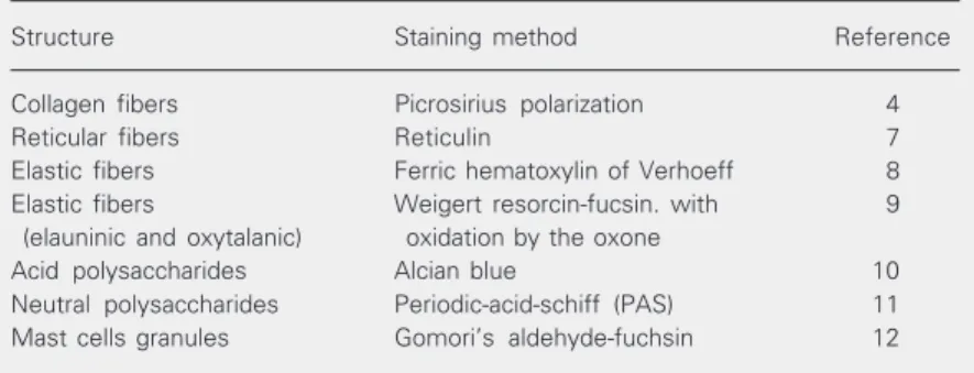
The histochemical methods used to iden-tify the structures in the mouse skin are pre-sented in Table 1. Skin fragments fixed in 95%
Table 1. Histochemical methods used to identify structures in the skin of hrUSP
mutants and BALB/c mice.
Structure Staining method Reference
Collagen fibers Picrosirius polarization 4
Reticular fibers Reticulin 7
Elastic fibers Ferric hematoxylin of Verhoeff 8
Elastic fibers Weigert resorcin-fucsin. with 9
(elauninic and oxytalanic) oxidation by the oxone
Acid polysaccharides Alcian blue 10
Neutral polysaccharides Periodic-acid-schiff (PAS) 11
alcohol were used for immunostaining, thus avoiding the antigen retrieval which is neces-sary for immunostaining in formalin-fixed tis-sues. After dewaxing and hydration, sections were treated for 30 min with 15% H2O2 in
absolute methanol for 30 min to block endog-enous peroxidase. Sections were washed and incubated overnight with the primary anti-bodies (Table 2) in a humid chamber at 4ºC, followed by incubation with a biotinylated secondary antibody and the streptavidin-bi-otin-peroxidase complex (Duet kit, Dako, Glostrup, Denmark). Peroxidase activity was then detected with a solution containing di-aminobenzidine and hydrogen peroxide. Sec-tions were counterstained with hematoxylin and mounted in synthetic resin.
Statistical analysis
Differences between means were tested by ANOVA, with the level of significance set at P < 0.05.
Results
Gross observations
At birth, hrUSP hairless mice were
indis-tinguishable from wild-type BALB/c mice. However, from the 3rd week of life on, hairless mice started to lose hair continu-ously, and became completely devoid of a hair coat by 45 days of age. However, they kept their vibrissae. In parallel with the hair loss, skin folding started to become evident. These folds covered the eyes, causing visual difficulties. Besides showing increased thick-ness, the skin of the hrUSP hairless mouse
became irregular and viscous, and white cysts could easily be seen all over the body. The nails grew continuously and became curved.
Histopathology of the skin of the hrUSP mutant
The skin of BALB/c mice (Figure 1A),
Table 2. Primary antibodies used for immunohistochemical staining of the skin of hrUSP mutants and BALB/c mice, and their dilutions.
Antibody Source Dilution
Cytokeratins (AE1/AE3) BioGenex®, San Ramon, CA, USA 1:1000
Macrophages - HAM 56 Dako®, Carpinteria, CA, USA 1:50
Fibronectin Dako®, Carpinteria, CA, USA 1:1000
Laminin Dako®, Carpinteria, CA, USA 1:20
used as control, did not show any detectable change between the ages of 18 and 240 days.
hrUSP skin at 18 days of age (Figure 1B).
A discrete invagination of the epidermis, moderate hyperkeratosis and small dermal cysts and utricles restricted to the upper portion of the skin close to the epidermis were observed. The dermal cysts and utricles were filled with lamellae of keratin, and some still showed the presence of hairs. Sebaceous glands were normally seen to-gether with the hair follicles, dermal cysts and utricles. No other alteration could be seen in H&E-stained slides.
hrUSP skin at 30 days of age (Figure 1C).
The epidermis presented orthokeratotic kera-tosis. Dilated hair follicles were seen in the dermis, as well as larger utricles and dermal cysts when compared to 18 days of age. Dermal cysts were frequently seen in the superficial dermis and were frequently filled with keratin lamellae. Some atrophic hair follicles could be seen.
hrUSP skin at 90 days of age (Figure 1D).
The epidermal hyperkeratosis was more in-tense since utricles and the dermal cysts were more prominent and occupied most of the dermis, extending to the deep dermis (Figure 1D). Deeper dermal cysts showed less keratin. Functional hair follicles were not observed, but sebaceous glands were still seen around the hair follicles. The hypo-dermis was scant.
hrUSP skin at 180 days of age (Figure 1E).
Figure 1. Photomicrographs of skin from BALB/c (A) and hrUSP
mice at 18 days of age (B), 30 days (C), 90 days (D), 180 days (E), and 240 days (F) (H&E, 165X). See the increase in thick-ening and the formation of utricles and cysts in the dermis.
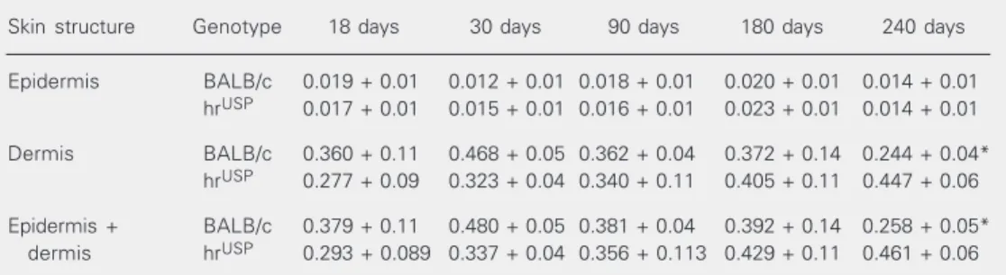
Table 3. Thickness of skin structures in male hrUSP and BALB/c mice of different ages.
Skin structure Genotype 18 days 30 days 90 days 180 days 240 days
Epidermis BALB/c 0.019 + 0.01 0.012 + 0.01 0.018 + 0.01 0.020 + 0.01 0.014 + 0.01 hrUSP 0.017 + 0.01 0.015 + 0.01 0.016 + 0.01 0.023 + 0.01 0.014 + 0.01
Dermis BALB/c 0.360 + 0.11 0.468 + 0.05 0.362 + 0.04 0.372 + 0.14 0.244 + 0.04* hrUSP 0.277 + 0.09 0.323 + 0.04 0.340 + 0.11 0.405 + 0.11 0.447 + 0.06
Epidermis + BALB/c 0.379 + 0.11 0.480 + 0.05 0.381 + 0.04 0.392 + 0.14 0.258 + 0.05* dermis hrUSP 0.293 + 0.089 0.337 + 0.04 0.356 + 0.113 0.429 + 0.11 0.461 + 0.06
Data are reported as means ± SD in cm for 5 mice of each strain per age group. *P < 0.05 compared to hrUSP
mice (Student t-test).
A B
C D
this time, skin adnexa (sebaceous glands) were only rarely observed.
hrUSP skin at 240 days of age (Figure 1F).
At this time, the dermis was almost com-pletely replaced by utricles and dermal cysts, which were very large, with thin walls, and contained almost no keratin. Dermal con-nective tissue was scant. The hypodermis and muscle layers were very thin. Epidermis, utricles, dermal cysts and also the sebaceous glands stained positively for the AE1/AE3 antibodies at all times studied.
Skin thickness during growth
Table 3 shows the skin thickness data for mutant and BALB/c mice. By 240 days of age the dermis of the mutants was signifi-cantly thicker than that of BALB/c mice.
Dermal connective tissue
Collagen. The picrosirius method asso-ciated with polarization showed that the der-mal collagen fibers were thick and strongly polarized to red or orange-yellow, suggest-ing the collagen type I pattern. Some thin fibers that polarize to green were seen, repre-senting collagen type III according to Junqueira et al. (4). The amount of dermal collagen fibers varied according to mouse age. In younger animals, collagen fibers were abundant in the dermis, but appeared to de-crease from 6 to 8 months of age, mainly because cysts and utricles occupied the der-mis.
Reticular fibers. Both mutant and BALB/ c mice presented thin argyrophilic fibers distributed as a meshwork in the dermis around the cysts and the hair follicles.
Elastic fibers. Elastic fibers were detected in the dermis of both mutant and BALB/c mice by the Weigert resorcin-fuchsin method, but not by the Verhoeff method. Thin dark brown elastic fibers were seen close to the dermo-epidermal junction, and thicker fi-bers were seen deeply in the dermis.
Inter-mediate thick fibers were also seen distrib-uted in the dermis. The positivity to Weigert staining and the thickness of the fibers sug-gested the presence of oxitalanic, elauninic and elastic fibers in the skin of both mutant and BALB/c mice. The amount and distribu-tion of these fibers did not vary with the phenotype or age of the mice.
Glycosaminoglycans. Acid glycosami-noglycans, stained blue by the Alcian blue method, were equally seen in the dermis of both mutant and BALB/c mice. Neutral gly-cosaminoglycans were detected by the PAS method in the basal membranes of the epi-dermis, sebaceous glands, cysts, and utricles. No difference was seen between mutant and wild-type BALB/c mice.
Fibronectin and laminin. Immunohisto-chemical staining of fibronectin and laminin was seen in the basal layer of the epidermis, as well as in the cysts and utricles of the mutant mice. No differences were detected in the thickness or distribution of these com-ponents between mutant and wild-type BALB/c mice.
Mast cells and macrophages. Mast cells were stained by the Gomori method (alde-hyde-fuchsin), in the skin of both mutant and wild-type BALB/c mice. These cells were scattered in the dermis, without any prefer-ential distribution. No differences were ob-served between phenotypes or ages. Macro-phages of both mutant and BALB/c mice stained positively with HAM 56 antibodies. No differences were observed in the number or distribution of macrophages between phe-notypes or ages.
Discussion
wrinkled and the nails are grotesquely curved. Rhino mutants have similar but more severe gross lesions similar those of hr/hr mice, and rhino mice have skin of normal appearance and hair at birth and lose hair from head to tail from 12 days of age on. Homozygous animals become devoid of all general body hair and fail to develop any new hair through-out life. The skin becomes progressively loose, forming folds, flaps and ridges. The skin surface expands after the filling of abor-tive hair follicles with cornified debris from the utricles. The utricles become enlarged and are grossly visible as uniform, white, subcutaneous nodules (2).
In the present study, we characterized the ENU-induced hrUSP mutant (3) in terms of
the skin of these mice by histological, his-tochemical and immunohishis-tochemical meth-ods. Our goal was to study the distribution of the skin components and to determine which of the histopathological alterations of the skin paralleled the clinical alterations ob-served during growth. Our mutants were indistinguishable from wild-type BALB/c mice at birth since their first hair coat devel-oped normally. However, they started to lose hair after the first hair cycle, becoming com-pletely naked from the age of 16 days like some other hairless mutants (2). As the mice aged, they developed wrinkled skin and long curved nails. Other alterations in these mice were hyperplasia of the lymph nodes and a membranous glomerulonephritis seen in older animals, as described previously (3). Histological examination of the skin showed a thin epidermis formed by a stratified epi-thelium and a dermis rich in dermal cysts. Our results show that these cysts probably originated from hair follicles, still keeping the sebaceous glands attached to them. Cysts were already present at 18 days and enlarged progressively through life. Growth of the hair follicles turned the skin progressively thicker, so that at 240 days (or 8 months) it was significantly thicker compared to the skin of wild-type BALB/c mice of the same
age. This increase in skin thickness can be accounted for by the enlargement of cysts since the epidermis showed no other differ-ences during the period studied when com-pared to the skin of BALB/c mice.
To study the distribution of skin compo-nents, connective tissue, extracellular ma-trix and cells were selectively stained. The expression of fiber components was studied using the picrosirius, reticulin, Weigert, and Verhoeff histochemical stains, and by im-munohistochemical staining for fibronectin and laminin. The staining patterns of the extracellular matrix of mutant micedid not differ qualitatively from those of BALB/c mice. However, it is possible that quantita-tive differences would have been observed if quantification methods had been applied. The same could be true for the noglycan component. Neutral glycosami-noglycans were visualized by PAS staining, while acid glycosaminoglycans were visual-ized by Alcian blue staining. About the con-nective tissue cells, mast cells and macro-phages were scattered in the dermis, with no difference between mutant and BALB/c mice. It has been reported that the amount of cellular matrix is reduced with aging, as is also the case for collagen located close to dermal cysts (5). The hairless skin was used as a model to study the modulation of col-lagen, glycosaminoglycans, and fibronectin following treatment with corticosteroids and retinoids, preventing skin atrophy. The re-ticular fibers which are present close to the hair follicles in the controls, were seen to-gether with the dermal cysts in the mutant. Changes in the structure and distribution of the dermis components were also observed. There was no defined structural pattern and the distribution of elastic fibers, of the fun-damental amorphous substance and of neu-tral polysaccharides was also altered.
Generalized acanthosis and orthokeratosis of the skin affecting the entire body of the mouse, a characteristic pattern for other mutants, were also seen in our mice.
An interesting finding was that the le-sions of hairless skin begin at the end of the first hair cycle when the dermal papilla fails to follow the contracting follicles and be-comes isolated in the reticular dermis and hypodermis. The presence of cysts was de-tected in the skin of 18-day-old mice which had small dermal cysts that grow with aging, filling the whole dermis. The structure and constitution of the dermal cysts of rhino
mice were described by Bernerd et al. (6), who found strong similarities to sebaceous glands and outer sheet roots.
While the hairless mutants have been largely used as models to test products that reduce skin aging, damage by UVB rays, and changes in skin due to acid treatment, no description of the skin of these mice appears to have been reported. We have shown here that all the normal skin components are pres-ent in hairless skin although their distribu-tion is altered by the presence of dermal cysts.
References
1. Sundberg JP & King LE (2000). Skin and its appendages: normal anatomy and pathology of spontaneous, transgenic and target mouse mutations. In: Ward JM, Mahler JF, Maronpot RR & Sundberg JP (Editors), Pathology of Genetically Engineered Mice. Iowa State University Press, Ames, IO, USA.
2. Sundberg JP (1994). The hairless (hr) and rhino (hrrh) mutations,
chromosome 14. In: Sundberg JP (Editor), Handbook of Mouse Mutations with Skin and Hair Abnormalities: Animal Models and Biomedical Tools. CRC Press, Boca Raton, FL, USA.
3. Massironi SMG, Dagli MLZ, D’Império-Lima MR, Alvarez JM & Kipnis TL (1994). A new mutant hairless mouse with lymph node hyperplasia and late onset of autoimmune pathology. Brazilian Jour-nal of Medical and Biological Research, 27: 2401-2405.
4. Junqueira LCU, Bignolas G & Brentani RR (1979). Differential stain-ing plus polarization microscopy: a specific method for collagen detection in tissue sections. Histochemical Journal, 11: 447-455. 5. Schwarz E, Mezick JA, Gendimenico GJ & Kligman LH (1994). In
vivo prevention of corticosteroid-induced skin atrophy by tretinoin in the hairless mouse is accompanied by modulation of collagen, glycosaminoglycans, and fibronectin. Journal of Investigative
Der-matology, 102: 241-246.
6. Bernerd F, Schwiezer J & Dermarchez M (1996). Dermal cysts of the rhino mouse develop into unopened sebaceous glands. Ar-chives of Dermatological Research, 288: 586-595.
7. Gordon H & Sweets HH (1936). A simple method for the silver impregnation of reticulum. American Journal of Clinical Pathology, 12: 545-552.
8. Verhöeff FH (1908). Some new staining methods of wide applicabil-ity, including a rapid differential stain for elastic tissue. Journal of the American Medical Association, 50: 876-877.
9. Weigert C (1989). Über eine Methode zur Färbung elastischer Fasern. Zentrablatt für Allgemeine Pathologie und Pathologische Anatomie, 9: 289-292.
10. Lison L (1960). Histochimie et Cytochimie Animales. 3rd edn. Gauthier-Villars, Paris, France.
11. Michalany J (1980). Técnica Histológica em Anatomia Patológica. E.P.U., São Paulo, SP, Brazil.