Receptor TrkB Are Dwarfed, and Are Similar to Mice with
a MAPK14 Deletion
Michele R. Hutchison*
Department of Pediatrics, University of Texas Southwestern Medical Center, Dallas, Texas, United States of America
Abstract
Long bone growth results from ordered chondrocyte development within the cartilagenous growth plate. Chondrocytes are recruited from a resting pool to proliferate along the long axis of the bone, until various signals trigger differentiation and hypertrophy. We have shown previously that the neurotrophin receptor TrkB is expressed in growth plate chondrocytes, where the tyrosine kinase receptor regulates the pace of hypertrophic differentiation by modulating the activities of ERK and p38 MAP kinases. To investigate the physiological relevance of TrkB to bone growthin vivo, we generated mice with a targeted disruption of the receptor, and compared them to mice targeted for MAPK14, the gene for p38a. The TrkB mutant and p38amutant mice showed a similar degree of dwarfism and delayed hypertrophic differentiation. To extend these findings, we showed that both the TrkB and p38a mutant mice have altered expression of Runx2 and Sox9, two key transcription factors required for skeletogenesis. The data providesin vivoevidence for the role of TrkB in bone growth, supports the role of p38 downstream of TrkB, and suggests that Runx2 and Sox9 expression is regulated by this pathway at the growth plate.
Citation:Hutchison MR (2013) Mice with a Conditional Deletion of the Neurotrophin Receptor TrkB Are Dwarfed, and Are Similar to Mice with a MAPK14 Deletion. PLoS ONE 8(6): e66206. doi:10.1371/journal.pone.0066206
Editor:Frank Beier, University of Western Ontario, Canada
ReceivedDecember 16, 2012;AcceptedMay 5, 2013;PublishedJune 11, 2013
Copyright:ß2013 Hutchison. This is an open-access article distributed under the terms of the Creative Commons Attribution License, which permits unrestricted use, distribution, and reproduction in any medium, provided the original author and source are credited.
Funding:This work was funded by National Institutes of Health Grants K08DK073447 and R03DK089151 to MRH. The funders had no role in study design, data collection and analysis, decision to publish, or preparation of the manuscript.
Competing Interests:The author has declared that no competing interests exist.
* E-mail: Michele.hutchison@utsw.edu
Introduction
The rate of longitudinal bone growth is determined by the pace of endochondral ossification that occurs within the cartilaginous growth plates located at the ends of long bones and vertebrae [1– 3]. Resting chondrocytes within the reserve zone are recruited to enter the proliferative zone, wherein they divide along the long axis of the bone. A large number of factors, acting mainly in a paracrine manner, signal the cells to cease proliferation and begin differentiating within the pre-hypertrophic zone; terminally differentiated cells are found in the hypertrophic zone, wherein glycogen accumulation leads to dramatic cell hypertrophy. We previously reported that the TrkB receptor tyrosine kinase and its ligand, brain-derived neurotrophic factor (BDNF) are expressed in growth plate chondrocytes, where their interaction inhibits proliferation and promotes chondrocytic differentiation [4].
TrkB is widely expressed in neuronal tissue, where BDNF regulates neuronal survival and differentiation in peripheral and central nervous systems, and maintains synaptic plasticity, particularly in the hippocampus and hypothalamus [5–7]. TrkB and BDNF are also expressed in non-neuronal cells such as vascular endothelial cells, immune cells, and osteoblasts [8–10]. When activated, TrkB stimulates MAP (mitogen-activated protein) kinase pathways, which occupy a critical place in many intracellular pathways that transfer extracellular signals to intracellular effectors such as transcription factors [11,12]. Whereas in neural tissues TrkB activates the ERK MAPK
pathway, in growth plate (GP) chondrocytes the ability of TrkB to enhance hypertrophic differentiation requires the increased activity of p38 MAPK and reduced activity of ERK. In both primary bovine GP chondrocytes and the cell line ATDC5, BDNF attenuates ERK activity while increasing that of p38, and BDNF-induced chondrocytic differentiation is blocked by p38 inhibition. The in vivo significance of these observations has not been demonstrated previously. In the present study we generated mice with targeted disruptions of either TrkB or MAPK14 (the gene for
p38a), and demonstrated that the mice are similarly dwarfed. The
dwarfism is due to impaired transition to hypertrophy, as cell proliferation within the growth plate was unaffected. The TrkB
mutant mice have reduced expression of p38a and reduced p38
activation at the GP. Thus TrkB, acting through p38a, is required for normal long bone growth in mice.
Results
Dwarfism inTrkBflox/flox;Col2a1-cremice
BecauseTrkB2/2mice die within 3–4 days of life [5,6], the cre recombinase approach was used to conditionally inactivate TrkB in cells of chondrocytic lineage by crossingTrkBflox/floxmice with
Col2a1-cretransgenic mice. At every generation, TrkBflox/floxmice were crossed with TrkBflox/flox;Col2a1-cre mice containing only a
single Col2a1-cre transgene. The TrkBflox/flox;Col2a1-cre mice
be active as early as 8.5 dpc [13]. All mutant mice were initially viable; however, approximately 20% of the females were severely
dwarfed at birth (Fig. 1a), and none of these extremely dwarfed
females survived past 2 weeks. Most of these severely dwarfed mice did not survive past post-natal day (P) 3. Similar severe dwarfism at birth was not noted among the male mutants, who demonstrated growth defects at age 3.5 weeks. The remaining female mutants showed abnormal growth by 4.5–5 weeks of age. By 12 weeks of age, nose-to-rump lengths and nose-to-tail lengths of mutant males
were approximately 70–75% that of the TrkBflox/flox males;
however, there was no difference in body weights. Female mutants
by 12 weeks were approximately 80% the size of theTrkBflox/flox
females.
Dwarfism inMAPK14flox/flox;Col2a1-cremice
Whereas global deletions of p38 isoforms beta, gamma and delta lead to fertile mice with no discernible phenotype [14,15], global deletions of the alpha isoform (MAPK14) are early embryonic lethal [16]. We crossed the floxed MAPK14 mice with
theCol2a1-cremice to conditionally delete p38afrom the growth plate. TheMAPK14flox/flox;Col2a1-cremice were also obtained at the expected Mendelian ratio of 50%. Dwarfism in both genders was apparent by 3–4 weeks of age, and by 12 weeks of age weights, nose-to-rump lengths and nose-to-tail lengths of mutant males were 75–80% that of theMAPK14flox/floxmice (Fig. 2). The severe dwarfism seen in someTrkBflox/flox;Col2a1-crefemales was not noted
in the p38amutant mice.
Soft X-ray analysis showed that all long bones and vertebrae
were equally affected in the TrkB mutant mice (Fig. 3a). Lengths
of tibiae, femurs and vertebrae in mutant males at 6 months of age were approximately 75–78% that of their normal littermates. The naso-occipital length of the calvarium was not significantly affected. As was seen for the TrkB mutant mice, radiographic
analysis of the p38a mutants showed femur length to be
approximately 78% of normal, with tibial and vertebral lengths about 75% that of their normal littermates (Fig. 3b). Whereas the
p38a mutant mice had body weights concordant with their
lengths, the TrkB mutant mice were the same weight as the control
Figure 1. Growth defects inTrkBloxp/loxp;Col2a1-cremice.A, Gross appearance of TrkB mutant female and control female littermate at 10 days of age. B–D, mean nose-to-tail lengths and body weights6SD for male (B,C) and female (D,E) mice from 1 to 12 weeks after birth;TrkBloxp/loxp(
N
),TrkBloxp/loxp;Col2a1-cre
(#), n = 23 for mutants, 22 for controls. doi:10.1371/journal.pone.0066206.g001
mice, making them proportionally heavier; this difference in body weights is seen in the radiographs in Figure 3.
Growth plate abnormalities inTrkBflox/flox;Col2a1-cre
mutant mice
Tibiae and femurs of mice at 4 weeks of age were examined histologically. The overall morphology of the GPs from the mutant mice was similar to that of the TrkBflox/flox
mice, but significant differences were apparent. The width of the GP at each age
examined of male mutant mice was 78.865.7% (p,0.01) of
TrkBflox/flox males. When the percentage of the GP made up of
hypertrophic cells was assessed, the male mutant mice consistently showed a reduction in the width of the hypertrophic zone (HZ), such that inTrkBflox/flox
mice the HZ was 51.864.2% of the total
width, whereas the mutant mice displayed a HZ width that was
40.263.7% of the epiphyseal width (p,0.02 for both
compari-sons). Thus the total width of the reserve, proliferative and pre-hypertrophic zones was unchanged in the mutant mice. Immu-nohistochemical staining for TrkB was present in the reserve and
proliferative zones of the TrkBflox/flox mice, but reduced in the
mutant mice (Fig. 4). Staining for BrdU to assess for cell
proliferation showed that, within the proliferative zone, the percentage of positive cells was not significantly different between the TrkBflox/flox;Col2a1-cre mutant mice and their TrkBflox/flox
littermates (not shown).
Growth plate abnormalities inMAPK14flox/flox;Col2a1-cre
mutant mice
Tibiae at 4 weeks were examined; as seen in the TrkB mutant mice, the GP morphology of the MAPK14 mutant mice was normal, but staining for p38, normally localized to the proliferative and pre-hypertrophic zones, was largely absent in the mutant mice (Fig. 5). Overall width of the GP in mutant mice was reduced to
75.464.9% (p,0.01) that of normal littermate controls. Unlike
what was seen in the TrkB mutant mice, the MAPK14 mutant mice showed narrowing of both the proliferative and hypertrophic zones. The total number of BrdU positive cells appeared to be
reduced in the p38a mutant mice, but the percent of positively
staining cells within the proliferative zone was not significantly different from control mice (not shown).
Inhibition of TrkB signaling reduces expression of Sox9 and Runx2 in vitro
To determine whether TrkB signaling is necessary for the expression of Sox9 and Runx2 –transcription factors known to be
Figure 2. Growth defects inMAPK14loxp/loxp;Col2a1-cremice.Mean nose-to-tail lengths and body weights
6SD for male (A,B) and female (C,D) mice from 1 to 12 weeks after birth,MAPK14loxp/loxp(
N
),MAPK14loxp/loxp;Col2a1-cre(#), n = 19 for mutants, 20 for controls. doi:10.1371/journal.pone.0066206.g002essential for growth plate development– we initially used the ATDC5 cell line, which in the presence of high-dose insulin and
25mg/ml ascorbate recapitulates the entire chondrogenic
pro-gram over a 14 day period [17,18]. Because we were interested in the zone at which the transition from proliferation to differenti-ation occurs, we focused on the analogous time frame in differentiating ATDC5 cells, which in our hands is between 6–9 days after insulin addition. Cells were cultured in the presence of insulin for 6 days prior to the addition of two TrkB inhibitors, K-252a and AG879. We have previously shown that activation of the MAPK p38 is necessary for TrkB function in chondrocytes, so the p38 inhibitor SB203580 was included for comparison. Both of the TrkB inhibitors showed changes in levels of expression of chondrocyte-specific markers that were similar to that seen with the p38 inhibitor. ColX and Col2a1 RNA levels were markedly reduced after incubation from day 6–9 with either the TrkB or p38 inhibitors, as compared to levels seen after 9 days in the presence of insulin with no inhibitors (Fig. 6). The Trk inhibitors reduced Sox9 and Runx2 RNA levels to a similar degree as seen with the p38 inhibitor SB203580. Wortmannin was included as a general kinase inhibitor control because we had previously seen no affect of this PI3K inhibitor on ColX or Col2a1 mRNA levels [4]. Again, Wortmannin did not affect ColX or Col2a1 RNA levels, but transcript levels for Runx2 and Sox9 were decreased, suggesting that PI3K might have a role in regulating the expression of these transcription factors.
Disruption of TrkB or MAPK14 reduces expression of Sox9 and Runx2 in vivo
To determine whether disruption of TrkB would also affect the expression of chondrocyte markers, mutant mice and littermates were sacrificed at 1 week of age, and tibiae were excised and freed of adherent tissue. Under a dissection microscope, GPs from the proximal tibiae were excised for RNA isolation and analysis.
Figure 7ashows that TrkB mRNA was reduced to 1561.9% in proximal tibial growth plates from TrkB mutant mice as compared to those from their floxed littermates, and the expression of p38a
was reduced to 5162.2% . The expression levels of the other p38
isoforms (ß, c and d) were unchanged. Col2a1 and ColX
expression were reduced in the mutant mice to 5563.6%, and
1962.8% of wild type levels, respectively. Runx2 expression was
reduced to 2262.9% and Sox9 to 5165.4% of wild type levels.
The MAP14 mutant mice were also examined for differences in gene expression patterns at the GP of the proximal tibiae. As
shown in Figure 7b, the expression of Col2a1 was not
significantly reduced in the mutant mice, but ColX expression
was reduced to 1362.7%. As was seen in the TrkB mutant mice,
the MAPK14 mutants displayed reduced expression of Runx2 and
Sox9 to 3565.1% and 3865.6%, respectively. The expression of
p38a was reduced to 1460.8% that of normal whole proximal
tibial GP in the MAPK14 mutant mice; TrkB expression was not
affected. The expression levels of p38 isoformsb,c andd were
very low in both the normal and mutant mice (not shown),
suggesting that p38ais the predominant p38 MAPK present at the
growth plate in mice. The fact that global knockout mice for either
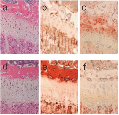
Figure 4. Histological analysis of long bones of TrkB mutants.A, B, and C are stained histological sections from a 4 week old maleTrkBloxp/loxp control; D, E and F are from a maleTrkBloxp/loxp;Col2a1-crelittermate. A, D, H&E staining; B, E, ColX immunostaining; C, F, TrkB immunostaining. doi:10.1371/journal.pone.0066206.g004
p38b, c or d were normally grown supports this conclusion [14,15].
To further explore the connection between TrkB signaling and p38 MAPK activation, the proximal tibiae of 4 week old male
TrkBflox/flox and TrkBflox/flox;Col2a1-cre mice were examined for differences in levels of active p38 protein. Immunohistochemistry performed with antisera against the dually-phosphorylated and active form of p38 showed that in normal mice p38 activity appears throughout the proliferative zone, with the greatest levels
at the pre-hypertrophic zone (Fig. 8). The TrkB mutant mice,
however, appear to have significantly reduced active p38 present at the GP, which is consistent with the reduced amount of total
p38amRNA seen in the GPs of TrkB mutant mice (Fig. 7).
IGF-I levels are not affected in mutant mice
To rule out the possibility that the observed dwarfism in the mutant mice was due to perturbations in the growth hormone/ IGF-I axis, serum from 8 week old mice was assessed for IGF-I
concentrations by ELISA. TheTrkBflox/floxmice had IGF-I levels
that were not significantly different from that of the TrkB mutant
mice, at 390633 ng/ml and 375626 ng/ml, respectively.
Simi-larly,MAPK14flox/floxmice had serum IGF-I levels of 412628 ng/
ml, and the IGF-I levels inMAPK14flox/flox;Col2a1-cre mice were
403631 ng/ml.
Figure 5. Histological analysis of long bones of p38amutants.A, B, and C are stained histological sections from a 4 week old male MAPK14loxp/loxp
control; D, E and F are from a maleMAPK14loxp/loxp;Col2a1-cre
littermate. A, D, H&E staining; B, E, ColX immunostaining; C, F, p38 immunostaining.
doi:10.1371/journal.pone.0066206.g005
Figure 6. Effect of TrkB inhibition on Runx2 and Sox9 expression in ATDC5 cells.The murine chondrocytic osteosarcoma cells were cultured in differentiation media containing 10mg/ml insulin
and 25mg/ml ascorbic acid; media was changed every other day for 6
days, at which time the indicated kinase inhibitors were added for an additional 3 days. Real-time RT-PCR was used to quantify mRNA levels of the marker proteins. Data points were calculated using theDDCt method and represent the mean 6SD of real-time data from five sample pairs, expressed as fold difference from insulin alone (the calibrator). *,P,0.001.
Discussion
The neurotrophin receptor TrkB is required for normal bone growth
BDNF was originally thought to be exclusively expressed in the CNS [19], where it stimulates neurite outgrowth and promotes neuronal cell differentiation and survival. BDNF is now recognized to be produced by a much wider variety of cell types, including vascular endothelial cells, leukocytes, adipocytes, and osteoblasts [8–10]. Studies showing that BDNF and TrkB are expressed in bone and cartilage, and that their expression is altered during bone healing, have implicated BDNF as a mediator of bone health [10,20–23]. We have shown that TrkB is expressed in epiphyseal growth plate cartilage, and that BDNF positively regulates chondrocyte differentiation, suggesting that TrkB and BDNF also have a role in long bone growth.
In the present work we demonstrate the role of TrkB in longitudinal growth by removing TrkB (both full and truncated forms, see the Methods section) from the epiphyseal growth plates of mice. The mutant mice are dwarfed, with shortened tails and extremities due to a delay in hypertrophic differentiation within the growth plate. TrkB mutants have narrowed growth plates, largely due to narrowing of the hypertrophic zone, as proliferative capacity within less-differentiated cells is unaffected. These histological differences were apparent by 4 weeks of age in males. It is unclear why a significant number of the female mutant mice
were severely dwarfed at birth. TheCol2a1-cretransgene is active
as early as 8.5 dpc [13], so it is possible that the severe dwarfism at birth is due to early disruption of TrkB expression in these mice. Some TrkB expression was detectible within the growth plates of all TrkBloxp/loxp;Col2a1-cre mice examined, whether by real time PCR analysis of RNA levels, or immunohistochemical methods. It is possible that the relatively mild dwarfism phenotype seen in most of the mice was due to retained expression of TrkB. Unfortunately we were unable to obtain histological samples from the severely dwarfed mice.
Figure 7. Expression of Runx2 and Sox9 in the growth plates of mutant mice.The growth plates from the proximal tibias of mutant and control littermates at day 7 after birth were dissected under a microscope, and isolated RNA analyzed by real time RT PCR for the expression of the two transcription factors as well as Col2a1, ColX, TrkB, and p38a. A, TrkB mutants compared to control littermates; B, p38a mutants compared to control littermates. Data points represent the mean6SD of six samples, expressed as fold difference from control samples (calibrator); *,P,0.001, **,P,0.01.
doi:10.1371/journal.pone.0066206.g007
Figure 8. Histological analysis of TrkB mutants for phospho-p38.A, B, are stained histological sections from a 4 week old male
TrkBloxp/loxp
control; C, D are from a male TrkBloxp/loxp;Col2a1-cre littermate. A, C, H&E staining; B, D, phospho-p38 staining.
doi:10.1371/journal.pone.0066206.g008
TrkB is required for normal expression of transcription factors Sox9 and Runx2
That cell proliferation is preserved in the TrkB mutant mice is consistent with our previous observation in primary bovine chondrocytes, wherein inhibition of TrkB did not affect
prolifer-ationin vitro[4]. In the murine embryonal carcinoma-derived cell
line ATDC5, TrkB inhibition with either K252a or AG879 dramatically blocks chondrocyte differentiation as measured by Col2a1 and ColX expression, but does not alter IGF-I-stimulated proliferation (4). The role of TrkB as a regulator of chondrocyte differentiation is apparent in the mutant mice that display reduced Col2a1 and ColX expression at the growth plate. We questioned whether the delayed hypertrophic differentiation in the TrkB mutant mice might involve altered expression of key transcrip-tional regulators, the transcription factors Sox9 and Runx2.
Sox9 is a master regulator of early cartilage development, and its inactivation in mouse embryos results in essentially no appendicular cartilage formation [24]. At the growth plate, Sox9 is expressed in reserve and proliferative zone cells, but is not present in more differentiated chondrocytes [25–27]. Heterozy-gous mutations in Sox9 cause a severe form of chondrodysplasia in humans called camptomelic dysplasia [28,29]. The transcription factor Runx2 is required for osteoblast differentiation and early chondrogenic development [30,31], and mice lacking Runx2 have almost no skeletal development [31]. At the growth plate, Runx2 is excluded from the proliferative zone, but re-activated at the pre-hypertrophic zone and throughout terminal pre-hypertrophic differ-entiation of chondrocytes [32,33]. In humans, haploinsufficiency of Runx2 causes cleidocranial dysplasia [34–36]. Runx2 is required for pre-hypertrophic and hypertrophic differentiation in mice [32,37]. We found that in ATDC5 cells, TrkB inhibition greatly reduced the expression of both Runx2 and Sox9. Moreover, TrkB mutant mice have reduced expression of the transcription factors at the growth plate, with Runx2 at about 30% and Sox9 at 50% of normal, suggesting that TrkB might influence chondrocyte development at least partly by regulating the expression of these key transcription factors.
Mice with a conditional deletion of p38a are similar to the TrkB mutant mice
We previously showed that BDNF-stimulated chondrocyte differentiation is dependent on p38 activity [4]. Of the four p38 isoforms, global deletions of either p38b,cordresult in mice with no discernible growth defect; however, p38a-deficient mice die at e10.5 due to defective placentation [14–16,38,39]. In an effort to
demonstrate the role of p38aMAPK (MAPK14) downstream of
TrkB in growth plate development, we deleted MAPK14 from the epiphyseal growth plates of mice, and found that the resulting dwarfism phenotype is very similar to that seen in the TrkB
mutant mice. The p38amutant mice also display dwarfism with
shortened extremities and tails, and examination of tibial growth plate sections shows narrowing of both the proliferative and hypertrophic zones with preservation of chondrocyte proliferation. In cell culture, inhibition of p38 activity results in enhanced chondrocyte proliferation, and thus we anticipated that the MAPK14 mutant mice would have a widened proliferative zone and evidence of increased cell proliferation within that zone. However, the p38 mutant mice have growth plates with a reduced
proliferative zone width. In our hands, the expression of p38a
appears to be greatest in the pre-hypertrophic zone in normal mice, wherein cell proliferation ceases and differentiation begins. The expression of p38 at the pre-hypertrophic zone is consistent with the proposed role of p38 as a promoter of chondrocyte
hypertrophic differentiation, rather than a regulator of prolifera-tionper se[40]. To further cement the connection between TrkB and p38 activation, we also demonstrated that the level of activated p38 protein is decreased in the growth plates of TrkB mutant mice.
Chondrocyte-specific expression of constitutively active MEK6, the kinase just upstream of p38, also caused dwarfism in mice with a reduction of long bone length to about 80% that of control mice
[41]. As seen in our p38a mutant mice, the transgenic mice
expressing active MEK6 showed a delay in hypertrophic differentiation. Clearly p38 MAPK is involved in the regulation of hypertrophic differentiation, with either excessive or insufficient p38 activity being detrimental to the process.
p38ais also required for normal expression of Sox9 and Runx2
Both Runx2 and Sox9 expression were reduced at the proximal
tibial growth plate of p38amutant mice to less than 40% that of
their normal littermates. This result is consistent with the increase in Sox9 activity seen in the transgenic mice expressing active MEK6 [41]. We propose that the pro-differentiating activity of BDNF/TrkB operates through, at least partly, an increase in expression of Runx2 and Sox9 in response to p38 activation
(Figure 8). Of note, both p38aexpression and p38
phosphoryla-tion is reduced in the growth plates of TrkB mutant mice, whereas
TrkB expression is preserved in the p38a mutants. It is thus
possible that the reduced expression of Runx2 and Sox9 in the
TrkB mutant is mice is due to the reduced expression of p38a.
In summary, we propose a model wherein the neurotrophin receptor TrkB in growth plate chondrocytes is activated by BDNF to halt proliferation and promote differentiation via the activation of the MAPK p38. We have extended the model to include the key chondrocytic transcriptional regulators Sox9 and Runx2 as
potential targets of the BDNF/TrkB/p38 pathway (Fig. 9). The
current work validates thein vitromodel, showing that TrkB and
p38aare required for normal skeletal development and growth in
mice. TrkB and p38a both play a role in chondrocyte
differentiation, as proliferation was preserved, and hypertrophy was delayed at the growth plates of affected mice.
Methods
Generation of mice
TrkBflox/flox mutant mice [6] in a C57/B6 background (back-crosses to pure C57/B6 were performed initially) were crossed
with mice carrying theCol2a1-cretransgene [13] also on a C57/B6
background to eliminate TrkB expression from cells of
chondro-cytic lineage. For theTrkBmutant mice, exon 1 of the TrkB gene,
which encodes the signal peptide and the first 40 amino acids of the N terminus of TrkB, was floxed. In neural tissue, TrkB exists in its full-length form as well as truncated forms that lack the tyrosine kinase domain [42,43]; the truncated forms of TrkB are capable of transmitting intracellular signals, and may have dominant inhibitory effects on BDNF signaling [44,45]. In the TrkBflox/flox
mutant mice, both full-length and truncated forms of TrkB are targeted.
To conditionally delete p38afrom the growth plate,
MAPK14-flox/flox
mice were obtained from the RIKEN Bioresource center
(Tsukuba, Japan) and crossed with theCol2a1-cremice.
In all crosses, only mice carrying a singleCol2a1-cretransgene
Animal Care and Use Committee at UT Southwestern Medical Center.
Genotype in each case was assessed by PCR of genomic DNA
obtained from tail cuttings. Primers for cre were: 59
-GAT-GAGGTTCGCAAGAACCTG-39 and 59
-ATCCGCCGCA-TAACCAGTG-39; these primers produced a 300 bp product.
Primers used for TrkB were: 59
-ACACACACAGTATATTT-TACCA-39 and 59-CAAGAAGTCAGAGACCAGAGAGA-39;
approximately 300 bp and 500 bp products were produced for the WT and floxed gene, respectively. Primers for MAPK14 were:
59-AGCCAGGGCTATACAGAGAAAAACCCTGTG-39 and
59-ATGAGATGCAGTACCCTTGGAGACCAGAAG-39,
pro-ducing 210 bp and 260 bp products, respectively
Measurements
All mice were kept in a pathogen-free environment on a 12 h light-dark cycle. Litters were culled to a maximum of 7 pups per litter; once weaned, mice were housed at a maximum of 4 per cage. Water and standard chow were provided ad libidum. Starting at 7 days of age, weight, tail length, and nose-to-rump length was measured twice weekly until 12 weeks of age; individuals performing measurements were blinded to genotype.
Radiographic analysis
Male mice were sacrificed by isoflurane inhalation; skeletal morphology was imaged at 25 kVp for 0.4 sec using the small focal spot setting on a Lorad M III mammography system (Lorad Medical Systems, Danbury CT). Specific bones were measured using ImageJ [46].
Histology
Tissues from 4 week-old mice were fixed in 4%
paraformalde-hyde/0.01 M PBS, pH 7.4 for 24 hours at 4uC. Tissues from mice
7 days of age or younger were decalcified for 1 week in 0.5 M EDTA, pH 8.0; specimens from older animals were decalcified for 2 weeks prior to imbedding in paraffin. 7 mm sections were cut and stained with hematoxylin and eosin (H&E). Immunohisto-chemical staining was done with either rabbit anti-rat collagen X at a 1:50 dilution (Abcam, Cambridge, MA), or rabbit anti-human TrkB at 1:100 (Abcam). Rabbit anti-p38 (Sigma) was used at 1:100 dilution; rabbit anti-phospho-p38 (Cell Signaling) was used at 1:50 dilution. Immunostaining was performed with the Cell and Tissue Staining Kit from R&D Systems (Minneapolis, MN) according to the manufacturer’s instructions. For epiphyseal measurements (width, % width of hypertrophic zone), H&E stains analyzed by ImageJ by an individual blinded to genotype. Widths were analyzed at five points spaced evenly across the growth plate and the median values for width and %width of hypertrophic zone selected for each bone.
BrdU uptake
Mice at 4 weeks of age were injected intraperitoneally with
BrdU (100mg/g body weight) and were sacrificed 4 hours later.
Tibiae were fixed in 4% paraformaldehyde/0.01 M PBS, pH 7.4
for 24 hours at 4uC, decalcified in EDTA for 2 weeks, and
embedded in paraffin. BrdU uptake in cells was determined by BrdU labeling with the BrdU Labeling and Detection Kit II from Roche Diagnostics (Indianapolis, IN). Cell proliferation was assessed as the percentage of positive cells in the proliferative zone within the microscopic field.
ATDC5 cultures
ATDC5 cell culture and differentiation experiments were performed as described previously [17]. Briefly, cells were maintained in DMEM/F12 with 10% FBS (Invitrogen) plus penicillin/streptomycin/amphotericin B; once at confluence, differentiation was initiated by changing the medium to
DMEM/F12 with 5% FBS plus 10mg/ml insulin, 10mg/ml
human transferrin, and 10 ng/ml sodium selenite from Sigma (ITS). The medium was changed every other day. Protein kinase inhibitors were added between days 6–9. The inhibitors used were:
p38 inhibitor SB203580 (Calbiochem, San Diego, CA) at 10mM,
TrkB inhibitors K-252a (Calbiochem) and AG879 (Calbiochem) at
3 nM and 10mM, respectively. The PI3K inhibitor Wortmannin
(Sigma) at 100 nM was also used as a general control, as we had previously shown that Wortmannin had minimal effects on ATDC5 cell development [47].
RNA Isolation, cDNA Synthesis and real time RT PCR
ATDC5 cells were induced to differentiate with ITS; on day 6 post ITS addition, indicated kinase inhibitors were added. On day 9, cells were harvested into RNA STAT-60 (TelTest, Inc, Friendswood, TX), and RNA was extracted per the manufactur-er’s instructions. Tibial epiphyseal cartilage from 1 week old mice was dissected under a microscope and harvested into RNA STAT-60 as above. Genomic DNA was removed from each sample using
DNA-Free (Ambion, Austin, TX), and 2mg RNA was reverse
transcribed with the High Capacity cDNA archive kit (Applied Biosystems). Real time RT PCR was performed using the Roche LightCycler 480 following the manufacturer’s protocol. 18S detection was done using TaqMan in the LightCycler 480 Probes Master Mix (Roche) and 200 nM of each primer and probe. Other targets were analyzed using double-stranded DNA dye SYBR
Figure 9. Proposed model of BDNF/TrkB regulation of chon-drocyte differentiation via p38 activation.Unopposed IGF-I action favors suppression of Runx2 and Sox9 expression and proliferation, whereas BDNF binding to TrkB results in increased p38 activity, decreased ERK activity, increased Runx2 and Sox9 expression, and ultimately hypertrophic differentiation.
doi:10.1371/journal.pone.0066206.g009
Green with the LightCycler 480 SYBR Green 1 Master Mix (Roche) and 200 nM of each primer. Primers spanned intron/ exon boundaries wherever possible, and all RT-PCR reactions were confirmed to produce only a single PCR product by comparison of the melt curves at the completion of each PCR reaction. The primer sets for the mouse targets have been previously described [47]. Relative gene expression for each
mRNA was calculated by theDDCT method using the ‘‘control’’
sample (no ITS) as calibrator.
IGF-I serum levels
Mutant and normal littermates of both genders were sacrificed at 8 weeks of age, and blood obtained via cardiac puncture. Serum IGF-I levels were assessed with the Mouse/Rat Quantikine ELISA kit from R&D Systems (Minneapolis, MN).
Statistical Analyses
Data are expressed as means 6SD. For growth parameters,
differences betweenTrkBflox/floxandTrkBflox/flox;Col2a1-cremice and
between MAPK14flox/flox and MAPK14flox/flox;Col2a1-cre mice was determined by one way repeated measures ANOVA, and was considered significant atP,0.05. For the RNA expression studies, significance between groups was determined by standard one-way ANOVA (SigmaPlot 11.0, SyStat Software, Inc. San Jose, CA). Differences were considered significant atP,0.05 unless otherwise noted.
Acknowledgments
The author would like to thank M. Castro for her technical assistance, J. Shelton for his assistance with histological preparations, and P.C. White for his critical review of the manuscript.
Author Contributions
Conceived and designed the experiments: MRH. Performed the experiments: MRH. Analyzed the data: MRH. Contributed reagents/ materials/analysis tools: MRH. Wrote the paper: MRH.
References
1. Hunziker EB (1994) Mechanism of longitudinal bone growth and its regulation by growth plate chondrocytes. Microsc Res Tech 28: 505–519.
2. Kronenberg HM (2003) Developmental regulation of the growth plate. Nature 423: 332–336.
3. Nilsson A, Ohlsson C, Isaksson OG, Lindahl A, Isgaard J (1994) Hormonal regulation of longitudinal bone growth. Eur J Clin Nutr 48 Suppl 1: S150–158; discussion S158–160.
4. Hutchison MR (2012) BDNF Alters ERK/p38 MAPK Activity Ratios to Promote Differentiation in Growth Plate Chondrocytes. Mol Endocrinol. 5. Klein R, Smeyne RJ, Wurst W, Long LK, Auerbach BA, et al. (1993) Targeted
disruption of the trkB neurotrophin receptor gene results in nervous system lesions and neonatal death. Cell 75: 113–122.
6. Luikart BW, Nef S, Shipman T, Parada LF (2003) In vivo role of truncated trkb receptors during sensory ganglion neurogenesis. Neuroscience 117: 847–858. 7. Minichiello L, Calella AM, Medina DL, Bonhoeffer T, Klein R, et al. (2002)
Mechanism of TrkB-mediated hippocampal long-term potentiation. Neuron 36: 121–137.
8. Nakahashi T, Fujimura H, Altar CA, Li J, Kambayashi J, et al. (2000) Vascular endothelial cells synthesize and secrete brain-derived neurotrophic factor. FEBS Lett 470: 113–117.
9. Yoshimura S, Ochi H, Isobe N, Matsushita T, Motomura K, et al. (2010) Altered production of brain-derived neurotrophic factor by peripheral blood immune cells in multiple sclerosis. Mult Scler 16: 1178–1188.
10. Yamashiro T, Fukunaga T, Yamashita K, Kobashi N, Takano-Yamamoto T (2001) Gene and protein expression of brain-derived neurotrophic factor and TrkB in bone and cartilage. Bone 28: 404–409.
11. Cobb MH (1999) MAP kinase pathways. Prog Biophys Mol Biol 71: 479–500. 12. Pearson G, Robinson F, Beers Gibson T, Xu BE, Karandikar M, et al. (2001) Mitogen-activated protein (MAP) kinase pathways: regulation and physiological functions. Endocr Rev 22: 153–183.
13. Ovchinnikov DA, Deng JM, Ogunrinu G, Behringer RR (2000) Col2a1-directed expression of Cre recombinase in differentiating chondrocytes in transgenic mice. Genesis 26: 145–146.
14. Beardmore VA, Hinton HJ, Eftychi C, Apostolaki M, Armaka M, et al. (2005) Generation and characterization of p38beta (MAPK11) gene-targeted mice. Mol Cell Biol 25: 10454–10464.
15. Sabio G, Arthur JS, Kuma Y, Peggie M, Carr J, et al. (2005) p38gamma regulates the localisation of SAP97 in the cytoskeleton by modulating its interaction with GKAP. EMBO J 24: 1134–1145.
16. Allen M, Svensson L, Roach M, Hambor J, McNeish J, et al. (2000) Deficiency of the stress kinase p38alpha results in embryonic lethality: characterization of the kinase dependence of stress responses of enzyme-deficient embryonic stem cells. J Exp Med 191: 859–870.
17. Shukunami C, Shigeno C, Atsumi T, Ishizeki K, Suzuki F, et al. (1996) Chondrogenic differentiation of clonal mouse embryonic cell line ATDC5 in vitro: differentiation-dependent gene expression of parathyroid hormone (PTH)/ PTH-related peptide receptor. J Cell Biol 133: 457–468.
18. Shukunami C, Ishizeki K, Atsumi T, Ohta Y, Suzuki F, et al. (1997) Cellular hypertrophy and calcification of embryonal carcinoma-derived chondrogenic cell line ATDC5 in vitro. J Bone Miner Res 12: 1174–1188.
19. Ernfors P, Bengzon J, Kokaia Z, Persson H, Lindvall O (1991) Increased levels of messenger RNAs for neurotrophic factors in the brain during kindling epileptogenesis. Neuron 7: 165–176.
20. Asaumi K, Nakanishi T, Asahara H, Inoue H, Takigawa M (2000) Expression of neurotrophins and their receptors (TRK) during fracture healing. Bone 26: 625– 633.
21. Pinski J, Weeraratna A, Uzgare AR, Arnold JT, Denmeade SR, et al. (2002) Trk receptor inhibition induces apoptosis of proliferating but not quiescent human osteoblasts. Cancer Res 62: 986–989.
22. Kurihara H, Shinohara H, Yoshino H, Takeda K, Shiba H (2003) Neurotrophins in cultured cells from periodontal tissues. J Periodontol 74: 76– 84.
23. Kajiya M, Shiba H, Fujita T, Ouhara K, Takeda K, et al. (2008) Brain-derived neurotrophic factor stimulates bone/cementum-related protein gene expression in cementoblasts. J Biol Chem 283: 16259–16267.
24. Akiyama H, Chaboissier MC, Martin JF, Schedl A, de Crombrugghe B (2002) The transcription factor Sox9 has essential roles in successive steps of the chondrocyte differentiation pathway and is required for expression of Sox5 and Sox6. Genes Dev 16: 2813–2828.
25. Wright E, Hargrave MR, Christiansen J, Cooper L, Kun J, et al. (1995) The Sry-related gene Sox9 is expressed during chondrogenesis in mouse embryos. Nat Genet 9: 15–20.
26. Ng LJ, Wheatley S, Muscat GE, Conway-Campbell J, Bowles J, et al. (1997) SOX9 binds DNA, activates transcription, and coexpresses with type II collagen during chondrogenesis in the mouse. Dev Biol 183: 108–121.
27. Zhao Q, Eberspaecher H, Lefebvre V, De Crombrugghe B (1997) Parallel expression of Sox9 and Col2a1 in cells undergoing chondrogenesis. Dev Dyn 209: 377–386.
28. Wagner T, Wirth J, Meyer J, Zabel B, Held M, et al. (1994) Autosomal sex reversal and campomelic dysplasia are caused by mutations in and around the SRY-related gene SOX9. Cell 79: 1111–1120.
29. Foster JW, Dominguez-Steglich MA, Guioli S, Kwok C, Weller PA, et al. (1994) Campomelic dysplasia and autosomal sex reversal caused by mutations in an SRY-related gene. Nature 372: 525–530.
30. Ducy P, Zhang R, Geoffroy V, Ridall AL, Karsenty G (1997) Osf2/Cbfa1: a transcriptional activator of osteoblast differentiation. Cell 89: 747–754. 31. Komori T, Yagi H, Nomura S, Yamaguchi A, Sasaki K, et al. (1997) Targeted
disruption of Cbfa1 results in a complete lack of bone formation owing to maturational arrest of osteoblasts. Cell 89: 755–764.
32. Kim IS, Otto F, Zabel B, Mundlos S (1999) Regulation of chondrocyte differentiation by Cbfa1. Mech Dev 80: 159–170.
33. Yoshida CA, Yamamoto H, Fujita T, Furuichi T, Ito K, et al. (2004) Runx2 and Runx3 are essential for chondrocyte maturation, and Runx2 regulates limb growth through induction of Indian hedgehog. Genes Dev 18: 952–963. 34. Lee B, Thirunavukkarasu K, Zhou L, Pastore L, Baldini A, et al. (1997) Missense
mutations abolishing DNA binding of the osteoblast-specific transcription factor OSF2/CBFA1 in cleidocranial dysplasia. Nat Genet 16: 307–310.
35. Mundlos S, Otto F, Mundlos C, Mulliken JB, Aylsworth AS, et al. (1997) Mutations involving the transcription factor CBFA1 cause cleidocranial dysplasia. Cell 89: 773–779.
36. Otto F, Thornell AP, Crompton T, Denzel A, Gilmour KC, et al. (1997) Cbfa1, a candidate gene for cleidocranial dysplasia syndrome, is essential for osteoblast differentiation and bone development. Cell 89: 765–771.
37. Inada M, Yasui T, Nomura S, Miyake S, Deguchi K, et al. (1999) Maturational disturbance of chondrocytes in Cbfa1-deficient mice. Dev Dyn 214: 279–290. 38. Tamura K, Sudo T, Senftleben U, Dadak AM, Johnson R, et al. (2000)
39. Mudgett JS, Ding J, Guh-Siesel L, Chartrain NA, Yang L, et al. (2000) Essential role for p38alpha mitogen-activated protein kinase in placental angiogenesis. Proc Natl Acad Sci U S A 97: 10454–10459.
40. Stanton LA, Sabari S, Sampaio AV, Underhill TM, Beier F (2004) p38 MAP kinase signalling is required for hypertrophic chondrocyte differentiation. Biochem J 378: 53–62.
41. Zhang R, Murakami S, Coustry F, Wang Y, de Crombrugghe B (2006) Constitutive activation of MKK6 in chondrocytes of transgenic mice inhibits proliferation and delays endochondral bone formation. Proc Natl Acad Sci U S A 103: 365–370.
42. Allen SJ, Dawbarn D, Eckford SD, Wilcock GK, Ashcroft M, et al. (1994) Cloning of a non-catalytic form of human trkB and distribution of messenger RNA for trkB in human brain. Neuroscience 60: 825–834.
43. Armanini MP, McMahon SB, Sutherland J, Shelton DL, Phillips HS (1995) Truncated and catalytic isoforms of trkB are co-expressed in neurons of rat and mouse CNS. Eur J Neurosci 7: 1403–1409.
44. Eide FF, Vining ER, Eide BL, Zang K, Wang XY, et al. (1996) Naturally occurring truncated trkB receptors have dominant inhibitory effects on brain-derived neurotrophic factor signaling. J Neurosci 16: 3123–3129.
45. Baxter GT, Radeke MJ, Kuo RC, Makrides V, Hinkle B, et al. (1997) Signal transduction mediated by the truncated trkB receptor isoforms, trkB.T1 and trkB.T2. J Neurosci 17: 2683–2690.
46. Schneider CA, Rasband WS, Eliceiri KW (2012) NIH Image to ImageJ: 25 years of image analysis. Nat Methods 9: 671–675.
47. Hutchison MR, Bassett MH, White PC (2010) SCF, BDNF, and Gas6 are regulators of growth plate chondrocyte proliferation and differentiation. Mol Endocrinol 24: 193–203.