ISSN 1414-431X
www.bjournal.com.br
www.bjournal.com.br
Volume 45 (12) 1102-1340 December 2012
Braz J Med Biol Res, December 2012, Volume 45(12) 1262-1268
10.1590/S0100-879X2012007500139
doi:
Whole-body vibration decreases the proliferative response of
+
TCD4 cells in elderly individuals with knee osteoarthritis
R. Tossige-Gomes, N.C.P. Avelar, A.P. Simão, C.D.C. Neves, G.E.A. Brito-Melo, C.C. Coimbra,
E. Rocha-Vieira and A.C.R. Lacerda
Institutional Sponsors
The Brazilian Journal of Medical and Biological Research is partially financed by
Faculdade de Medicina de Ribeirão Preto Campus
Ribeirão Preto
Explore High - Performance MS Orbitrap Technology In Proteomics & Metabolomics
analiticaweb.com.br S C I E N T I F I C
BIOMEDICAL SCIENCES
AND
Whole-body vibration decreases the proliferative
response of TCD4
+
cells in elderly individuals
with knee osteoarthritis
R. Tossige-Gomes
1, N.C.P. Avelar
1,2, A.P. Simão
1,2, C.D.C. Neves
1,2,
G.E.A. Brito-Melo
2,3, C.C. Coimbra
2,4, E. Rocha-Vieira
2,5and A.C.R. Lacerda
1,21Departamento de Fisioterapia, Faculdade de Ciências Biológicas e da Saúde, Universidade Federal dos Vales do Jequitinhonha e Mucuri, Diamantina, MG, Brasil
2Programa Multicêntrico de Pós-Graduação em Ciências Fisiológicas,
Universidade Federal dos Vales do Jequitinhonha e Mucuri, Diamantina, MG, Brasil 3Departamento de Farmácia, Faculdade de Ciências Biológicas e da Saúde, Universidade Federal dos Vales do Jequitinhonha e Mucuri, Diamantina, MG, Brasil
4Departamento de Fisiologia e Biofísica, Faculdade de Ciências Biológicas, Universidade Federal de Minas Gerais, Belo Horizonte, MG, Brasil 5Departamento de Ciências Básicas, Faculdade de Ciências Biológicas e da Saúde,
Universidade Federal dos Vales do Jequitinhonha e Mucuri, Diamantina, MG, Brasil
Abstract
The aim of this study was to investigate the effect of adding whole-body vibration (WBV; frequency = 35 to 40 Hz; amplitude = 4 mm) to squat training on the T-cell proliferative response of elderly patients with osteoarthritis (OA) of the knee. This study was a randomized controlled trial in which the selected variables were assessed before and after 12 weeks of training. Twenty-six subjects (72 ± 5 years of age) were divided into three groups: 1) squat training with WBV (WBV, N = 8); 2) squat training
without WBV (N = 10), and 3) a control group (N = 8). Women who were ≥60 years of age and had been diagnosed with OA in
at least one knee were eligible. The intervention consisted of 12 uninterrupted weeks of squatting exercise training performed 3 times/week. Peripheral blood mononuclear cells were obtained from peripheral blood collected before and after training. The proliferation of TCD4+ and TCD8+ cells was evaluated by flow cytometry measuring the carboxyfluorescein succinimidyl ester
fluorescence decay before and after the intervention (∆). The proliferative response of TCD4+ cells (P = 0.02, effect size = 1.0)
showed a significant decrease (23%)in the WBV group compared to the control group, while there was no difference between
groups regarding the proliferative response of TCD8+ cells (P = 0.12, effect size = 2.23). The data suggest that the addition of
WBV to squat exercise training might modulate T-cell-mediated immunity, minimizing or slowing disease progression in elderly patients with OA of the knee.
Key words: Vibratory platform; Lymphocyte; Proliferation; Elderly; Osteoarthritis
Introduction
Correspondence: A.C.R. Lacerda, Rodovia MGT 367, km 583, No. 5000, 39100-000 Diamantina, MG, Brasil. E-mail: [email protected]
Received February 1, 2012. Accepted August 9, 2012. Available online September 7, 2012. Published December 17, 2012.
Osteoarthritis (OA) is a chronic, progressive and degenerative osteoarticular disease characterized by arthralgia, stiffness and limitations of articular function. OA most commonly occurs in the knee (1). The etiology of OA involves biomechanical, biochemical and genetic factors that contribute to the imbalance between the synthesis and destruction of articular cartilage (2). The degradation
of the articular cartilage initiates an inflammatory process
in which cytokine production acts as both a causative
agent and a consequence of the disease (2).
Although OA has been considered to be a local form
of joint inflammation by many researchers, there is now evidence indicating the involvement of a systemic inflam -matory response exhibited by T cells and the presence
of inflammatory markers in peripheral blood, including inflammatory cytokines and antibodies (3). Therefore,
osteoarthritis has been associated with an
Vibration training and T cell proliferative response 1263
which seems to be an important factor contributing to the change in the function of chondrocytes. This favors the development of an imbalance between anabolic and catabolic activities involved in the remodeling of the ex-tracellular matrix of cartilage (4). Moreover, the presence of activated T cells and cytokine transcripts in chronic joint lesions of patients with OA suggests that T cells
contribute to chronic inflammation in a large proportion
of these patients (5).
Recent studies have documented that the cellular components of the OA synovium include activated T and B cells as well as monocytes and macrophages, which are thought to be immunocompetent (5). The literature reports an increase in the proliferative response of
mono-nuclear cells under inflammatory conditions, such as
OA (6). As reported by Sakata et al. (7), the proliferative T-cell response in OA is greater compared to controls, and the TCD4+ cells, which play critical roles in
produc-ing the proinflammatory cytokines that contribute to the
degradation of articular cartilage, are involved in this augmented proliferative response (7).
Although the process of articular cartilage degradation is irreversible, non-pharmacological interventions have been indicated to relieve the signs and symptoms of the disease and to delay its progression. Whole-body vibration
(WBV) training has been recommended as an efficient
and alternative method for improving muscle strength and proprioception in volunteers with knee OA (8,9).
During this training modality, the individual stands on a platform that generates vertical sinusoidal vibrations. These mechanical stimuli are transmitted to the body to stimulate the primary endings of the muscle spindles,
which in turn activate α-motor neurons, resulting in muscle contractions comparable to the tonic vibration reflex (10).
Recently, our group demonstrated improvement in the functionality and the self-perception of the disease status in OA patients after a 12-week training program in which WBV was combined with squatting exercise (11), as well
as improvements in the inflammatory marker profiles of
elderly patients with knee OA, with reduction in the plasma concentration of soluble tumor necrosis factor receptor 1 (sTNFR1) and sTNFR2 (12). However, to the best of our knowledge, no study has investigated the effect of this modality of intervention on the proliferative response of T cells in elderly patients with knee OA.
Because activated TCD4+ cells may play critical roles in cartilage degradation in OA (7), it is believed that the addition of WBV to the squat exercises could reduce the proliferative response of TCD4+ cells since this training
modality improves functionality and the self-perception of the disease status in OA patients (11), and also improves
their inflammatory marker profiles (12). Therefore, the
aim of the present study was to investigate the effect of adding WBV to squat training on the T-cell proliferative response of elderly subjects with knee OA.
Material and Methods
This was a randomized controlled study in which the variables were assessed 48 h before and after a 12-week training program. For allocation of the participants to groups, a 1:1 ratio of randomization was applied. To minimize the chance of bias, we used opaque envelopes that were sealed and serially numbered. The envelopes were opened sequen-tially after the participant’s name and further details were written on each envelope, and they were kept in a locked and secure place. The allocation sequence was concealed from the researcher who enrolled and assessed the participants. Only one researcher involved in the randomization was aware of the group assignments. The study was approved by the Internal Review Board of the Universidade Federal dos Vales do Jequitinhonha e Mucuri.
The sample for this study consisted of elderly volunteers from the community, who lived in the city of Diamantina, MG, Brazil, and were sedentary according to the Inter-national Physical Activity Questionnaire (IPAQ) (13). The volunteers were recruited through the physiotherapy clinic of the Universidade Federal dos Vales do Jequitinhonha e Mucuri and through medical referrals. To participate in the study, volunteers were required to meet the following
inclusion criteria: age ≥60 years and a diagnosis of OA in
at least one knee according to clinical and radiographic criteria of the American College of Rheumatology (14). The
severity of OA was classified radiographically according
to the Kellgren and Lawrence scale (15) (grades 0-4, with 0 being normal and 4 representing severe OA). Grade 2
(definite osteophytes and the possible narrowing of the
joint space) was set as a cutoff to classify OA of the knee in our sample population. The Kellgren and Lawrence criteria were absence of any recent knee injury, no requirement for a walking aid, self-report of not having been submitted to any rehabilitation procedure in the previous three months, no regular (twice per week or more) use of aspirin or other
nonsteroidal anti-inflammatory medications, and no use of
corticosteroids or other medications known to affect immune function (15,16). Volunteers were excluded if they had any orthopedic, neurological, respiratory, or acute cardiac dis-eases that would interfere with the study or if they had any
cognitive deficit demonstrated by the Mini-Mental Status
Examination (17). A radiological evaluation was also per-formed to verify the OA diagnosis (18).
Of the 125 elderly individuals screened for eligibility,
26 satisfied the inclusion and exclusion criteria and were
of physical activity. To ensure the retention of patients in the control group, weekly phone calls were made to each
group member to confirm her routine activities.
Clinical and demographic data were collected from the participants in both groups (WBV and EXE) using an evaluation chart. All of the study patients (WBV, EXE, CON) underwent a clinical evaluation and blood sample collection 48 h prior to the initiation of the 12-week intervention program for the platform and squat groups. The blood samples were always collected at 8:00 am, followed by clinical testing. At the end of the training period, the volunteers in each of the three groups were reassessed. Peripheral blood was col-lected from the cubital vein at the beginning of the study and at the end of 12 continuous weeks of intervention to assess the proliferation of T lymphocytes. The intervention program consisted of performing squat exercise training with WBV or without WBV 3 times/week on alternate days.
Warm-up
Prior to each training session, both groups (WBV and EXE) completed a warm-up on a stationary cycle (Stone
Fitness, 2001) at 70% of the predicted maximum heart
rate based on each subject’s age; the heart rate was moni-tored using a Polar heart rate monitor (model F4) for 10 min. Immediately afterwards, the participants in the WBV group were positioned with their feet 28 cm apart (14 cm to the right and 14 cm to the left of the center of the vibra-tion stimulus), ready to begin the squat exercises on the vibratory platform, while the participants in the EXE group performed the same procedure without vibration.
Squat exercises
The structured program of squat exercises on the platform and squat groups was conducted 3 times/week, on alternate days, for 12 weeks. The squat exercise was
performed starting at approximately 10° of knee flexion and continuing until 60° of knee flexion was reached. For
each volunteer, a knee angle of 60° was measured before the series of exercises, and a barrier was imposed on the
buttocks to limit the degree of knee flexion.
The parameters of the vibration in the platform group were based on the principles of training load progression: the frequency ranged from 35 to 40 Hz, the amplitude was 4 mm and the acceleration ranged from 2.78 to 3.26 g. The choice of vibratory frequencies and the amplitude were set so that we obtained an acceleration range between 2 and
5 g, which was suggested to be sufficient for achieving
physiological effects by Delecluse et al. (19). Prior to data collection, the acceleration values of the platform were measured using the Mega accelerometer (b) (ZPP1-3D-BC, Acceleration Measuring Kit, Kuopio, Finland).
The volume load of squat training for the squat and platform groups was increased systematically during the 12-week intervention by increasing the time and number of sets (6 sets x 20 s to 8 sets x 40 s) and reducing the resting
time (40 to 25 s) (11). For patients in the platform group, the vibration acceleration was also increased by varying the vibratory frequency (35-40 Hz).
Whole-body vibration
For the volunteers in the WBV group, a commercially available vibration platform was used (FitVibe, GymnaUni-phy NV, Belgium) that produces vertical synchronous vibration, causing vibration in both legs while the platform moves predominately in the vertical direction. This results in simultaneous and symmetrical movement of both sides of the body during exposure (20).
The overload used in the present study was only body weight. However, to ensure control and progressive train-ing, a gradual increase in volume (increase in exercise time of sets and decrease in rest intervals between sets) and intensity (acceleration was increased by varying the vibration frequency from 35 to 40 Hz) was used during the 12 weeks of intervention.
Lymphocyte proliferative response
To evaluate the effect of training on the T-cell prolifera-tive response, a blood sample (approximately 10 mL) was collected into heparinized tubes from each volunteer 48 h before training and 48 h after the last training session. Blood collection was performed between 7:30 and 8:30 am after a 12-h fast using the following stringent blood-draw criteria: no reported infection or symptoms of infection for 7 days, adequate sleep (6-9 h), no exercise or alcohol use for 24 h, no topical corticosteroid use or aspirin for 48 h, no systemic antihistamines or corticosteroid use for 1 week, and no immunizations during the previous 3 weeks.
Peripheral blood mononuclear cells (PBMCs) were iso-lated by centrifugation using Ficoll-Hypaque (Sigma, USA), as described by Bicalho et al. (21). Next, 1 x 10⁷ cells/mL
were suspended in phosphate-buffered saline (PBS) with
0.1% bovine serum albumin (PBS/BSA, 0.1%) and labeled with carboxyfluorescein succinimidyl ester (CFSE, 10 µM),
as described by Lyons et al. (22). The cells were washed and resuspended in RPMI supplemented with 2 mM L-glu-tamine, antibiotic/antimycotic solution (100 IU/mL penicillin
G, 100 µg/mL streptomycin and 2.5 µg/mL fungizone) and 10% fetal calf serum. The cells were stimulated with the mitogen phytohemagglutinin (PHA, 1 µg/mL) for 5 days at 37°C and 5% CO2. After the culture period, the cells were
collected, washed in PBS and incubated with monoclonal
antibodies that were specific for human CD4 and CD8 and
conjugated with phycoerythrin (PE) and Cy-Chrome (CY), as described by Openshaw et al. (23).
Labeled cells were then analyzed by flow cytometry
(FACScan, Becton Dickinson, USA) using the region of small lymphocytes and blast cells (R1) according to the
profile of size and granularity [forward scatter (FSC) x side scatter (SSC)]. The fluorescence of CFSE was measured
CD8-Vibration training and T cell proliferative response 1265
CY on the FL3 channel. The compensation and gain settings were determined for each experiment based on unstained cells and
cells labeled with only one fluorescent dye.
Thirty-thousand events in the R1 region were acquired for later analysis using the Cell Quest software (Becton Dickinson).
The proliferative index was then calcu-lated from the data in the histograms using the following formula (24), considering that two cells with a given CFSE intensity emerged from the mitotic division of a single cell pos-sessing a CFSE intensity immediately above:
proliferative index = (100 - Y) / Y, where Y (%)
= X0 + X1/2 + X2/4 + X3/8 + X4/16 + X5/32 + X6/64 + X7/128; X0 represents the percent-age of T cells that did not divide (located in M1), and X1-7 represents the peak of gradual division (located in M2 to M7).
Statistical analysis
The SPSS statistical software, version 18.0, was used for the statistical analysis. P
values ≤0.05 were considered to be statisti
-cally significant. We performed the analysis between groups by variation (∆) within each
group, where post-value - pre-value = delta
value (∆). The Shapiro-Wilk test was used to evaluate the normalcy of the data [delta value (∆)]. Because the data were normally distrib -uted for the dependent variables, parametric tests were used for the statistical analysis. The differences between groups were tested by ANOVA. The Tukey post hoc test was used to verify the differences between the conditions.
To check the size of the differences be-tween the pre- and post-training periods and between groups, we analyzed the magnitude of the effects (25,26).
Sample size
The sample size necessary to detect a difference between groups of PBMCs with a proliferation index of 6.06 and a standard
deviation value of 1.87 (8) was calculated considering a
significance level of 0.05 and statistical power of 0.8. We
calculated that at least 2 participants would be required in each group.
For the dependent variable (lymphocyte proliferative response), the statistical power of the study was calculated from the results of the magnitude of the effect of the deltas obtained from comparison of the results before and after training (26,27). For comparison between groups, the mag-nitude of the effect was high for both TCD4+ (Cohen’s f =
1.00) and TCD8+ cells (Cohen’s f = 2.23), indicating that the
study had a statistical power above 85% for these variables,
and the minimum sample size to obtain a statistical power
of 80% is 5 individuals per group (27).
Results
To analyze cell proliferation using CSFE in flow cytom
-etry, the R1 region was defined by dot plots in graphs of
FSC x SSC and comprised lymphocytes and blast cells to
Figure 1. Analysis of cell proliferation using carboxyfluorescein succinimidyl
ester (CFSE) in flow cytometry. A, Control for unstimulated cultures; B,
phyto-hemagglutinin (PHA)-stimulated cultures; C, CFSE x CD4; D, CFSE x CD8; E,
CFSE-stained cells derived from unstimulated cultures; F, CFSE-stained cells in
cultures stimulated with PHA. SSC and FSC = side and forward scatter, respec
control for unstimulated (Figure 1A) and PHA-stimulated cultures (Figure 1B). The events in R1 were then analyzed
for fluorescence from dot plots of FL1 x FL2 (CFSE x CD4)
(Figure 1C) and FL1 x FL3 (CFSE x CD8, Figure 1D), from
which regions R2 and R3 were defined, which included the
CFSE+CD4+ cells and the CFSE+CD8+ cells, respectively. The events in R1, R2 and R3 were sequentially analyzed for
CFSE fluorescence intensity using histograms (Figure 1E and F). The M1 region was defined as a region of
CFSE-stained cells derived from unstimulated cultures, which represents the peak of quiescent cells
(Figure 1E). The M2 to M7 regions were
defined according to the peaks of different
CFSE intensities in cultures stimulated with PHA (Figure 1F).
There was no significant difference
between groups with respect to mean age
and anthropometric data. No significant
differences were found in the data col-lected prior to training between groups,
thus confirming the baseline homogeneity
of the groups (Table 1).
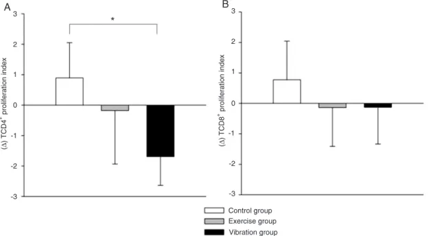
The variation (Δ) in the proliferative
response of TCD4+ cells (P = 0.02, effect
size = 1.00) was significantly reduced in the
WBV group compared to the CON group (Figure 2A), while there was no difference between groups regarding the variation
(Δ) in the proliferative response of TCD8+ cells (P = 0.12,
effect size = 2.23; Figure 2B).
Discussion
In this study, we investigated the effect of adding WBV to squat training on the T-cell proliferative response in elderly
subjects with knee OA, since OA is a chronic inflammatory
disease and, as we have previously demonstrated (11), WBV
improves the clinical condition of OA patients. The findings
Figure 2. Variation of the proliferation index (Δ) of A, TCD4+ and B, TCD8+ cells before and after exercise. Δ is reported for the vibration group (N = 8), exercise group (N = 10) and control group (N = 8). Data are reported as means ± SD.
*P ≤ 0.05 compared to control (ANOVA).
Table 1. Sample characterization and baseline variables of the whole-body vibration group (WBV), exercise group (EXE) and control group (CON).
Characteristics WBV (N = 8) EXE (N = 10) CON (N = 8)
Age (years) 75 ± 7 71 ± 4 72 ± 6
Body mass (kg) 72.75 ± 11.41 74.24 ± 9.95 63.71 ± 10.15
Height (m) 1.58 ± 0.05 1.57 ± 0.08 1.55 ± 0.08
Index of TCD4+ cells proliferation 3.20 ± 0.96 2.46 ± 1.22 1.42 ± 1.00 Index of TCD8+ cells proliferation 2.40 ± 1.28 2.79 ± 2.31 1.95 ± 1.42
Grade 2 (%) 20 20 37.5
Grade 3 (%) 30 30 37.5
Grade 4 (%) 50 50 25
Grades 2, 3, and 4 = severity of knee osteoarthritis in percent. Data are reported as
means ± SD. There were no statistically significant differences among groups for
Vibration training and T cell proliferative response 1267
of the present investigation demonstrated thatthe addition of WBV to squat exercise training reduces the proliferative response of TCD4+ cells but not the number of TCD8+ cells
in elderly subjects with knee OA. As we did not find any dif -ference in the proliferative response of TCD4+ cells between
the squat exercise and control groups, we conclude that the addition of WBV to squat exercises training is necessary to provide an additional overload to squat exercises.
Although OA has been considered to be primarily a disease of biomechanical alterations (28), accumulating
evidence supports the involvement of the inflammatory
response in the pathogenesis of the disease, with TCD4+
cells playing an important role in this process (5). Because chondrocytes respond to cytokines and chemokines
pres-ent in the synovial fluid and joint tissues, activated TCD4+
cells infiltrate the synovial membrane of patients with OA (5,29) and secrete inflammatory cytokines, such as IFN-α
and IL-2 (30,31), which directly or indirectly contribute to
the ongoing inflammation and destruction of joint cartilage
and decrease the anabolic activity of the joint. Considering this, we hypothesized that the addition of WBV to squat ex-ercise training could minimize or slow disease progression in elderly patients with OA of the knee because activated TCD4+ cells may play critical roles in inflammation and car
-tilage degradation in OA and seem to be directly involved in the immune response to autologous chondrocyte antigens (7). In this context, our group has previously reported that WBV training improves balance, gait performance and self-reported pain and reduces the serum levels of the soluble
forms of the TNF-α receptors sTNFR1 and sTNFR2 (12),
suggesting that this modality of training can improve the
inflammatory control of OA, thus allowing clinical improve -ment. The reduced proliferative response of TCD4+ cells in
OA patients following WBV training reported in the present study further supports our hypothesis.
WBV training did not modulate the proliferative response of TCD8+ cells in OA patients. The role of TCD8+ cells in
the pathogenesis of OA is less well understood than that of TCD4+ cells, although TCD8+ cells have been observed
in the synovial membranes of OA patients (31), and some investigators have suggested that these cells may exert cytotoxic activities against chondrocytes (8), thus contribut-ing to the destruction of joint cartilage. However, Kummer et al. (32) reported that up to 75% of the granzyme-positive
synovial lymphocytes are natural killer cells, and only less
than 5% are CD3+CD8+ cytotoxic cells, suggesting that
natural killer (NK), and not TCD8+ cells, may have some
role in cytotoxic cartilage destruction in OA. Although WBV training did not modulate the in vitro TCD8+ cell proliferative
response, it is possible that the lower TCD4+ cell response
may also attenuate the cytotoxic response of TCD8+ cells, considering the role that TCD4+ cells play in the
develop-ment of the primary TCD8+ cell response (33). This
pos-sibility should be experimentally investigated.
To the best of our knowledge, this is the first random -ized study to investigate the effect of the addition of WVB to squat exercise training on the proliferative response of T cells in elderly subjects with OA of the knee. We cannot say whether the effect of WBV on the proliferative response of T lymphocytes from patients with OA reported here results from a direct modulation of T-cell function by vibration or if this modulation is secondary to an improvement in neuro-muscular properties. Better neuroneuro-muscular control could result in greater joint stabilization, so that the shear stress on the cartilage would be smaller, resulting in reduced
produc-tion of proinflammatory cytokines and reduced stimulaproduc-tion
of T lymphocytes, a possibility that, however, has not yet been properly investigated.
The present study has several limitations, and the results must be interpreted within the context of its design. It is
important to note that specific frequencies and amplitudes
were used; therefore, the results cannot be extrapolated to other parameters of vibration. Furthermore, it should be
emphasized that PBMCs, rather than local inflammatory
cells, were used. Moreover, PHA is a general mitogen that stimulates the proliferation of a wide variety of lymphocyte clones, which could mask the responses of those clones
specifically involved in the immune response to OA.
Since the addition of WBV to squat exercise training de-creases the proliferative response of TCD4+ cells in elderly
subjects with OA of the knee, it is believed that this modality of training might modulate T-cell-mediated immunity in this population, minimizing or slowing the disease progression in elderly patients.
Acknowledgments
Research supported by SBFis, FAPEMIG (#APQ-00279-08), CNPq (#501083/2009-0), CAPES, and Santa Casa of Diamantina/MG.
References
1. Michael JW, Schluter-Brust KU, Eysel P. The epidemiology, etiology, diagnosis, and treatment of osteoarthritis of the knee. Dtsch Arztebl Int 2010; 107: 152-162.
2. Goldring SR, Goldring MB. The role of cytokines in cartilage matrix degeneration in osteoarthritis. Clin Orthop Relat Res
2004; 427: S27-S36.
3. Sakkas LI, Platsoucas CD. The role of T cells in the patho-genesis of osteoarthritis. Arthritis Rheum 2007; 56: 409-424.
4. Tetlow LC, Adlam DJ, Woolley DE. Matrix metalloproteinase
and proinflammatory cytokine production by chondrocytes of
degenera-tive changes. Arthritis Rheum 2001; 44: 585-594.
5. Sakkas LI, Scanzello C, Johanson N, Burkholder J, Mitra A, Salgame P, et al. T cells and T-cell cytokine transcripts in the synovial membrane in patients with osteoarthritis. Clin Diagn Lab Immunol 1998; 5: 430-437.
6. Vos K, Miltenburg AM, van Meijgaarden KE, van den Heu-vel M, Elferink DG, van Galen PJ, et al. Cellular immune response to human cartilage glycoprotein-39 (HC
gp-39)-derived peptides in rheumatoid arthritis and other inflamma
-tory conditions. Rheumatology 2000; 39: 1326-1331. 7. Sakata M, Masuko-Hongo K, Nakamura H, Onuma H,
Tsu-ruha JI, Aoki H, et al. Osteoarthritic articular chondrocytes stimulate autologous T cell responses in vitro. Clin Exp Rheumatol 2003; 21: 704-710.
8. Roelants M, Delecluse C, Verschueren SM. Whole-body-vibration training increases knee-extension strength and speed of movement in older women. J Am Geriatr Soc 2004; 52: 901-908.
9. Trans T, Aaboe J, Henriksen M, Christensen R, Bliddal H, Lund H. Effect of whole body vibration exercise on muscle strength and proprioception in females with knee osteoar-thritis. Knee 2009; 16: 256-261.
10. Cardinale M, Bosco C. The use of vibration as an exercise intervention. Exerc Sport Sci Rev 2003; 31: 3-7.
11. Avelar NC, Simão AP, Tossige-Gomes R, Neves CD, Rocha-Vieira E, Coimbra CC, et al. The effect of adding whole-body vibration to squat training on the functional performance and self-report of disease status in elderly patients with knee os-teoarthritis: a randomized, controlled clinical study. J Altern Complement Med 2011; 17: 1149-1155.
12. Simão AP, Avelar NCP, Tossige-Gomes R, Neves CDC, Mendonça VA, de Miranda AS, et al. Functional performance
and inflammatory cytokines after squat exercises and
whole-body vibration in elderly individuals with knee osteoarthritis.
Arch Phys Med Rehabil 2012; 93: 1692-1700.
13. Matsudo S, Araújo T, Matsudo V, Andrade D, Andrade E, Oliveira LC, et al. International physical activity question-naire (IPAQ): study of validity and reability in Brazil. Rev Bras Med Esporte 2001; 6: 5-12.
14. Hinton R, Moody RL, Davis AW, Thomas SF. Osteoarthritis: diagnosis and therapeutic considerations. Am Fam Physi-cian 2002; 65: 841-848.
15. Kellgren JH, Lawrence JS. Radiological assessment of osteo-arthrosis. Ann Rheum Dis 1957; 16: 494-502. 16. Schiphof D, de Klerk BM, Koes BW, Bierma-Zeinstra S.
Good reliability, questionable validity of 25 different classifi
-cation criteria of knee osteoarthritis: a systematic appraisal.
J Clin Epidemiol 2008; 61: 1205-1215.
17. Brucki SM, Nitrini R, Caramelli P, Bertolucci PH, Okamoto
IH. [Suggestions for utilization of the mini-mental state ex
-amination in Brazil]. Arq Neuropsiquiatr 2003; 61: 777-781. 18. Davies AP, Glasgow MM. Imaging in osteoarthritis: a guide to
requesting plain X-rays of the degenerate knee. Knee 2000; 7: 139-143.
19. Delecluse C, Roelants M, Verschueren S. Strength increase after whole-body vibration compared with resistance train-ing. Med Sci Sports Exerc 2003; 35: 1033-1041.
20. Cochrane DJ. Vibration exercise: the potential benefits. Int
J Sports Med 2011; 32: 75-99.
21. Bicalho HM, Gontijo CM, Nogueira-Machado JA. A simple technique for simultaneous human leukocytes separation. J Immunol Methods 1981; 40: 115-116.
22. Lyons AB, Hasbold J, Hodgkin PD. Flow cytometric analysis
of cell division history using dilution of carboxyfluorescein diacetate succinimidyl ester, a stably integrated fluorescent
probe. Methods Cell Biol 2001; 63: 375-398.
23. Openshaw P, Murphy EE, Hosken NA, Maino V, Davis K, Murphy K, et al. Heterogeneity of intracellular cytokine syn-thesis at the single-cell level in polarized T helper 1 and T helper 2 populations. J Exp Med 1995; 182: 1357-1367. 24. Angulo R, Fulcher DA. Measurement of Candida-specific
blastogenesis: comparison of carboxyfluorescein succin
-imidyl ester labelling of T cells, thymidine incorporation, and CD69 expression. Cytometry 1998; 34: 143-151.
25. Cohen J. A power primer. Psychol Bull 1992; 112: 155-159. 26. Cohen J. The Earth is Round (p<.05). Am Psychol 1994; 49:
997-1003.
27. Portney LG, Watkins MP. Foundations of clinical research: applications to practice. 3rd edn. Upper Saddle River: Pren-tice Hall; 2008.
28. Ishii H, Tanaka H, Katoh K, Nakamura H, Nagashima M,
Yoshino S. Characterization of infiltrating T cells and Th1/
Th2-type cytokines in the synovium of patients with osteoar-thritis. Osteoarthritis Cartilage 2002; 10: 277-281.
29. Dolhain RJ, ter Haar NT, Hoefakker S, Tak PP, de Ley M, Claassen E, et al. Increased expression of interferon (IFN)-gamma together with IFN-(IFN)-gamma receptor in the rheumatoid synovial membrane compared with synovium of patients with osteoarthritis. Br J Rheumatol 1996; 35: 24-32. 30. Kahle P, Saal JG, Schaudt K, Zacher J, Fritz P, Pawelec G.
Determination of cytokines in synovial fluids: correlation with
diagnosis and histomorphological characteristics of synovial tissue. Ann Rheum Dis 1992; 51: 731-734.
31. Kennedy TD, Plater-Zyberk C, Partridge TA, Woodrow DF, Maini RN. Morphometric comparison of synovium from patients with osteoarthritis and rheumatoid arthritis. J Clin Pathol 1988; 41: 847-852.
32. Kummer JA, Tak PP, Brinkman BM, van Tilborg AA, Kamp AM, Verweij CL, et al. Expression of granzymes A and B in synovial tissue from patients with rheumatoid arthritis and osteoarthritis. Clin Immunol Immunopathol 1994; 73: 88-95.