RESEARCH ARTICLE / ARTIGO
Detection of
Xanthomonas axonopodis
pv.
phaseoli
in �ean
in �ean
seeds by f���� �y���e��y�� ������s�������� ��d d��e�� ���b�e
��� �y���e��y�� ������s�������� ��d d��e�� ���b�e
counting
Nilvanira D. Tebaldi1, Jeroen Peters2, Ricardo M. Souza1, Luiz G. Chitarra3, Patricia van der Zouwen2, Jan Bergervoet2 & Jan van der Wolf2
1Departamento de Fitopatologia, Universidade Federal de Lavras, 37200-000, Lavras, MG, Brazil; 2Plant Research
International, 6700 AA, Wageningen, The Netherlands; 3Embrapa Algodão, 58.107-720, Campina Grande, PB, Brazil
Author for correspondence: Ricardo M. Souza, e-mail: [email protected]
Part of the Thesis of the first author. Universidade Federal de Lavras.Universidade Federal de Lavras. Lavras MG. 2005.
ABSTRACT
Flow cytometric analysis of immuno-stained cells (immuno-FCM) was compared to immunofluorescence microscopy (IF) and dilution plating on a semi-selective medium, for quantitative detection of Xanthomonas axonopodis pv. phaseoli (Xap) in bean seed extracts. Cell concentrations of Xap between 103-107 CFU/mL were added to healthy bean seed extracts. A flow cytometry sorting procedure was developed to separate immuno-stained Xap cells from crude seed extracts and confirming by PCR. FCM was evaluated for direct viable counting (DVC) of Xap using combinations of propidium iodide (PI) and carboxy fluorescein diacetate (cFDA) or PI and SYTO 9 and also the combination of immuno-FCM and PI. Dilution plating and IF allowed detection of Xap in bean seed extracts in a range of 103-106 CFU/mL and immuno-FCM from 104-106 CFU/mL. Sorted cells could be detected in crude seed extracts by PCR without further extraction. FCM also allowed quantification of viable cells of Xap after DVC procedures; the red fluorescent dye propidium iodide was used to identify dead cells in combination with the green fluorescent dyes cFDA or SYTO 9, these identifying live cells. The combination of immuno-FCM and PI could be more promising and reliable to detect this pathogen in seeds.
Key words: seed pathology, flow sorting, PCR-amplification, viability probes, immunofluorescence, bacteria. RESUMO
Detecção de Xanthomonas axonopodis pv. phaseoli em sementes de feijão usando citometria de fluxo em combinação com anticorpo e sondas fluorescentes de viabilidade
A combinação do uso do citômetro de fluxo (FCM) e de anticorpo policlonal (imuno-FCM) foi comparada à microscopia de imunofluorescência (IF) e ao plaqueamento em meio de cultura semi-seletivo, para a detecção de Xanthomonas axonopodis pv. phaseoli (Xap) em sementes de feijão. Concentrações de Xap variando de 103 a 107 CFU/mL foram adicionados aos extratos de sementes. Um método de separação pelo citômetro de fluxo foi desenvolvido para a detecção de Xap em extratos de semente e posterior confirmação por PCR. Para avaliação da viabilidade das células foram usadas sondas fluorescentes, iodeto de propídio (PI)/carboxi diacetato de fluoresceína (cFDA) e PI/SYTO 9 e também, a combinação de imuno-FCM e PI. Em meio semi-seletivo e IF foram detectadas 103-106 UFC/mL e no FCM 104-106 UFC/mL, em extratos de sementes artificialmente infestados. Xap somente foi detectada em extratos de sementes por PCR, após o processo de separação pelo FCM. Foi possível pelo FCM a quantificação e identificação de células viáveis (verde fluorescente) e células mortas (vermelho fluorescente) de Xap, pelas sondas cFDA/SYTO 9 e PI, respectivamente. A combinação de immuno-FCM e PI poderá ser uma técnica promissora e segura para a detecção deste patógeno em sementes.
Palavras-chave: patologia de sementes, PCR, sondas de viabilidade, imuno-fluorescência, bactéria.
INTRODUCTION
Xanthomonas axonopodis pv. phaseoli (Smith) Vauterin, Hoste, Kersters & Swinges (Xap) is a seed-transmitted plant-pathogenic bacterium which causes common blight of bean (Phaseolus vulgaris L.) (Saettler & Perry, 1972). This disease causes major economic losses
in commercial bean production worldwide, especially in tropical areas (Hall, 1994). To prevent common blight, disease-free seeds should be used. Therefore, testing of seed lots for the presence of the pathogen is the only efficient way to avoid spread of the disease.
cell-staining (Malin et al., 1983) and also enzyme-linked immunosorbent assay (ELISA) (Van Vuurde et al., 1983; Alvarez & Lou, 1985). Neither method discriminates between viable and non-viable cells. The specificity of the reaction is highly dependent on the quality of the antibodies.
For detection and identification of Xap, PCR-amplification methods have also been described (Audy et al., 1994; Toth et al., 1998). In general, PCR assays are rapid and highly specific, but quantification is difficult and amplification is prone to inhibition by contaminants present in seed samples (Van der Wolf & Schoen, 2004).
Flow cytometry (FCM) is a technique which allows high-speed multiparameter analysis and quantification of particles, such as bacterial cells. The analysis is based on size and granularity, and can be based on emission of fluorescent light, after staining with a fluorescent dye. FCM has already been used in combination with antibody staining (immuno-in combination with antibody staining (immuno-FCM) for the detection of Clavibacter michiganensis subsp.
michiganensis (Smith) Davis, Gillaspie, Vidaver & Harris in tomato seed extracts, and Xanthomonas campestris pv.
campestris (Pammel) Dowson in cabbage seed extracts (Chitarra et al., 2006). In theory, FCM can replace visual observation and quantification of bacteria in IF cell-staining (Diaper & Edwards, 1994; Bunthof et al., 2001).
FCM can also be used to analyze bacterial cells after direct viable count staining (DVC), by using fluorescent probes distinguishing viable from non-viable cells. Currently,Currently, different fluorescent probes are available for DVC, targeting various processes, including enzyme activity, respiration, pH gradient, membrane potential and the integrity of the cell membrane (Rechinger & Siegumfeldt, 2002). In contrast to plating techniques, DVC methods allow the detection of cells in a viable but non culturable state (VBNC). Cells in a VBNC state have already been found for the taxonomically related X. campestris pv. campestris in sterile soil (Ghezzi & Steck, 1999) and may also exist for Xap.
The combination of FCM and DVC methods with fluorescent probes has been used for the detection of bacteria in water (Lebaron et al., 1998), food (Gunasekera et al., 2000) and phytopathogenic bacteria such as C.
michiganensis subsp. michiganenis and X. campestris pv.
campestris (Chitarra et al., 2006). The aim of this work was to evaluate FCM methods for the detection and quantification of Xap in crude bean seed extracts and to compare with IF and dilution plating; to evaluate different fluorescent probes for direct viable count staining of Xap and to develop a FCM sorting method for PCR amplification directly on sorted sample fluid, without the need of DNA purification.
MATERIAL AND METHODS
Seed lots
Two Xap-free (cvs. Carioca and Perola) and three naturally infected bean seed lots (cvs. Roxo, Valente and Vermelho) were used. They were produced in the States of Minas Gerais and Santa Catarina, respectively, in Brazil.
Additionally one Xap-free seed lot (cv. Nuria) produced in the Netherlands was used. The contamination level of the seed lots was determined based on plate count results.
Bacterial strain and growth conditions
Xap 510 (NCPPB 1811) isolated from bean was grown on medium 523 (Kado & Heskett, 1970) for 48 hmedium 523 (Kado & Heskett, 1970) for 48 h at 28ºC. Cells were resuspended in phosphate buffered saline (PBS) (8 g of NaCl, 2.7 g of Na2HPO4.12H2O, and
0.4 g of NaH2PO4.2H2O, per liter, pH 7.2) prior to use in the
artificially contaminated seed extract. For experiments on direct viable counting, Xap was grown to the exponential phase in Nutrient Broth (Oxoid, England) at 28ºC for 24 h, while shaking at 250 rpm. Cells were centrifuged at 10,000 g for 3 min and washed twice in 0.01 M PBS or in 0.85% (w/v) NaCl. Cells were resuspended either in PBS for staining with carboxy fluorescein diacetate (cFDA) or in 0.85% (w/v) NaCl for staining with SYTO 9. The optical density was measured with a spectrophotometer at 600 nm and adjusted by diluting with PBS or NaCl to approximately 0.14 in order to obtain a suspension of 5 x 10⁷ CFU/mL.
Immuno-FCM
The polyclonal antibody Xcph 103 (Plant Research International) was purified using protein G sepharose fast flow (Amersham Biosciences, 2002). Crude serum was diluted one time with 40 mM sodium phosphate pH 7.0 and passed through a 0.2 µm filter. The protein G sepharose instructions were followed using 40 mM sodium phosphate pH 7.0 as a binding buffer and 0.1 M glycine-HCl pH 2.7 as elution buffer. Eluted IgG fractions were pooled and buffer exchanged to PBS using PD-10 columns (Amersham Biosciences, 2002), resulting in an IgG fraction of 6 mg/ mL. The antibodies were labeled with Alexa 488 (λex 488
nm, λem 519 nm) (Invitrogen, Breda, the Netherlands) and
purified form free dyes according to protocol. For FCM, the optimal dilution of the antibody was determined at 1:100.
In an artificially contaminated seed extract, a pure culture of Xap was diluted in PBS to concentrations between 5 x 10⁷ and 5 x 10³ CFU/mL. For this, 50 �L of each Xap�L of each Xap of each Xap
concentration was added to 450 �L of a 10 or 100 times diluted�L of a 10 or 100 times diluted a 10 or 100 times diluted seed extract. Naturally infected seed extracts were also diluted 10 or 100 times before staining. Samples (500 µL) were stained by incubation with 20 �L pre-immune serum and 5 �L Alexa20 �L pre-immune serum and 5 �L Alexa5 �L Alexa 488-conjugated antibodies at room temperature for 20 min in the dark. Cells were spun down at 16,000 g for 5 minutes, washed, resuspended in PBS and analyzed by FCM.
Dilution plating
per liter, after sterilizing add 50 mg/L of cephalexin, 10 mg/L of 5-fluorouracil, 0.4 mg/L of cycloheximide and 10 mL of Tween 80). After incubation for 72 or 96 hours at 28ºC, the number of CFU/mL of suspected colonies was determined. For viability studies, ten-fold serial dilutions of pure culture samples were prepared in 0.01 M PBS (pH 7.2) and triplicate aliquots of 100 µL of the dilutions were plated on 523 medium, to determine the number of culturable cells. The colonies were counted 48 h after incubation at 28ºC. after incubation at 28ºC.after incubation at 28ºC.
Fluorescence microscopy
To detect bacteria in seed extracts, three replicates of the 10-fold diluted seed extract were spread on a microscope slide. Subsequently, samples were incubated with primary antibody Xcph 103 (dilution 1:900) and secondary goat-anti rabbit antibodies conjugated with fluorescein isothiocyanate (FITC) (Sigma) (dilution 1:40) and the number of positive cells was determined. The bacterium suspensions after direct viable counting were immobilized on a glass surface, which was coated with 20 �L of a poly-L-lysine solution (0.1 mg/mL, 100.000 MW) by incubating slides for 10 min at room temperature. Subsequently, glasses were dried with paper and 5 µL of stained cells were added and covered with an 18 mm square slip and observed under a fluorescence microscope (Leitz, Laborlux D). Labeled cells were visualized when agitated by the blue light (495 nm), usingusing a 100x objective magnification, 10x ocular magnification. Photomicrographs were taken with a digital camera (Leica DFC 320, Software Leica IM500).
Flow cytometric analysis analysis
Flow cytometry was performed with a Coulter EPICS XL-MCL flow cytometer (Beckman-Coulter Electronics, Epics XL MCL) equipped with a 15 mW Argon ion laser at 488 nm. A band pass filter of 530 nm (515 to 545 nm) was used to collect the green fluorescence (FL1) and a band pass filter of 585 nm (564 to 606 nm) was used to collect the red fluorescence (FL3).
In seed extracts, cells were separated from the background on the basis of their side and forward scatter characteristics. Green fluorescence emission was measured with antibody labeling with Alexa 488. For direct viable For direct viableFor direct viable staining, green fluorescence emission (FL1) was measured for cFDA and SYTO 9 and red fluorescence emission (FL3) for PI. For combined immunostaining and DVC staining with PI, the green fluorescence was measured by FL1 and the red fluorescence was measured by FL3. The density of labeled cells present in each sample was calculated based on the number of events and volume (µL) of the suspension analyzed per second. Four subsamples of three bean seedFour subsamples of three bean seed lots naturally infected with Xap (Roxo, Valente, Vermelho) were analyzed in four independent experiments by immuno-FCM, dilution plating on semi selective medium (XCP1) and IF-microscopy.
FCM-sorting of the seed extract
Immuno-FCM was used in combination with cell sorting to isolate Xap from seed extracts, in order to improve detection of Xap by PCR and dilution plating. CellCell sorting was done on samples of naturally-infected and Xap-free seed extracts, stained with 100-fold diluted Alexa 488-conjugated antibodies, in the presence of 25 fold-diluted pre-immune serum, at room temperature for 20 minutes in the dark. Cells were sorted by a hypersorter (Beckman Coulter, Epics Altra) based on the green fluorescence of the cells at 530 nm. Cells were counted by flow cytometry. From the samples before and after sorting 10 µL was analyzed by PCR and 50 µL by plating on medium 523. Approximately 105 cells were sorted per sample. The identity of suspected The identity of suspected
colonies on medium 523 was confirmed by PCR.
PCR-amplification
PCR assays were performed in a 50 �L reaction mixture containing: 1 �L bacteria cell suspension, 1 X reaction buffer (Life Technologies), 1 U Taq DNA
polymerase, 0.3 �mol/L of each primer X4c (5’-GGCAACACCCGATCCCTAAAACAGC-3’) and X4e (5’-CGCCGGAAGCACGATCCTCGAAG-3’), 100 �mol/L of each dNTPs and 0.75 mmol/L MgCl2 (Toth
et al., 1998). PCR amplification was performed in a thermocycler (Perkin Elmer, Applied Biosystems 9600, Norwalk, USA) under the following conditions: 37 cycles at 94ºC for 30 s, 65ºC for 30 s and 72ºC for 1 min, with a final extension of 72ºC for 10 min. The amplified product was analyzed by gel electrophoresis on a 1% (w/ v) agarose gel and visualized by staining with ethidium bromide.
Direct viable counting
Two direct viable staining procedures were evaluated based on the use of fluorescent probes. In the first cFDA and PI were used, and in the second SYTO 9 and PI. Bacterial suspensions were heat-treated inBacterial suspensions were heat-treated in a water bath at 90ºC for 20 min to kill cells. After the treatment, dead cells were no longer culturable ondead cells were no longer culturable on medium 523. Non-treated and heat-treated bacterialNon-treated and heat-treated bacterial cells were mixed such that populations were obtainedmixed such that populations were obtained with 100, 80, 50, 20 and 0% viable cells. BacteriumBacterium was fixed with glutaraldehyde (GTA) and labeled with cFDA (λex 492 nm, λem 517 nm) (Invitrogen, Breda, the
Netherlands), according to the protocol described by Morono et al. (2004). Cells were also labeled with propidium iodide (PI) (λex 535 nm, λem 617 nm), which
resuspended in 0.01 M PBS pH 7.2. Cell suspensions were also double labeled with SYTO 9 in combination with PI, using the Live/ Dead BacLight Bacterial Viability Kit (Invitrogen, Breda, Netherlands). SYTO 9 (λex 485
nm, λem 498 nm) can permeate intact cell membranes.
Once inside the cell, it binds to nucleic acids. SYTO 9 and PI were used in a final concentration of 5 µM and 30 µM, respectively, in 990 µL of 0.85 % NaCl and 10 �L of cell suspension, according to the manufacturer’s instructions. Samples varying in the percentage of viable cells and heat-killed cells were stained and analyzed by FCM and fluorescence microscopy.
Immunostaining in combination with direct viable staining
Samples of Xap pure culture were prepared varying the percentage of viable cells, by mixing live and heat-killed cells (100, 80, 50, 20 and 0% of viable cells); they were then 10-fold diluted with PBS and plated in medium 523. The suspensions were stained with 100-fold diluted antibodies conjugated with Alexa 488 (green fluorescence) and 10 �M of PI (red fluorescence) and incubated for 20 min in the dark. Samples were analyzed by FCM and fluorescence microscopy.
RESULTS
Comparison of immuno-FCM, dilution plating and IF-microscopy
A linear relation was found between the concentrations of Xap added to the seed extracts, and the number of cells counted by IF and immuno-FCM. The dynamic range for dilution plating and IF was between 103and106 CFU/mL,
and for immuno-FCM between 104and106 CFU/mL (Figure
1). In undiluted seed extracts, the Xap could not be detected
FIGURE 1 - Detection of Xanthomonas axonopodis pv. phaseoli
added to bean seed extracts by immuno-flow cytometry (FCM) and immunofluorescence microscopy (IF). Cell densities (CFU/ mL) were determined by dilution plating on XCP1 medium.
due to a high background (results not shown). FCM analysis could only detect Xap in bean seed extracts after addition of pre-immune serum, which blocked non-specific reactions of antibodies and reduced the background (Figure 2), and the detection level was 104 CFU/mL.
Immuno-FCM detected Xap in all four subsamples at a density of 106��107 CFU/mL (Figure 3). Densities of
Xap determined by IF in cv. Roxo and cv. Valente were similar to those in immuno-FCM, but in cv. Vermelho densities were lower in three out of four subsamples. Xap was detected by dilution plating in all subsamples except in sample 3 of cv. Valente and sample 2 and 4 of cv. Vermelho. The number of CFU/mL determined by dilution plating was often lower than that determined by IF and immuno-FCM. From two subsamples of three supposedly healthy seed lots
FIGURE 2 - Immuno-FCM density plots of ten-fold serial dilutions (106, 105, 104, 103 CFU/mL) of Xanthomonas axonopodis pv. phaseoli
in a ten-fold diluted artificially contaminated seed extract. A) No pre-immune serum added. B) Pre-immune serum added as a blocking agent. The green fluorescent particles are indicated with a circle. Between brackets, the number of green fluorescent particles is given. FL1 = green fluorescence, FS = forward scattering.
FCM
IF
10
7
10
6
10
5
10
4
10
3
10
2
Cells/mL
CFU/mL 5,9x10
6
4,5x10
5
7,8x10
4
7,2x10
of cultivars Carioca, Perola and Nuria, Xap was detected by dilution plating in only one subsample of cv. Carioca in a low density of 102 CFU/mL. In immuno-FCM and
IF-microscopy, relatively high densities of fluorescent cells (105 CFU/mL) were found in both subsamples of cultivars
Carioca, Perola and Nuria.
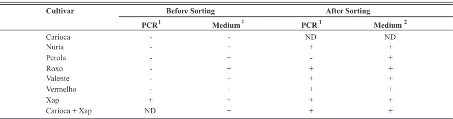
FCM-sorting and PCR
Prior to sorting, Xap was detected by plating in all samples, except in the cv. Carioca, but Xap could not be detected in the crude seed extracts by PCR (Table 1). After sorting, Xap was detected by PCR and by plating in all samples analyzed, except in the supposed Xap-free seed lot cv. Perola. Xap was also detected in the sorted fluids on plates, although cell numbers were low and saprophytic bacteria were still present (data not shown).
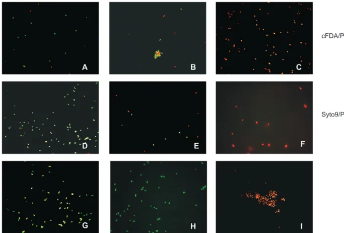
Direct viable counting
In the fluorescence microscopy with both dual staining procedures, in samples with 100% of viable cells most were green and only a few were red (Figure 4A and D). In samples with 50% of viable cells, 50% of the cells were green and 50% were red (Figure 4B and E). In samples with 0% of viable cells, all cells were red (Figure 4C and F). The Figure 4G, H and I shows viable cells stainediable cells stained only with cFDA or SYTO9 and dead cells only with PI, respectively. In the FCM analysis the viable cellsIn the FCM analysis the viable cells (green fluorescence) were identified by FL1 and the dead (red fluorescence) by FL3. A linear relation (R2 ≥
0.97) was found between the percentage of viable (green fluorescent) and heat-killed cells (red fluorescent), both after dual staining with cFDA and PI and with SYTO 9 and PI (Figure 5).
FIGURE 3 - Detection of Xanthomonas axonopodis pv. phaseoli in naturally infected
seed lots of the cultivars Roxo, Valente and Vermelho, by immuno-flow cytometry (FCM), immunofluorescence microscopy (IF) and dilution plating. Four different subsamples of each seed lot were tested independently.
0 1 2 3 4 5 6 7 8
FCM
IF
Plating
Roxo Valente Vermelho
1 2 3 4 1 2 3 4 1 2 3 4
l
o
g
C
F
U
/
m
L
Cultivar Before Sorting After Sorting
PCR
1
Medium
2
PCR
1
Medium
2
Carioca - - ND ND
Nuria - + + +
Perola - + - +
Roxo - + + +
Valente - + + +
Vermelho - + + +
Xap + + + +
Carioca + Xap ND + + +
TABLE 1 - Detection of Xanthomonas axonopodis pv. phaseoli (Xap) in bean seed extracts by PCR and dilution plating on medium 523,
before and after sorting
Immunostaining in combination with direct viable counting
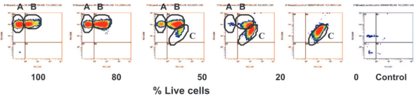
Immuno-FCM could distinguish two clusters in 100% of viable cells, a cluster of green fluorescent particles (A) and a small cluster of particles emitting both green and red fluorescence (B) (Figure 6). In 80% of viable cells,
cluster B increased in size. In 50% and 20% of live cells, a third cluster (C) appeared of red particles with a low level of green fluorescence (A). In samples containing heat-killed cells with 0% viable cells, only red fluorescent particles in cluster (C) were detected. Fluorescence microscopy distinguished viable (green) and dead (red) cells (Figure 7).
FIGURE 4 - Photomicrographs of mixtures of viable and heat-killed cells of Xanthomonas axonopodis pv. phaseoli stained
with fluorescent probes. Suspensions contained 100% (A, D), 50% (B, E) and 0% (C, F) of viable cells double stained with carboxyfluorescein diacetate (cFDA) and propidium iodide (PI) or with SYTO9 and PI, respectively. Viable cells were also stained only with cFDA (G) or SYTO9 (H), and dead cells only with PI (I). Cells were visualized by fluorescence microscopy using blue light for excitation.
y = -0.9103x + 88.081 R
2 = 0.9959 y = 0.9511x + 4.3461 R
2 = 0.999
0 20 40 60 80 100
0 20 40 60 80 100
% Dead cells (heat-treated)
%
L
iv
e
c
e
ll
s
y = -0.7577x + 72.549
R
2
= 0.9814
y = 0.8709x + 11.531
R
2
= 0.9781
0 20 40 60 80 100
0 20 40 60 80 100
% Dead cells (heat-treated )
%
L
i
v
e
c
e
l
ls
FIGURE 5 - Linear relation between ratios of viable and dead (heat-treated) cells and the number of fluorescent particles after dual
staining with cFDA and PI (A) or Syto 9 and PI (B) for pure culture of Xanthomonas axonopodis pv. phaseoli as estimated by flow cytometry. (♦= red cells (%) and □ = green cells (%)).
DISCUSSION
Immuno-FCM allowed rapid quantification of Xapmmuno-FCM allowed rapid quantification of Xap in bean seed extracts. The procedure included a short incubation with antibodies and it could be completed within one hour. It was faster and less time consuming than IF, in which visual observations are less objective and can be tiresome and laborious.
The detection limit of immuno-FCM in a ten-fold diluted bean extract was ca. 104 and the dynamic range
was from 104 to 106 cells per mL. Similar results have
been found for detection of Salmonella typhimurium in eggs and milk with immuno-FCM (McClelland & Pinder,
1994; Gunasekera et al., 2000). The detection level of immunofluorescence microscopy (IF) was ten times lower, whereas dilution plating was even more sensitive. Although the use of pre-immune rabbit serum considerably reduced non-specific binding of antibodies to sample particles, still a certain level of auto-fluorescent particles remained present, which was responsible for a relatively high background in immuno-FCM.
In naturally-infected seed lots, Xap was detected in most of them by immuno-FCM, IF and dilution plating. The absence of CFU on the semi-selective medium in some subsamples of Valente and Vermelho cultivars may be explained by the presence of non-culturable cells,
FIGURE 6 - Flow cytometry density plots of green fluorescent and red fluorescent particles, after staining different percentages of live
and dead cells of Xanthomonas axonopodis pv. phaseoli with antibodies conjugated with Alexa 488 (green fluorescence) and propidium iodide (red fluorescence). A) Cluster of green fluorescent particles. B) Cluster of green and red fluorescent particles. C) Cluster of red fluorescent particles. Control, cells in PBS.
FIGURE 7 - Mixture of 50% of live and dead
which may be dead or in a viable but non-culturable (VBNC) state. According to Gehzzi & Steck (1999) X. campestris pv. campestris is able to enter the VBNC state. Conditions that can induce non-culturability differ depending on the organism and include various factors, such as starvation, water stress, salinity, visible light and temperature (McDougald et al., 1998). Cells are able to exit the VBNC state and return to an actively metabolizing state when conditions become favorable for the pathogen (McDougald et al., 1998). If Xap are present in a VBNC state in the seed extract, dilution plating will underestimate the actual number of cells, potentially able to cause blight on beans. The dry and cool conditions in which seeds are stored for long periods may induce non-culturability, although Xap has been isolated by dilution plating from seed stored for 15 years at 10ºC (Neergaard, 1979).
The densities estimated in immuno-FCM and IF were largely similar for 8 out of 12 subsamples. For four subsamples higher densities were found in immuno-FCM. This discrepancy may be explained by the high sensitivity of the laser-based flow cytometric measurement. A weak fluorescence caused by low numbers of molecules of the fluorochrome (e.g. <1000 FITC molecules per particle) is detected already. Cross-reactions or non-specific bindings may therefore result in false-positive reactions in FCM (Clarke & Pinder, 1998). For several plant-pathogenic bacteria cross-reactions with saprophytic bacteria have been described (Franken et al., 1992). Therefore, the use of antibodies with a higher specificity, such as monoclonal antibodies, is desirable in immuno-FCM.
Also in the supposed pathogen-free seed lots (typical) fluorescent cells were detected by IF and immuno-FCM. In contrast, in two subsamples tested from three seed lots, only in one subsample of cv. Carioca a few colonies were found on plates. In the FCM-sorting experiments, however, Xap was also found in low densities in a subsample of cv. Nuria and cv. Perola. It can be concluded that these supposedly Xap-free samples contained low levels of culturable cells, which makes it more likely that at least part of the fluorescent cells found at 105 CFU/mL by IF and immuno-FCM in cv. Perola and cv.
Nuria are non-culturable cells of Xap.
The potential of flow sorting was explored for isolation from bean extracts to enhance detection of Xap by PCR or dilution plating. Xap could not be detected directly in crude seed extracts by PCR due to the presence of inhibitors. After sorting, Xap could be detected directly in the sampled sorting fluid, indicating that PCR-inhibiting compounds were largely removed. The sorting procedure, however, could be optimized further, as dilution plating on a non-selective agar medium showed that during the sorting procedure other bacteria had also been sorted.
Two DVC methods were evaluated and found suitable to distinguish viable from dead cells of Xap, a
dual staining method with cFDA and PI, and with SYTO 9 and PI. DVC methods on the basis of dual staining DVC methods on the basis of dual stainingDVC methods on the basis of dual stainingdual staining with cFDA and PI have been described for other bacterial species (Bunthof et al., 2001; Ben Amor et al., 2002; (Bunthof et al., 2001; Ben Amor et al., 2002; Hoefel et. 2003a-b, Chitarra et al., 2006). cFDA, whichcFDA, which is an esterified fluorogenic substrate assessing bacterium esterase activity, was able to stain specifically viable cells of Xap. In line with Morono et al. (2004), it was found that the use of glutaraldehyde prevented the leakage of cF out of the cell, improving the efficacy to discriminate viable from dead cells (results not shown). PI specifically stained dead cells of Xap. PI is membrane impermeable and only stains dead bacteria with damaged membranes (Haugland, 2002).
PI is also used in the LIVE/DEAD bacterial viability kit in combination with SYTO 9, which is a green fluorescent, membrane permeable nucleic acid stain, which stains bacteria irrespective of their viability. PI and SYTO 9 have been used for the direct enumeration of physiologically active bacteria in drinking water (Boulos et al., 1999) and also in other fields of bacteriological research (Lebaron et al., 1998; Auty et al., 2001). For). For DVC of Xap, the use of cFDA and PI is preferred over the use of PI and SYTO 9, because cFDA in contrast to SYTO 9 is able to discriminate viable from non-viable cells. Samples need to be measured immediately after staining cells with SYTO 9/PI, to avoid an increase in dead red fluorescent cells. In particular, SYTO 9 was toxic to Xap. The number of non-staining cells decreased from 1.7x10⁷ CFU/mL to 1x 10CFU/mL to 1x 10 to 1x 10⁶ CFU/mL after staining after staining
with SYTO 9/PI plated on medium 523.
Studies on DVC methods should preferably be done with liquid media rather than solid agar media. The bacterial suspensions from liquid growth medium contain a higher percentage of culturable cells than an agar medium (data not shown). Possibly, cells were stressed on agar plates or died from limited nutrients and the build-up of toxic products (Clarke & Pinder, 1998). TheThe combination of immuno-FCM and DVC techniques was evaluated to distinguish live and dead cells of Xap. This would also allow use of immuno-FCM after seed treatments carried out to eliminate seed-borne pathogens, such as heat treatments. This dual staining method was able to distinguish viable from dead cells in pure cultures, but not in seed extract, due to a high background of red fluorescent particles.
ACKNOWLEDGEMENTS
REFERENCES
Alvarez AM, Lou K (1985) Rapid identification of Xanthomonas1985) Rapid identification of Xanthomonas campestris pv. campestris by ELISA. Plant Disease 69:1082-1086.
Amersham Biosciences (2002) Protein G Sepharose 4 fast. In: Antibody Purification Handbook. Life Science News 12. Audy P, Laroche A, Saindon G, Huang HC, Gilbertson RL (1994) Detection of the bean common blight bacteria, Xanthomonas campestris pv. phaseoli and X. c. phaseoli var. fuscans, using the polymerase chain reaction. Phytopathology 84:1185-1192. Auty MAE, Gardiner GE, Mcbrearty SJ, O’Sullivan EO, Mulvihill DM, Collins JK, Fitzgerald GF, Station C, Ross RP (2001) Direct in situ viability assessment of bacteria in probiotic dairy products using viability staining in conjunction with confocal scanning laser microscopy. Applied andApplied and Environmental Microbiology 67:420-425..
Ben Amor K, Breeuwer P, Verbaarschot P, Rombouts FM, Akkermans ADL, De Vos, WM, Abee T (2002) Multiparametric2002) Multiparametric flow cytometry and cell sorting for the assessment of viable, injured, and dead Bifidobacterium cells during bile salt stress. Applied and Environmental Microbiology 68:5209-5216. Boulos L, Prévost M, Barbeau B, Coallier J, Desjardins R (1999) Live/Dead® Baclight™: application of a new rapid staining method for direct enumeration of viable and total bacteria in drinking water. Journal of Microbiological MethodsJournal of Microbiological Methods 37:77-86..
Bunthof CJ, Bloemen K, Breeuwer P, Rombouts FM, Abee TP, Rombouts FM, Abee T (2001) Flow cytometric assessment of viability of lactic acid bacteria. Applied and Environmental Microbiology 67:2326-2335.
Chitarra LG, Breeuwer P, Abee T, Van Den Bulk RW (2006) The use of fluorescent probe to assess viability of the plant pathogenic bacterium Clavibacter michiganensis subsp. michiganensis by flow cytometry. Fitopatologia Brasileira 31:349-356.
Clarke RG, Pinder AC (1998) Improved detection of bacteria byImproved detection of bacteria by flow cytometry using a combination of antibody and viability markers. Journal of Applied Microbiology 84:577-584. Diaper JP, Edwards C (1994) The use of fluorogenic esters to detect viable bacteria by flow cytometry. Journal of Applied Bacteriology 77:221-228.
Franken AALM, Zilverentant JF, Boonekamp PM, Schots ASchots A (1992) Specificity of polyclonal and monoclonal antibodies for the identification of Xanthomonas campestris pv. campestris. Netherlands Journal of Plant Pathology 98:81-94.
Ghezzi JI, Steck TR (1999) Induction of the viable but non-culturable condition in Xanthomonas campestris pv. campestris in liquid microcosms and sterile soil. FEMS Microbiology 30:203-208.
Goszczynska T, Serfontein JJ (1998) Milk-Tween agar, a(1998) Milk-Tween agar, a semiselective medium for isolation and differentiation of Pseudomonas syringae pv. syringae, Pseudomonas syringae pv. phaseolicola and Xanthomonas axonopodis pv. phaseoli. Journal of Microbiological Methods 32:65-72.
Gunasekera TS, Attfield PV, Veal DA (2000) A flow cytometry2000) A flow cytometry method for rapid detection and enumeration of total bacteria
in milk. Applied and Environmental Microbiology 66:1228-Applied and Environmental Microbiology 66:1228-1232..
Hall R (1994) Compendium of bean diseases. Saint Paul MN. APS Press.
Haugland RP (2002) Handbook of fluorescent probes and research products. 9th ed. Oregon. Eugene Molecular Probes. Hoefel D, Grooby WL, Monis PT, Andrews S, Saint CP (2003a) A comparative study of carboxyfluorescein diacetate succinimidyl ester as indicators of bacterial activity. Journal of Microbiological Methods 52:379-388.
Hoefel D, Grooby WL, Monis PT, Andrews S, Saint CP (2003b) Enumeration of water-borne bacteria using viability assays and flow cytometry: a comparison to culture-based techniques. Journal of Microbiological Methods 55:585-597. Kado CI, Heskett MG (1970) Selective media for isolation of Agrobacterium, Corynebacterium, Erwinia, Pseudomonas and Xanthomonas. Phytopathology 60:969-976.
Lebaron P, Parthuisot N, Catala P (1998) Comparison of blue nucleic acid dyes for flow cytometric enumeration of bacteria in aquatic systems. Applied and Environmental Microbiology 64:725-1730.
Malin EM, Roth DA, Belden EL (1983) IndirectEL (1983) Indirect immunofluorescent staining for detection and identification of Xanthomonas campestris pv. phaseoli in naturally infected bean seed. Plant Disease 67:645-647.
McClelland RG, Pinder AC (1994) Detection of Salmonella typhimurium in dairy products with flow cytometry and monoclonal antibodies. Applied and Environmental Microbiology 60:4255-4262.
McDougald D, Rice SA, Weichart D, Kjelleberg S (1998) Nonculturability: adaptation or debilitation? FEMS Microbiology Ecology 25:1-9.
Morono Y, Takano S, Miyanaga K, Tanji Y, Unno H, Hori K (2004) Application of glutaraldehyde for the staining of esterase-active cells with carboxyfluorescein diacetate. Biotechnology Letters 26:379-383.
Neergaard P (1979) Seed Pathology. London. MacMillan Press. Rechinger KB, Siegumfeldt H (2002) Rapid assessment of cell viability of Lactobacillus delbrueckii subsp. bulgaricus by measurement of intracellular pH in individual cells using fluorescence ratio imaging microscopy. International Journal of Food Microbiology 78:53-60.
Saettler AW, Perry SK (1972) Seed transmitted bacterial diseases in Michigan navy beans, Phaseolus vulgaris. Plant Disease Reportes 56:378-381.
Sheppard JW, Roth DA, Saettler AW (1989) Detection of Xanthomonas campestris pv. phaseoli in bean. In: Saettler AW, Schaad NW, Roth DA (Eds.) Detection of bacteria in seed and other planting material. Saint Paul MN. APS Press. pp. 17-29. Toth IK, Hyman LJ, Taylor R, Birch PRJ (1998) PCR-based detection of Xanthomonas campestris pv. phaseoli var. fuscans in plant material and its differentiation from X .c. pv. phaseoli. Journal of Applied Microbiology 85:327-336.
Van Vuurde JWL, Van Den Bovenkamp GW, Birnbaum Y (1983) Immunofluorescence microscopy and enzyme-linked immunosorbent assay as potential routine tests for the detection