UNIVERSIDADE FEDERAL DO CEARÁ
FACULDADE DE FARMÁCIA, ODONTOLOGIA E ENFERMAGEM PROGRAMA DE PÓS-GRADUAÇÃO EM ODONTOLOGIA ÁREA DE CONCENTRAÇÃO: CLÍNICA ODONTOLÓGICA
LIDIANE COSTA DE SOUZA
EFEITO DAS PROANTOCIANIDINAS NA LONGEVIDADE DE RESTAURAÇÕES ADESIVAS: ENSAIO CLÍNICO ALEATORIZADO E
DUPLO-CEGO
LIDIANE COSTA DE SOUZA
EFEITO DAS PROANTOCIANIDINAS NA LONGEVIDADE DE RESTAURAÇÕES ADESIVAS: ENSAIO CLÍNICO ALEATORIZADO E DUPLO-CEGO
FORTALEZA – CE 2018
Tese apresentada ao Programa de Pós-Graduação Odontologia da Universidade Federal do Ceará, como requisito parcial para a obtenção do título de Doutor em Odontologia. Área de concentração: Clínica Odontológica.
LIDIANE COSTA DE SOUZA
EFEITO DAS PROANTOCIANIDINAS NA LONGEVIDADE DE RESTAURAÇÕES ADESIVAS: ENSAIO CLÍNICO ALEATORIZADO E
DUPLO-CEGO
Aprovada em: ___/___/______.
BANCA EXAMINADORA
________________________________________ Prof. Dr. Vicente de Paulo Aragão Saboia (Orientador)
Universidade Federal do Ceará (UFC)
_________________________________________ Prof. Dr.Romulo Rocha Régis
Universidade Federal do Ceará (UFC)
_________________________________________ Prof. Dr. Victor Pinheiro Feitosa
Universidade Federal do Ceará (UFC) e Faculdade Paulo Picanço _________________________________________ Prof. Dr. Francisco Cláudio Fernandes Alves e Silva
Centro Universitário Christus (UniChristus) _________________________________________
Prof. Dr. Jiovanne Rabelo Neri
Universidade de Fortaleza (UNIFOR) e Centro Universitário Christus (UniChristus)
A Deus. Aos meus pais, Pedro e Maria José. Ao meu esposo, Daniel.
AGRADECIMENTOS ESPECIAIS
A Deus e a Nossa Senhora, que são minha fortaleza, agradeço por cada dia da minha vida.
Aos meus pais, Pedro e Maria José, que tanto amor me dão, por tudo que fizeram e fazem por mim e por nossa família. Minha querida mãe, agradeço pela convivência diária também em nosso consultório, por ser paciente e dedicada ao nosso trabalho.
Aos meus irmãos, Pedro Henrique e João Felipe; minhas cunhadas, Indira e Paloma; e ao meu sobrinho, Pedro Filho, por todos os momentos felizes que me fizeram recarregar as energias para os desafios diários e por serem família para mim.
Ao meu esposo, Daniel, minha grande inspiração e incentivo, por todo companheirismo, amor, paciência e toda dedicação que tem por mim.
A todos os meus familiares (avós, tios, tias, primos, primas, sogra), os quais sempre acreditaram e torceram por mim, pelo carinho e orações.
Ao meu orientador, Prof. Dr. Vicente de Paulo Aragão Saboia, por todos esses 10 anos de ensinamentos, por ser um mestre sempre presente e incentivador de meu crescimento.
Ao Professor Victor Pinheiro Feitosa, por sua presteza, amizade e estar sempre disposto a compartilhar seus conhecimentos.
Aos Profs. Dr. Alessandro Dourado Loguercio e Jorge Perdigão, por toda a colaboração que deram ao nosso estudo.
A todos os professores do Programa de Pós-Graduação em Odontologia da Universidade Federal do Ceará, pelos valiosos ensinamentos durante o curso de Doutorado.
À querida amiga Nara Sousa Rodrigues, por sua presença e apoio constantes. Por toda sua colaboração no planejamento e na execução deste trabalho, que também é seu. Sua ajuda e companheirismo foram essenciais. Minha admiração por você é crescente.
À amiga Diana Cunha, por sua disponibilidade quando, mesmo ainda não estando no Programa de Pós-Graduação, se dispôs a ajudar. Por ser sempre solidária, responsável e dedicada. Alguém com quem pude contar desde o Mestrado.
Às amigas e aos amigos, Cecília Atem, Nara Sena, Deborah Magalhães, Lívia Barros, Mara Assef, Lucas Soares, Rafael Monteiro, Matheus Sebbe, Roseline Ramos, Bárbara Lima, Jéssica Monte e Daisy Nantes, pela cooperação nos retornos das avaliações clínicas, por tornarem o trabalho menos cansativo.
Ao grupo de pesquisa, pelos que já se foram, na pessoa da Fabianni - e pelos que ainda estão, pela convivência, aprendizado, diversão e amizade.
Ao Técnico de Laboratório David Queiroz, por ser sempre prestativo e por sua dedicação ao nosso Programa de Pós-Graduação.
A todos os meus alunos e colegas professores que, nos últimos anos, fizeram com que eu descobrisse uma nova paixão: a docência.
A minha profissão, Odontologia, meu sonho de criança, hoje minha realidade que me subsidia na realização dos meus objetivos.
AGRADECIMENTOS
À Universidade Federal do Ceará, por meio do Reitor Prof. Dr. Henry de Holanda Campos.
À Faculdade de Farmácia, Odontologia e Enfermagem (FFOE/UFC), na pessoa de sua diretora Profa. Dra Lidiany Karla Azevedo Rodrigues Gerage.
Ao Curso de Odontologia, na pessoa de seu Coordenador Prof. Dr. Juliano Sartori Mendonça.
Ao Programa de Pós-Graduação em Odontologia da Universidade Federal do Ceará, na pessoa de seu Coordenador Prof.Dr..Vicente de Paulo Aragão Saboia.
Aos membros da banca examinadora pela disponibilidade, além da presteza em avaliar este trabalho, enriquecendo-o.
Às funcionárias da secretaria de Pós-Graduação em Odontologia da Universidade Federal do Ceará, Lúcia Ribeiro Marques Lustosa, Janaine Marques Leal e Joana Karla de Assis Pinheiro,pelo auxílio como também pela disponibilidade.
À Fundação Cearense de Apoio ao Desenvolvimento Científico e Tecnológico – FUNCAP e à Coordenação de Aperfeiçoamento de Pessoal de Nível Superior – CAPES, pela concessão da bolsa de estudo.
À IVOCLAR/VIVADENT pela doação das resinas compostas utilizadas neste trabalho.
À Fundação Cearense de Apoio ao Desenvolvimento Científico e Tecnológico – FUNCAP pelo apoio financeiro conseguido pela aprovação no edital de cooperação internacional (CI3-0093-000670100/14)
“Não há lugar para a sabedoria onde não há paciência.”
RESUMO
As proantocianidinas (PAs) são agentes naturais capazes de estabelecer ligações cruzadas com o colágeno dentinário, inibir atividades proteolíticas das colagenases e que têm mostrado efeitos positivos na resistência à biodegradação, propriedades mecânicas e estabilidade estrutural da dentina, in vitro. Esta tese é constituída por dois
capítulos que objetivaram avaliar in vivo o efeito das PAs na longevidade de
restaurações adesivas de lesões cervicais não cariosas (LCNCs) através de um ensaio clínico aleatorizado e duplo-cego. O capítulo 1 avaliou a PA, na forma de solução aquosa, aplicada na dentina previamente à aplicação do sistema adesivo e após o condicionamento ácido, nas concentrações de 2% (PA2) e 5% (PA5) (em peso). O capítulo 2 avaliou a PA incorporada ao sistema adesivo nas concentrações de 2% (EX2) e 5% (EX5) (em peso). Para ambos, foram selecionados 45 pacientes com 3 lesões cada, dando um total de 135 LCNC por estudo. Um sistema adesivo comercial convencional simplificado, aplicado de acordo com as recomendações do fabricante, foi usado como grupo controle nos estudos. As LCNC de ambos os estudos foram restauradas com resina composta e as restaurações foram avaliadas após o polimento e nos períodos de 6 e 24 meses, utilizando-se os critérios da United States Public Health Service (USPHS)
critério FDI, mas nenhuma restauração foi considerada como tendo uma discrepância clinicamente relevante. Quanto à descoloração marginal, para o critério FDI, observou-se uma diferença significativa entre a avaliação inicial e a avaliação de 24 meobservou-ses para todos os grupos. Os resultados do capítulo 2 mostraram que o grupo EX5 apresentou uma significativa queda na taxa de retenção (15%) após 6 meses. Após 24 meses, tanto o grupo EX2 (27%) quanto o grupo EX5 (29%) apresentaram taxa de retenção significativamente menor que o grupo controle. Quanto a adaptação marginal, todos os grupos apresentaram discrepância significativa ao longo do tempo, somente para o critério FDI. Todos os grupos apresentaram aumento da pigmentação marginal ao longo do tempo para os dois critérios avaliados, mas somente o grupo EX5 apresentou diferença estatística quando comparado aos demais grupos nos períodos de 6 e 24 meses. Nos dois estudos, nenhuma restauração apresentou sensibilidade pós-operatória ou recorrência de cárie. Desta forma, conclui-se que a PA aplicada previamente ou incorporada ao sistema adesivo não apresentou vantagens clínicas após 24 meses de avaliação.
ABSTRACT
Proanthocyanidins (PAs) are natural agents capable of crosslinking dentin collagen, inhibit collagenase proteolytic activities and have shown positive effects on the resistance to biodegradation, mechanical properties and structural stability of dentin in vitro. This thesis consists of two chapters that aimed to evaluate in vivo the effect of
compared to the other groups in the periods of 6 and 24 months. In both studies, no restoration showed postoperative sensitivity or recurrence of caries. In this way, it was concluded that PA applied previously or incorporated into the adhesive system did not present clinical advantages after 24 months of evaluation.
SUMÁRIO
1 INTRODUÇÃO GERAL--- 14
2 PROPOSIÇÕES--- 18
2.1 Objetivo Geral--- 18
2.2 Objetivos Específicos--- 18
3 CAPÍTULOS --- 20
4 CAPÍTULO 1 --- 22
5 CAPÍTULO 2 --- 59
8 CONCLUSÃO GERAL --- 97
REFERÊNCIAS --- 99
APÊNDICE A - TERMO DE CONSENTIMENTO LIVRE E ESCLARECIDO --- 106
APÊNDICE B- FICHA DE CLASSIFICAÇÃO DAS LESÕES --- 108
APÊNDICE C - FICHA DE AVALIAÇÃO DAS RESTAURAÇÕES --- 109
ANEXO A – PARECER DO COMITÊ DE ÉTICA EM PESQUISA--- 111
1 INTRODUÇÃO GERAL
A manutenção da estabilidade da união da interface formada entre sistemas adesivos e a dentina é tema de diversos estudos, pois a degradação dessa interface é a principal responsável pelo insucesso clínico de restaurações com resinas compostas (DE MUNCK et al., 2005).
A união de monômeros resinosos ao substrato dentinário é mais crítica quando comparada à união ao esmalte. Isto é uma consequência da complexa composição daquele tecido, constituído, em volume, por 50% de mineral, 30% de componentes orgânicos e 20% de água (MARSHALL, 1993). Dentre os componentes orgânicos, o colágeno tipo I representa 90% da matriz dentinária. As fibras de colágeno apresentam ligações cruzadas exógenas intra e intermoleculares que são responsáveis pela forma e coesão da estrutura dentinária (STENZEL; MIYATA; RUBIN, 1974). Na ocorrência de desmineralização, a rede de fibrilas de colágeno e proteínas não-colagenosas é a responsável por preservar a forma e a dimensão da dentina (MACIEL et al., 1996) e é nesse substrato, entre os espaços interfibrilares, que os
monômeros resinosos deveriam infiltrar-se para formar uma interface de união compacta e homogênea, chamada camada híbrida (MARSHALL et al., 1997).
Entretanto, o que ocorre na realidade é uma incompleta infiltração e encapsulamento das fibrilas de colágeno por esses monômeros, deixando parte delas expostas. Desta forma, as fibrilas ficam mais susceptíveis à degradação (WANG; SPENCER, 2002; WANG; SPENCER, 2003) durante a vida útil da restauração, tanto por processos físicos (mecânicos e térmicos) e químicos (agentes ácidos, saliva) (BRESCHI et al.,
2008) quanto biológicos (ação de metaloproteinases da matriz extracelular e catepsinas) (AGEE; ZHANG; PASHLEY, 2000; DE MUNCK et al., 2009). Portanto,
a formação de uma rede de colágeno insolúvel, resistente e estável teria um papel importante na longevidade da interface de união dentina/resina frente à degradação inerente às condições do meio bucal (BEDRAN-RUSSO et al., 2009;
BEDRAN-RUSSO et al., 2010).
(CHERUKUPALLI; REDDY, 2016; LIU et al., 2009; MOREIRA et al., 2017; SUNG et al., 2003). Estudos recentes têm utilizado um agente natural capaz de estabelecer
ligações cruzadas com o colágeno dentinário: a proantocianidina (PA) (AL-AMMAR; DRUMMOND; BEDRAN-RUSSO, 2009; BEDRAN-RUSSO et al., 2008;
BEDRAN-RUSSO et al., 2014; CASTELLAN et al., 2010a; CASTELLAN et al.,
2010b; HASS et al., 2016a; MACEDO; YAMAUCHI; BEDRAN-RUSSO, 2009;
SCHEFFEL et al., 2014; XIE; BEDRAN-RUSSO; WU, 2008). As proantocianidinas
(PAs) são parte de um grupo específico de compostos polifenólicos, formados por subunidades de flavan-3-ol, pertencentes à categoria conhecida como taninos condensados (HAN et al., 2003). São encontradas em uma grande variedade de
vegetais, frutas, flores, nozes, sementes e cascas (FERREIRA; SLADE, 2002). As PAs apresentam atividades antibacteriana, anti-inflamatória e antialérgica, bem como ações vasodilatadoras (AFANAS'EV et al., 1989; BUENING et al. 1981;
KOLODZIEJ et al. 1995), o que tem levado ao aumento do interesse pelo estudo
deste composto em áreas da saúde. Além disso, as PAs são capazes de inibir significantemente atividades proteolíticas das enzimas como as colagenases e elastases (MAFFEI et al., 1994) e a progressão de cáries artificiais em dentina
radicular (WALTER et al. 2008, XIE et al., 2008). As PAs são agentes antioxidantes
naturais e também podem aumentar a síntese de colágeno, pois, embora tenham uma atividade inibitória para a maioria das enzimas, são capazes de facilitar a hidroxilação da prolina através da ativação da enzima hidroxilase (MAFFEI et al., 1994).
Outras vantagens inerentes a PA são sua baixa citotoxicidade, seu baixo custo e sua fácil obtenção (AL-AMMAR; DRUMMOND; BEDRAN-RUSSO, 2007; BEDRAN-RUSSO et al., 2009), uma vez que são encontradas abundantemente na
natureza, como em sementes de uva e de cacau, açaí, canela e oxicoco (COS et al.,
2004). Por todas essas características, vários estudos in vitro têm investigado o efeito
de extratos ricos em PAs sobre a resistência à biodegradação, propriedades mecânicas e estabilidade estrutural de dentina coronal (BEDRAN-RUSSO et al., 2011;
CASTELLAN et al., 2010b)
o colágeno intacto, mesmo depois de ter sido clivado por uma enzima (WEADOCK; OLSON; SILVER, 1983).
Em restaurações adesivas, a PA pode ser utilizada de algumas maneiras, como: um primer adicional, aplicado após o condicionamento ácido e antes da aplicação do
adesivo (AL-AMMAR et al., 2009; CASTELLAN et al., 2013; FANG et al., 2012),
adicionada ao sistema adesivo (EPASINGHE et al., 2012; EPASINGHE et al., 2015;
GREEN et al., 2010; LIU; WANG, 2013; VENIGALA et al., 2016) ou até mesmo
adicionada ao agente de condicionamento ácido (HASS et al., 2016b). HECHLER et al. (2012) avaliaram o desempenho a longo prazo da PA aplicada como primer ou
incorporada ao sistema adesivo utilizando o Single Bond (3M ESPE, St. Paul, MN) e verificaram que, após 52 semanas de exposição à digestão por colagenase, a resistência à microtração da interface resina/dentina foi significativamente maior em relação ao controle quando a PA foi utilizada como um primer, ao passo que a PA
incorporada ao sistema adesivo não mostrou diferença significativa em relação ao controle no tempo avaliado.
Embora as metodologias de testes in vitro com protocolos de envelhecimento
possam prever o desempenho de um material (AMARAL et al., 2007; HEINTZE;
ROUSSON, 2011; VAN MEERBEEK et al., 2010), ensaios in vivo continuam sendo
imprescindíveis para avaliar a melhor eficácia clínica de adesivos e/ou técnicas. Em virtude da demanda restauradora e da facilidade de acesso e visualização para posterior avaliação, as lesões cervicais não cariosas (LCNCs) têm sido largamente usadas em estudos clínicos para materiais adesivos. As LCNCs são comuns na cavidade oral e têm sido encontradas em mais de 85% dos pacientes que procuram tratamento odontológico (LEVITCH et al., 1994). Sabe-se que a dentina
esclerótica das LCNCs pode ser mais desafiadora para a união do que a dentina coronal, por isso tal substrato proporciona uma boa superfície para se testar as qualidades de um adesivo (TAY et al., 2000) ou de um protocolo de adesão.
2 PROPOSIÇÃO
2.1Objetivo Geral
• Avaliar clinicamente o efeito na longevidade de restaurações de lesões cervicais não cariosas (LCNCs) de uma solução aquosa contendo proantocianidinas a 2% ou a 5%, aplicada como pré-tratamento da dentina, ou da inclusão de proantocianidina nessas mesmas concentrações (em peso) em um sistema adesivo convencional simplificado.
2.2 Objetivos Específicos
• Avaliar clinicamente, através dos critérios da United States Public Health Service (USPHS) modificados e da Federação Internacional de Odontologia
(FDI), o efeito da aplicação de solução aquosa de proantocianidinas a 2 % ou a 5% antes da aplicação de um sistema adesivo convencional simplificado, ou da incorporação de proantocianidinas 2% ou 5% (em peso) no sistema adesivo, após a confecção das restaurações e nos períodos de 6 e 24 meses.
• Comparar a taxa de retenção, pigmentação marginal, adaptação marginal, sensibilidade pós-operatória e recorrência de cárie em restaurações de LCNCs quando uma solução aquosa de proantocianidinas a 2% ou a 5% é aplicada antes da aplicação de um sistema adesivo convencional simplificado, nos períodos após a confecção das restaurações e após 6 e 24 meses.
3 CAPÍTULOS
Esta tese está baseada no artigo 46 do Regimento Interno do Programa de Pós-graduação em Odontologia da Universidade Federal do Ceará que regulamenta o formato alternativo para dissertações de Mestrado e teses de Doutorado e permite a inserção de artigos científicos de autoria ou coautoria do candidato. Por se tratarem de pesquisas envolvendo seres humanos, ou partes deles, o projeto de pesquisa deste trabalho foi submetido à apreciação do Comitê de Ética em Pesquisa da Universidade Federal do Ceará, através da submissão no site da plataforma Brasil, tendo sido aprovado (Anexos A) e foi também inscrito no site do Registro Brasileiro de Ensaios Clínicos (Anexo B). Assim sendo, esta tese é composta por 2 capítulos citados abaixo:
• Capítulo 1
Título: Two-year clinical evaluation of proanthocyanidin-primer performance in non-carious cervical lesions: a double-blind randomized clinical trial
Autores: SOUZA LC, RODRIGUES NS, CUNHA DA, FEITOSA VP, SANTIAGO SL, REIS A, LOGUERCIO AD, SABOIA VPA. Periódico: Clinical Oral Investigations*
• Capítulo 2
Título: Two-year clinical evaluation of proanthocyanidin incorporation in two-step etch-and-rinse adhesive system
Autores: SOUZA LC, RODRIGUES NS, CUNHA DA, FEITOSA VP, SANTIAGO SL, REIS A, LOGUERCIO AD, SABOIA VPA. Periódico: Journal of Dentistry**
Normas das revistas
* http://www.springer.com/medicine/dentistry/journal/784?detailsPage=pltci_1060698
Two-year clinical evaluation of proanthocyanidin-primer performance in non-carious
cervical lesions: a double-blind randomized clinical trial
Lidiane Costa de Souza1,2, DDS, MCs
1 Postgraduate Program of Dentistry - Faculty of Pharmacy, Dentistry and Nursing, Federal
University of Ceará, Fortaleza, Ceará, Brazil
2Paulo Picanço School of Dentistry, Fortaleza, Brazil Nara Sousa Rodrigues1, DDS, MCs
1 Postgraduate Program of Dentistry - Faculty of Pharmacy, Dentistry and Nursing, Federal
University of Ceará, Fortaleza, Ceará, Brazil Diana Araújo Cunha1, DDS, MCs
1 Postgraduate Program of Dentistry - Faculty of Pharmacy, Dentistry and Nursing, Federal
University of Ceará, Fortaleza, Ceará, Brazil Victor Pinheiro Feitosa2, DDS, MCs, PhD
2Paulo Picanço School of Dentistry, Fortaleza, Brazil Sérgio Lima Santiago1, DDS, MCs, PhD
1 Postgraduate Program of Dentistry - Faculty of Pharmacy, Dentistry and Nursing, Federal University of Ceará, Fortaleza, Ceará, Brazil
Alessandra Reis3, DDS, MCs, PhD
3School of Dentistry, Department of Restorative Dentistry, State
3School of Dentistry, Department of Restorative Dentistry, State
University of Ponta Grossa, Ponta Grossa, Parana, Brazil Vicente de Paulo Aragão Saboia1, DDS, MCs, PhD
1 Postgraduate Program of Dentistry - Faculty of Pharmacy, Dentistry and Nursing, Federal University of Ceará, Fortaleza, Ceará, Brazil
Running Title: Proanthocyanidin pre-treatment on resin/dentin adhesion
Keywords: Dentin-bonding agents; Non-carious cervical lesions; Proanthocyanidin; Longevity; Clinical trial.
CORRESPONDING AUTHOR
Vicente de Paulo Aragão Saboia, Department of Restorative Dentistry - Faculty of Pharmacy, Dentistry and Nursing, Federal University of Ceará, Fortaleza, Ceará, Brazil
R. Gilberto Studart, 770/901, Cocó, Fortaleza, CE, Brazil Zip Code: 60190-750
Two-year clinical evaluation of proanthocyanidin-primer performance in non-carious cervical lesions: a double-blind randomized clinical trial
ABSTRACT
Objective: This double-blind randomized clinical trial evaluated the influence of
pre-treatment with proanthocyanidin (PA) from grape seed extract on clinical behavior of simplified etch-and-rinse adhesive placed in non-carious cervical lesions (NCCL) over 6- and 24-month, using two evaluation criteria: FDI and USPHS.
Methods: A total of 135 restorations were randomly placed in 45 patients. The
NCCLs were phosphoric acid etched for 15 s and distributed into 3 groups: Control (PA0) - adhesive ExciTE F (Ivoclar Vivadent) applied following the manufacturer's recommendations; PA2 and PA5 groups – 2wt% and 5wt% PA solution, respectively, were applied for 60 s and washed for 30 s prior to application of the adhesive ExciTE F. The resin composite was placed incrementally and light-cured. The restorations were evaluated at baseline, 6 and 24 months. Statistical analyses were performed using appropriate tests (=0.05).
Results: The retention rates were 98% (PA0), 98% (PA2) and 83% (PA5) after
Conclusion: The application of proanthocyanidin as primer did not present clinical
advantages after 24 months of clinical evaluation, regardless of the concentration used.
Clinical Relevance: Scientific literature shows that the collagen crosslinking agent,
proanthocyanidin, can stabilize and reinforce the collagen fibrils of the dentin matrix in vitro, increasing the durability of the dentin-resin interface. However, no improvements were found clinically after 24 months herein.
Keywords: Dentin-bonding agents; Non-carious cervical lesions; Proanthocyanidin; Longevity; Clinical trial.
1. Introduction
The degradation of resin–dentin bond interface created with hydrophilic bonding agents occurs by hydrolytic, enzymatic and fatigue degradation processes [1, 2]. Significant number of resin-sparse collagen fibrils can be found at the hybrid layer [3-5] and this matrix is one of the main degradation patterns found in unsuccessful adhesive restorations [2, 6-9].
In recent years, several alternatives have been proposed to preserve the durability of the resin-dentin interface in vitro and in vivo. Among them, the
application of collagen cross-linkers is an emerging and interesting option to increase the longevity of resin/dentin interfaces, as they can increase the resistance of collagen fibrils from dentin matrix alone with inhibitting inactivate host-derived metalloproteases (MMPs) [10-12].
natural polyphenolic compound known as a potent antioxidant and great scavenger of proteins, with low toxicity [14, 15].
Grape seed extract is one of the most used sources of proanthocyanidin. [14, 16, 17]. The use of grape seed extract improved the ultimate tensile strength, stiffness [18, 19], and long-term stability [20, 21] of dentin collagen. In addition to its cross-linking effect, proanthocyanidin has also been shown to inhibit the synthesis of several MMPs from macrophages and inhibit the catalytic activity of MMP-1 and MMP-9 [14, 22].
However, the application of PA-based agents, often made as an extra bonding step with 10 min, 1 h or longer durations [18, 20, 23-25], which turns makes clinical application unfeasible. Recently, PA preconditioners were used in shorter treatment duration (60 s or 120 s), a clinically applicable time [26], showing increase of the cross-linking degree and ultimate tensile strength of demineralized dentin [27]. Unfortunately, to extent of our knowledge, no clinical trials were conducted to predict the effect of PA applied as pre-treatment on clinical performance of adhesive restorations in non-carious cervical lesions (NCCLs).
The description of the experimental design followed the Consolidated Standards of Reporting Trials (CONSORT) statement [28].
2.1 Ethics approval
The local Ethics Committee on Investigations Involving Human Subjects reviewed and approved the protocol and issued a consent form for this study (protocol #640.695). Written informed consent was obtained from all patients prior to starting the treatment.
2.2 Protocol registration
This clinical trial was registered in http://www.ensaiosclinicos.gov.br/ clinical registry under protocol #RBR-366MBJ. All participants were informed about the nature and objectives of the study.
2.3 Trial design, settings and location of data collection
This was a double-blind, equal allocation rate, split-mouth randomized clinical trial. The study was carried out in the clinics of the School of Dentistry at the local University from November 2014 to January 2017.
Recruitment
Patients were recruited as they seek for treatment in the clinics of Dentistry of the local university. No advertisement was made for participant recruitment. Patients were recruited in the order in which they reported for the screening session, thus forming a sample of convenience.
Eligibility criteria
The evaluations were performed using a mouth mirror, an explorer and a periodontal probe. Participants had to be in good general health, older than 18 years old, have an acceptable oral hygiene level and present at least 20 teeth under occlusion.
Participants were required to have at least three NCCLs to be restored in three different teeth. These lesions had to be non-retentive, deeper than 1 mm, and involving both the enamel and dentin of vital teeth without mobility. The cavosurface margin could not involve more that 50% of enamel [29]. Patients with extremely poor oral hygiene, severe or chronic periodontitis, or heavy bruxism habits were excluded from the study as they would receive other treatments before restorative intervention.
2.4 Sample size calculation
The adhesive two-step etch-and-rinse ExcitTE F (Ivoclar Vivadent AG, Schaan, Liechtenstein) was used in the present study. The reported percentage of retention of the Excite adhesive system is 73% after 5 years of clinical evaluation [30]. Considering 5% alpha, an 80% power and a monocaudal two-sided test, the minimum sample size was 43 restorations per group to find a 22% difference between the tested groups.
2.5 Random sequence generation and allocation concealment
The randomization was done on an intra-individual basis so that each subject ended up with three restorations. These randomization schemes were performed using software available at http://www. sealedenvelope.com.
guaranteed the concealment of the random sequence. In all cases, the tooth with the highest tooth number received the treatment described first, while the tooth with the next number in sequence received the treatment mentioned second and the next tooth received the treatment mentioned third.
2.6 Interventions: restorative procedure
Forty-five patients were selected for this study and all received dental prophylaxis with a suspension of pumice and water in a rubber cup and signed an informed consent before the restorative procedures.
The degree of sclerotic dentin from the NCCLs was measured according to the criteria described by Swift and others [31] (Table 1). The cavity dimensions in millimeters (height, width, and depth), the geometry of the cavity (evaluated by profile photograph and labeled at <45o, 45o-90o, 90o<135o, and >135o)[32], the presence of an antagonist, and the presence of attrition facets were observed and recorded. Pre-operative sensitivity was also evaluated by applying an air-blast for 10 s from a dental syringe placed 2 cm from the tooth surface and with an explorer. These features were recorded to allow comparison of the baseline features of the dentin cavities among experimental groups.
One operator restored all teeth. All participants received three restorations, one of each experimental group in different lesions previously selected according to the inclusion criteria.
Before restorative procedures, the operator cleaned all lesions with pumice and water in a rubber cup, followed by rinsing and drying. Then, shade selection was made using a shade guide (Ivoclar Vivadent AG, shade guide, Schaan, Liechtenstein, German). The tooth to be restored was isolated with cotton rolls and a light cured gingival barrier (Top Dam, FGM, Joinville, Santa Catarina, Brazil). The operator did not prepare any additional retention or bevel in the class V cavity. The teeth were distributed in 3 groups and the adhesive ExciTE F (Ivoclar Vivadent, Schaan, Liechtenstein) was applied as described below. The materials, compositions and application modes are described in Table 2.
- PA0 (Control) – The 37% phosphoric acid (Condac acid, FGM, Brazil) was
applied for 15 s. Then, cavities were rinsed thoroughly for 15 s, keeping dentin visible moist slightly with absorbent paper. One coat of adhesive was gently scrubbed on the entire enamel and dentin surface for approximately 10 s each, according to the manufacturer’s recommendations (Table 2). Then, the solvent was evaporated by gentle air stream for 5 s and light cured for 10 s at 1250 mW/cm2 (Emitter A Schuster, Santa Maria, RS, Brazil).
- PA2 – After the phosphoric acid procedure, 2% proanthocyanidin (V.
- PA5 - After the phosphoric acid, 5% proanthocyanidin (V. vinifera,
Meganatural Gold, Madera, CA, USA) solution was applied for 1 minute in the dentin using a disposable applicator, washed for 30 s, removing excess moisture with absorbent paper. Then, ExciTE F was applied according to the control group.
The resin composite Empress Direct (Ivoclar Vivadent, Schaan, Liechtenstein) was used in up to three increments, each one being lightly cured for 20 s at 1250 mW/cm2. The restorations were finished immediately with fine and extra-fine #3195 diamond burs (KG Sorensen, Barueri, SP, Brazil) under constant water-cooling. After one-week, each one was finished and polished with slow-speed polishing points (Jiffy Polishers, Ultradent, South Jordan, UT, USA).
2.7 Calibration procedures for clinical evaluation
For training purposes, two experienced and calibrated examiners observed 10 photographs that were representative of each score for each criterion. They evaluated 10 patients each on two consecutive days. These subjects had cervical restorations but were not part of this project. An intra-examiner and inter-examiner agreement of at least 85% was necessary before beginning the evaluation [33]. In case of disagreement between the examiners, consensus was obtained.
2.7.1 Blinding
The examiners, who were not involved with the restoration procedures and therefore blinded to the group assignment, performed the clinical evaluation. Patients were also blinded to group assignment in a double-blind randomized clinical trial design.
An individual standardized paper case report form was used for each evaluator at each recall time so that evaluators were kept blinded to earlier evaluations during the follow-up recalls. Intraoral color photographs were collected at baseline and at the recall appointments to aid in the evaluation, if necessary. Clinical photographs consisted of digital images obtained using a Nikon D90X camera with a 105-mm Medical Nikon lens (Nikon Inc., Melville, NY, USA).
The restorations were evaluated by World Federation criteria (FDI) [34] and the classical United States Public Health Service (USPHS) criteria (adapted by Bittencourt and others [35] and Perdigão and others [36]) at baseline and after 6 and 24 months of clinical service. Only the clinically relevant measures for evaluation of adhesive performance were used and scored (Tables 3 and 4). The primary clinical endpoint was restoration retention/fracture, but the following secondary endpoints were also evaluated: marginal staining, marginal adaptation, postoperative sensitivity, and recurrence of caries.
These variables were ranked according to FDI criteria into clinically very good, clinically good, clinically sufficient/ satisfactory, clinically unsatisfactory but repairable, and clinically poor (replacement required) [34] and in the USPHS criteria into Alfa, Bravo and Charlie. [35]. Both examiners evaluated all the restorations once and independently. When disagreements occurred during the evaluations, they had to reach a consensus before the participant was dismissed. The restoration retention rates were calculated according to the ADA guidelines [37]. Cumulative failure percentage = [(PF + NF) / (PF + RR)] X 100%, where PF is the number of previous failures before the current recall, NF is the number of new failures during the current recall, and RR is the number of currently recalled restorations.
The statistical analyses followed the intention-to-treat protocol according to CONSORT (Consolidated Standards of Reporting Trials) suggestion [28]. Descriptive statistics were used to describe the distributions of the evaluated criteria. Statistical analysis for each individual item was performed for each evaluation criteria (FDI and USPHS criteria).
The differences in the ratings of the three groups after 6 and 24 months were tested with Friedman repeated-measures analysis of variance by rank (=0.05), and differences in the ratings of each group at baseline and after 6 and 24 months were evaluated using the Wilcoxon test (=0.05). Cohen’s kappa statistics was used to test inter-examiner agreement. In all statistical tests, we pre-set the level of significance to 5%.
3. Results
The restorative procedures were implemented exactly as planned and no modification was performed. Seventeen out of 62 patients were not enrolled in the study because they did not fulfill the inclusion criteria (Figure 1). Thus, 45 subjects were selected. All baseline details relative to the research subjects and characteristics of the restored lesions are displayed in Table 5. All research subjects were evaluated at the baseline and at 6 months and two patients with three restorations each did not attend the 24-month recall rate (Figure 1), because one moved to another city and other could not return due to health problems.
3.1 Retention
were lost at 24 months. According to FDI and USPHS criteria, the 24-month retention rates (95% confidence interval) were 93% (82 – 98%) for PA0; 89% (76 – 95%) for PA2; and 70% (55 – 88%) for PA5.
When the data from 6-month results from each group were compared with their baseline findings, a significant difference was found only for PA5 (p = 0.03; Tables 6 and 7). Also, when the retention rate of the PA5 was compared with PA0 and PA2, significant differences in the retention rates were detected after 6 months (p = 0.001; Tables 6 and 7). When the data from 24-month results from each group were compared with their baseline findings, a significant difference was found only for PA5 (p = 0.03; Tables 6 and 7). Also, when the retention rate of the PA5 was compared with PA0, significant differences in the retention rates were detected after 24 months (p = 0.001; Tables 6 and 7).
3.2 Post-operative sensitivity
No restorations showed post-operative sensitivity immediately after restorative procedures according to the FDI and USPHS criteria. After 6- and 24-month, no restoration showed post-operative sensitivity using both the FDI and USPHS criteria (Tables 6 and 7).
3.3 Marginal adaptation
However, significant worsening for all three groups of marginal adaptation was observed within all groups over time (baseline vs. 6-month and baseline vs. 24-month) (p < 0.05; Tables 6 and 7). Despite the high number of the restorations with lack of marginal adaptation in the FDI criteria, none of them was considered to have clinically relevant discrepancies in the marginal adaptation even after 6- and 24-month of clinical evaluation (Table 6).
When the USPHS criteria were used, only 8 restorations were scored as Bravo
for marginal adaptation (p > 0.05) at the 6-month recall. After 24-month recall, 8 restorations were scored as Bravo for marginal adaptation (p > 0.05). No significant
difference was detected between any pair of groups at the 6- and 24-month recalls and between recall times within group (p > 0.05).
3.4 Marginal discoloration
For the FDI criteria, 12 restorations at the 6-month recall were considered to have minor discrepancies. After 24-month recall, 42 restorations were considered to have minor discrepancies (clinically good and satisfactory).
A significant difference between baseline vs. 6-month recall was observed for the group PA2 using FDI criteria (p < 0.05; Tables 6). However, a significant difference between baseline vs. 24-month recall was observed for all groups using FDI criteria (p < 0.05; Tables 6). It worth to mention that, after 24-month recall, no significant differences were observed between groups (p>0.05; Tables 6 and 7).
When the USPHS criteria were used, only 7 restorations at 6-month recall were scored as Bravo for marginal staining (p > 0.05). After 24-month recall, 21
restorations were scored as Bravo for marginal staining. A significant difference
USPHS criteria (p < 0.05; Tables 6). After 24-month recall, only PA2 showed a significant higher marginal staining when compared with PA5 (p = 0.001; Tables 6 and 7).
3.5 Recurrence of caries
No restoration showed recurrence of caries at the 6- and 24-month clinical recall using the FDI and the USPHS criteria.
3.6 General Overview
When the FDI criteria for ‘acceptable’ vs. ‘not acceptable’ restorations were
applied, only 21 restorations were ranked as ‘not acceptable’ due to loss of the restorations, the majority for the PA5 group (Table 8).
4. Discussion
Among the clinical parameters for the evaluation of an adhesion protocol or the performance of any restorative material in the NCCL, the retention rate is the most important, since the restoration loss do not allow an evaluation of other parameters [38]. Regarding this parameter, PA5 group showed significant reduction in the retention rate (17%) when compared with its baseline and when compared with PA0 (2%) and PA2 (2%) groups, after 6-month. After 24-month, this reduction was even greater for PA5 group (30%) and also increased for PA2 group (11%), although it was not statistically significant, which leads to rejection of the null hypothesis.
dose dependent [41]. In this study, concentrations of 2% (PA2) and 5% (PA5) were used and a greater drop in retention rate was observed when the highest concentration of PA was used. A justification for this can be that PA has a free radical scavenging effect, which can disturb the free radical polymerization of the resin, inhibiting the ideal resin polymerization [42], especially within collagen mesh.
The use of non-carious cervical lesions is particularly valuable for clinical studies because it is difficult to study this type of lesion in vitro. These lesions have
quite variable etiology and their prevalence is increasing as the adult population continues to age [43, 44]. Besides, the sclerotic dentin on the surface of the NCCLs is more challenging than coronal dentin to adhesion, so such a substrate provides a good surface to test the qualities of an adhesive system [45].
FDI criteria, as well the USPHS criteria, are parameters for evaluating dental restorations. FDI criteria were published in 2007 by FDI [34, 46] and since then, some studies [38, 47, 48] have used them. It has been concluded that the FDI criteria is more sensitive than the USPHS criteria modified for identifying small variations in the clinical outcomes when evaluating restorations of NCCLs [38, 47, 48]. This finding was corroborated in the present study, as the marginal adaptation was only statistically significant for all three groups over time (baseline vs. 6-month) when FDI criteria were used.
One hundred and three restorations exhibited some marginal adaptive discrepancies in the 24-month recall. All groups showed a significant worsening in this criteria over time (baseline vs 6-month vs 24-month), however, none restauration
and clinically acceptable [51] and the simple procedure of restoration re-polishing can amend these discrepancies without causing any damage to the integrity of the restoration [52].
When using FDI criteria, PA2 group showed more marginal discoloration after 6 months compared to baseline and this difference was not seen for the other groups. After 24 months no differences were observed among the groups but all of them showed increase in marginal discoloration compared to baseline. When USPHS was used, the groups PA0 and PA2 presented a significant difference compared their baseline with 24-month recall and PA2 showed a significant higher marginal discoloration than PA5, in this period. The PA5 group had great loss of restorations, which may have underestimated the statistical results for this assessment, since these criteria were not evaluated in the lost restorations. Moreira et al. (2017) [53] showed that the PA-treated dentin samples were brownish in color.
PA grape-seed solutions have a darker color and it can be attributed to their oxidative properties and the presence of high different molecular weight polymer polyphenols, which may justify the color change [10, 39, 54]. Studies have attempted to purify PA by extracting oligomeric or dimeric substances that would be more related to the benefits of PA in dental procedures and [55,56] might cause less undesirable changes.
possible benefits from pre-treatment with 2% PA, as observed in some in vitro
studies, only will be noted after more follow-up time. 5. Conclusion
The application of proanthocyanidin as primer did not present clinical advantages after 24 months of clinical evaluation, regardless of the concentration used.
Acknowledge
This investigation was supported by research grants from FUNCAP (Grant# CI3-0093-000670100/14) and CAPES (scholarship). The authors would like to thank the generous donations of resin composite Empress Direct by Ivoclar Vivadent.
6. References
[1] Reis A, Carrilho M, Breschi L, Loguercio AD (2013) Overview of clinical alternatives to minimize the degradation of the resin-dentin bonds. Oper Dent 38: E1–E25. doi:10.2341/12-258-LIT.
[2] Breschi L, Mazzoni A, Ruggeri A, Cadenaro M, Di Lenarda R, De Stefano Dorigo E (2008) Dental adhesion review: Aging and stability of the bonded interface. Dent Mater 24: 90–101. doi:10.1016/j.dental.2007.02.009.
[3] Hashimoto M, Ohno H, Kaga M, Endo K, Sano H, Oguchi H (2000) In vivo Degradation of Resin-Dentin Bonds in Humans Over 1 to 3 Years. J Dent Res 79: 1385–1391. doi:10.1177/00220345000790060601.
[4] Hashimoto M, Ohno H, Sano H, Tay FR, Kaga M, Kudou Y, Oguchi H, Araki Y, Kubota M (2002) Micromorphological changes in resin-dentin bonds after 1 year of water storage. J Biomed Mater Res 63: 306–311. doi:10.1002/jbm.10208.
electron microscopy. Biomaterials 24: 3795–3803. doi:10.1016/S0142-9612(03)00262-X.
[6] Mazzoni A, Pashley DH, Nishitani Y, Breschi L, Mannello F, Tjäderhane L, Toledano M, Pashley EL, Tay FR (2006) Reactivation of inactivated endogenous proteolytic activities in phosphoric acid-etched dentine by etch-and-rinse adhesives. Biomaterials 27: 4470–4476. doi:10.1016/j.biomaterials.2006.01.040.
[7] Mazzoni A, Carrilho M, Papa V, Tjäderhane L, Gobbi P, Nucci C, Di Lenarda R, Mazzotti G, Tay FR, Pashley DH, Breschi L (2011) MMP-2 assay within the hybrid layer created by a two-step etch-and-rinse adhesive: Biochemical and immunohistochemical analysis. J Dent 39: 470–477. doi:10.1016/j.jdent.2011.04.004. [8] Kostoryz EL, Dharmala K, Ye Q, Wang Y, Huber J, Park JG, Snider G, Katz JL,
Spencer P (2009) Enzymatic biodegradation of HEMA/BisGMA adhesives formulated with different water content. J Biomed Mater Res - Part B Appl Biomater 88: 394– 401. doi:10.1002/jbm.b.31095.
[9] Tay FR, Pashley DH (2003) Have dentin adhesives become too hydrophilic? J Can Dent Assoc 69: 726–731. doi:10.1016/S0109-5641(03)00110-6.
[10] Bedran-Russo AK, Pauli GF, Chen SN, McAlpine J, Castellan CS, Phansalkar RS, Aguiar TR, Vidal CMP, Napotilano JG, Nam JW, Leme AA (2014) Dentin biomodification: Strategies, renewable resources and clinical applications. Dent Mater 30: 62–76. doi:10.1016/j.dental.2013.10.012.
[11] Liu Y, Liu W, Sun G, Wei X, Yi D (2009) Calcification resistance of procyanidin-treated decellularized porcine aortic valves in vivo. Heart Surg Forum 12: 24-29. doi:10.1532/HSF98.20081103.
[13] Perdigão J, Reis A, Loguercio AD (2013) Dentin adhesion and MMPs: A comprehensive review. J Esthet Restor Dent 25: 219–241. doi:10.1111/jerd.12016. [14] Han B, Jaurequi J, Tang BW, Nimni ME (2003) Proanthocyanidin: a natural
crosslinking reagent for stabilizing collagen matrices. J Biomed Mater Res A 65: 118– 124. doi:10.1002/jbm.a.10460.
[15] Ye X, Krohn RL, Liu W, Joshi SS, Kuszynski CA, McGinn TR, Bagchi M, Preuss HG, Stohs SJ, Bagchi D (1999) The cytotoxic effects of a novel IH636 grape seed proanthocyanidin extract on cultured human cancer cells. Mol Cell Biochem 196: 99– 108. http://www.ncbi.nlm.nih.gov/pubmed/10448908.
[16] Liu Y, Chen M, Yao X, Xu C, Zhang Y, Wang Y (2013) Enhancement in dentin collagen’s biological stability after proanthocyanidins treatment in clinically relevant
time periods. Dent Mater 29: 485–492. doi:10.1016/j.dental.2013.01.013.
[17] Castellan CS, Bedran-Russo AK, Antunes A, Pereira PNR (2013) Effect of dentin biomodification using naturally derived collagen cross-linkers: One-year bond strength study. Int J Dent. doi:10.1155/2013/918010.
[18] Bedran-Russo AKB, Pashley DH, Agee K, Drummond JL, Miescke KJ (2008) Changes in stiffness of demineralized dentin following application of collagen crosslinkers. J Biomed Mater Res - Part B Appl Biomater 86: 330–334. doi:10.1002/jbm.b.31022.
[19] Bedran-Russo AKB, Pereira PNR, Duarte WR, Drummond JL, Yamauchi M (2007) Limitations in bonding to dentin and experimental strategies to prevent bond degradation. J Biomed Mater Res B Appl Biomater 80: 268–272. doi:10.1002/jbm.b.30593.
[21] Castellan CS, Bedran-Russo AK, Karol S, Pereira PNR (2011) Long-term stability of dentin matrix following treatment with various natural collagen cross-linkers. J Mech Behav Biomed Mater 4: 1343–1350. doi:10.1016/j.jmbbm.2011.05.003.
[22] Song SE, Choi BK, Kim SN, Yoo YJ, Kim MM, Park SK, Roh SS, Kim CK (2003) Inhibitory effect of procyanidin oligomer from elm cortex on the matrix metalloproteinases and proteases of periodontopathogens. J Periodontal Res 38: 282– 289. http://www.ncbi.nlm.nih.gov/pubmed/12753366.
[23] Karol S, Bedran-Russo AK (2010) Nanomechanical properties of demineralized dentin treated with collagen cross linkers. Dent Mater. doi:10.1016/j.dental.2009.11.083.
[24] Bedran-Russo AKB, Castellan CS, Shinohara MS, Hassan L, Antunes A (2011) Characterization of biomodified dentin matrices for potential preventive and reparative therapies. Acta Biomater 7: 1735–1741. doi:10.1016/j.actbio.2010.12.013.
[25] Aguiar TR, Vidal CMP, Phansalkar RS, Todorova I, Napolitano JG, McAlpine JB, Chen SN, Pauli GF, Bedran-Russo AK (2014) Dentin Biomodification Potential Depends on Polyphenol Source. J Dent Res 93: 417–422. doi:10.1177/0022034514523783.
[26] Fang M, Liu R, Xiao Y, Li F, Wang D, Hou R, Chen J (2012) Biomodification to dentin by a natural crosslinker improved the resin-dentin bonds. J Dent 40: 458–466. doi:10.1016/j.jdent.2012.02.008.
[27] Liu R, Fang M, Xiao Y, Li F, Yu L, Zhao S, Shen L, Chen J (2011) The effect of transient proanthocyanidins preconditioning on the cross-linking and mechanical properties of demineralized dentin. J Mater Sci Mater Med 22: 2403–2411. doi:10.1007/s10856-011-4430-4.
doi:10.4103/0976-500X.72352.
[29] Loguercio AD, Reis A, Barbosa AN, Roulet JF (2003) Five-year double-blind randomized clinical evaluation of a resin-modified glass ionomer and a polyacid-modified resin in noncarious cervical lesions. J Adhes Dent 5: 323–332. http://www.ncbi.nlm.nih.gov/pubmed/15008339.
[30] Franco EB, Benetti AR, Ishikiriama SK, Santiago SL, Lauris JRP, Jorge MFF, Navarro MFL (2006) 5-year Clinical Performance of Resin Composite Versus Resin Modified Glass Ionomer Restorative System in Non-carious Cervical Lesions. Oper Dent 31: 403–408. doi:10.2341/05-87.
[31] Swift EJ, Perdigão J, Heymann HO, Wilder AD, Bayne SC, May KN, Sturdevant JR, Roberson TM (2001) Eighteen-month clinical evaluation of a filled and unfilled dentin adhesive. J Dent 29: 1–6. http://www.ncbi.nlm.nih.gov/pubmed/11137632.
[32] Da Costa TRF, Loguercio AD, Reis A (2013) Effect of enamel bevel on the clinical performance of resin composite restorations placed in non-carious cervical lesions. J Esthet Restor Dent 25: 346–356. doi:10.1111/jerd.12042.
[33] Schmalz G, Ryge G (2005) Reprint of Criteria for the clinical evaluation of dental restorative materials. Clin Oral Investig 9: 215–232. doi:10.1007/s00784-005-0018-z. [34] Hickel R, Peschke A, Tyas M, Mjör I, Bayne S, Peters M, Hiller KA, Randall R,
Vanherle G, Heintze SD (2010) FDI World Dental Federation - clinical criteria for the evaluation of direct and indirect restorations. Update and clinical examples. J Adhes Dent 12: 259–272. doi:10.3290/j.jad.a19262.
[35] Dalton Bittencourt D, Ezecelevski IG, Reis A, Van Dijken JW, Loguercio AD (2005) An 18-months’ evaluation of self-etch and etch & rinse adhesive in non-carious cervical lesions. Acta Odontol Scand 63: 173–178. doi:10.1080/00016350510019874. [36] Perdigão J, Sezinando A, Monteiro PC (2012) Laboratory bonding ability of a
http://www.ncbi.nlm.nih.gov/pubmed/22988685.
[37] ADA Council on Scientific Affairs (2001) Revised American Dental Association acceptance program guidelines: dentin and enamel adhesives. American Dental Association, Chicago, pp 1–9.
[38] Perdigão J, Kose C, Mena-Serrano A, De Paula E, Tay L, Reis A, Loguercio A (2014) A New Universal Simplified Adhesive: 18-Month Clinical Evaluation. Oper Dent 39: 113–127. doi:10.2341/13-045-C.
[39] Scheffel D, Hebling J, Scheffel R, Agee K, Turco G, de Souza Costa C, Pashley D (2014) Inactivation of Matrix-bound Matrix Metalloproteinases by Cross-linking Agents in Acid-etched Dentin. Oper Dent 39: 152–158. doi:10.2341/12-425-L.
[40] Liu Y, Tjäderhane L, Breschi L, Mazzoni A, Li N, Mao J, Pashley DH, Tay FR (2011) Limitations in Bonding to Dentin and Experimental Strategies to Prevent Bond Degradation. J Dent Res 90: 953–968. doi:10.1177/0022034510391799.
[41] Seseogullari-Dirihan R, Mutluay MM, Pashley DH, Tezvergil-Mutluay A (2017) Is the inactivation of dentin proteases by crosslinkers reversible? Dent Mater 33: 62–68. doi:10.1016/j.dental.2016.09.036.
[42] Kalachandra S, Turner DT (1989) Water sorption of plasticized denture acrylic lining materials. Dent Mater 5: 161–164. http://www.ncbi.nlm.nih.gov/pubmed/2637211. [43] Bartlett DW, Shah P (2006) A Critical Review of Non-carious Cervical (Wear)
Lesions and the Role of Abfraction, Erosion and Abrasion. J Dent Res 85: 306–312. doi:10.1177/154405910608500405.
[44] Wood I, Jawad Z, Paisley C, Brunton P (2008) Non-carious cervical tooth surface loss: A literature review. J Dent 36: 759–766. doi:10.1016/j.jdent.2008.06.004.
interfacial ultrastructure and microtensile bond strength evaluation. J Adhes Dent 2: 9–28. http://www.ncbi.nlm.nih.gov/pubmed/11317411.
[46] Hickel R, Roulet JF, Bayne S, Heintze SD, Mjör IA, Peters M, Rousson V, Randall R, Schmalz G, Tyas M, Vanherle G (2007) Recommendations for conducting controlled clinical studies of dental restorative materials. Science Committee Project 2/98--FDI World Dental Federation study design (Part I) and criteria for evaluation (Part II) of direct and indirect restorations includi. J Adhes Dent 9: 121–147. http://www.ncbi.nlm.nih.gov/pubmed/18341239.
[47] Mena-Serrano A, Kose C, De Paula EA, Tay LY, Reis A, Loguercio AD, Perdigão J (2013) A new universal simplified adhesive: 6-month clinical evaluation. J Esthet Restor Dent 25: 55–69. doi:10.1111/jerd.12005.
[48] Piva F, Coelho-de-Sousa FH, Ribeiro CS (2009) A deciduous teeth composite restoration clinical trial using two methods. J Dent Res.
[49] Loguercio AD, Andrade E, Paula D, Hass V, Luque-martinez I, Reis A, Perdigão J (2015) A new universal simpli fi ed adhesive : 36-Month randomized double-blind clinical trial. J Dent 43: 1083–1092. doi:10.1016/j.jdent.2015.07.005.
[50] Lawson NC, Robles A, Fu C, Paul C, Sawlani K, Burgess JO (2015) Two-year clinical trial of a universal adhesive in total-etch and self-etch mode in non-carious cervical lesions. J Dent 43: 1229–1234. doi:10.1016/j.jdent.2015.07.009.
[51] Peumans M, De Munck J, Mine A, Van Meerbeek B (2014) Clinical effectiveness of contemporary adhesives for the restoration of non-carious cervical lesions. Dent Mater 30: 1089–1103. doi:10.1016/j.dental.2014.07.007.
[52] Hickel R, Brüshaver K, Ilie N (2012) Repair of restorations – Criteria for decision making and clinical recommendations. Dent Mater 29: 28–50. doi:10.1016/j.dental.2012.07.006.
Lomonaco D, Mazzetto SE, Feitosa VP (2017) Efficacy of new natural biomodification agents from Anacardiaceae extracts on dentin collagen cross-linking. Dent Mater 33: 1103–1109. doi:10.1016/j.dental.2017.07.003.
[54] Machado AC, Dezan Junior E, Gomes-Filho JE, Cintra LTA, Ruviére DB, Zoccal R, Damante CA, Jardim Junior EG (2012) Evaluation of tissue reaction to Aroeira (Myracrodruon urundeuva) extracts: a histologic and edemogenic study, J. Appl. Oral Sci. 20 (2012) 414–418. doi:10.1590/S1678-77572012000400005.
[55] Phansalkar RS, Nam JW, Chen SN, McAlpine JB, Napolitano JG, Leme A, Vidal CM, Aguiar T, Bedran-Russo AK, Pauli GF (2015) c. Fitot. 101:169-78. doi: 10.1016/j.fitote.2014.12.006.
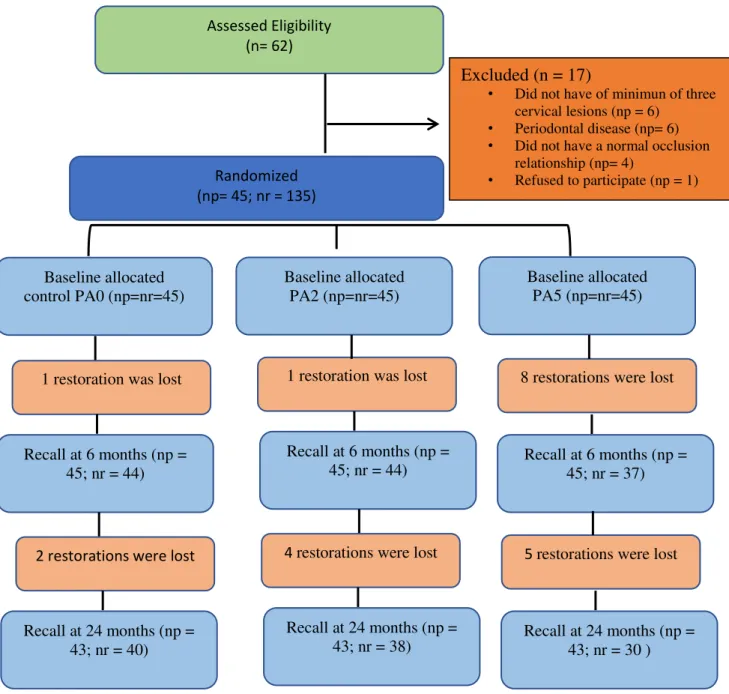
Figure 1 – Flow diagram. Np: number of patients, Nr: number of restorations. PA2 = 2% proanthocyanidin solution; PA5: 5% proanthocyanidin solution.
Excluded (n = 17)
• Did not have of minimun of three cervical lesions (np = 6)
• Periodontal disease (np= 6) • Did not have a normal occlusion
relationship (np= 4)
• Refused to participate (np = 1) Assessed Eligibility
(n= 62)
Randomized (np= 45; nr = 135)
Baseline allocated control PA0 (np=nr=45)
Baseline allocated PA2 (np=nr=45)
Baseline allocated PA5 (np=nr=45)
1 restoration was lost 1 restoration was lost 8 restorations were lost
Recall at 6 months (np = 45; nr = 44)
Recall at 6 months (np =
45; nr = 44) Recall at 6 months (np = 45; nr = 37)
2 restorations were lost 4 restorations were lost 5 restorations were lost
Recall at 24 months (np = 43; nr = 40)
Recall at 24 months (np =
Legends of Tables:
Table 1 - Dentin sclerosis scale.
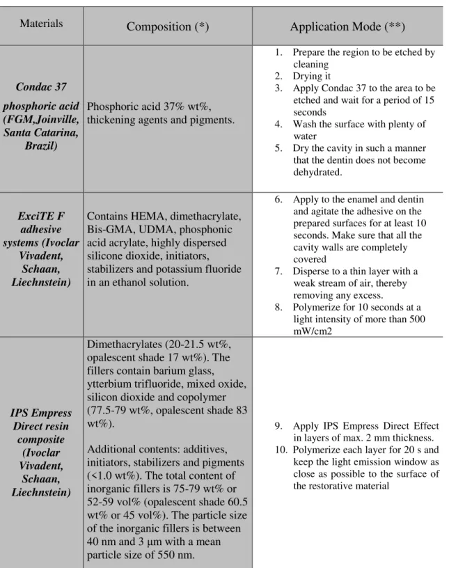
Table 2 –Materials, composition and application mode.
Table 3 - World Dental Federation (FDI) criteria used for clinical evaluation [34].
Table 4 - Modified United States Public Health Service (USPHS) criteria according to Bittencourt and others [35] and Perdigão and others [36].
Table 5 - Distribution of noncarious cervical lesions according to research subject (gender and age) and characteristics of Class V lesions (shape, cervicoincisal size of the lesion, degree of sclerotic dentin, presence of antagonistic, presence of attrition facets, presence of preoperative sensitivity, and tooth and arch distribution).
Table 6 - Number of evaluated restorations for each experimental group (PA0 [no pretreatment with PA], PA2 [2% proanthocyanidin applicated before the adhesive system] and PA5 [5% proanthocyanidin applicated before the adhesive system] classified according to the World Dental Federation (FDI) criteria[34].
Table 7 - Number of evaluated restorations for each experimental group (PA0 [no pretreatment with PA], PA2 [2% proanthocyanidin applicated before the adhesive system] and PA5 [5% proanthocyanidin applicated before the adhesive system] classified according to the adapted United States Public Health Service (USPHS) criteria [35], [36].
Table 1
Dentin sclerosis scale*
CATEGORY CRITERIA
1 No sclerosis present; dentin is light yellowish or whitish, with little discoloration; dentin is opaque, with little translucency or transparency
2 More sclerosis than in category 1 but less than halfway between categories 1 and 4
3 Less sclerosis than in category 4 but more than halfway between categories 1 and 4
4
Significant sclerosis present; dentin is dark yellow or even discolored (brownish); glassy appearance, with significant translucency or transparency evident
* Adapted from Swift and co
Table 2 - Materials, composition and application mode.
Materials Composition (*) Application Mode (**)
Condac 37 phosphoric acid (FGM,Joinville, Santa Catarina,
Brazil)
Phosphoric acid 37% wt%, thickening agents and pigments.
1. Prepare the region to be etched by cleaning
2. Drying it
3. Apply Condac 37 to the area to be etched and wait for a period of 15 seconds
4. Wash the surface with plenty of water
5. Dry the cavity in such a manner that the dentin does not become dehydrated. ExciTE F adhesive systems (Ivoclar Vivadent, Schaan, Liechnstein)
Contains HEMA, dimethacrylate, Bis-GMA, UDMA, phosphonic acid acrylate, highly dispersed silicone dioxide, initiators, stabilizers and potassium fluoride in an ethanol solution.
6. Apply to the enamel and dentin and agitate the adhesive on the prepared surfaces for at least 10 seconds. Make sure that all the cavity walls are completely covered
7. Disperse to a thin layer with a weak stream of air, thereby removing any excess. 8. Polymerize for 10 seconds at a
light intensity of more than 500 mW/cm2 IPS Empress Direct resin composite (Ivoclar Vivadent, Schaan, Liechnstein)
Dimethacrylates (20-21.5 wt%, opalescent shade 17 wt%). The fillers contain barium glass, ytterbium trifluoride, mixed oxide, silicon dioxide and copolymer (77.5-79 wt%, opalescent shade 83 wt%).
Additional contents: additives, initiators, stabilizers and pigments (<1.0 wt%). The total content of inorganic fillers is 75-79 wt% or 52-59 vol% (opalescent shade 60.5 wt% or 45 vol%). The particle size of the inorganic fillers is between 40 nm and 3 μm with a mean particle size of 550 nm.
9. Apply IPS Empress Direct Effect in layers of max. 2 mm thickness. 10. Polymerize each layer for 20 s and
keep the light emission window as close as possible to the surface of the restorative material
(*) HEMA = 2-hydroxyethyl methacrylate Bis-GMA = bisphenol glycidyl methacrylate; UDMA = urethane dimethacrylate
Table 3
Esthetic Property Functional Properties Biological Properties 1. Staining margin 2. Fractures
and retention
3. Marginal
adaptation 4. Postoperative (hyper-) sensitivity 5. Recurrence of caries
1. Clinically very
good 1.1 No marginal staining 2.1 Restoration retained, no fractures / cracks 3.1 Harmonious outline, no gaps, no discoloration. 4.1 No
hypersensitivity. 5.1 No secondary or primary caries
2. Clinically good (after correction very good
1.2 Minor marginal staining, easily removable by polishing. 2.2 Small hairline crack. 3.2.1 Marginal gap (50 μm). 3.2.2 Small marginal fracture removable by polishing. 4.2 Low
hypersensitivity for a limited period of time
5.2 Very small and localized demineralization. No operative treatment required 3.Clinically sufficient / satisfactory (minor shortcomings with no adverse effects but not adjustable without damage to the tooth)
1.3 Moderate marginal staining, not esthetically unacceptable.
2.3 Two or more or larger hairline cracks and/or chipping (not affecting the marginal integrity).
3.3.1 Gap < 150 μm not removable 3.3.2. Several small enamel or dentin fractures
4.3.1 Premature / slightly more intense 4.3.2 Delayed/weak sensitivity; no subjective complaints, no treatment needed.
5.3 Larger areas of demineralization, but only preventive measures necessary (dentine not exposed) 4. Clinically unsatisfactory (repair for prophylactic reasons) 1.4 Pronounced marginal staining; major intervention necessary for improvement 2.4 Chipping fractures which damage marginal quality; bulk fractures with or without partial loss (less than half of the restoration).
3.4.1 Gap > 250 μm or dentine/base exposed. 3.4.2. chip fracture damaging margins 3.4.3 Notable enamel or dentine wall fracture
4.4.1 Premature/ very intense 4.4.2 Extremely delayed/weak with subjective complaints 4.4.3 Negative Sensitivity Intervention necessary but not replacement.
5. 4 Caries with cavitation (localized and accessible and can be repaired
5. Clinically poor (replacement necessary)
1.5 Deep marginal staining not accessible for intervention. 2.5 (Partial or complete) loss of restoration.
3.5 Filling is loose but in situ.
4.5 Very intense, acute pulpitis or non vital. Endodontic treatment is necessary and
restoration has to be replaced.
5.5 Deep secondary caries or exposed dentine that is not accessible for repair of restoration.
Acceptable or not acceptable (n, % and reasons
Table 4
Marginal staining Retention Fracture Marginal adaptation Postoperative sensitivity
Recurrence of caries
Alfa No discoloration
along the margin Retained None Restoration is continuous with existing anatomic form.
No postoperative sensitivity directly after the restorative process and during the study period
None
evidence of caries
contiguous with the margin
Bravo Slight and
superficial staining (removable, usually localized)
Partially retained
Small chip, but clinically
acceptable
Detectable V-shaped defect in enamel only. Catches explorer going both ways.
-- --
Charlie Deep staining
cannot be polished away
Missing Failure due to bulk restorative fracture
Detectable V-shaped defect to dentin-enamel junction
Sensitivity present at any time during the study period
Table 5
Characteristics of research subjects Number of
lesions Gender distribution
Male 28
Female 17
Age distribution (years)
20-29 06
30-39 11
40-49 9
> 49 19
Characteristics of Class-V lesions Number of
lesions
PA0 PA2 PA5
Shape (degree of angle)
< 45 1 2 2
45-90 10 12 15
90-135 19 18 16
> 135 15 13 12
Cervico-incisal height (mm)
< 1.5 2 7 7
1.5-2.5 28 22 25
> 2.5 15 16 13
Degree of sclerotic dentin
1 22 19 22
2 13 16 15
3 9 9 6
4 1 1 2
Presence of antagonist
Yes 45 45 45
No 00 00 00
Attrition facet
Yes 43 41 42
No 2 4 3
Pre-operative sensitivity (spontaneous)
Yes 00 00 00
No 45 45 45
Pre-operative sensitivity (air dry)
Yes 24 21 24
No 21 24 21
Tooth distribution Anterior
Incisor 6 5 9
Canines 9 14 5
Posterior
Premolar 28 23 29
Molar 2 3 2
Arc distribution
Maxillary 20 19 20