INTRODUCTION
Despite significant improvements in adhesive systems and composite resins in the last decades, some limitations are still present with these materials, such as polymerization shrinkage and questionable longevity of the adhesive interface. These critical factors can affect the fracture strength of restored teeth, being a problem for composite restorations, especially those placed in posterior teeth (1,2).
The removal of dental structure on cavity preparation has a direct correlation with the decrease in the resistance to fracture (3,4). However, when prepared teeth are restored with adhesive materials, there could
Influence of Adhesive System and Bevel Preparation
on Fracture Strength of Teeth Restored With
Composite Resin
Fábio Herrmann COELHO-DE-SOUZA1
Analice da Cunha ROCHA2
Alessandro RUBINI2
Celso Afonso KLEIN-JÚNIOR3
Flávio Fernando DEMARCO4
1Department of Conservative Dentistry, Federal University of Rio Grande do Sul, Porto Alegre, RS, Brazil 2Clinical Practice, Pelotas, RS, Brazil
3Department of Dentistry, Lutheran University of Brazil, Cachoeira do Sul, RS, Brazil 4Department of Operative Dentistry, Federal University of Pelotas, Pelotas, RS, Brazil
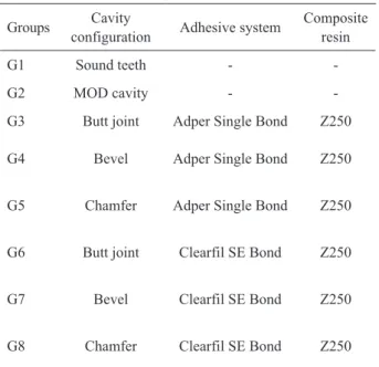
The aim of this study was to evaluate the fracture strength of teeth with different cavosurface margin cavity preparations and restored with composite resin and different adhesive systems. Eighty premolars were randomly divided in 8 groups, as follow: G1- sound teeth; G2- MOD preparation (no restoration); G3- Adper Single Bond without bevel preparation (butt joint); G4- Adper Single Bond with bevel preparation; G5- Adper Single Bond with chamfer preparation; G6- Clearfil SE Bond without bevel (butt joint); G7- Clearfil SE Bond with bevel preparation; G8- Clearfil SE Bond with chamfer preparation. The adhesive systems were applied according to manufacturers’ instructions. Composite resin (Filtek Z250) was incrementally placed in all cavities. After 24 h, the specimens were tested in a universal testing machine at a crosshead speed of 0.5 mm/min. Data were analyzed statistically by ANOVA and Tukey’s test (fracture strength) and Fisher’s exact test (fracture pattern). The confidence level was set at 95% for all tests. Prepared and non-restored teeth showed the worst performance and G4 exhibited the highest fracture strength among all groups (p<0.05). In conclusion, all restorative treatments were able to recover the fracture strength of non-restored teeth to levels similar to those of sound teeth. Using a total-etch adhesive system with bevel preparation significantly improved the resistance to fracture.
Key Words: bevel, adhesive systems, fracture strength, composite resins.
have a partial or total recovering of the fracture strength, depending on the type of adhesive system and restorative technique employed (4,5).
While bonding to enamel is more stable (6), bonding to dentin is more complex to achieve because it is a moist substrate with different regional composition (1,7). Beveling of the cavosurface margin increases enamel area, exposing a more favorable adhesion substrate (8). Beveling of cavity margins also provides stability to the adhesive interface and has been suggested to improve the restoration retention (9), avoid microleakage (8) and increase the fracture strength of restored teeth (10).
Different types of adhesive systems with different
surface treatments are available in the market, namely etch-and-rinse, self-etch primers and all-in-one systems (1). Within the range of these adhesive systems, different mechanism of actions will be made with the substrate, which will ultimately affect the restoration performance. Etch-and-rinse systems usually show better performance in relation to bond strength in enamel (11).
Stress produced by polymerization shrinkage and biodegradation can disrupt the adhesive interface, impairing marginal sealing and could lead to restoration failure (12,13). However, the presence of a bevel can prevent these adverse effects, sustaining bonding at the composite/enamel interface (8,14,10). Nevertheless, it is still unclear what type of bevel is preferred to improve restoration performance. While Coelho-de-Souza et al. (10) showed good results with bevel preparation in relation to fracture strength and marginal adaptation, Peixoto et al. (14) found that chamfer preparation improved marginal seal, and other studies (9,15) showed similarities between bevel or chamfer preparations.
The aim of this study was to evaluate the influence of marginal cavity preparation (bevel or chamfer bevel preparations) and adhesive systems (etch-and-rinse or self-etch) over the fracture strength of teeth restored with direct composite resin.
MATERIAL AND METHODS
Tooth Selection and Preparation
The research protocol had the approval of the Research Ethics Committee of the Lutheran University of Brazil (Protocol 098H). Eighty sound human premolars were selected. Following soft tissue removal, the teeth were stored in 10% formalin solution for 15 days. The inclusion criteria for the premolars were based on crown dimensions: 9.0-9.6 mm buccolingual distance; 7.0-7.4 mm mesiodistal distance and 7.7-8.8 mm cervico-occlusal distance (4). Finally, the teeth should be free of cracks under microscopy examination (10× magnification). The selected teeth were stored in distilled water at 37°C, which was periodically changed through the study.
The teeth had their roots embedded in a PVC matrix, using acrylic resin (Artigos Odontológicos Clássico Ltda., São Paulo, SP, Brazil), until 1 mm below of the cementoenamel junction. Ten premolars were not prepared (Group 1) and served as positive controls.
In the remaining teeth, standard Class II MOD
cavities were prepared. Diamond burs (#4137; KG Sorensen, Barueri, SP, Brazil) were mounted in a Galloni Machine (S. Colombano, Milano, Italy) to obtain a standardized cavity preparation. Burs were replaced after 2 cavity preparations to ensure high cutting efficiency. The occlusal box was 4-mm deep (without axial wall) and 2 mm in the buccolingual dimension. The cervical walls were located in enamel (1 mm above the cementoenamel junction). Teeth were randomly allocated to 7 different groups (n=10). For those teeth where a bevel preparation was done, a # 2135 (KG Sorensen) diamond bur was used to create a 45º bevel around the cavosurface angle, with 1 mm of extension. When a chamfer preparation was required, # 4137 (KG Sorensen) diamond burs were used to produce a rounded angle in the entire cavosurface region.
The cavities were restored with composite resin Filtek Z250 (3M ESPE, St. Paul, MN, USA), using either an etch-and-rinse (Adper Single Bond; 3M ESPE) or a self-etch adhesive system (Clearfil SE Bond; Kuraray, Tokyo, Japan), as described in Table 1.
The materials were used following manufacturers’ instructions, and the restorations were placed using an incremental technique. XL 3000 halogen unit (3M ESPE) with irradiance above 450 mW/cm², as constantly monitored by a curing radiometer (Curing Radiometer Model 100; Kerr/Demetron, Danbury, CT, USA), was used for light activation purposes.
Table 1. Experimental groups, type of cavity preparation, adhesive system and composite resin used.
Groups Cavity
configuration Adhesive system
Composite resin
G1 Sound teeth -
-G2 MOD cavity -
-G3 Butt joint Adper Single Bond Z250
G4 Bevel Adper Single Bond Z250
G5 Chamfer Adper Single Bond Z250
G6 Butt joint Clearfil SE Bond Z250
G7 Bevel Clearfil SE Bond Z250
After restoration conclusion, material excess was removed using #11 or 12 scalpel bladed and restorations were polished immediately using Enhance system (Dentsply, York, PA, USA) (16).
Fracture Strength
Specimens were tested after storage for 24 h in distilled water. Axial compression was performed in a universal testing machine (Pantec Versat 500, São Paulo, SP, Brazil) using an 8-mm metal sphere at a crosshead speed of 0.5 mm/min. Care was taken to maintain the sphere in contact with dental structure, without touching the restorative material. The fracture strength was reported in Newtons.
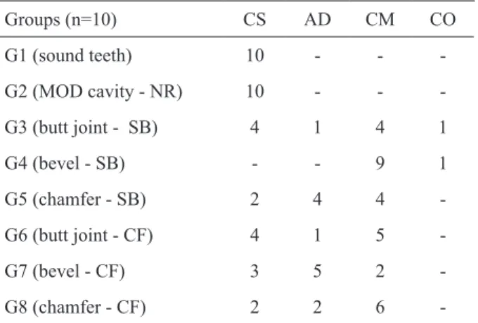
After fracture, the specimens were examined under a stereoscopic microscopy with 40× magnification (Kyowa, Tokyo, Japan) to evaluate the fracture patterns, as follows: cohesive fracture of the tooth - CS, adhesive fracture at the interface - AD, cohesive failure of the restorative material - CM, and complete fracture of the specimens involving the two cusps and the restorative material - CO.
Statistical Analysis
Fracture strength data were analyzed statistically by parametric tests (ANOVA and Tukey’s tests), while fracture pattern data were analyzed by non-parametric Fisher’s exact test. The confidence level was set at 95% for all tests.
RESULTS
Fracture Strength
Data from fracture strength test for the different groups are disclosed in Table 2.
G2 (MOD preparation - no restoration) and G4 (bevel - SB) had the lowest and the highest fracture strength values (p<0.05), respectively. All other groups (including G1 - sound teeth) had intermediate values, without significant differences from each other (p>0.05). The failure patters for the different groups are observed in Table 3.
The analysis of the failure patterns showed that more cohesive fractures were observed for G4 (bevel - SB) when compared to G5 (chamfer SB) (p=0.029). It was also observed that G7 (bevel - CF) had significantly more adhesive fractures than G4 (p=0.004).
DISCUSSION
The overall results of this study demonstrated that a bevel placed around the cavosurface margin together with the use of etch-and-rinse adhesive system produced the highest fracture strength, improving the resistance to values superior to those of the sound teeth.
Previous studies (3,4) have demonstrated a decreased fracture strength for prepared teeth due to the
Table 3. Fracture patterns observed for different groups after fracture resistance test.
Groups (n=10) CS AD CM CO
G1 (sound teeth) 10 - -
-G2 (MOD cavity - NR) 10 - -
-G3 (butt joint - SB) 4 1 4 1
G4 (bevel - SB) - - 9 1
G5 (chamfer - SB) 2 4 4
-G6 (butt joint - CF) 4 1 5
-G7 (bevel - CF) 3 5 2
-G8 (chamfer - CF) 2 2 6
-NR = No restoration; SB - Adper Single Bond; CF - Clearfil SE Bond; CS: cohesive fracture of the tooth; AD: adhesive fracture at the interface; CM: cohesive failure of the restorative material; CO: complete fracture of the specimens involving the two cusps and the restorative material.
Table 2. Fracture strength (N) for the different groups.
Groups (n=10) Mean ± SD
G1 (sound teeth) 1034.60 ± 247.96a
G2 (MOD cavity - NR) 320.30 ± 53.56c
G3 (butt joint - SB) 1021.20 ± 173.59a
G4 (bevel - SB) 1750.60 ± 175.99b
G5 (chamfer - SB) 919.20 ± 224.85a
G6 (butt joint - CF) 1044.70 ± 249.21a
G7 (bevel - CF) 909.10 ± 155.38a
G8 (chamfer - CF) 1051.50 ± 206.56a
cusp deflection increase and specially because of tooth structure removal (4,10). The findings of our study had confirmed these previous results.
In spite of the type of preparation, adhesive restorations improved the resistance to fracture to a level similar to the sound teeth and the resistance was significantly higher than that observed for the prepared and non-restored teeth. Corroborative results are present in the literature (4,10).
Bevel preparation brings several advantages to the restorative procedures: the removal of the aprismatic superficial enamel layer, which is also richer in fluoride content, favoring the acid etching; increasing the free surface energy, favoring surface wetting; enhancing the surface area of exposed enamel; providing better marginal seal; better esthetic results, making difficult to detect the interface; and improving the material retention (8,14,17). Bevel (8,10,17) and chamfer preparations (14,15) have previously been tested and both have been shown to improve the performance of restorations compared to non-beveled preparations. In the present study, non-beveled (butt joint) preparations exhibited similar fracture strength to that of cavities with beveled or chamfered margins, except for G4.
In a recent study, using direct and indirect composite restorations, Coelho-de-Souza et al. (10) observed an improved resistance to fracture when using beveled preparations compared to non-beveled preparations. These direct restorations were made with incremental technique, as well as in this study, according to the C-factor principles (12). In the present study, the bevel preparation combined with the etch-and-rinse adhesive system produced a resistance higher than that observed for sound teeth. Basically, the bevel preparation helped to produce a higher area of enamel in the restoration margin, which after acid etching could improve the fracture strength. However, this beneficial effect was not observed when the self-etch system was employed.
Moreover, the use of chamfer has not improved the performance of restorations compared to non-beveled preparations. When a bevel is prepared, the enamel prisms are exposed more favorably to the acid etching, and there will be a larger area in the cuspids covered by the composite. Peixoto et al. (14) showed that a chamfer preparation could improve the marginal seal compared to bevel preparation. Reis et al. (9) and Gandhi and Nandlal (15) found similar results for bevel and chamfer preparations in relation to restoration retention in anterior
teeth. Chamfered or beveled preparations restored with the self-etch adhesive system exhibited similar fracture strength in the present study.
Generally, the use of a self-etch system in this study showed a resistance to fracture similar to the use of an etch-and-rinse system. However, there was no significant improvement in fracture strength with the combination bevel preparation and self-etch system, differently from the combination bevel preparation and etch-and-rinse adhesive system. Probably, the reason is the absence of one separated conditioning step in the self-etch agent, and, consequently, this lack of conditioning did not additionally increase the bonding resistance (6). In a field-emission scanning electron microscopic study, it was observed deep interprismatic etching pattern for total-etch adhesive, whereas the self-etch systems resulted in an etching pattern ranging from absent to moderate. It was previously reported that commercial self-etch adhesives performed better on prepared enamel than on unprepared enamel. However, etch-and-rinse step made before a self-etch adhesive system use can really improve the bond strength to enamel (18).
In the present study, fracture pattern analysis showed more prevalence for composite cohesive fractures in G4 (bevel - SB), as reported elsewhere (10), this group presenting the highest fracture strength values. Bevel could improve bond strength in at enamel interface, changing the breakable point to composite resin. Moreover, the G5 (chamfer - SB) and G7 (bevel - CF) groups exhibited more adhesive failures, which could be associated to a smaller enamel area in G5 (6) and to the use of the self-etch system (G7) without etching and rinse step (1,19).
Bevel preparation is a simple, fast and safe step in cavity preparation, which produces beneficial effects for composite restorations (8,17,10,20). Based on the findings of the present study, the use of bevel associated with previous conditioning could be advocated as the outstanding strategy to improve the fracture strength of posterior (premolar) teeth. The use of bevel preparation with a total-etch adhesive systemin direct restorative procedures improved the restoration performance, in relation to fracture strength. However, this study is an in vitro experiment and the longitudinal clinical evaluation (20) remains the ideal parameter to ultimately determine the best restorative technique.
method to improve fracture strength; 2. All cavity preparation configurations were able to recover the fracture strength to values similar to those of sound teeth; 3. Self-etch or etch-and-rinse adhesive systems exhibited similar performances when used in butt joint and or chamfer preparations; 4. For the self-etch system, the cavity preparation did not influence the fracture strength.
RESUMO
O objetivo deste estudo foi determinar, in vitro, a resistência à fratura de pré-molares superiores com diferentes preparos do ângulo cavossuperficial e restaurados com resina composta com distintos sistemas adesivos. Foram selecionados 80 dentes, divididos em 8 grupos: G1- hígidos; G2- preparos M.O.D.; G3- Single Bond sem bisel; G4- Single Bond com bisel reto; G5- Single Bond com bisel chanfrado; G6- Clearfil SE Bond sem bisel; G7- Clearfil SE Bond com bisel reto e G8- Clearfil SE Bond com bisel chanfrado. Os grupos 3-8 foram restaurados com resina composta Z250, pela técnica incremental. Os corpos-de-prova foram submetidos ao teste de resistência à fratura em máquina universal de ensaios, a uma velocidade de 0,5 mm/min. Os dados foram analisados estatisticamente pelos testes ANOVA, Tukey e exato de Fisher (α=0,05). O grupo 2 apresentou resistência inferior aos demais e o grupo 4 mostrou-se mais resistente à fratura do que os demais grupos (p<0,05). Concluiu-se que o bisel reto foi o preparo mais efetivo quando associado a adesivos dentinários com condicionamento ácido prévio. Ambos adesivos dentinários usados foram capazes de devolver a resistência perdida com o preparo cavitário.
REfERENCES
1. Van Meerbeek B, De Munck J, Yoshida Y, Inoue S, Vargas M, Vijay P, et al.. Adhesion to enamel and dentin: current status and future challenges. Oper Dent 2003;28:215-235.
2. Pires-de-Souza FC, Brubi Filho B, Casemiro LA, Garcia LF, Consani S. Polymerization shinkage stress of composites photoativated by different light sources. Braz Dent J 2009;20:319-324.
3. Mondelli J, Steagall L, Ishikiriama A, Navarro MFL, Soares FB. Fracture strength of human teeth with cavity preparations. J Prosthet Dent 1980;43:419-422.
4. Habekost LV, Camacho GB, Azevedo EC, Demarco FF. Fracture resistance of thermal cycled and endodontically treated premolars with adhesive restorations. J Prosthet Dent 2007;98:186-192. 5. Morimoto S, Vieira GF, Agra CM, Sesma N, Gil C. Fracture
strength of teeth restored with ceramic inlays and overlays. Braz Dent J 2009;20:143-148.
6. Perdigão J, Gomes G, Lopes MM. Influence of conditioning time on enamel adhesion. Quintessence Int 2006;37:35-41.
7. Yang B, Ludwig K, Adelung R, Kern M. Micro-tensile bond strength of three luting resins to human regional dentin. Dent Mater 2006;22:45-56.
8. Opdam NJM, Roeters JJM, Kuijs R, Burgerdijk RCW. Necessity of bevels for box only class II composite restoration. J Prosthet Dent 1998;80:274-279.
9. Reis A, Francci C, Loguercio AD, Carrilho MRO, Rodrigues Filho LE. Re-attachment of anterior fractured teeth: fracture strength using different techniques. Oper Dent 2001;26:287-294. 10. Coelho-de-Souza FH, Camacho GB, Demarco FF, Powers J.
Fracture resistance and gap formation of MOD restoration: influence of restorative technique, bevel preparation and water storage. Oper Dent 2008;33:37-43.
11. Goracci C, Bertelli E, Ferrari M. Bonding to worn or fractured incisal edges: shear bond strength of new adhesive systems. Quintessence Int 2004;35:21-27.
12. Carvalho RM, Pereira JC, Yoshiyama M, Pashley DH. A review of polymerization contraction: the influence of stress development versus stress relief. Oper Dent 1996;21:17-24.
13. Sano H. Microtensile testing, nanoleakage and biodegradation of resin-dentin bonds. J Dent Res 2006;85:11-14.
14. Peixoto RT, Poletto LT, Lanza MD, Buono VT. The influence of occlusal finish line configuration on microleakage of indirect composite inlays. J Adhes Dent 2002;4:145-150.
15. Gandhi K, Nandlal B. Effect of enamel preparations on fracture resistance of composite resin build-up of fractures involving dentine in anterior bovine teeth: an in vitro study. J Indian Soc Pedod Prev Dent 2006;24:69-75.
16. Cenci MS, Venturini D, Pereira-Cenci T, Piva E, Demarco FF. The effect of polishing techniques and time on the surface characteristics and sealing ability of resin composite restorations after one-year storage. Oper Dent 2008;33:165-172.
17. Schmidlin PR, Wolleb K, Imfeld T, Gygax M, Lussi A. Influence of beveling and ultrasound application on marginal adaptation of box-only class II (slot) resin composite restorations. Oper Dent 2007;32:291-297.
18. Rotta M, Bresciani P, Moura SK, Grande RH, Hilgert LA, Baratieri LN, et al.. Effects of phosphoric acid pretreatment and substitution of bonding resin on bonding effectiveness of self-etching systems to enamel. J Adhes Dent 2007;9:537-545.
19. Moura SK, Pelizzaro A, Dal Bianco K, De Goes MF, Loguercio AD, Reis A, et al.. Does the acidity of self-etching primers affect bond strength and surface morphology of enamel? J Adhes Dent 2006;8:75-83.
20. Coelho-de-Souza FH, Klein-Jr CA, Camargo J, Beskow T, Blestrin M, Demarco FF. Double-blind randomized clinical trial of posterior composite restorations with and without bevel: 6-month follow-up. J Contemp Dent Pract 2010;11:1-7.