Shrimp diet and skin healing strength in rats
Dieta com camarão e resistência cicatricial
da pele, em ratos
Elizabeth Lage BORGES1
Fernanda Kelley Silva PEREIRA1
Jacqueline Isaura ALVAREZ-LEITE2
Luiz Ronaldo ALBERTI3
Mônica Alves Neves Diniz FERREIRA4
Andy PETROIANU3
A B S T R A C T
Objective
Surgical scar tensile strength may be influenced by several factors such as drugs, hormones and diet. The purpose of the present study was to determine the influence of a shrimp-enriched diet on the tensile strength of rat scars.
Methods
Forty male Wistar rats were submitted to a 4 cm dorsal skin incision and the wounds were sutured with 5-0 nylon interrupted suture. The animals were divided into two groups: Group 1 (control) received a regular diet, and Group 2 (experimental) received a shrimp-enriched diet. The two diets contained the same amounts of proteins, lipids and carbohydrates. The rats in each group were divided into two subgroups according to the time of assessment of the scar tensile strength: subgroup A, studied on the 5th postoperative day, and
subgroup B, studied on the 21st postoperative day. Results
The tensile strength of the scar on the 5th postoperative day was lower in the animals that received the shrimp-enriched-diet (303.0, standard error of mean= 34.1) than in the control group (460.1, SEM = 56.7) (p<0.05).
1
Universidade Federal de Minas Gerais, Instituto de Ciências Biológicas, Departamento de Fisiologia e Biofísica. Av. Antônio Carlos, 6627, Bloco A4, Sala 249, Pampulha, 31270-010, Belo Horizonte, MG, Brasil. Correspondência para/Correspondence to: E-mail: <[email protected]>.
2
Universidade Federal de Minas Gerais, Instituto de Ciências Biológicas, Departamento de Bioquímica e Imunologia. Belo Horizonte, Minas Gerais, Brasil.
3
Universidade Federal de Minas Gerais, Faculdade de Medicina, Departamento de Cirurgia. Belo Horizonte, Minas Gerais, Brasil.
4
Conclusion
A shrimp diet reduces the tensile strength of the scar. The next step of this study will be to clarify the mechanism in which shrimp affects tensile strength.
Indexing terms: diet; wound healing; eating; rats.
R E S U M O
Objetivo
A resistência cicatricial da pele pode ser influenciada por diversos fatores como medicamentos, hormônios e dieta. Este trabalho foi delineado para determinar a influência da dieta com camarão na resistência cicatricial na pele.
Métodos
Quarenta ratos machos Wistar foram submetidos a incisão (4cm) e suturas interrompidas da pele dorsal, com fio de nylon 5-0, e foram divididos em dois grupos: o Grupo 1 (controle) recebeu uma dieta convencional e Grupo 2 (experimental), recebeu dieta com adição de com camarão. As duas dietas continham quantidades semelhantes de proteína, lipídeos, e carboidratos. Os ratos de cada grupo foram divididos em dois subgrupos de acordo com os distintos períodos pós-operatórios de avaliação da resistência tecidual: subgrupo A, estudado no 5° dia pós-operatório, e subgrupo B, estudado no 21° dia pós-operatório.
Resultados
A resistência cicatricial da pele no 5° dia pós-operatório foi menor nos animais que receberam dieta suplementada com camarão (303,0, erro padrão da média=34,1), quando comparada ao grupo controle (460,1, erro padrão=56,7) (p<0,05).
Conclusão
A dieta suplementada com camarão reduziu a resistência cicatricial da pele de ratos. Dando continuidade ao estudo, será averiguado o mecanismo pelo qual ocorre essa redução.
Termos de indexação: dieta; cicatrização de feridas; ingestão de alimentos; ratos.
I N T R O D U C T I O N
Skin wound healing involves a cascade of cellular and molecular events in which biological processes such as proliferation, differentiation and
cell migration play pivotal roles1-5. The process may
be influenced by factors such as vitamin C,
hormones, diet, and systemic or local diseases6-9.
Some hormones and other mediators, mainly angiotensin II and angiotensin-(1-7), accelerate skin
repair by means of keratinocyte proliferation10. In
contrast, obstructive jaundice and glucocorticoids inhibit the healing of jejunal anastomoses and skin
wounds11.
Crustaceans are a common source of coastland diet and folk culture mentions that food based on crustaceans interferes with wound healing. However, no information is available regarding this topic in the scientific literature.
Therefore, a study of the influence of a shrimp--rich diet on skin healing should be relevant. The results of the present investigation showed that the addition of shrimp to the diet (33% of the diet) reduces the tensile strength of healing wounds.
M E T H O D S
Forty male Wistar rats weighing 210-236 g were housed individually in acrylic metabolic cages (Nalgene, Rochester, NY, USA) with free access
to food and water available ad libitum. The animals
were maintained under standard laboratory conditions of a 12/12-h light-dark cycle and temperature of 25°C.
based on Association of Official Analytical
Chemists (AOAC)12 with modifications, in order
to maintain the composition of both, experimental and control diets, and to avoid interferences on the results. The protein concentration of dried shrimp flour (made from shell as well as the flesh of the shrimp) was 33.5mg/100mg as determined
by the method of Lowry et al.13. Both diets contained
the same amounts of proteins, lipids and carbohydrates.
To determine if the salty taste of the shrimp diet induced an increase in food intake, in another experimental stage, we added salt to the regular diet so that it would contain the same amount of salt as the shrimp-enriched diet.
After a four-day adaptation period, the animals were randomly divided into two groups: Group1 (control) received a regular rat chow, and Group 2 (experimental) received a diet enriched 33% with shrimp. The rats of each group were divided into two subgroups according to the time
that the tensile strength of the scar was studied:
rats of subgroup A were investigated on the 5th
postoperative day and rats of subgroup B on the
21st postoperative day (times that are well
acknowledged in literature6-7). Food and water
intake were assessed daily during the entire experimental period.
Under general anesthesia with intraperitoneal thionembutal (40mg/kg), all rats were submitted to a 4cm incision in the dorsal thoracic skin. The wound was closed with four interrupted sutures using 5-0 nylon suture.
After 5 or 21 days, the rats were anesthetized (thionembutal 40mg/kg), the skin fragment containing the scar was cross-sectionally removed and the tensile strength of the scar was determined. Each skin segment was 3cm long and 1cm wide and included the scar in its middle part. The nylon suture was carefully removed to avoid damage to the skin and the two ends of the skin sample were lifted with two Duval clamps. One clamp was suspended and fixed on a support and the other was connected to a plastic container, which was filled with distilled water at the rate of 1.4 liter/minute. The tensile strength of the wound was estimated by the total weight of the plastic container, of one clamp and of the amount of
water at the time of scar rupture11.
Another wound sample was removed, fixed in Bouin solution, dehydrated and embedded in paraffin. Sections of 5 µm were prepared and stained with hematoxylin-eosin (HE) for light microscopy analysis. Other sections were stained with Picrosirius solution and examined by polarization microscopy in order to identify
collagen fibers14-15.
Serum sodium and potassium ion concentrations were assessed by flame photometry (FC-180, CELM, Brasil).
The present investigation was in agreement with the Ethical Principles in Animal Experimentation, adopted by the Declaration of Helsinki (2000) and by the Ethics Committee in Animal Experimentation (CETEA/UFMG). Table 1. Diet composition (%), nutrient contents (g/100g), and
energy density (kcal/g).
aComposition: 30mg niacin, 15mgpantothenic acid, 6mg vitamin B6, 5mg thiamine, 6mg riboflavin, 2mg folic acid, 750µg vitamin K, 200µg D-biotin, 25µg vitamin B12, 4000µg vitamin A, 1000µg vitamin D3, and
75µg vitamin E.
bComposition: essential minerals -35mg iron, 5000mg calcium, 1561mg
phosphate, 3600mg potassium, 300mg sulfur, 1019mg sodium, 1571mg chlorine, 507mg magnesium, 30mg zinc, 10mg manganese, 5mg copper, 0.2mg iodine, 0.15molybdenum, 0.15mg selenium, and potentially beneficial minerals -5mg silicone, 1mg chromium, 1mg fluorine, 0.5mg nickel, 0.1mg lithium, and 0.1mg vanadium.
Corn starch Casein Cellulose Soy bean oil Vitamin mixturea
Mineral mixtureb
Choline Dry-shrimp-flour Nutrient content Energy density 57 20 10 5 1 5 1 0
Protein 16.00 Lipid 5.00 Carbohydrate 52.00
3.25 46.9 09.1 01.0 03.0 01.0 05.0 01.0 33.0
Protein 18.33 Lipid 5.33 Carbohydrate 42.8
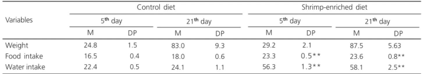
Table 2. Average food intake (g/day), water intake (mL), and weight of the animals (g) on the 5th and 21st postoperative days.
Weight Food intake Water intake
5th day
Control diet 24.8 16.5 22.4 1.5 0.4 0.5
Data are reported as means and standard error of mean Control (n=11) and Experimental group (n=12) on the 5th day. Control (n=8) and Experimental group (n= 9) on the 21st day. **p<0.001 compared with control.
Variables
M DP
21th day 83.0 18.0 24.1 9.3 0.6 1.1 M DP
5th day
Shrimp-enriched diet 29.2 23.3 56.3 2.1** 0.5** 1.3** M DP
21th day 87.5 23.6 58.1 5.63** 0.8**0 2.5**0 M DP
Table 3. Average food intake (g/day) and water intake (mL), on the 5th postoperative day.
Data are reported as means and standard error of mean. Control (n=4), and Experimental group (n= 4), on the 5th day. *p<0.05 compared with control.
Food intake Water intake
Variables Control diet (5
th day)
15.5 19.3
1.2 1.0
M DP
Salt-enriched diet (5th day) 17.1
28.8
0.4*
0.9*
M DP
Figure 1. Water volume necessary to cause scar rupture on the 5th (A) and 21st (B) postoperative days. Data are reported as means and standard error of mean. Control (n=11), and Experimental group (n=12), on the 5th day. Control (n=8), and Experimental group (n=9), on the 21st day. The rats that received the shrimp--enriched diet presented a lower tensile strength than the control group (p<0.05) on the 5th postoperative
day.
The results (mean and standard error of mean-SEM) obtained for the two groups were
compared by the Student t-test and Mann-Whitney
rank sum test, with the level of significance set at
p<0.05.
R E S U L T S
The animals did not present any abnormality during the experimental period. No sign of toxicity was verified.
Table 2 shows total food intake, water
intake, and weight of the animals on the 5th and
21st postoperative days. Food and water intake
were higher in the group that received the
shrimp--enriched diet (p<0.001). No difference in body
weight was observed between the groups (Table 2).
The tensile strength of the skin segment from the two groups is presented in Figure 1 A and B. The skin of the rats that received the shrimp--enriched diet demonstrated a lower tensile strength
on the 5th postoperative day (p<0.05) (Figure 1A).
Table 3 shows the total food and water intake, and weight of the animals that received a
salt-enriched diet on the 5th postoperative day. The
salt-enriched diet did not affect food intake.
1000 800 600 400 200 0 Ruptur
e tension (g)
A
*
Control5thday Experimental day 5th
Ruptur
e tension (g)
3000 2500 2000 1500 1000 500 0 B
Control21thday
Serum sodium and potassium levels (mM) did not differ significantly between the two groups
on the 5th (n=7) and 21st (n=4) days: serum sodium
on the 5th postoperative day (125.9, SEM=1.7
versus 127.6, SEM=2.7) and on the 21st day (121.8,
SEM=0.8 versus 119.8, SEM=0.4); serum
potassium on the 5th postoperative day (6.0,
SEM=0.1 versus 6.2, SEM=0.1) and on the 21st
day (6.0, SEM=0.3 versus 6.5 and 0.2). Data are
reported as means and SEM. The two groups were homogeneous after being submitted to the specific
diets (control vs. experimental shrimp-enriched
diet).
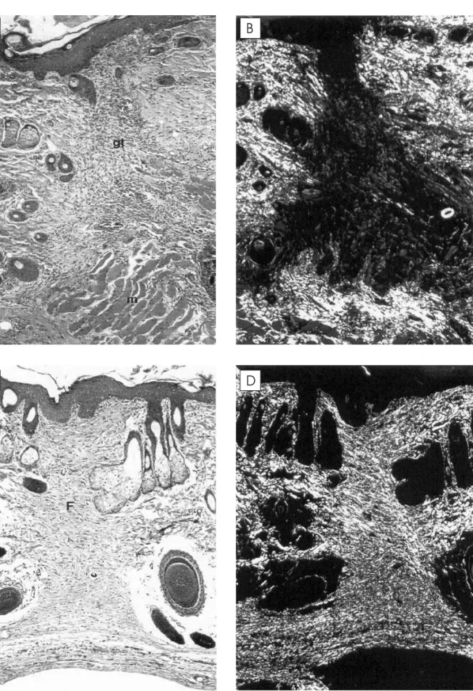
No histological difference was found between the groups (control and experimental)
on the 5th and 21st postoperative days. On the 5th
postoperative day, the dermis showed loose connective tissue fibers, neovascularization and
granulation tissue (Figure 2A). On the 21st day,
the healing tissue was mature and fibrous with a less intense inflammatory reaction (Figure 2C).
Both groups presented type III collagen on the 5th
postoperative day, but type I fibers were predominant after 21 days (Figures 2B and D).
D I S C U S S I O N
In the healing process, fine and disorganized
collagen fibers appear first (type III)16-17, being then
replaced with thicker fibers, with the progressive occurrence of organization of type I collagen. Collagen type, more than the amount of collagen fibers, is important in maintaining the strength of
healed tissue17. The present findings agree with
literature data showing the occurrence of type III collagen in granulation tissue and its later replacement with type I collagen in the fibrous tissue of the mature scar. This replacement provides more resistance to mechanical tension.
The hypothesis of increased food intake
induced by the salty taste of the shrimp18 was not
verified in this investigation. The consequent enhancement of water consumption observed in
tables 2 and 3 for the shrimp-enriched diet and salt-enriched diet respectively, suggests an osmotic pressure regulation. The rats of the salt-enriched diet group did not increase the food intake, and no ionic alteration occurred in either group with the shrimp-enriched diet and its control.
Shrimp is also rich in chitin fiber, increasing the total amount of feces in the group that ate shrimp; unfortunately this was only observed, not measured. Chitin is called an animal fiber because of its low digestibility in the animal gastrointestinal tract. However, the percentage of cellulose (10%) was reduced in the shrimp diet to compensate the percentage of chitin present in it. Although the total energy of the two diets differs in about 9%, the result suggests that the increased intake of the shrimp-enriched diet was also due to the reduced energy density of the diet caused by the animal fiber. The animals tried to compensate the lower energy value of this diet by consuming more food.
In conclusion, these data suggest that, even though the groups receiving the two diets ingested the same amount of energy, the shrimp-enriched diet reduced the tensile strength of the scar of the animals, in agreement with folk belief. The next step of this study will be to clarify the mechanism in which shrimp affects tensile strength.
A C K N O W L E D G M E N T S
Fernanda K.S. PEREIRA was sponsored by Conselho Nacional de Desenvolvimento Científico e Tecnológico (CNPq).
C O N T R I B U T O R S
R E F E R E N C E S
1. Ortonne JP, Clevy JP. Physiology of cutaneous cicatrisation. Rev Prat. 1994; 44(13):1733-7. 2. Kirsner RS, Eaglstein WH. The wound healing
process. Dermatol Clin. 1993; 11(4):629-40. 3. Hunt TK, Hopf H, Hussain Z. Physiology of wound
healing. Adv Skin Wound Care. 2000; 13(2 Suppl): 6-11.
4. Yamaguchi Y, Yoshikawa K. Cutaneous wound healing: an update. J Dermatol. 2001; 28(10): 521-34.
5. Scheithauer M, Riechelmann H. Review part I: basic mechanisms of cutaneous wound healing. Laryngorhinootologie. 2003; 82(1):31-5. 6. Petroianu A, Souza SD, Martins SG, Alberti LR,
Vasconcellos LS. Influência da vitamina C e da hidrocortisona sobre a tensão anastomótica jejunal em ratos. Acta Cir Bras. 2000; 15(4):215-9. 7. Petroianu A, Souza SD, Martins SG, Alberti LR. Influence of ascorbic acid on anastomosis and in jejunal loop in rat. Arq Gastroenterol. 2001; 38(1):48-52.
8. Arantes VN, Okawa RY, Silva AA, Barbosa AJ, Petroianu A. Effect of methylprednisolone on jejunal anastomotic tension. Arq Gastroenterol. 1994; 31(3):97-102.
9. Werner S, Grose R. Regulation of wound healing by growth factors and cytokines. Physiol Rev. 2003; 83(3):835-70.
10. Rodgers K, Xiong S, Felix J, Roda N, Espinoza T, Maldonado S, et al. Development of angiotensin (1-7) as an agent to accelerate dermal repair. Wound Repair Regen. 2001; 9(3):238-47. 11. Arantes VN, Okawa RY, Fagundes-Pereyra WJ,
Barbosa AJA, Petroianu A. Influence of obstructive
jaundice on wound and jejunal anastomosis healing in rats. Rev Col Bras Cir. 1999; 26(5): 269-73.
12. Cunnif P, editor. Official Methods of Analysis of AOAC International. 16th ed. Arlington, Virginia: AOAC; 1995. Chapter 45.
13. Lowry OH, Rosebrough NJ, Farr AL, Randall RJ. Protein measurement with the Folin phenol reagent. J Biol Chem. 1951; 193(1):265-75. 14. Junqueira LC, Cossermelli W, Brentani R.
Differential staining of collagens type I, II and III by sirius red and polarization microscopy. Arch Histol Jpn. 1978; 41(3):267-74.
15. Junqueira LC, Bignolas G, Brentani RR. Picrosirius staining plus polarization microscopy, a specific method for collagen detection in tissue sections. Histochem J. 1979; 11(4):447-55.
16. Andrade GB, Montes GS, Conceição GM, Saldiva PH. Use of the Picrosirius-polarization method to age fibrotic lesions in the hepatic granulomas produced in experimental murine schistosomiasis. Ann Trop Med Parasitol. 1999; 93(3):265-72. 17. Rabau MY, Hirshberg A, Hiss Y, Dayan D. Intestinal
anastomosis healing in rat: collagen concentration and histochemical characterization by Picrosirius red staining and polarizing microscopy. Exp Mol Pathol. 1995; 62(3):160-5.
18. Yamasaki K, Marubayashi U, Reis AM, Coimbra CC. Preferential saline or water intake by pinealectomized, adrenalectomized, and pinealectomized-adrenalectomized male rats. Braz J Med Biol Res. 1990; 23(11):1177-80.
Received on: 30/11/2005