260 261
260 261
Effect of irradiation mode and filling technique on resin/dentin
bonding strength in class I cavities
Efeito do modo de ativação e da técnica restauradora na
resistência de união entre compósito e dentina em cavidades
classe I
Alex José Souza dos Santos* Marcelo Giannini**
Luis Alexandre Maffei Sartini Paulillo*** José Roberto Lovadino****
Ricardo Marins de Carvalho*****
ABSTRACT: Factors such as light-curing mode, filling technique and cavity configuration may affect the bonding strength to dentin. This study evaluated the effect of irradiation mode and filling technique on resin/dentin bond-ing strength on the buccal wall of class I cavities in human teeth. Occlusal enamel was removed to expose a flat dentin surface. Occlusal cavities (4 x 3 x 3 mm) were prepared in dentin. The adhesive Single Bond was applied according to the manufacturer’s instructions and TPH Spectrum composite resin was placed using the follow-ing: oblique incremental, horizontal incremental or bulk filling techniques. The composite resin was light-cured either by continuous (600 mW/cm² for 40 s) or Soft-Start (250 mW/cm² for 10 s + 600 mW/cm² for 30 s) modes. Specimens of the control group were obtained by bonding the material to the flat exposed buccal wall of the cavity (C-factor = 1). The teeth were stored in water at 37oC for 24 h and prepared for microtensile testing. Bonded beams
of approximately 0.8 mm² were obtained from the buccal wall and tested with a tension of 0.5 mm/min. Results were analyzed by two-way ANOVA, Tukey’s test and Dunnett’s test (α = 0.05). Incremental placement techniques with both irradiation modes produced higher bonding strength values than the bulk technique (p < 0.05). Bond-ing strength tested in the cavities had lower values than those obtained in flat dentin surfaces (control group) (p < 0.05), except for incremental fillings using stepped irradiation. Bonding strength to the cavity walls depends on the filling technique and on the irradiation mode of composite resins.
DESCRIPTORS: Composite resins; Dentin; Dental cements; Dental enamel.
RESUMO: Fatores como o modo de ativação, a técnica restauradora e a configuração cavitária podem afetar a re-sistência adesiva à dentina. Este estudo avaliou os efeitos dos modos de ativação e das técnicas restauradoras na resistência de união compósito/dentina na parede vestibular de restaurações classe I. O esmalte oclusal dos den-tes foi removido para expor uma superfície dentinária planificada. Cavidades oclusais (4 x 3 x 3 mm) foram prepa-radas em dentina. O adesivo Single Bond foi aplicado de acordo com as instruções do fabricante, e o compósito TPH Spectrum, inserido através de três diferentes técnicas: oblíqua incremental, horizontal incremental ou incremento único. O compósito foi ativado utilizando o método contínuo (600 mW/cm² por 40 s) ou “Soft-Start” (250 mW/cm² por 10 s + 600 mW/cm² por 30 s). O grupo controle foi obtido pela união somente à parede vestibular planificada (fator C = 1). Os dentes foram armazenados por 24 h em água a 37oC e preparados para o ensaio de
microtra-ção (0,5 mm/min). Os espécimes apresentaram na secmicrotra-ção transversal uma área de união de aproximadamente 0,8 mm². Os resultados foram analisados pela ANOVA (dois fatores), pelos testes de Tukey e Dunnett (α = 0,05). A técnica incremental seguida da ativação com ambas as formas de fotopolimerização produziu maiores valores de resistência de união que a técnica de incremento único (p < 0,05). A resistência de união sempre foi menor quando a restauração foi confeccionada em cavidade (p < 0,05), exceto quando foram utilizadas ativação com “Soft-Start” e técnicas incrementais. A resistência de união às paredes cavitárias é dependente da técnica de inserção e do modo de ativação da resina composta.
DESCRITORES: Resinas compostas; Dentina; Cimentos dentários; Esmalte dentário.
* PhD Student; **Assistant Professor; ***Associate Professor ****Chairman, Professor – Department of Restorative Dentistry, School of Dentistry of Piracicaba, State University of Campinas.
260 261
260 261
INTRODUCTION
Composite resins have been widely used in restorative and esthetic dentistry as direct restor-ative materials. However, the restorrestor-ative technique in three-dimensional cavities consists of sensitive steps such as adhesive application, composite in-sertion and photo-activation techniques2,18.
Despite recent development in resin-based restorative materials, the amount of shrinkage reported still poses a clinically relevant problem. The monomers used in restorative dentistry are methacrylate- or acrylate-based, and they under-go shrinkage during polymerization. If the stress resulting from composite shrinkage exceeds the bonding strength to the cavity wall, marginal fail-ures can occur due to disruption of the composite-tooth adhesive interface4,7,19. If bonded interfaces
remain intact, residual forces might transfer stress to adjacent tooth structures, possibly resulting in enamel fractures9,23.
Bonding failure is a common cause of replace-ment of composite resin restorations. Studies have reported that high values of contraction stress for specific configurations of cavity preparation can af-fect the integrity of the restoration-cavity interface. This might explain the large number of early bond failures4,6,15. Different filling and photo-activation
techniques have been developed in an attempt to reduce the shrinkage potential and produce mini-mal interfacial stress development2,18.
Soft-start polymerization or stepped light in-tensity uses an initial low-output inin-tensity of light followed by a higher intensity of light13,22, and
in-cremental insertion of composite resin reduces the volume of the restorative material that shrinks un-der photo-activation8,12,17. Both methods have been
advocated because they minimize stress generated during polymerization and improve the marginal sealing and cavity wall adaptation of restorations18.
The purpose of this study was to evaluate the in-fluence of two irradiation modes and three fill-ing techniques on the tensile bond strength of a composite resin to the dentin of the buccal wall of box-like class I cavities, using the microtensile bond test. The null hypothesis tested was that ir-radiation modes and filling techniques do not affect the tensile bonding strength.
MATERIALS AND METHODS
Cavity preparationThirty-two sound human third molars that had been refrigerated in a solution of 0.05% thymol
(LabSynth, Diadema, Brazil) for no longer than two months after extraction were cleaned of gross debris and placed in distilled water for twenty-four hours before beginning the experiment. The teeth used in this study were obtained under the proto-col 039/2002, which was analyzed and approved by the Ethical Committee in Research, School of Dentistry of Piracicaba, State University of Campi-nas.
The occlusal enamel was removed using a diamond saw (Isomet, Buehler Ltd., Lake Bluff, USA), under water-cooling, to expose superficial dentin parallel to the occlusal surface (Figure 1A). Standardized uniform box-shaped class I cavities were prepared with a precision cavity preparation device on flat occlusal surfaces of twenty-four teeth. The preparations were outlined with diamond burs (#2143 - KG Sorensen Ind. e Com. Ltda., Barueri, Brazil) using a high-speed handpiece (Extra-Torque 605, Kavo do Brasil S.A., Joinville, Brazil) and copious air-water spray (Figure 1B). The bucco-lingual width of the preparations was 4 mm, the mesio-distal width was 3 mm and the pulpal floor was prepared to a depth of 3 mm (Figure 1C).
Eight teeth from the control group (corre-sponding to flat buccal dentin surfaces) were pre-pared the same way, but cavities were enlarged after preparation (Figure 1D). The cavity prepara-tion device was adjusted to remove all surround-ing dental structures, leavsurround-ing a flat buccal dentin area (3 mm x 4 mm) with the same texture for the bonding procedures.
Restorative procedures
Single Bond (3M/ESPE, St. Paul, USA) was applied according to the manufacturer’s instruc-tions and TPH Spectrum composite resin (Dent-sply Caulk, Milford, USA) was placed either using oblique incremental, horizontal incremental or bulk filling techniques. For the oblique incremental technique, the composite resin was applied to the cavity and cured in three oblique increments, as suggested by Pollack17 (1987) in class II cavities.
Each layer was light cured for 40 s from the oc-clusal surface.
262 263
262 263
of the control group and cured for 40 seconds (each layer).
The restorative composite resin was light-cured (Degulux Soft-Start, Degussa, Hanau, Ger-many) either by continuous (600 mW/cm² for 40 s) or stepped (250 mW/cm² for 10 s + 600 mW/cm² for 30 s) irradiation modes. The intensity of 250 mW/cm² was obtained with the light tip 13 mm distant from the resin, and the 600 mW/ cm² intensity with the tip touching the restorative material. The light intensity was measured using a radiometer (Demetron Corp., Danbury, USA).
Micro-tensile testing
After 24 h of storage in water at 37oC, the
re-stored specimens were buccal-lingually and me-sio-distally serially sectioned (Figure 2A and 2B) with a saw (Isomet, Buehler Ltd., Lake Bluff, USA) resulting in bonded beams with a cross-sectional area of approximately 0.8 mm² (Figure 2C). Three or four specimens were selected from each restored teeth and tested in a universal testing machine (4411, Instron Co., Canton, USA) at 0.5 mm/min until failure in the bonding system occurred (Fig-ure 2D).
The cross-sectional area for each tested speci-men was measured to the nearest 0.01 mm with a digital caliper (727-6/150, Starret, Itu, Brazil) to
A
C
Cavity preparation for experimental groups
D Cavity preparation for
control groups Enamel
Dentin
B
FIGURE 1 - Schematic representation of cavity preparation. (A) Removal of occlusal enamel; (B) cavity preparation using a diamond bur and a high-speed handpiece; (C) prepared class I cavity for bulk or incremental fillings; (D) control group cavity design with a flat buccal dentin surface for bonding. calculate the tensile bond strength and express results in MPa. Results were analyzed by two-way ANOVA (irradiation mode and filling technique), Tukey’s test and Dunnett’s test at the 0.05 level of significance. All statistical analyses were per-formed using SAS for personal computers (SAS Institute, Cary, NC, USA).
RESULTS
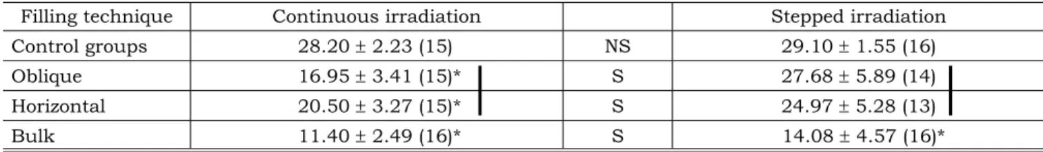
Two-way ANOVA (Table 1) indicated that there were statistically significant differences for the vari-ables filling technique (p = 0.0002) and irradiation mode (p = 0.0034), but failed to identify any inter-action between variables (F = 1.91; p = 0.1766).
The mean tensile bonding strength and standard deviation values are shown in Table 2. Tukey’s test showed that the bulk filling technique presented significantly lower bonding strength val-ues than the incremental filling techniqval-ues (hori-zontal and oblique), using both irradiation modes (p < 0.05). The photo-activation with stepped light intensity resulted in significantly higher bonding strength values than the conventional continu-ous irradiation for all filling techniques, except for control groups (p < 0.05).
incre-262 263
262 263
mentally placed in three-dimensional cavities and cured with the stepped light intensity mode were similar to that of controls groups, which were re-stored in flat surfaces (p > 0.05).
DISCUSSION
During the curing process, the time when the resin polymer develops a higher elastic modulus is called the gel point. Before this point and dur-ing the early phase of photo-polymerization, the polymer is in a flexible and fluid state. In the pre-gel phase, no tension is generated at the tooth/ composite interface, because the stress can be relieved by the composite flow. After the formation of a more rigid structure with higher modulus (gel-point), the strain developed during further cure is transmitted to the bonded interface and adjacent tooth structures if contraction of the material is hindered by adhesion to the walls of three-dimen-sional cavities4,7,19.
Reduction of the shrinkage potential of com-posite resins by manufacturers and restorative techniques used by dentists are fundamental to avoid resin-based restorations with interfacial stress development, which compromises their longevity18. This study tested filling restorative
techniques and irradiation modes to overcome
the stress developed during the curing process of composite resins in class I cavities.
The soft-start or step-curing irradiation mode comprises the application of low-intensity light during initial phases of the curing process, followed by final cure with full intensity. This can provide time for the composite flow to occur, minimizing the shrinkage effects. Reduction in light intensity promotes reduction in the number of free radicals formed, lowering the overall rate of polymerization and allowing shrinkage strain in the polymer to be relieved by increased flow capability of the mate-rial18. Afterwards, high light intensity is applied to
complete the polymerization process and provide proper mechanical properties10,25.
Since the volumetric contraction of the com-posite can cause debonding forces and a part of this stress can be compensated during the initial phase of polymerization by the flow of the dental composite3,4, the extending in time of the pre-gel
phase with stepped irradiation can preserve the marginal integrity of the restoration10,13,22,26 due
to a reduction in the polymerization contraction stress5,11,24.
The stepped irradiation mode used with incre-mental filling techniques to restore class I cavities provided bonding strength values similar to those of the control group. The initial low-intensity light may avoid the detrimental shrinkage effects on the
Specimens C
Composite resin B
AComposite resin
Micro-tensile bonding test
Composite resin D
Dentin
Enamel
264 265
264 265
TABLE 1 - Two-way analysis of variance.
Source DF Sum of squares Mean square F value p > F
Filling technique 2 511.63 255.81 13.70 0.0002*
Irradiation mode 1 213.01 213.01 11.41 0.0034*
Interaction 2 71.40 35.70 1.91 0.1766
*Statistical significance at the level of 5%. DF: degrees of freedom.
bonded interface, producing bonding strength val-ues similar to those obtained on flat surfaces and without cavity configuration influence. Both incre-mental methods provide higher bonding strength values than the bulk technique. However, when continuous irradiation was used, they did not match control group values.
Two factors may have contributed to the lower bonding strength values obtained with the bulk filling technique: increased polymerization con-traction stress due to great volume of composite and decreased effectiveness of polymerization at deeper portions of the composite8. The competition
between the composite-dentin bonding strength and the polymerization contraction stress can be avoided, inserting composite resin in increments to reduce the volume of the resin and the stress generated on the cavity walls1,12,15,17.
In this study, the orientation of the dentin-al tubules on the buccdentin-al wdentin-all was pardentin-allel to the bonded interface. Studies have shown that the bonding strength of the resin is not uniform inside a cavity, and the direction of the dentinal tubules appears to be an important variable regarding the formation of a hybrid layer in dentin16,21. The
in-fluence of tubule direction can result in higher bonding strength values for bonding performed with tubules oriented parallel to the surface14. The
microtensile method allowed the measurement of bonding strength at small bonded areas and at a specific wall of a three-dimensional cavity20.
The class I cavity design was chosen because it resembles clinical situations with complex cav-ity preparation and restoration; and the results showed that the shrinkage potential of the com-posite and the cavity configuration factor affected the bonding strength. In flat surfaces, the large free surface allows resin to flow during the cur-ing process, not compromiscur-ing the bondcur-ing as observed in the control group27. Negative effects
on the restoration-cavity interfaces promoted by forces of polymerization contraction can be con-trolled to some extent by using stepped irradiation and incremental filling restorative techniques in clinical restorations. Thus, the results lead us to reject the null hypothesis, because the irradiation mode and incremental filling techniques did affect the bonding strength to the dentin of the buccal wall of box-like class I cavities.
CONCLUSIONS
According to the methodology employed and based on the obtained results and on the statisti-cal analysis, it can be concluded that:
1. Soft-start or step-curing irradiation mode ap-plied following incremental techniques can improve bonding strength values of composite resin to buccal wall dentin of box-like class I cavities.
2. Bulk placement of composite resins is not recommended as a restorative procedure for dental cavities.
TABLE 2 - Mean tensile bond strength values in MPa ± standard deviations (N).
Filling technique Continuous irradiation Stepped irradiation
Control groups 28.20 ± 2.23 (15) NS 29.10 ± 1.55 (16)
Oblique 16.95 ± 3.41 (15)* S 27.68 ± 5.89 (14)
Horizontal 20.50 ± 3.27 (15)* S 24.97 ± 5.28 (13)
Bulk 11.40 ± 2.49 (16)* S 14.08 ± 4.57 (16)*
264 265
264 265
ACKNOWLEDGMENTS
The authors are indebted to the Laboratory of Dental Materials (School of Dentistry of Piracicaba, State University of Campinas – FOP-UNICAMP)
13. Mehl A, Hickel R, Kunzelmann KH. Physical properties and gap formation of light-cured composites with and with-out softstart-polymerization. J Dent 1997;25:321-30. 14. Ogata M, Okuda M, Nakajima M, Pereira PNR, Sano
H, Tagami J. Influence of the direction of tubules on bond strength to dentin. Oper Dent 2001;26:27-35.
15. Opdam NJM, Feilzer AJ, Roeters JJM, Smale I. Class I occlusal composite resin restorations: in vivo post-operative sensitivity, wall adaptation, and microleakage. Am J Dent 1998;11:229-34.
16. Phrukkanon S, Burrow MF, Tyas MJ. The effect of den-tine location and tubule orientation on the bond strengths between resin and dentine. J Dent 1999;27:265-74. 17. Pollack BF. Class II composites: 1987 thoughts and
techniques. NY State Dent J 1987;53:25-7.
18. Rueggeberg F. Contemporary issues in photocuring. Compend Contin Educ Dent 1999;Suppl:4-15.
19. Sakaguchi RL, Berge HX. Reduced light energy density decreases post-gel contraction while maintaining degree of conversion in composites. J Dent 1998;26:695-700. 20. Sano H, Shono T, Sonoda H, Takatsu T, Ciucchi B,
Carvalho R, et al. Relationship between surface area for adhesion and tensile bond strength – evaluation of a micro-tensile bond test. Dent Mater 1994;10:236-40.
21. Schüpbach P, Krejci I, Lutz F. Dentin bonding: effect of tubule orientation on hybrid-layer formation. Eur J Oral Sci 1997;105:344-52.
22. Uno S, Asmussen E. Marginal adaptation of a restor-ative resin polymerized at reduced rate. ScandJ Dent Res 1991;99:440-4.
23. van Dijken JW, Horstedt P, Waern R. Directed polym-erization shrinkage versus a horizontal incremental filling technique: interfacial adaptation in vivo in class II cavities. Am J Dent 1998;11:165-72.
24. Watts DC, al Hindi A. Intrinsic soft-start polymeriza-tion shrinkage-kinetics in an acrylate-based resin-compos-ite. Dent Mater 1999;15:39-45.
25. Yap AUJ, Ng SC, Siow KS. Soft-start polymerization: influence on effectiveness of cure and post-gel shrinkage. Oper Dent 2001;26:260-6.
26. Yoshikawa T, Burrow MF, Tagami J. A light curing method for improving marginal sealing and cavity wall adaptation of resin composite restorations. Dent Mater 2001;17:359-66.
27. Yoshikawa T, Sano H, Burrow MF, Tagami J, Pashley DH. Effect of dentin depth and cavity configuration on bond strength. J Dent Res 1999;78:898-905.
Received for publication on Jan 14, 2004 Accepted for publication on Jun 18, 2004
REFERENCES
1. Ben-Amar AR, Liberman R, Nordenberg D, Metzger Z. The effect of retention grooves on gingival marginal leakage in class II posterior composite resin restorations. J Oral Rehabil1988;15:325-31.
2. Carvalho RM, Pereira JC, Yoshiyama M, Pashley DH. A review of polymerization contraction: the influence of stress development versus stress relief. Oper Dent 1996;21:17-24.
3. Davidson CL, de Gee AJ. Relaxation of polymerization con-traction stress by flow in dental composites. J Dent Res 1984;63:146-8.
4. Davidson CL, de Gee AJ, Feilzer AJ. The competition be-tween teeth composite-dentin bond strength and the po-lymerization contraction stress. J Dent Res 1984;63:1396-9.
5. Ernst CP, Kürschner R, Rippin G, Willershausen B. Stress reduction in resin-based composites cured with a two-step light-curing unit. Am J Dent 2000;13:69-72.
6. Feilzer AJ, de Gee AJ, Davidson CL. Setting stress in com-posite resin in relation to configuration of the restoration. J Dent Res1987;66:1636-9.
7. Feilzer AJ, Dooren LH, de Gee AJ, Davidson CL. Influence of light intensity on polymerization shrinkage and integrity of restoration-cavity interface. Eur J Oral Sci 1995;103:322-6.
8. Figueiredo Reis A, Giannini M, Ambrosano GM, Chan DC. The effects of filling techniques and a low-viscosity com-posite liner on bond strength to class II cavities. J Dent 2003;31:59-66.
9. Kanca J 3rd, Suh BI. Pulse activation: reducing resin-based
composite contraction stresses at the enamel cavosurface margins. Am J Dent 1999;12:107-12.
10. Koran P, Kürschner R. Effect of sequential versus continuous irradiation of a light-cured resin composite on shrinkage, viscosity, adhesion and degree of polymeriza-tion. Am J Dent 1998;10:17-22.
11. Lim BS, Ferracane JL, Sakaguchi RL, Condon JR. Reduction of polymerization contraction stress for den-tal composites by two-step light-activation. Dent Mater 2002;18:436-44.
12. Lutz E, Krejci I, Oldenburg TR. Elimination of polym-erization stresses at the margins of posterior composite resin restorations: a new restorative technique. Quintes-sence Int 1986;17:777-84.