Correspondence: Nevreste Çelikbilek, Ankara Atatürk Training and Research Hospital, Ankara, Turkey E-mail: [email protected]
Received: Received: 24 November 2014, Accepted: 28 March 2015 Copyright © Journal of Microbiology and Infecious Diseases 2015, All rights reserved
JMID doi: 10.5799/ahinjs.02.2015.02.0178
R E S E A RC H A RT I C L E
Evaluation of Anti-Nuclear antibody test results in clinical practice
Nevreste Çelikbilek1, Birsen Özdem1, Ziya Cibali Açıkgöz2
1 Ankara Atatürk Training and Research Hospital, Ankara, Turkey
2 Yıldırım Beyazıt University Medical Faculty, Ankara, Turkey
ABSTRACT
Objective: Aim of this study is to evaluate anti-nuclear antibody (ANA) test results obtained between 2009 and 2011.
Methods: Of a totally 5068 cases tested for ANA by indirect immunoluorescence method (IIFA), randomly chosen
982 ANA-positive cases were reviewed in terms of gender, level and pattern of luorescence, anti-dsDNA (anti-double stranded DNA) and anti-extractable nuclear antigen (ENA) proile. Anti-dsDNA levels and anti-ENA proiles were deter -mined by enzyme linked immune assay (ELISA) and immune-blotting (IB), respectively.
Results: Sex distribution of ANA positive patients was determined as 756 (77%) females and 226 (23%) males. Fifty per
cent of the cases were from rheumatology department, 20% from gastroenterology and 30% from other units. Fluo -rescence levels were considered borderline or weak positive in 62.6% of the samples. The most frequent patterns were homogeneous (23%), speckled (22%), homogeneous-speckled (15.5%) and nucleolar (13.5%). Anti-dsDNA were studied in 759 ANA positive patients and 66 (8.7%) samples were found positive, being 44 of them (68.8%) with homogeneous pattern and the rest with speckled, nucleolar, nuclear dots, centromeric or midbody patterns. Totally 131 (31.6%) of 414 samples studied for anti-ENA proile were found positive. The irst four frequent proiles were SSA (34.4%), SSA-SSB (16.8%), Scl70 (16%) and Sm/RNP (9.2%).
Conclusion: Our results are similar with the current related literature. It is known that autoantibodies can be detectable before clinical symptoms being apparent, especially in SLE. Therefore, borderline or weak luorescence levels should also be reported and the patients having them should be followed-up carefully. J Microbiol Infect Dis 2015;5(2): 63-68
Key words: Antinuclear antibody, indirect immune-luorescence assay, extractable nuclear antigen antibodies
Anti-nükleer antikor test sonuçlarının klinik uygulamada değerlendirilmesi
ÖZET
Amaç: Bu çalışmanın amacı 2009-2011 yıllarında laboratuvarımızda yapılan ANA (antinükleer antikor) tetkik sonuçları -nın retrospektif olarak değerlendirilmesidir.
Yöntemler: Bu dönemde laboratuvarımızda toplam 5068 serum örneğinde indirekt immünoloresans antikor yöntemiy -le (IIFA) ANA varlığı araştırıldı. Rastge-le örnek-leme i-le seçi-len ANA-pozitif 982 olgu cinsiyet dağılımı, ışıma düzey-leri ve paternleri, dsDNA sonuçları ve ekstrakte edilebilir nükleer antijen (ENA) proilleri açısından incelendi. ANA ölçümü için doku olarak HEP-2 ve maymun karaciğeri hücrelerini birlikte içeren ticari IIFA kiti kullanıldı. Hasta serumlarının 1/100 su -landırım titresi ile çalışıldı. Sonuç verilirken ışıma titresi ve derecesi ile birlikte homojen, benek, sentromer gibi paternler de rapor edildi. Anti-dsDNA düzeylerinin tayininde Enzim İmmün Assay (ELISA) yöntemi kullanıldı. 20 IU/mL üzerindeki değerler pozitif kabul edildi. Anti-ENA proili immünoblot yöntemi ile bakıldı.
Sonuç: Sonuçlarımız ilgili literatür sonuçlarıyla benzerdir. Özellikle SLE’de, klinik semptomlardan önce otoantikorların
pozitileşebildiği bilinmektedir. Bu olasılık göz önünde bulundurularak serum örneklerinde zayıf/sınırda ışıma gözlenen hastaların yakın takibe alınmasını ve bu ışıma düzeylerinin sonuç raporunda belirtilmesi gerektiğini düşünmekteyiz.
Anahtar kelimeler: Antinükleer antikor, indirekt immün-lörasan tetkiki, ayrıştırılabilir nükleer antijen antikorlar
Medizinische Labordiagnostica AG, Germany) was used for screening ANA. Test was performed with 1/100 dilution of the serum samples. The results were reported as negative or positive with the luo-rescence levels and patterns like homogeneous, granular, nucleolar, etc.
Detection of anti-dsDNA levels
ELISA (Organtec Diagnostika GmbH, Germany) was performed for testing anti-ds DNA. Levels above 20 IU/mL were considered positive according to the test protocol.
Detection of anti-ENA proiles
Immunblotting was done by using Euroline Anti ENA proile plus 1 IgG assay (Euroimmun Medizinische Labordiagnostica AG, Germany).
All tests were performed by using the instruc-tions in the kit inserts.
RESULTS
Gender distribution of ANA positive patients was determined as 756 (77%) females and 226 (23%) males.
Table 1 shows the distribution of ANA positive patients to the medical departments. The majority of the ANA positive patients were from rheumatol-ogy (50%), gastroenterolrheumatol-ogy (20%) and physical medicine and rehabilitation (5%) departments. Oth-er ANA positive patients wOth-ere from pulmonary dis-eases, dermatology, neurology, internal disdis-eases, hematology, nephrology, infectious diseases, endo-crinology and other departments.
The luorescence levels and the patterns of ANA positive samples were shown in Table 2 and 3 respectively. The most frequent four patterns were homogeneous (23%), granular (22%), homo-geneous-granular (15.5%) and nucleolar (13.5%) in our study. The majority of ANA positive samples (62.6%) had only borderline or weak luorescence level in our study. The indicative luorescence levels were found as; 1+ (24.5%), 2+ (7%), 3+ (3.3%) and 4+ (2.3%).
INTRODUCTION
Anti-nuclear antibody (ANA) is a common name for the antibodies against the contents of the cell nucleus. The detection of ANA is used as screening test for the diagnosis of autoimmune diseases es-pecially for rheumatologic disorders. Approximately 25% of the community has ANA positivity but the prevalence of signiicantly elevated levels is about 2.5% which indicates an autoimmune disease. The gold standard for the detection of ANA is indirect immune-luorescence assay (IIFA) that has a lot of advantages like the patterns which indicate cer-tain diseases.1-3 There are three types of patterns; nuclear (homogeneous, granular, nuclear lamine, centromeric, nuclear dots, proliferating cell nuclear antigen), cytoplasmic (granular, ilaments like actin, vimentin, cytokeratin, lysosomal-like, Golgi appara-tus) and mitotic (spindle, midbody, centrosomoses). As the antibodies not only against to the nuclear parts but also cytoplasmic and mitotic elements, the terminology of ANA was discussed for a change to appropriate term like anticellular antibodies.4,5 After a positive result, a further examination is done with double stranded DNA (dsDNa) and anti-extractable nuclear antigen (anti-ENA) proile which contain speciic antigens like SSA/Ro, SSB/La, Sm, Scl-70 for clarifying the diagnosis.1-6
The aim of this study is to look for features of ANA results including gender and department of the patients, ratio of positiveness, luorescence titre levels and patterns, anti-dsDNA and anti-ENA pro-iles during a three year period. For this reason, the results of ANA tests obtained between 2009-2011, were evaluated retrospectively.
METHODS
5068 serum samples were tested for ANA by IIFA between 2009 and 2011. Randomly chosen 982 ANA-positive cases were reviewed in terms of sex, level and pattern of luorescence, anti-dsDNA and ENA proile.
Detection of ANA levels
Table 1. The distribution of ANA positive samples accord-ing to the departments
Department Number %
Rheumatology 491 50.0
Gastroenterology 196 20.0
Physical Medicine and Rehabilitation 49 5.0
Pulmonary Diseases 40 4.1
Dermatology 40 4.1
Neurology 39 4.0
Internal Diseases 29 3.0
Hematology 29 3.0
Nephrology 9 0.9
Infectious Diseases 20 2.0
Endocrinology 20 2.0
Other 20 2.0
Total 982 100
Table 2. The distribution of luorescence levels of ANA positive samples
Fluorescence levels Number %
Borderline 352 36.0
Weak 259 26.6
1+ 238 24.5
2+ 68 7.0
3+ 31 3.3
4+ 21 2.3
Mix 3 0.3
Total 982 100
Table 3. The distribution of patterns of ANA positive sam-ples
Patterns Number %
Homogeneous 226 23.0
Granular 212 22.0
Homogeneous/granular 152 15.5
Nucleolar 133 13.5
Midbody 37 3.7
Cytoplasmic granular 36 3.6
Homogeneous/nucleolar 32 3.2
Nuclear dots 23 2.3
Granular/nucleolar 22 2.2
Centromere 18 1.8
Nuclear lamine 12 1.2
Other 80 8.0
Total 982 100
Anti-dsDNA was ordered from 759 out of 982 (77.3 %) ANA positive patients; 66 (8.7%) of them were positive. Homogenous pattern was deter-mined in 44 (68.8%) anti-dsDNA positive samples. Granular, nucleolar, nuclear dots, centromeric and midbody patterns were determined from the rest of the positives.
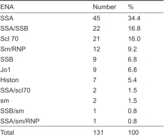
Anti-ENA proile was ordered from 414 (42%) of the ANA positive patients and 131 (31.6%) of them were found positive. The distribution of the ex-tractable anti-nuclear antigens was shown in Table 4. According to our study, the most frequent four an-tigens were SSA (34.4%), SSA-SSB (16.8%), Scl70 (16%) and Sm/RNP (9.2%) respectively.
Table 4. The distribution of ENA results
ENA Number %
SSA 45 34.4
SSA/SSB 22 16.8
Scl 70 21 16.0
Sm/RNP 12 9.2
SSB 9 6.8
Jo1 9 6.8
Histon 7 5.4
SSA/scl70 2 1.5
sm 2 1.5
SSB/sm 1 0.8
SSA/sm/RNP 1 0.8
Total 131 100
DISCUSSION
The autoantibodies which are seen in autoimmune diseases are against to nuclear and cytoplasmic components of the cells. The target antigens are ri-bonucleoproteins for anti-Sm, anti-RNP, anti-SSA/ Ro, anti-SSB/La; DNA topoisomerase Type 1 for anti-Scl70; centromere for anti-centromere trans-fer; histidyl-tRNA synthetase for Jo-1 and double-stranded DNA for anti-dsDNA.1
which is the major test to ind an autoimmune dis-ease.7
ANA positivity rate found in our female patients (77%) is consistent results with the knowledge of the autoimmune diseases are more frequent in women.8,9 This predominance was researched by Leo and et al. According to their study, the hormone proile, fetal microchimerism and some strategic genes which are on the sex chromosomes are play-ing role on this relationship.10
As expected, the most of the positive ANA re-sults were from rheumatology department (50%) in our study. This result is similar with the recent study of Karakeçe et al. which was done in an university hospital in Turkey.11 ANA is a very valuable test for the diagnosis of SLE (93% sensitivity) and sclero-derma (85% sensitivity). It is also important for diagnosing Sjögren’s syndrome, secondary Rayn-aud, polymyositis/dermatomyositis and rheumatoid arthritis.12
Surprisingly, the second frequent ANA positivity rate was in patients of the gastroenterology depart-ment (20%) in our study. The presence of ANA is found in a lot of chronic hepatobiliary diseases like viral hepatitis, drug induced hepatic disease, prima-ry biliaprima-ry cirrhosis, primaprima-ry sclerosing cholangitis, non-alcoholic steatohepatitis, and alcoholic hepa-titis. The main reason of ANA positivity is not the stimulation by an immunogen but the destruction of the hepatic cells because of the inlammation and necrosis. ANA is a very important diagnostic criteri-on especially in type 1-autoimmune hepatitis, alcriteri-ong with anti-smooth muscle cell antibody. The most frequent patterns are homogeneous and granular in autoimmune hepatitis and nuclear lamine is the second one.13-15
Of all ANA positive samples, 62.6% had only borderline or weak luorescence level in this study. As the clinical association of borderline/weak luo-rescence levels are a subject of discussion, some researchers did not reported because they ac-cepted as negative. Low titers of autoantibodies are seen in healthy people, relatives of the autoimmune patients and patients who have chronic inlamma-tory disease or cancer without having an autoim-mune base. These kinds of antibodies are usually in IgM type of low afinity and polyreactive.16 Most of the ANA positive people don’t developed an autoim-mune disease and this is consisted with low preva-lence of rheumatologic disorders (5-7%) despite the rate of ANA presence.9 ANA positivity can be detect-ed 20-30% at 1/40, 10% at 1/80 and 5% at 1/160
dilution in healthy people.17 Li et al. suggest that the persistence of the positive ANA may be a part of the component of the normal immune response.9 In the study of Mariz et al., ANA positive healthy people were followed up for four years of period and none of them developed any symptoms. Along the period, 72.5% of the ANA positivity persisted on the same level, while 27.5% of them dropped below 1/80 and were reported as negative. It has been emphasized that this follow up was done only from the healthy people who had ANA-Hep-2 patterns that were not speciic for acute rheumatic diseases. The writers suggested that the low ANA positivity of the patterns like homogeneous and centromere which are relat-ed only to autoimmune diseases must be followrelat-ed intensely.18 The common recommendation is to re-port the luorescence levels above 1+ as positive. The increase of ANA by age was proved by several studies.16,19 In our laboratory, IIFA is performed at 1/100 dilution and all patterns even with the bor-derline luorescence level are reported and the judgment of the importance of the positivity is left to the clinicians. This type of reporting might give a chance to the SLE patients whose ANA is positive considerably before the clinical symptoms which is not a rare probability and these patients must be followed carefully.20 Of all ANA positive patients with borderline/weak luorescence level, 43.3% were from rheumatology department in this study.
The most frequent four patterns were homoge-neous (23%), granular (22%), homogehomoge-neous-granu- homogeneous-granu-lar (15.5%) and nucleohomogeneous-granu-lar (13.5%) in this study. This was similar with the results of other studies from Turkey. Güdücüoğlu et al. reported 152 homoge-neous, 96 nucleolar, 82 granular pattern out of 367 ANA positive patients.8 The most and dominantly seen pattern was found as homogeneous (51.2%) and this was followed by ine granular (6%), ho-mogeneous/ine granular (6%) and homogeneous/ nucleolar (6%) by Yılmaz et al. [1]. Likewise, Yumuk et al. reported homogeneous as the most frequent pattern and the second one was granular.21 Also Karakeçe et al. found the most frequent patterns as nuclear (56.2; ine and coarse granular, homoge-neous and nuclear membrane), nucleolar (16.2%), mitotic (14%) and cytoplasmic (13.6%).11
granular and chromosomal granular type of anti-DFS. This is an autoantibody which can be seen in some of the dermatologic disorders like atopic dermatitis, psoriatic conditions, asthma, interstitial cystitis and rheumatologic diseases like Sjögren’s syndrome but its clinical importance is questionable as this pattern also present in 10% of normal popu-lation.22,23
Anti-dsDNA was positive in 8.7% of ANA posi-tive patients in the study. The major pattern of anti-dsDNA positive samples was homogeneous (68.8%). The most seen pattern from SLE disease is homogeneous (60-70%) which shows the auto-antibody presence against dsDNA and our results were parallel to the literature.1,21,24 On the other hand, it must be considered that homogeneous pat-tern may also point to the autoantibodies against histon and nucleosomes. The presence of anti-dsD-NA has prognostic value as the titer of anti-dsDanti-dsD-NA is an important criterion of disease activity and also shows a correlation with lupus nephritis.24-26
First step of the algorithm of ANA and speciic antibody testing in the diagnosis of rheumatic dis-eases is to screen ANA. The second step is testing for anti-ENA proile from positive samples. Anti-ENA proile test is an immunoblotting assay that uses only known antigens.27-29 Therefore, ANA with IIFA is more sensitive than anti-ENA proile test. Accord-ing to our study, the most frequent four antigens were SSA (34.4%), SSA-SSB (16.8%), Scl70 (16%) and Sm/RNP (9.2%) respectively. Anti-Sm antibod-ies are mostly found in SLE patients but they can only detected in 25-30% of them. Similarly, Scl70 is 100% speciic for the diagnosis of systemic sclero-sis. If SSA or/and SSB are detected, the result will direct us not only to diagnose Sjögren’s syndrome but also to sub acute cutaneous SLE and neona-tal lupus syndrome.30 Some studies reported that some ENAs especially anti-SSA/Ro and anti-SSB/ La antibodies can be missed on IIFA, although oth-ers demonstrated borderline luorescence might contain these antibodies.31 It should not be forgotten that anti-ENA proile may be negative depending on the positive ANA pattern.
Our three years’ experience of testing autoanti-bodies was shared in this study. Reliable test results are very important for the health of the patients with autoimmune disorders. For being a dependable lab-oratory, having enough knowledge and experience about the chosen methods of autoantibody tests is mandatory. It should be remembered that clinic sta-tus of the patients are very important for considering the results of autoimmune tests especially ANA.32
A good relationship with the clinicians is also an indispensable component of conidential analy-sis and reporting.
REFERENCES
1. Yılmaz Ö, Karaman M, Ergon MC, ve ark. Konnektif doku has -talıklarının tanısında Antinükleer (ANA) ve Anti-double stran -ded DNA (anti-dsDNA) antikorlarının önemi. T Parazitol Derg 2005;29:287-290.
2. Yumuk Z, Çalışkan Ş, Gündeş S, Willke A. Anti-nükleer anti -korların araştırılması ve saptanmasında kullanılan teknikler. Türk Mikrobiyol Cem Derg 2005;35:40-44.
3. Afşar İ, Şener AG, Vural A, ve ark. Anti nükleer antikorların po -zitif saptandığı hastalarda immunoblotting test sonuçlarının değerlendirilmesi. Türk Mikrobiyol Cem Derg 2007;37:39-42. 4. Kumar Y, Bhatia A, Minz RW. Antinuclear antibodies and their detection methods in diagnosis of connective tissue diseas-es: a journey revisited. Diagnostic Pathol 2009; 4:1. Avail-able from: http://www.diagnosticpathology.org/content/4/1/1. 5. Agmon-Levin N, Damoiseaux J, Kallenberg C, et al. Interna-tional recommendations for the assessment of autoantibod-ies to cellular antigens referred to as anti-nuclear antibodautoantibod-ies. Ann Rheum Dis 2014;73:17-23.
6. Greidinger EL, Hoffman RW. Antinuclear antibody testing: methods, indications, and interpretation. doi: 10.1309/VUB -90VTPMEWV3W0FLabMedicine 2003;34:113-117. 7. Avaniss-Aghajani E, Sophia B, Sarkissian A. Clinical value
of multiplexed bead-based immunoassays for detection of autoantibodies to nuclear antigens. Clin Vaccine Immunol 2007;14:505-509.
8. Güdücüoğlu H, Yaman G, Çıkman A, et al. Retrospective evaluation of immunoblotting (IB) test results in anti-nuclear antibody positive patients. Turkish J Clin Lab 2011;2:59-62. 9. Li Q, Karp D, Quan J, et al. Risk factors for ANA positivity in
healthy persons. Artritis Res Ther 2011;13:R38.
10. Lleo A, Battezzati PM, Semli C, et al. Is autoimmunity a mat -ter of sex? Autoimmun Rev 2008;7:626-630.
11. Karakeçe E, Atasoy AR, Çakmak G, ve ark. Bir üniversite hastanesinde antinükleer antikor pozitilikleri. Turk J Immu -nol 2014;2:5-8.
12. Habash-Besaiso D, Yale S, Glurich I, Goldberg J. Serologic testing in connective tissue disorders. Clin Med and Re -search 2005;3:190-193.
13. Ardeniz Ö. Otoimmün karaciğer hastalıklarında antinükleer antikorların değerlendirilmesi ve klinik uygulamadaki yeri. Güncel Gastroenteroloji 2006;10:187-192.
14. Aydemir S, Tekin İ, Engin H, ve ark. Non alkolik steatohepatitli hastalarda antinükleer antikor prevalansı ve önemi. Akade -mik Gastroenteroloji Dergisi 2005;4:158-161.
15. Yumuk Z, Sayan M, Çalışkan Ş. Kronik hepatit C hastala -rında otoantikorların HCV RNA düzeyi ile ilişkisi. İnfeksiyon Derg 2008;22:29-34.
16. Ulvestad E, Kanestrom A, Madland TM, et al. Evaluation of diagnostic tests for antinuclear antibodies in rheumatological practise. Scand J Immunol 2000;52:309-15.
17. Keren D. Guidelines for the Clinical Use of ANA and Related Speciic Autoantibody Testing. Article Archives 2000;11:2. 18. Mariz H, Sato E, Barbosa S, et al. Pattern on the antinuclear
19. Hurme M, Korkki S, Lehtimaki T, et al. Autoimmunity and longevity: presence of antinuclear antibodies is not associ-ated with the rate of inlammation or mortality in nonagerians. Mech Ageing Dev 2007;128:407-408.
20. Arbuckle MR, McClain MT, Rubertone MV, et al. Develop -ment of autoantibodies before the clinical onset of systemic lupus erithematosus. N Engl J Med 2003;349:1526-1533. 21. Yumuk Z, Çalışkan Ş. Evaluation of diagnostic autoantibody
tests used in clinical laboratories. Türk Mikrobiyol Cem Derg 2008;38:37-41.
22. Ganapathy V, Casiano CA, Autoimmunity to the nuclear auto -antigen DFS70 (LEDGF): what exactly are the autoantibod -ies trying to tell us? Arthritis Rheum 2004;50:684-688. 23. Watanabe A, Kodera M, Sugiara K, et al. Anti-DFS70 an
-tibodies in 597 healthy hospital workers. Arthritis Rheum 2004;50:892-900.
24. Hughes R, Ul-Hassan S. Anti-dsDNA antibodies: their role in the detection and monitoring of SLE. CLI 2006;7:12-17. 25. Biesen R, Dahnrich C, Rosemann A, et al. Anti-dsDNA-NcX
ELISA: dsDNA-loaded nucleosomes improve diagnosis and monitoring of disease activity in systemic lupus erythemato-sus. Arthritis Res Ther 2011;13:R26.
26. Ece A, Şahin C. Çocuk romatolojisi pratiğinde laboratuar test -lerinin kullanımı. J Clin Exp Invest 2013;4:258-261. 27. Tanyel T, Tutkak H, Önen I, Laleli Y. Düzen laboratuarına
başvuran ANA pozitif hastalarda gözlenen paternlerin sıklı -ğı. Proceedings of the 33. Türk Mikrobiyoloji Kongresi Kitabı; Ekim 21-25, 2008; Bodrum, Türkiye; 2008.p.823.P170. 28. Gonzalez C, Guevara P, Alarcon I, et al. Antinuclear
antibod-ies (ANA) screening by enzyme immunoassay with nuclear Hep-2 cell extract and recombinant antigens: analytical and clinical evaluation. Clin Biochem 2002;35:463-469.
29. Verstegen G, Duyck MC, Meeus P, et al. Detection and identi -ication of antinuclear antibodies (ANA) in a large community hospital. Acta Clin Belg 2009;64:317-323.
30. Birtane M, Diagnostic role of anti-nuclear antibodies in rheu -matic diseases. Turk J Rheumatol 2012;27:79-89.
31. Hoffman IEA, Peene I, Veys EM, De Keyser F. Detection of speciic antinuclear reactivities in patients with negative anti-nuclear antibody immunoluorescence screening tests. Clini -cal Chemistry 2002;48:2171-2176.