Revista da Sociedade Brasileira de Medicina Tropical 48(3):258-264, May-Jun, 2015
http://dx.doi.org/10.1590/0037-8682-0037-2015
Major Article
INTRODUCTION
Corresponding author: Dr. Rania Kishk. Department of Microbiology and Immunology/Faculty of Medicine/Suez Canal University.Km 4.5, Circular Road, Ismailia, Egypt.
Phone: 0020 102 509-9921 e-mail: [email protected] Received 1 February 2015 Accepted 24 April 2015
Occult HBV infection status among chronic hepatitis C
and hemodialysis patients in Northeastern Egypt:
regional and national overview
Mohamed Mandour
[1], Nader Nemr
[2], Atef Shehata
[3], [4], Rania Kishk
[3],
Dahlia Badran
[5]and Nashaat Hawass
[2][1]. Department of Clinical Pathology, Faculty of Medicine, Suez Canal University, Ismailia, Egypt. [2]. Department of Endemic and Infectious Diseases, Faculty of Medicine, Suez Canal University, Ismailia, Egypt. [3]. Department of Microbiology and Immunology, Faculty of Medicine, Suez Canal University, Ismailia, Egypt. [4]. Department of Microbiology and Immunology, Faculty of Medicine, Jazan University, Jazan, Kingdom of Saudi Arabia. [5]. Department of Medical Biochemistry, Faculty of Medicine, Suez Canal University, Ismailia, Egypt.
ABSTRACT
Introduction: Occult hepatitis B infection (OBI) is considered to be one of the major risks for patients suffering from end-stage renal disease (ESRD) on regular hemodialysis (HD) and patients with chronic hepatitis C virus (HCV) infection. This study compared the prevalence of OBI among these two high-risk groups in the Suez Canal region, Northeastern Egypt, to obtain a better national overview of the magnitude of OBI in this region. Methods: Serum samples were collected from 165 HD patients and 210 chronic HCV-infected patients. Anti-HCV antibody, hepatitis B surface antigen (HBsAg), total hepatitis B core (anti-HBc) antibody, and hepatitis B surface antibody (anti-HBs) were detected by enzyme-linked immunosorbent assay (ELISA). HCV RNA was detected using a quantitative real-time RT-PCR assay, and HBV was detected using a nested PCR.
Results: All patients were negative for HBsAg. A total of 49.1% and 25.2% of the patients in the HD and HCV groups, respectively, were anti-HBc-positive. In addition, more anti-HBs-positive patients were detected in the HD group compared to the HCV group (52.1% and 11.4%, respectively). Three cases were positive for HBV DNA in the HD group, while eighteen positive cases were
detected in the HCV group. Both study groups showed signifi cant differences in serum alanine aminotransferase (ALT) and aspartate
aminotransferase (AST) level as well as anti-HBc, anti-HBs and HBV-DNA positivity. Conclusions: OBI was more prevalent among chronic HCV patients than HD patients in the Suez Canal region, Egypt, with rates of 8.5% and 1.8%, respectively. However, more precise assessment of this infection requires regular patient follow-up using HBV DNA detection methods.
Keywords: Occult hepatitis B. HCV. Hemodialysis. Egypt. Anti-HBc.
The continual existence of hepatitis B surface antigen (HBsAg) in blood has been established as a marker of overt hepatitis B virus (HBV) infection. Occult hepatitis B infection
(OBI) is defi ned as the continuous existence of the HBV genome
in liver tissues and/or serum in the absence of serum HBsAg. This phenomenon could result from the persistent presence of covalently closed circular deoxyribonucleic acid (cccDNA), which accumulates in the nuclei of hepatocytes and acts as a template for HBV transcription(1). The risk of OBI transmission is decreased by the transfusion of blood components containing hepatitis B surface antibodies (anti-HBs)(2) (3).
Although Africa is considered to be a highly endemic region for HBV, it is considered to have an intermediate prevalence (2-6%) in Egypt. This discrepancy is mostly due to the introduction of HBV vaccination into compulsory immunization programs in Egypt in 1991 and the availability of this vaccine to populations at high risk for HBV infection. However, a considerable incidence of OBI has been reported in Egypt(4).
OBI may occur in serologically positive patients with markers of prior HBV infection (anti-HBs and/or hepatitis B core antibody (anti-HBc) as well as serologically negative patients without markers of previous exposure. Generally, approximately 20% of OBI patients are serologically negative, whereas 80% are serologically positive for one or more markers of prior infection(5). OBI is primarily spread among hemodialysis (HD) patients, individuals under going frequent blood transfusions, and intravenous drug abusers(6) (7) (8) (9) (10) (11) (12). Patients on chronic HD represent an important risk group for acquiring OBI, which may be attributed to the persistent exposure to blood-borne infections through the dialysis process and the need for frequent blood transfusions(13).
METHODS
(low or undetectable serum HBsAg)(14) (15). Whereas some studies have failed to demonstrate any association between OBI and HCV(16) (17), others have reported a high prevalence of OBI in chronically infected HCV patients(18) (19) (20). Similarly, a close association between OBI infection and the existence of anti-HCV antibodies among HD patients was reported(21).There is a broad range of clinical implications for OBI because it carries the potential for HBV transmission through blood transfusions, organ transplantations and HD. Moreover, OBI can result in fulminant hepatitis, poor responses to interferon/ribavirin (IFN/RBV) antiviral therapy, progression of hepatocellular carcinoma (HCC) and acute exacerbation of chronic hepatitis B(22).
The prevalence of OBI in HD patients varies markedly from one locality to another worldwide, with a range of 0% to 36 %(13). In Egypt, the prevalence was reported at 4% in Minia and Assuit (Upper Egypt) and reached 26.8% among patients in Alexandria (North Egypt)(23) (24). Even with these recent data addressing the prevalence of OBI in patients on HD in Egypt, more comprehensive data may be needed to evaluate OBI among chronic HCV carriers and to determine whether the prevalence differs following HD, as both HCV carriers and chronic HD patients may serve as a potential source for the spread of OBI in the community.
Our study investigated the prevalence of occult hepatitis B in two groups of patients who were potentially immunocompromised and at higher risk for acquiring OBI (chronic HCV-infected patients and patients on regular HD due to end-stage renal disease (ESRD) using serological tests and nested polymerase chain reaction (PCR). A highly specifi c and sensitive nested PCR method was used to test for OBI.
Patients
This study included two groups. The fi rst group included
165 patients with ESRD who had been undergoing regular HD for more than 6 months and were HCV-negative (negative for anti-HCV antibody).The second group included 210 patients with chronic HCV infection. The patients were selected from two HD centers in the Suez Canal region, Northeastern Egypt. HCV diagnosis was based on the detection of serum anti-HCV
antibodies and confi rmed by the detection of serum ribonucleic
acid (HCV/RNA). Patients with acute or chronic HBV infection (as determined by positive HBsAg), other causes of liver diseases (i.e., autoimmune hepatitis and continued alcohol abuse), or currently being treated with IFN and/or ribavirin were excluded. The medical history was collected for the entire study population, including HBV vaccination history and clinical and biochemical assessments. The study was conducted in accordance with the ethical standards of the Declaration of Helsinki. The study was approved by the research ethical committee of the Faculty of Medicine, Suez Canal University, and informed written consent was obtained from all patients included in this study.
Samples
Blood samples were collected from all patients. The serum was separated and stored at -80°C prior to analysis. Both
alanine aminotransferase (ALT) and aspartate aminotransferase (AST) levels were measured in all samples using a colorimetric
method. All patients were screened for human immunodefi ciency
virus (HIV) 1 and 2 with commercial kits (AxSYM HIV 1/2gO, Abbott Diagnostics, Wiesbaden, Germany).
Hepatitis C virus testing
A third-generation enzyme-linked immunosorbent assay (ELISA; HCV 3.0 ELISA Ortho, Raritan, NJ, USA) was used to test for anti-HCV antibodies according to the manufacturer’s instructions. HCV RNA was detected using a quantitative real time reverse transcription polymerase chain reaction (RT-PCR) assay with a detection limit of 33.6IU/ml (Artus HCV RG RT-PCR, Qiagen, Hilden, Germany).
Hepatitis B virus testing
Serology: HBsAg was detected by ELISA (ETI-MAK-2 PLUS, Diasorin, Italy) and further confirmed with the Microparticle Enzyme Immunoassay (MEIA) kit (AxSYM HBsAg V2, Abbott). Other serological markers (i.e., anti-HBs and total anti-HBc) were screened using standard commercially available enzyme immunoassays according to the manufacturer’s instructions (AB-AUK PLUS kits and ETI-AB-COREK PLUS; DiaSorin, Saluggia, Italy, respectively).
Nested PCR amplification of HBV region S: DNA was extracted from 200µl of serum using the QIAamp Min Elute Virus Spin Kit (QIAGEN, Inc., Hilden, Germany) and re-suspended in 100µl of a storage buffer provided by the kit manufacturer.
According to the method of Sugauchi el al.(25), the S region of the HBV genome (681bp) was amplifi ed using a nested PCR with4
primers targeting the S region; the assay has a lower limit of detection
of 100 copies/ml. The fi rst PCR used the primer pair (nucleotide
positions 18-989) (IS1)5'-AAGCTCTGCTAGATCCCAGAGT-3' and (HS4R) 5'-CATACTTTCCAATCAATAGG-3'. The PCR was performed in a 25µl volume containing 5µl of DNA template, 12.5µl of One Taq® Quick-Load®, 2× Master Mix [20mM Tris-HCl, 22mM KCl, 22mM NH4Cl, 1.8mM MgCl2, 5% glycerol, 0.06% IGEPAL CA-630, 0.05% Tween 20, 0.2mM dNTPs, 1×xylene cyanol, 1×tartrazine, and 25 units/ml OneTaq® DNA Polymerase, pH 8.9 at 25°C] (New England Biolabs), 25µM of each primer, and PCR-grade water. The cycling conditions consisted of 7 min at 96°C and 45 cycles of 96°C for 45 s, 55°C for 45 s and 72°C for 1 min. The second PCR (nucleotide positions 414-989) used the primers (SB1) 5'- TGCTGCTATGCCTCATCTTC-3' and (HS4R) 5'- CATACTTTCCAATCAATAGG-3'. The PCR was performed in a 25µl reaction mixture volume containing 2µl
of DNA template (from the fi rst PCR product), 25µM of each
primer, 12.5µl of One Taq® Quick-Load® 2× Master Mix (New England Bio labs), and PCR-grade water. The cycling
conditions were the same as those described forthe fi rst PCR.
RESULTS Rev Soc Bras Med Trop 48(3):258-264, May-Jun, 2015
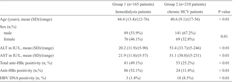
TABLE 1 - Demographic and laboratory data of the study group populations.
Group 1 (n=165 patients) Group 2 (n=210 patients)
hemodialysis patients chronic HCV patients P value Age (years), mean (SD)/(range) 44.4 (13.4)/(12-76) 40.6 (9.1)/(17-56) < 0.01 Sex (n,%)
male 89 (53.9%) 141 (67.2%)
female 76 (46.1%) 69 (32.8%) 0.01
ALT in IU/L, mean (SD)/(range) 20.2 (11.9)/(5-90) 53.4 (33.7)/(5-246) < 0.01 AST in IU/L, mean (SD)/(range) 21.9 (11.0)/(5-57) 51.1 (30.0)/(5-231) < 0.01 Total anti-HBc positivity (n, %) 81 (49.1%) 53 (25.2%) < 0.01 Anti-HBs positivity (n,%) 86 (52.1%) 24 (11.4%) < 0.01 HBV DNA positivity (n, %) 3 (1.8%) 18 (8.5%) < 0.01
HCV: hepatitis C virus; SD: standard deviation; n: number; ALT: alanine aminotransferase; AST: aspartate aminotransferase; IU/L: international unit per liter; anti-HBc: hepatitis B core antibody; anti-HBs: hepatitis B surface antibody; HBV DNA: hepatitis B virus deoxyribonucleic acid.
Gel electrophoresis: The resulting PCR amplicons were electrophoresed in 1.5% agarose gels in 1× Tris-borate-ethylene diaminetetraacetic acid (EDTA) buffer and stained with ethidium bromide. The images were captured using the Syngene G: Box documentation system (Syngene, UK).
Statistical analysis
Statistical Package for Social Science (SPSS) version 16 was used for data processing and analysis. The results are expressed in ranges, percentages and mean (SD). For comparisons between the study groups, the Student’s t-test was employed for quantitative variables, and the Chi-square test was used for categorical (qualitative) variables. P values less than 5% were
considered to be statistically signifi cant.
General characteristics of the studied populations
The current study included two groups. The fi rst group
included 165 patients with ESRD who received regular HD. These patients ranged from 12 to 76 years in age with a mean (SD) of 44.35 years (13.40 years); additionally, 53.9% of the patients were male and 46.1% were female. The second group included 210 patients with chronic HCV infection. These patients ranged from17 to 56 years with a mean (SD) of 40.61 years (9.09 years); additionally, 67.2% of the patients were male
and 32.8% were female. There were signifi cant differences in
age and gender between the study groups.
The ALT values ranged from 5 to 90 with a mean (SD) of 20.17 (11.89) in the HD group and from 5 to 246 with a mean (SD) of 53.43 (33.67) in the chronic HCV group. The AST means (SD) were 21.93 (10.98) and 51.07 (30.01) in the HD and HCV patients, respectively. Both the ALT and AST
levels were signifi cantly different between the HD and chronic
HCV groups (Table 1).
Hepatitis B serological status
All the patients were negative for HBsAg. Positive cases
for total anti-HBc were signifi cantly higher in the HD group
compared to the HCV group (49.1% and 25.2%, respectively). Moreover, the number of positive cases for anti-HBs was higher in the HD group compared to the HCV group (52.1% and 11.4%,
respectively). The results revealed signifi cant differences in
anti-HBc and anti-HBs seropositivity in both groups, with p values < 0.01 for both markers (Table 1).
In both study populations, males were more common than females among the anti-HBc-positive cases. Table 2 shows
that serum ALT and AST levels were signifi cantly higher in
the HCV group, where as the HD group showed more positive anti-HBs cases than the HCV group (63% and 18.9%, respectively).
Nested PCR results
All the DNA samples extracted from patients in the current study were subjected to nested PCR targeting the S region of
the hepatitis B genome. The resulting amplifi ed product was
approximately 681bp in size, as shown in Figure 1. Highly
signifi cant variation was observed in positive cases between the
two groups (p value <0.01); only 3 cases were positive for HBV DNA in the HD group, while 18 cases were positive in the HCV group (Table 1). The OBI prevalence was 1.8% and 8.5% in the HD and HCV groups, respectively. The characterization of HBV DNA-positive cases in both study groups is shown in Table 3. All three cases in the HD group were negative for anti-HBs, and only two cases were positive for anti-HBc; in contrast, more than half (55.5%) of the cases in the HCV group were positive for anti-HBs, and all cases were positive for anti-HBc. There
were no signifi cant differences in age, gender, ALT and AST
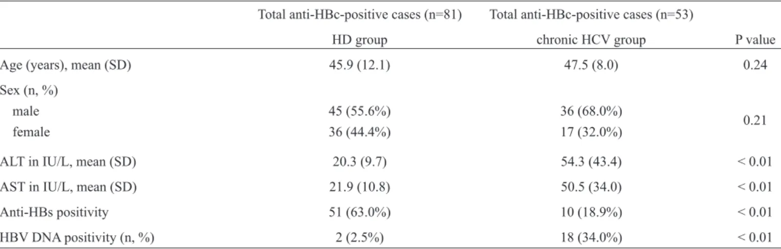
TABLE 2 - Demographic and laboratory data for the total anti-HBc-positive cases.
Total anti-HBc-positive cases (n=81) Total anti-HBc-positive cases (n=53)
HD group chronic HCV group P value Age (years), mean (SD) 45.9 (12.1) 47.5 (8.0) 0.24 Sex (n, %)
male 45 (55.6%) 36 (68.0%)
0.21
female 36 (44.4%) 17 (32.0%)
ALT in IU/L, mean (SD) 20.3 (9.7) 54.3 (43.4) < 0.01 AST in IU/L, mean (SD) 21.9 (10.8) 50.5 (34.0) < 0.01 Anti-HBs positivity 51 (63.0%) 10 (18.9%) < 0.01 HBV DNA positivity (n, %) 2 (2.5%) 18 (34.0%) < 0.01
anti-HBc: hepatitis B core antibody; HD: regular hemodialysis; HCV: hepatitis C virus; SD: standard deviation; n: number; ALT: alanine aminotransferase; AST: aspartate aminotransferase; IU/L: international unit per liter (IU/L); anti-HBs: hepatitis B surface antibody; HBV DNA: hepatitis B virus deoxyribonucleic acid.
TABLE 3 - Characterization of HBV DNA-positive cases in the two study groups.
HBV DNA-positive cases(n=3) HBV DNA-positive cases (n=18)
HD group chronic HCV group P value Age (years), mean (SD)/(range) 47.6 (7.37)/(42-56) 44.3 (8.9)/(26-56) 0.49 Sex (n, %)
male 1 (33.3%) 13 (22.2%) 0.51
female 2 (66.7%) 5 (77.8%)
ALT in IU/L, mean (SD)/range 19.3 (18.0)/(7-40) 39.0 (21.2)/(10-81) 0.11 AST in IU/L, mean (SD)/range 17 (6.0)/(10-21) 21.5 (11.3)/(8-49) 0.47 Anti-HBs positivity (n, %) 0 (0.0%) 10 (55.5%) 0.22 Total anti-HBc positivity (n, %) 2 (66.7%) 18 (100.0%) 0.14
HBV DNA: hepatitis B virus deoxyribonucleic acid; HD: regular hemodialysis; HCV: hepatitis C virus; SD: standard deviation; n: number; ALT: alanine aminotransferase; AST: aspartate aminotransferase; IU/L: international unit per liter (IU/L); anti-HBs: hepatitis B surface antibody; anti-HBc: hepatitis B core antibody.
M 1 2 3 4 5 6 7 M 8 9 10 11 12 13 14 15 bp
1,000
600
300
DISCUSSION
HBV infection is a major health concern due to its widespread prevalence, with 350,000 million infected individuals worldwide(26). HBV infection leads to major complications, including fulminant hepatitis, chronic liver disease and HCC,
which carry the signifi cant burden of high health costs. OBI
is a variant of HBV infection that occurs when patients show no HBsAg in the serum but persistent HBV DNA in the liver tissue with or without detection of the HBV genome in the serum. Patients with ESRD on HD and chronic HCV-infected patients are highly susceptible to acquiring HBV infection. Our study was designed to measure the magnitude of this infection in these two groups of patients in a large district in our country. The current study showed marked variations in patients’ ages, because we included variable age groups (from children to elderly patients with a range of 12 to 76 years in the HD group and from 17 to 56 years in the HCV group). This approach enabled a more accurate assessment of OBI in our patients, as these samples were representative of the age groups in the general population.
Our results showed signifi cantly different ALT and AST
levels between the study groups. The levels were more elevated in the HCV group, which may be attributed to hepatic dysfunction due to chronic hepatitis.
Anti-HBc constitutes the fi rst antibody response to HBV
infection, and its detection in the serum may denote acute or chronic infection. Patients remain anti-HBc-positive after recovery, and this antibody therefore serves as the only serologic marker for HBV during the window period of infection. As a result, this marker is considered one of the most valuable serological markers for the diagnosis of OBI(27) (28) (29) and has been proposed as a surrogate screening marker for OBI diagnosis when viral DNA detection methods cannot be applied(26). In contrast, the anti-HBs antibody appears late in infection after the disappearance of HBsAg and has a protective neutralizing effect. Anti-HBs is the only HBV serologic marker that can be detected after vaccination against this virus.
In the current study, the rates of detection of both
anti-HBs and anti-HBc differed signifi cantly between the study
populations, and both markers were detected at higher levels
in the HD group compared to the HCV group. This fi nding can
be explained either by a true difference in the infection rate between groups or by immune unresponsiveness or a weak immune response to HBV antigens as a result of the suppression of replication and expression of this virus in HCV co-infected patients(14) (15). Moreover, the positivity for both an ti-HBc and anti-HBs could be explained by either recovery from previous acute or chronic HBV infection(26)or OBI seropositivity in which HBsAg cannot be detected in the serum because it is cleared to an undetectable level(30). Additionally, the increased number of anti-HBs-positive patients in the HD group may be a result of vaccination against HBV, which is routinely performed in HD patients in our region. As shown in Table 2, a considerable number of the anti-HBc-positive patients were negative for both anti-HBs and HBV DNA; this result may be due to either
recovery from previous HBV infection or true occult HBV infection without detectable DNA in the serum. Thus, the rate of DNA detection may increase if liver biopsies are examined.
The detection of HBV DNA is considered the most defi nitive
diagnostic tool for the detection of occult hepatitis infection. In our study, we used nested PCR because this method is simple and accurate, easy to interpret, can detect even very low amounts of viral DNA and has been previously applied on a wide scale for the detection, genotyping and phylogenetic analysis of HBV(31) (32) (33) (34). Specifi cally, we targeted the S region of the HBV genome in patient serum samples; this region was selected
as a target for amplifi cation by PCR because it was found to
be more sensitive for the detection of HBV DNA in serum(26). Our nested PCR demonstrated that the prevalence of OBI was 1.8% and 8.5% in the HD and HCV groups, respectively; the
difference in these prevalence rates was statistically signifi cant.
Marked variation was found by comparing these rates with OBI prevalence rates reported in other related national or international studies(23) (24) (35) (36) (37) (38) (39).This variation may be attributed to differences in the sample size of the study populations, demographics, immunologic status of the studied patients, endemicity of HBV infection, levels of viral DNA in the blood, sampling conditions and the types of diagnostic tool(s) used for diagnosis(40). This comparison also revealed that the prevalence of OBI in our target HD patients was relatively low, possibly due to the application of safety precautions during the dialysis process in these units and the implementation of the routine vaccination program. Additionally, the low rate can be attributed to the use of serum samples to detect HBV DNA in the current study. The likelihood of detecting DNA is much lower in serum samples than in liver tissue, although the use of liver biopsies for the detection
of OBI is diffi cult, especially in HD patients. Moreover, the
higher prevalence rate of OBI in the chronic HCV group can be explained by the occurrence of coinfection with HBV, as both of these viruses share common routes of infection(41).
Two of the three HBV DNA-positive patients in the HD group were seropositive for anti-HBc alone, while the third patient was seronegative. In contrast, all HBV DNA-positive cases in the HCV group were positive for anti-HBc, and more than half were positive for anti-HBs. This result is in accordance with
the fi nding that OBI is more prevalent in seropositive patients
who are positive for anti-HBc and/or anti-HBs(23) (29) (37) (42) (43). The occurrence of occult infection despite the presence of neutralizing an ti-HBs can result from mutations affecting the S region of the HBV genome(39). However, the absence of all HBV serological markers (even anti-HBc) in some HBV DNA-positive patients strengthens the proposal that the detection of viral DNA is the gold standard for the diagnosis of OBI. Therefore, the presence of seronegative cases should be given consideration during the management of HD and HCV patients. From the current study, we can conclude that OBI occurs in HD and chronic hepatitis C patients in our area at rates of 1.8% and 8.5%, respectively. Despite these relatively low rates, this infection is considered to be a real health problem in these debilitated patients and necessitates regular follow-up for the early detection of OBI using DNA detection methods.
REFERENCES
The authors declare that there is no confl ict of interest. CONFLICT OF INTEREST
1. Alavian SM, Bagheri-Lankarani K, Mahdavi-Mazdeh M, Nourozi S. Hepatitis B and C in dialysis units in Iran: changing the epidemiology. Hemodial Int 2008; 12:378-382.
2. Allain JP. Occult hepatitis B virus infection: implications in transfusion. Vox Sang 2004; 86:83-91.
3. Romero M, Madejon A, Fernandez-Rodriguez C, Garcia-Samaniego J. Clinical signifi cance of occult hepatitis B virus infection. World J Gastroenterol 2010; 17:1549-1552.
4. Attia MA. Prevalence of hepatitis B and C in Egypt and Africa. Antivir Ther 1998; 3 (suppl III):1-9.
5. Aghakhani A, Banifazl M, Kalantar E, Eslamifar A, Ahmadi F, Razeghi E, et al. Occult hepatitis B virus infection in hemodialysis patients with isolated hepatitis B core antibody: a multicenter study. Ther Apher Dial 2010; 14:349-353.
6. Fang Y, Shang QL, Liu JY, Li D, Xu WZ, Teng X, et al. Prevalence of occult hepatitis B virus infection among hepatopathy patients and healthy people in China. J Infect 2009; 58:383-388.
7. Georgiadou SP, Zachou K, Liaskos C, Gabeta S, Rigopoulou EI, Dalekos GN. Occult hepatitis B virus infection in patients with autoimmune liver diseases. Liver Int 2009; 29:434-442.
8. Ramezani A, Banifazl M, Eslamifar A, Aghakhani A. Serological pattern of anti-HBc alone infers occult hepatitis B virus infection in high-risk individuals in Iran. J Infect Dev Ctries 2010; 4:658-661. 9. Shire NJ, Rouster SD, Rajicic N, Sherman KE. Occult hepatitis
B in HIV-infected patients. J Acquir Immune Defi c Syndr 2004; 36:869-875.
10. Tamori A, Hayashi T, Shinzaki M, Kobayashi S, Iwai S, Enomoto M, et al. Frequent detection of hepatitis B virus DNA in hepatocellular carcinoma of patients with sustained virologic response for hepatitis C virus. J Med Virol 2009; 81:1009-1014. 11. Torbenson M, Thomas DL. Occult hepatitis B. Lancet Infect Dis
2002; 2:479-486.
12. Toyoda H, Hayashi K, Murakami Y, Honda T, Katano Y, Nakano I, et al. Prevalence and clinical implications of occult hepatitis B viral infection in hemophilia patients in Japan. J Med Virol 2004; 73:195-199.
13. Gutierrez-Garcia ML, Fernandez-Rodriguez CM, Lledo-Navarro JL, Buhigas-Garcia I. Prevalence of occult hepatitis B virus infection. World J Gastroenterol 2011;17:1538-1542.
14. Chu CM, Yeh CT, Liaw YF. Low-level viremia and intracellular expression of hepatitis B surface antigen (HBsAg) in HBsAg carriers with concurrent hepatitis C virus infection. J Clin Microbiol 1998; 36:2084-2086.
15. Shih CM, Lo SJ, Miyamura T, Chen SY, Lee YH. Suppression of hepatitis B virus expression and replication by hepatitis C virus core protein in HuH-7 cells. J Virol 1993; 67:5823-5832.
16. Besisik F, Karaca C, Akyuz F, Horosanli S, Onel D, Badur S, et al. Occult HBV infection and YMDD variants in hemodialysis patients with chronic HCV infection. J Hepatol 2003; 38:506-510. 17. Kanbay M, Gur G, Akcay A, Selcuk H, Yilmaz U, Arslan H,
et al. Is hepatitis C virus positivity a contributing factor to occult hepatitis B virus infection in hemodialysis patients? Dig Dis Sci 2006; 51:1962-1966.
18. Raimondo G, Caccamo G, Filomia R, Pollicino T. Occult HBV infection. Semin Immunopathol 2013;35:39-52.
19. Raimondo G, Pollicino T, Cacciola I, Squadrito G. Occult hepatitis B virus infection. J Hepatol 2007; 46:160-170.
20. Torbenson M, Kannangai R, Astemborski J, Strathdee SA, Vlahov D, Thomas DL. High prevalence of occult hepatitis B in Baltimore injection drug users. Hepatology 2004; 39:51-57.
21. Di Stefano M, Volpe A, Stallone G, Tartaglia L, Prato R, Martinelli D, et al. Occult HBV infection in hemodialysis setting is marked by presence of isolated antibodies to HBcAg and HCV. J Nephrol 2009; 22:381-386.
22. Mrani S, Chemin I, Menouar K, Guillaud O, Pradat P, Borghi G, et al. Occult HBV infection may represent a major risk factor of non-response to antiviral therapy of chronic hepatitis C. J Med Virol 2007; 79:1075-1081.
23. Abu El Makarem MA, Abdel Hamid M, Abdel Aleem A, Ali A, Shatat M, Sayed D, et al. Prevalence of occult hepatitis B virus infection in hemodialysis patients from egypt with or without hepatitis C virus infection. Hepat Mon 2012; 12:253-258.
24. Elgohry I, Elbanna A, Hashad D. Occult hepatitis B virus infection in a cohort of Egyptian chronic hemodialysis patients. Clin Lab 2012; 58:1057-1061.
25. Sugauchi F, Orito E, Ohno T, Kato H, Suzuki T, Hashimoto T, et al. Liver transplantation-associated de novo hepatitis B virus infection: application of molecular evolutionary analysis. Intervirology 2002; 45:6-10.
26. Raimondo G, Allain JP, Brunetto MR, Buendia MA, Chen DS, Colombo M, et al. Statements from the Taormina expert meeting on occult hepatitis B virus infection. J Hepatol 2008; 49:652-657. 27. Kang SY, Kim MH, Lee WI. Occult hepatitis B virus infection in
Korean patients with isolated anti-HBc. Arch Virol 2014; 159:227-233.
28. Ocana S, Casas ML, Buhigas I, Lledo JL. Diagnostic strategy for occult hepatitis B virus infection. World J Gastroenterol 2011; 17:1553-1557.
29. Urbani S, Fagnoni F, Missale G, Franchini M. The role of anti-core antibody response in the detection of occult hepatitis B virus infection. Clin Chem Lab Med 2010; 48:23-29.
30. Lok AS, McMahon BJ. Chronic hepatitis B. Hepatology 2001; 34:1225-1241.
31. He J, Wu J, Du S. Nested polymerase chain reaction (PCR) and multiple cloned antibody capture PCR for the examination of serum hepatitis B virus DNA in negative hepatitis B surface antigen patients suffering from liver diseases. Zhonghua Nei Ke Za Zhi 1996; 35:537-541.
32. Panigrahi R, Biswas A, De BK, Chakrabarti S, Chakravarty R. Characterization of antiviral resistance mutations among the Eastern Indian Hepatitis B virus infected population. Virol J 2013;10:56.
33. Sugauchi F, Mizokami M, Orito E, Ohno T, Kato H, Suzuki S, et al. A novel variant genotype C of hepatitis B virus identified in isolates from Australian Aborigines: complete genome sequence and phylogenetic relatedness. J Gen Virol 2001; 82:883-892. 34. Sun S, Zhou H, Zhou B, Hu Z, Hou J, Sun J. Sensitivity and specifi city
of nested PCR pyrosequencing in hepatitis B virus drug resistance gene testing. Nan Fang Yi Ke Da Xue Xue Bao 2012; 32:610-613. 35. Albuquerque AC, Coelho MR, Lemos MF, Moreira RC. Occult
hepatitis B virus infection in hemodialysis patients in Recife, State of Pernambuco, Brazil. Rev Soc Bras Med Trop 2012; 45:558-562. 36. Cacciola I, Pollicino T, Squadrito G, Cerenzia G, Orlando ME,
37. Keyvani H, Agah S, Kabir A, Alavian SM. Prevalence and risk factors of isolated anti-HBc antibody and occult hepatitis B infection in hemodialysis patients: a nationwide study. Ann Hepatol 2013; 12:213-219.
38. Levast M, Larrat S, Thelu MA, Nicod S, Plages A, Cheveau A, et al. Prevalence and impact of occult hepatitis B infection in chronic hepatitis C patients treated with pegylated interferon and ribavirin. J Med Virol 2010; 82:747-754.
39. Minuk GY, Sun DF, Greenberg R, Zhang M, Hawkins K, Uhanova J, et al. Occult hepatitis B virus infection in a North American adult hemodialysis patient population. Hepatology 2004; 40: 1072-1077.
40. Yoo JH, Hwang SG, Yang DH, Son MS, Kwon CI, Ko KH, et al. Prevalence of occult hepatitis B virus infection in hemodialysis patients. Korean J Gastroenterol 2013;61:209-214.
41. Chen LW, Chien RN, Yen CL, Chang JJ, Liu CJ, Lin CL. Therapeutic effects of pegylated interferon plus ribavirin in chronic hepatitis C patients with occult hepatitis B virus dual infection. J Gastroenterol Hepatol 2010; 25:259-263.
42. Levicnik-Stezinar S, Rahne-Potokar U, Candotti D, Lelie N, Allain JP. Anti-HBs positive occult hepatitis B virus carrier blood infectious in two transfusion recipients. J Hepatol 2008; 48:1022-1025.
43. Shi Y, Wu YH, Wu W, Zhang WJ, Yang J, Chen Z. Association between occult hepatitis B infection and the risk of hepatocellular carcinoma: a meta-analysis. Liver Int 2012; 32:231-240.