Drosophila
Cbp53E Regulates Axon Growth
at the Neuromuscular Junction
Kimberly R. Hagel1, Jane Beriont1, Charles R. Tessier2*
1Department of Biological Sciences, University of Notre Dame, Notre Dame, Indiana, United States of America,2Department of Medical and Molecular Genetics, Indiana University School of Medicine-South Bend, South Bend, Indiana, United States of America
Abstract
Calcium is a primary second messenger in all cells that functions in processes ranging from cellular proliferation to synaptic transmission. Proper regulation of calcium is achieved through numerous mechanisms involving channels, sensors, and buffers notably containing one or more EF-hand calcium binding domains. TheDrosophilagenome encodes only a sin-gle 6 EF-hand domain containing protein, Cbp53E, which is likely the prototypic member of a small family of related mammalian proteins that act as calcium buffers and calcium sen-sors. Like the mammalian homologs, Cbp53E is broadly though discretely expressed throughout the nervous system. Despite the importance of calcium in neuronal function and growth, nothing is known about Cbp53E’s function in neuronal development. To address this deficiency, we generated novel null alleles ofDrosophilaCbp53E and examined neuro-nal development at the well-characterized larval neuromuscular junction. Loss of Cbp53E resulted in increases in axonal branching at both peptidergic and glutamatergic neuronal ter-minals. This overgrowth could be completely rescued by expression of exogenous Cbp53E. Overexpression of Cbp53E, however, only affected the growth of peptidergic neuronal pro-cesses. These findings indicate that Cbp53E plays a significant role in neuronal growth and suggest that it may function in both local synaptic and global cellular mechanisms.
Introduction
Calcium is a major second messenger in all cells and is vital for cellular function. Given this importance, all aspects of calcium homeostasis including influx, efflux, intracellular mobiliza-tion, and storage are tightly controlled processes[1]. Excitable cells, such as neurons, are partic-ularly susceptible to changes in calcium dynamics, as calcium is required for synaptic vesicle fusion and neurotransmitter release [2]. Even small defects in calcium homeostasis can affect synaptic transmission and result in profound consequences ranging from individual neuronal dysfunction to aberrant circuit formation and altered behavior [3–5]. Therefore, understanding how calcium is regulated is critical for understanding neuronal development and function. An important class of calcium regulators that function in neural development are the calcium buff-ering proteins.
a11111
OPEN ACCESS
Citation:Hagel KR, Beriont J, Tessier CR (2015)
DrosophilaCbp53E Regulates Axon Growth at the Neuromuscular Junction. PLoS ONE 10(7): e0132636. doi:10.1371/journal.pone.0132636
Editor:William D Phillips, University of Sydney, AUSTRALIA
Received:April 17, 2015
Accepted:June 16, 2015
Published:July 13, 2015
Copyright:© 2015 Hagel et al. This is an open access article distributed under the terms of the
Creative Commons Attribution License, which permits unrestricted use, distribution, and reproduction in any medium, provided the original author and source are credited.
Data Availability Statement:All relevant data are within the paper.
Funding:This work was supported by a Biomedical Research Enhancement Grant from Indiana University School of Medicine. The funders had no role in study design, data collection and analysis, decision to publish, or preparation of the manuscript.
Proteins that function as calcium buffers are thought to bind and sequester intracellular cal-cium ([Ca2+]i) in order to modulate its spatiotemporal availability as a second messenger sig-naling molecule[6]. There are many vertebrate calcium buffering proteins including
parvalbumin, calretinin, calbindin-D9k and calbindin-D28k. Each of these proteins contains one or more EF-hand binding domains, a motif capable of binding calcium ions, though not all EF-hand domains may be active under physiological conditions[7]. Moreover, the affinity of these domains for calcium may be different across different buffering proteins. Thus, some cal-cium buffering proteins are considered slow buffers and others fast buffers, depending on sev-eral factors including their affinity for calcium, binding kinetics, and their cytoplasmic mobility[8]. This distinction is particularly important in neurons, which have both pre- and postsynaptic compartments capable of responding to membrane depolarization through multi-ple calcium influx and calcium mobilizations pathways[9]. Thus, different aspects of calcium homeostasis may be regulated by different buffering proteins.
The two major vertebrate calcium buffers, calbindin-D28k (CB) and calretinin (CR), share 58% identity to each other and are members of a small family of 6 EF-hand domain containing proteins that are classically characterized as fast calcium buffers[10,11]. A third protein, secreta-gogin shares significant homology to these buffers, but has only been characterized as a calcium sensor [12,13]. Calcium sensors typically bind target proteins to affect signaling in response to changes in [Ca2+]i. Indeed, CB and CR were once thought to function exclusively as buffers, but now both have been shown to bind effector proteins under different calcium-dependent situa-tions [14,15]. CB and CR are expressed throughout the brain in discrete, mostly exclusive pat-terns, predominantly in the cerebellum, hippocampus, and cortex [16–19]. Each protein affects Purkinje cell development, a function responsible for motor coordination defects found in CB and CR knockout mice [20–22]. At the cellular level, CB mitigates calcium influx transient amplitude in dendrites, while CR can affect the timing of certain functional spikes [21–23]. Neu-ronal function is also impaired in the hippocampus where loss of CB or CR leads to defects in long-term potentiation as well as animal failures in spatial learning tests [24,25]. In addition to roles in synaptic plasticity, these buffers seem to have neuroprotective roles in neurons involved in Parkinson’s disease (PD) and Alzheimer’s disease (AD)[26–30]. Interestingly, the neuropro-tective functions may be in part due to a direct interaction with target binding proteins which can affect downstream signaling cascades[31]. Therefore, CB and CR are multifunctional pro-teins that regulate calcium homeostasis to control neuronal development and function.
Drosophilaare now routinely being used as models for understanding both normal neuronal development and disease states such as PD and AD[32]. However, little work has been done to understand the role of calcium buffers in this well-established model organism. TheDrosophila genome encodes only a single 6 EF-hand domain containing protein, Cbp53E [33]. Cbp53E shares nearly the same homology to all 3 members of the mammalian family, having 45%, 46%, and 45% protein sequence identity with CB, CR, and secretagogin respectively. Although Cbp53E was cloned over two decades ago, virtually nothing is known about its function [33,
34]. We have generated two novel null alleles of theDrosophilaCbp53E gene in order to
deter-mine its role in neuronal development. We describe Cbp53E-dependent effects on axon growth which seem to implicate both local synaptic and global cellular functions for this highly con-served protein.
Materials and Methods
Fly stocks and genetics
The control strain wasw1118. Cbp53E mutants are:Cbp53EMi22,Cbp53EMi41, andCbp53EMi2. The 2ndchromosome deficiency is Df(2R)ED2751. Animals used in rescue experiments are:
elav-GAL4/+ (control) and elav-GAL4/+;Cbp53EMi22and elav-GAL4/+; UAS-Cbp53E/+ and elav-GAL4/+;Cbp53EMi22; UAS-Cbp53E/+ (rescue), 24B-GAL4/+ (control) andCbp53EMi22; 24B-GAL4 and 24B-GAL4/UAS-Cbp53E andCbp53EMi22; 24B-GAL4/UAS-Cbp53E (rescue). Flies used for motor neuron analysis were: UAS-mCD8GFP/+;D42-GAL4/+ and flies used for 24B-GAL4 analysis were: UAS-mCD8GFP/+;24B-GAL4/+.
Cbp53E mutant allele generation
All lines used in mutant generation were acquired from the BloomingtonDrosophilaStock Center. The transposon insertion Mi{ET1}Cbp53EMB00150was mobilized by crossing it to a heat shock inducible Minos transposase P{hsILMiT}2.4 using standard techniques[35]. Flies were screened for loss of GFP expression present on the transposon containing chromosome and subsequently outcrossed to remove the transposase. Individual progeny were then crossed to a second chromosome deficiency line. To determine the effect of transposon mobilization on Cbp53E expression, brains from the progeny were stained for Cbp53E by immunohis-tochemistry. Mutant linesCbp53EMi22andCbp53EMi41were then sequenced using PCR prim-ers around the transposon insertion site to produce a PCR product approximately 2kb in length. The primers used were:
forward:50-CCCATTTCCCATTGCATTTCTAAGAGC-30;
reverse:50-GCCAGTGTTAGCAGAATCATGGTCAGT-30. Sequence analysis revealed that
Cbp53E sequences remained intact around the insertion, but that approximately 7.6kb of the 30-most portion of the transposon had been removed from each mutant line.
Transgenic generation
RNA was extracted fromw1118flies using Trizol (Invitrogen) and then used to make cDNA by Superscript III Reverse Transcriptase (Invitrogen) as previously[36]. The following cloning primers were used to amplify Cbp53E cDNA from the total cDNA withNot1andKpnI restric-tion sites:
forward:50-AAATTTGCGGCCGCTTCTCCAAAGATA-30;
reverse:50-AATGGTACCTTTTGGAGTGCTGACCCC-30. The resultant PCR product
encompassed the entire Cbp53E mRNA including untranslated regions. The PCR product was purified and digested withNot1andKpn1and ligated into a similarly digested pUAS-T vector (DrosophilaGenomics Resource Center). The recombined plasmid was purified and microin-jected intow1118Drosophilaembryos by Genetic Services INC (Waltham MA). Transformants were identified and mapped to chromosomes using standard techniques. UAS-Cbp53E was balanced over TM6 for stock maintenance.
Immunohistochemistry
Developmental Studies Hybridoma Bank). Secondary antibodies used (all at 1:2000 dilutions): AlexaFluor 647 goat anti-mouse (Invitrogen), AlexaFluor 488 goat anti-rabbit (Invitrogen), AlexaFluor 488 goat anti-mouse (Jackson Laboratories), and Cy3 goat anti-guinea pig (Invitrogen).
Quantitative RT-PCR
cDNA was generated as described above from heads of mutant and control animals. POWER SybrGreen (Applied Biosystems) was then used according to the manufacturer’s instructions to perform quantitative PCR for Cbp53E expression using a BioRad CFX Connect Real Time Sys-tem. Primers for Cbp53E detection were:
forward:50-GCCAACAAGGACGGACGTCTGCAGTT-30;
reverse:50-GGCATCATAGTCGTCCTTCTTGACCAA-30. GAPDH2 was used as the
refer-ence control with the following primers:
forward:50-CCGATGCGACCAAATCCATTGATA-30;
reverse:50-CGCTCAAAATTTCTCAGCCATCAC-30. Cycling parameters were: 96°C-2min,
(94°C-30sec, 60°C-30sec, 72°C-30sec) X40, 72°C-1min. TheΔΔCt method was used to calcu-late relative fold changes of Cbp53E expression in mutant versus control animals.
Confocal microscopy
Z-stacks of segment 3, muscle 4, muscle 6/7 and muscle 12/13 neuromuscular junctions and whole brains were acquired on a Zeiss LSM 710 confocal microscope under 10X (1.3na), 40X (1.3na) or 63X (1.4na) magnification. ImageJ and Zeiss ZEN software were used to analyze maximum intensity projections or single slice sections of Cbp53E expression or axonal elabora-tions. Branches were defined as at least two boutons connected to the NMJ arbor as before [38].
Statistical Analyses
ANOVAs were performed on all data sets followed by a post-hoc Newman-Keuls multiple comparisons test using GraphPad Prism software. All error bars are given as Standard Error Mean. Significance definitions are0.01<p<0.05,0.001<p<0.01,0.001<p.
Results
Cbp53E is theDrosophilahomolog of the mammalian CB/CR family of 6 EF-hand domain containing proteins[33]. Though neuronal expression of Cbp53E was reported long ago, noth-ing is known about how this protein affects neuronal development [33,34]. In order to begin to parse the function of Cbp53E, we attempted to identify null Cbp53E alleles by screening existing transposable elements within this locus using anti-Cbp53E antibodies [37]. All inser-tion lines retained a small amount of residual Cbp53E expression as measured by whole brain immunohistochemistry (data not shown). We therefore identified the Minos transposable ele-ment MiET1 located in the 30portion of the Cbp53E gene and mobilized it through standard
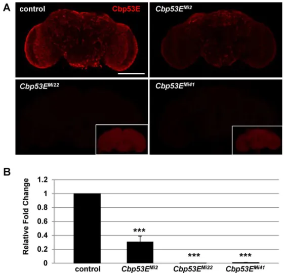
techniques to generate new mutant alleles [35]. Three alleles with reduced Cbp53E expression were identified through this process. Cbp53E is normally expressed throughout the adult brain; however theCbp53EMi2allele exhibited substantially reduced expression while staining in theCbp53EMi22andCbp53EMi41alleles was completely absent (Fig 1A). These findings were confirmed at the RNA level as quantitative RT-PCR revealed essentially no Cbp53E mRNA present inCbp53EMi22andCbp53EMi41heads whileCbp53EMi2animals only showed an approximately 70% reduction in Cbp53E mRNA relative to controls (Fig 1B). Thus,
Cbp53EMi22andCbp53EMi41were considered null alleles, since each completely lacked Cbp53E expression. None of the alleles exhibited any overt developmental phenotypes, and all were homozygous viable. Preliminary experiments could not identify any molecular or structural defects inCbp53EMi2, and since it retained some Cbp53E expression, only the two true null alleles were considered for further experimentation.
Drosophila
Cbp53E is expressed at the neuromuscular junction
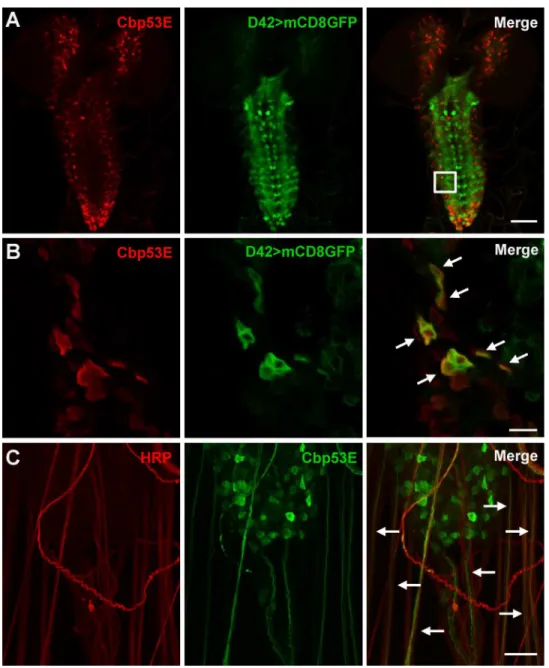
TheDrosophilalarval system has been extensively used to characterize the effects of genetic mutations on neuronal growth [39]. Since Cbp53E expression has mostly been characterized in adult animals, we used immunohistochemistry to determine where Cbp53E was localized in larval neuronal tissues. In the third instar larval brain, Cbp53E was expressed in a broad but discrete pattern but was completely absent fromCbp53EMi22null animals (Fig 2AandS1 Fig). In order to characterize some of this pattern, we specifically identified motor neurons by
Fig 1. Generation of novel loss of function Cbp53E alleles.A) A Minos transposable element in the 30
portion of the CBP53E locus was mobilized to generate 3 new alleles with reduced Cbp53E expression in the adult brain.Cbp53EMi2
shows residual Cbp53E, while allelesCbp53EMi22
andCbp53EMi41
are complete nulls. Insets show the same image with increased detector gain settings. Scale bar is 100μm. B) Quantitative RT-PCR of Cbp53E mRNA levels normalized to GAPDH2 from the indicated genotypes and reported as the relative fold change to control levels.***p<0.001.
expressing GFP from a motor neuron specific GAL4-driver and co-stained with anti-Cbp53E antibodies. While the expression patterns were not identical, high magnification of single slice images revealed significant colocalization of GFP and Cbp53E in subsets of cells (Fig 2B. Pear-son’s coefficient = 0.993). Strong Cbp53E expression was also visualized exiting the central ner-vous system in nearly every nerve bundle (Fig 2C). We therefore followed the nerves out to the peripheral nervous system to determine potential synaptically localized Cbp53E.
Fig 2. Expression of Cbp53E in the larval brain.A) Representative maximum intensity projection of a third instar larval brain from animals expressing UAS-mCD8GFP (green) from a D42-GAL4 motor neuron specific driver. Anti-Cbp53E staining (red) shows discrete expression patterns. Scale bar is 50μm. B) A single slice from the magnified region outlined in the merge panel in (A). Significant colocalization of anti-Cbp53E (red) and GFP (green) is seen in many cells (arrows). Scale bar is 10μm. C) Strong Cbp53E expression (green) can be seen in nerve fibers labeled with anti-HRP (red) as they exit the larval central nervous system (arrows). Scale bar is 20μm.
doi:10.1371/journal.pone.0132636.g002
The larval neuromuscular junction (NMJ) is comprised of axons innervating muscles and forming multiple types of synaptic boutons which can be characterized by their size and neuro-transmitter properties. Type Ib and Is boutons are primarily glutamatergic, while type II and type III boutons are peptidergic [40–43]. The strongest Cbp53E expression at the NMJ was seen in type II and type III synaptic boutons. Type II boutons are readily identified by their small, bead-like morphology, but type III boutons can only be conclusively characterized by the presence of insulin [41]. We therefore co-stained tissues with the neuronal marker anti-HRP, anti-Cbp53E, and anti-insulin antibodies and observed strong overlap of Cbp53E and insulin on muscle 12 in control animals (Fig 3A). Notably,Cbp53EMi22animals showed no Cbp53E NMJ localization (S2 Fig). Expression of Cbp53E in type II boutons, however, was more variable. We could find many instances of strong Cbp53E expression in type II boutons on many muscles including muscles 4, 6/7 and 12/13. One example of strong expression is shown on muscles 6/7 inFig 3B. Sometimes, however, expression in type II boutons was very weak as seen in the type II innervation present on muscle 4 (Fig 3C). There was no obvious pat-tern to this discrepancy in expression levels. Often times, type II boutons on different muscles in the same abdominal segment stained differently for Cbp53E. Furthermore, we examined NMJs of muscles 4, 6/7, and 12/13 and could not find any expression of Cbp53E in type I gluta-matergic synapses. Thus, levels of Cbp53E that are detectable by immunohistochemistry seem to be associated specifically with peptidergic synapses at the NMJ.
Drosophila
Cbp53E regulates neuronal development
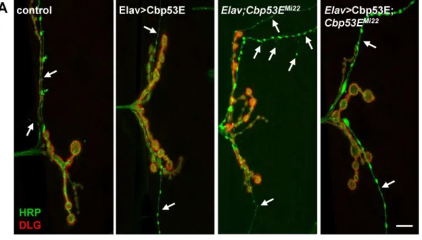
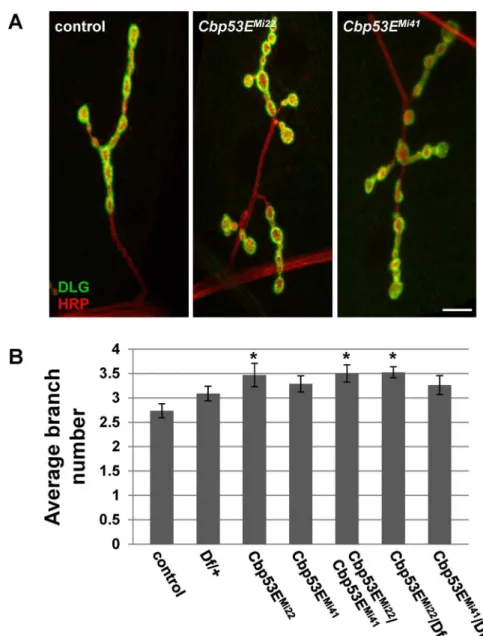
TheDrosophilalarval neuromuscular junction is a well-characterized model of neuronal devel-opment [44]. Since Cbp53E is localized to at least one class of NMJs we wanted to determine whether loss of function of Cbp53E could alter the growth of the NMJ. Since Cbp53E is most strongly expressed in type II and type III boutons on muscle 12, we combined the analysis of these innervations in control and multiple mutant lines. No reproducible differences in the number of type II/III synaptic boutons between control animals and eitherCbp53EMi22or Cbp53EMi41mutants could be identified (data not shown). We therefore focused on quantifying the axon branching patterns of type II/III innervations. BothCbp53EMi22andCbp53EMi41 mu-tant alleles showed an increase in type II/III neuronal branching relative to controls (Fig 4. con-trol: 4.7 +/- 0.37, n = 10;Cbp53EMi22: 6.8 +/- 0.31, n = 10, vs. control: p<0.05;Cbp53EMi41: 7.7 +/- 0.46, n = 9, vs. control: p<0.001). This finding remained consistent in theCbp53EMi22/ Cbp53EMi41trans-heterozygote (Cbp53EMi22/Cbp53EMi41: 7.1 +/-0.54, n = 9, vs. control: p<0.01). While a heterozygote deficiency line spanning the Cbp53E genomic locus exhibited a slight increase in branching, each Cbp53E allele in combination with the deficiency produced more significant increases in branch number (Df/+: 6.0 +/- 0.43, n = 12, vs. control: p<0.05; Cbp53EMi22/Df: 6.6 +/- 0.43, n = 14, vs. control: p<0.01;Cbp53EMi41/Df: 6.6 +/- 0.48, n = 10, vs. control: p<0.01). None of the mutant allele combinations were significantly different than the heterozygote deficiency line alone (all comparisons p>0.05) which is likely due to the large deletion affecting many genes. Thus, Cbp53E mitigates the extent of type II/III axonal arboriza-tion at the neuromuscular juncarboriza-tion.
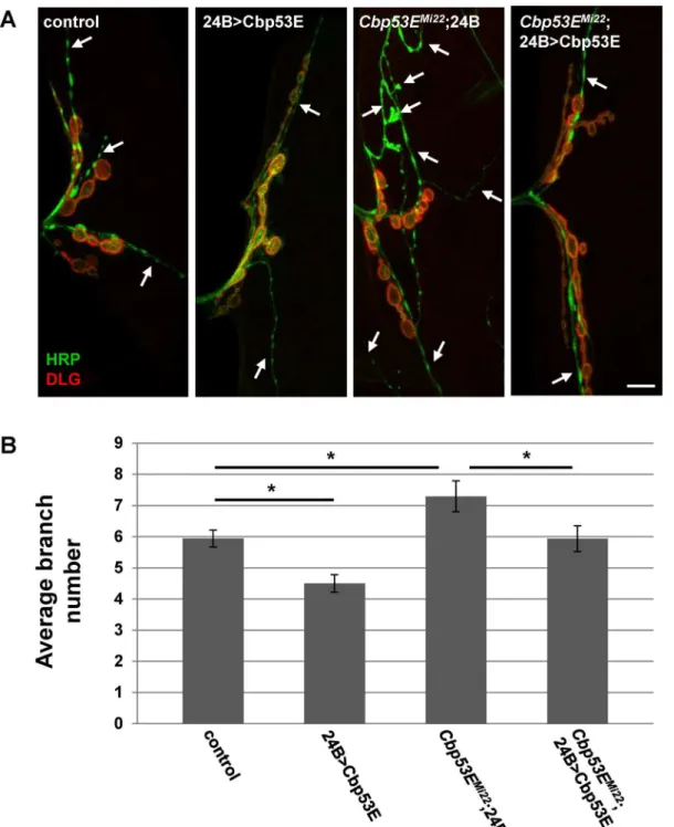
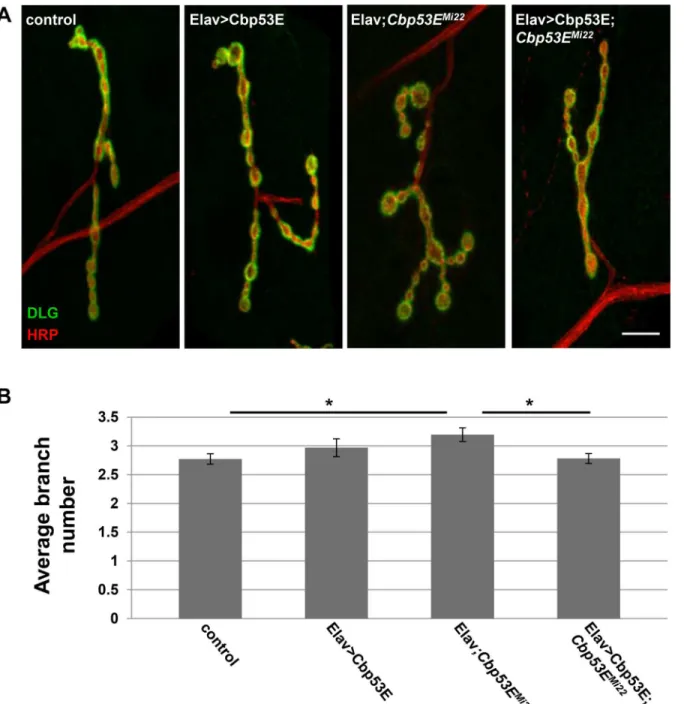
II/III axon overgrowth (Elav/+;Cbp53EMi22: 7.7 +/- 0.51, n = 15, vs. control: p<0.05; Elav>Cbp53E;Cbp53EMi22: 6.1 +/- 0.44, n = 16, vs Elav/+;Cbp53EMi22: p<0.05). We also expressed Cbp53E postsynaptically at the NMJ with the muscle specific driver 24B-GAL4 (Fig 6). In addition to rescuing the overgrowth seen inCbp53EMi22neurons, postsynaptic expression of Cbp53E alone resulted in a decrease in type II/III axonal branching (control: 5.9 +/- 0.27, n = 17; 24B>Cbp53E: 4.5 +/- 0.28, n = 13, vs. control: p<0.05,Cbp53EMi22;24B/+: 7.3 +/- 0.5, n = 17, vs. control: p<0.05;Cbp53EMi22;24B>Cbp53E: 5.9 +/- 0.42, n = 16, vs.
Fig 3. Expression of Cbp53E at the larval neuromuscular junction.A) Insulin peptide (purple) and Cbp53E (red) co-label type III boutons on muscle 12 of the NMJ. HRP (green) labels all neurons. B) A representative maximum intensity projection of strong Cbp53E staining (green) in type II boutons on muscles 6/7. C) A representative single slice image of weak Cbp53E staining (green) in type II boutons (arrow) on muscle 4. Cbp53E can also be seen in the nerve fibers around most muscles (arrowhead). Scale bars in all panels are 20μm.
doi:10.1371/journal.pone.0132636.g003
Fig 4. Loss of Cbp53E increases axon branching of type II/III terminals.A) Representative maximum intensity projections of muscle 12 innervations. All synapses are labeled with anti-HRP (green) while type I synapses are eliminated from analysis with anti-DLG labelling (red). Arrows point to type II/III axons branches of the indicated genotypes. Scale bar is 10μm. B) Quantitation of the average branch number of type II/III innervations from the indicated genotypes.*0.01<p<0.05,**0.001<p<0.01,***p<0.001.
Cbp53EMi22;24B/+: p<0.05). To ensure that rescue of branching defects in the Cbp53E nulls was not due to a potentially leaky transgene, we compared the number of branches from each driver in the mutant background with the transgene in the mutant background and could not detect any differences (Cbp53EMi22;24B/+: 7.3 +/- 0.5, n = 17; Elav/+;Cbp53EMi22: 7.7 +/- 0.51, n = 15; Cbp53EMi22; Cbp53E/+: 7.2 +/- 0.4, n = 17. ANOVA p = 0.79). Thus, neuronal growth defects stemming from genetic loss of function alleles of Cbp53E can be recovered by either
Fig 5. Neuronal expression of transgenic Cbp53E rescues type II/III axon branching defects.A) Representative maximum intensity projections of muscle 12 innervations labeled with anti-HRP (green) and anti-DLG (red). Arrows point to type II/III axon branches of the indicated genotypes. Scale bar is 10μm. B) Quantitation of the average branch number of type II/III innervations from the indicated genotypes.*0.01<p<0.05.
doi:10.1371/journal.pone.0132636.g005
pre- or postsynaptic introduction of transgenic Cbp53E. However, only the postsynaptic com-partment at the NMJ is sensitive to excessive Cbp53E expression with respect to type II/III innervations.
Even though we could not detect Cbp53E expression in type I glutamatergic synapses, we wanted to determine if these synapses were also affected by loss of function of Cbp53E. We
Fig 6. Muscle expression of transgenic Cbp53E rescues type II/III axon branching defects.A) Representative maximum intensity projections of muscle 12 innervations labeled with anti-HRP (green) and anti-DLG (red). Arrows point to type II/III axon branches of the indicated genotypes. Scale bar is 10μm. B) Quantitation of the average branch number of type II/III innervations from the indicated genotypes.*0.01<p<0.05.
therefore quantified type Ib innervations at the well-characterized larval muscle 4. This gluta-matergic synaptic arborization has a simple, easily-quantified pattern of innervation branches and boutons and has frequently been used as a model for glutamatergic neuronal development
[45–48]. As with type II/III boutons on muscle 12, we were unable to identify any changes in
type Ib bouton number between control animals and Cbp53E mutant alleles. We therefore quantified type Ib axon branch numbers in controls and all mutant allele combinations as before. Only theCbp53EMi22null mutant animals showed a significant increase in the average number of axon branches within the muscle 4 arbor (Fig 7. control: 2.7 +/- 0.14, n = 17; Cbp53EMi22: 3.5 +/- 0.24, n = 16, vs. control: p<0.05;Cbp53EMi41: 3.3 +/- 0.17, n = 14, vs. con-trol: p>0.05). The excessive branching seen inCbp53EMi22was, however, present in all trans-heterozygote combinations, though not in the deficiency trans-heterozygote line alone (Df/+: 3.1
+/-Fig 7. Loss of Cbp53E increases type Ib axonal branching at the NMJ.A) Representative maximum intensity projections of muscle 4 type Ib innervations from the indicated genotypes stained with anti-DLG (green) and anti-HRP (red). Scale bar is 10μm. B) Quantitation of the average branch number of type Ib innervations from the indicated genotypes.*0.01<p<0.05.
doi:10.1371/journal.pone.0132636.g007
0.15, n = 17, vs. control: p>0.05;Cbp53EMi22/Cbp53EMi41: 3.5 +/-0.18, n = 16, vs. control: p<0.05;Cbp53EMi22/Df: 3.5 +/- 0.11, n = 20, vs. control: p<0.05). As seen at type II/III NMJs, none of the allelic combinations were significantly different from the heterozygote deficiency line (p>0.05). Thus, we conclude that theCbp53EMi41allele is somewhat weaker than the Cbp53EMi22allele. Nevertheless, in addition to regulating peptidergic terminal formation, Cbp53E also seems to mitigate the extent of glutamatergic axonal growth at the neuromuscular junction.
We next characterized the effects of both pre- and postsynaptic overexpression of Cbp53E on muscle 4 type Ib axon branches. Surprisingly, unlike type II/III innervations on muscle 12, there were no differences detected in branch number of type Ib arbors between control and either Cbp53E overexpression condition (Figs8and9) (Neuronal expression: control: 2.8 +/-0.09, n = 22; Elav>Cbp53E: 3.0 +/- 0.15, n = 16, vs. control: p>0.05. Muscle expression: con-trol: 2.9 +/- 0.13, n = 23; 24B>Cbp53E: 2.8 +/-0.19, n = 19, vs. control: p>0.05). However, as was seen in peptidergic terminals, Cbp53E can function both pre-and postsynaptically as evi-denced by the targeted ability to rescue theCbp53EMi22null overgrowth (Elav/+;Cbp53Emi22: 3.2 +/- 0.12, n = 28 vs. control: p<0.05; Elav>Cbp53E;Cbp53EMi22: 2.8 +/- 0.09, n = 32, vs. Elav/+;Cbp53Emi22: p<0.05;Cbp53EMi22;24B/+: 3.2 +/-0.17, n = 21, vs. control: p<0.05; Cbp53EMi22;24B>Cbp53E: 2.6 +/- 0.16, n = 20, vs.Cbp53EMi22;24B/+: p<0.05). As with type II/III NMJs we did not detect any difference between the driver lines in the mutant background and the transgene in the mutant background indicating that this rescue is likely not the result of a leaky transgene (Elav/+;Cbp53Emi22: 3.2 +/- 0.12, n = 28;Cbp53EMi22;24B/+: 3.2 +/-0.17, n = 21;Cbp53EMi22; Cbp53E/+ 3.5 +/- 0.3, n = 17. ANOVA p = 0.4). Thus, loss of function of Cbp53E results in an increase in axon branching of glutamatergic innervations, which can be rescued by expressing Cbp53E either in neurons or in the muscle. However, irrespective of cel-lular compartment, overexpression of Cbp53E alone does not seem to affect glutamatergic neu-ronal development.
Discussion
For over 100 years,Drosophilahave been a key model organism in understanding genes and pathways involved in organismal development. Due to the strong evolutionary conservation of many genes, researchers are now turning toDrosophilato mimic the genetic alterations found in human diseases, including neurodevelopmental disorders such as Rett Syndrome and Angelman Syndrome and neurodegenerative disorders such as Parkinson’s disease and Alzheimer’s disease
[32,49,50]. Despite this increased effort to exploitDrosophilafor translational research, little
Cbp53Enull mutants exhibit overgrowth of terminal axon branches in both peptidergic and glutamatergic synapses (Figs4and7). It is therefore possible that the excessive branching at two different classes of synaptic terminals occurs via two distinct mechanisms. Cbp53E is local-ized to type II/III synaptic boutons and therefore may participate directly in synaptic mecha-nisms regulating neuronal growth. However, Cbp53E does not appear to be expressed at type I larval NMJs (Fig 3). While we cannot rule out that Cbp53E is expressed at type I synapses below the levels of our detection, since loss of function of Cbp53E still affects type I NMJ
Fig 8. Neuronal expression of transgenic Cbp53E rescues type Ib axon branching defects.A) Representative maximum intensity projections of muscle 4 type Ib innervations from the indicated genotypes stained with anti-DLG (green) and anti-HRP (red). Scale bar is 10μm. B) Quantitation of the average branch number of type Ib innervations from the indicated genotypes.*0.01<p<0.05.
doi:10.1371/journal.pone.0132636.g008
growth, a more global, rather than synaptic mechanism may direct events at these synapses. Alternatively, it is possible that localization of Cbp53E to type II/III synapses is not at all directly related to its function in neuronal growth but serves some other uncharacterized pur-pose. In this scenario, somatic control of axon growth by Cbp53E could govern both peptider-gic and glutamaterpeptider-gic arbor development. Further experiments directed at separating soma and synaptic mechanisms, however, are required to distinguish between these possibilities.
Even though we could not detect Cbp53E endogenously in the the larval muscle, it is inter-esting that both pre- and postsynaptic expression of Cbp53E is able to rescue all axonal branch-ing defects in Cbp53E null animals. One important caveat to these findbranch-ings is that while the
Fig 9. Muscle expression of transgenic Cbp53E rescues type Ib axon branching defects.A)
Representative maximum intensity projections of muscle 12 innervations labeled with anti-DLG (green) and anti-HRP (red). Scale bar is 10μm. B) Quantitation of the average branch number of type Ib innervations from the indicated genotypes.*0.01<p<0.05.
24B-gal4 driver used in this study is often used to target transgene expression in the muscle, it does also express transgenes in some neurons. We examined this expression pattern in the lar-val system, however, and could not find any overlap between neurons which express 24B-gal4 and endogenous Cbp53E either in cell soma, or at the NMJs examined in this study (S3 Fig). Nevertheless, it remains possible that neurons expressing transgenic Cbp53E from the 24B-gal4 driver could produce some circuit based effects which alter the axon development of the motor neurons studied here.
Somewhat mitigating this interpretation, however, is the finding that postsynaptic muscle overexpression of Cbp53E is able to reduce axon branching of type II/III terminals while pre-synaptic overexpression has no effect (Figs5and6). This result would suggest that any effect of presynaptic Cbp53E expression from the 24B-gal4 driver is offset by Cbp53E being present in the muscle. It is unclear why reduction of axon branch elaboration is limited to a postsynaptic function of Cbp53E. It is possible that there is a level of functional Cbp53E saturation that is already met by endogenous Cbp53E in neurons. Since muscles do not express appreciable lev-els of Cbp53E, this hypothetical threshold is not met in muscles thereby allowing transgenic Cbp53E to alter axon growth. The mechanism of this apparent retrograde function remains to be determined.
It is also interesting that peptidergic type II/III terminals are more sensitive than the type I glutamatergic terminals to levels of Cbp53E. Specifically, the effect of Cbp53E overexpression on axon growth does not occur at type I NMJs. Since Cbp53E is expressed at type II/III synap-ses but not type I synapsynap-ses, it is possible that this result does in fact point to a specific synaptic control mechanism of axon growth that is dependent on Cbp53E. However, the differences in growth effects on these terminals could also be due to the inherent functional differences of the synapses. It is known for example that the neurexin/neuroligin system modulates in order to differentially regulate the formation of different classes of synapses [52]. Differential regulation of these molecules in the context of Cbp53E null animals could potentially explain why overex-pression of Cbp53E uniquely affects type I and type II/III NMJs. More functional characteriza-tion of Cbp53E in these pre- and postsynaptic contexts will be necessary to elucidate the exact mechanism of these axon growth defects.
We have used theDrosophilaNMJ as a model of neuronal growth to begin to understand the role of Cbp53E in the developing nervous system. Though data reported here support the hypothesis that Cbp53E regulates neuronal development, it is clear that many questions still remain about Cbp53E function. The CB/CR family of calcium binding proteins possesses well-characterized calcium buffering properties, however the calcium buffering capacity of Cbp53E remains to be empirically determined. Nevertheless, the animals we have described can now be used to investigate the diverse contributions of the CB/CR proteins inDrosophilagenetic mod-els of human diseases. In particular, it will be interesting to determine whether this single gene can provide the neuroprotective functions of the mammalian family in degenerative disorders such as PD and AD. Incorporating genetic alterations of Cbp53E will surely enhance the value of these and other models and potentially uncover important calcium-dependent mechanisms underlying each disease.
Supporting Information
S1 Fig. Expression of Cbp53E is absent inCbp53EMi22larval brains.Control (A) and Cbp53EMi22(B) larval brains were stained with anti-Cbp53E and anti-HRP antibodies and then imaged under identical conditions. Scale bar is 10μm.
(TIF)
S2 Fig. Expression of Cbp53E is absent inCbp53EMi22muscle 12 NMJs.Control (A) and Cbp53EMi22(B) larval segment 3 muscle 12 NMJs were stained with Cbp53E and anti-HRP antibodies and then imaged under identical conditions. Scale bar is 10μm.
(TIF)
S3 Fig. 24B-gal4 drives expression in some neurons but is postsynaptic at the NMJ.(A) Lar-val brains from animals using 24B-gal4 to drive expression of mCD8GFP were stained with anti-Cbp53E antibodies. Each slice panel is a single representative optical section from 3 differ-ent depths of the same brain. The final panel is a maximum intensity projection of the image stack. Arrows indicate neurons expressing GFP from the 24B-gal4 driver. We could not find any cells expressing both GFP and Cbp53E. (B) Muscle 4 NMJ from the animals in (A) stained with HRP to denote the presynaptic compartment and DLG to denote the postsynaptic com-partment. GFP overlaps only with DLG. (C) Muscle 12 NMJ from the animals in (A) stained with anti-Cbp53E antibodies. GFP does not appear in type II/III synapses containing Cbp53E, thus indicating only postsynaptic expression at this synapse. Scale bars are 10μm.
(TIF)
Acknowledgments
We would like to thank Andrew Bantel for his experimental assistance and manuscript review. Work was performed at the Indiana University School of Medicine-South Bend Imaging and Flow Cytometry Core Facility. Stocks obtained from the BloomingtonDrosophilaStock Center (NIH P40OD018537) were used in this study.
Author Contributions
Conceived and designed the experiments: CRT. Performed the experiments: KRH JB CRT. Analyzed the data: KRH JB CRT. Wrote the paper: KRH CRT.
References
1. Bronner F. Extracellular and intracellular regulation of calcium homeostasis. ScientificWorldJournal. 2001; 1:919–25. Epub 2003/06/14. doi:10.1100/tsw.2001.489PMID:12805727.
2. Kavalali ET. The mechanisms and functions of spontaneous neurotransmitter release. Nat Rev Neu-rosci. 2015; 16(1):5–16. doi:10.1038/nrn3875PMID:25524119.
3. Bachstetter AD, Webster SJ, Tu T, Goulding DS, Haiech J, Watterson DM, et al. Generation and behav-ior characterization of CaMKIIbeta knockout mice. PLoS One. 2014; 9(8):e105191. doi:10.1371/ journal.pone.0105191PMID:25127391; PubMed Central PMCID: PMC4134274.
4. Waites CL, Garner CC. Presynaptic function in health and disease. Trends Neurosci. 2011; 34(6):326–37. Epub 2011/05/21. doi:10.1016/j.tins.2011.03.004S0166-2236(11)00054-3 [pii]. PMID:21596448. 5. Suo D, Park J, Young S, Makita T, Deppmann CD. Coronin-1 and calcium signaling governs
sympa-thetic final target innervation. J Neurosci. 2015; 35(9):3893–902. doi:10.1523/JNEUROSCI.4402-14. 2015PMID:25740518; PubMed Central PMCID: PMC4348186.
6. Gilabert JA. Cytoplasmic calcium buffering. Adv Exp Med Biol. 2012; 740:483–98. doi: 10.1007/978-94-007-2888-2_20PMID:22453955.
7. Schwaller B, Meyer M, Schiffmann S. 'New' functions for 'old' proteins: the role of the calcium-binding proteins calbindin D-28k, calretinin and parvalbumin, in cerebellar physiology. Studies with knockout mice. Cerebellum. 2002; 1(4):241–58. Epub 2003/07/26. doi:10.1080/147342202320883551PMID: 12879963.
8. Schwaller B. Cytosolic Ca2+ buffers. Cold Spring Harb Perspect Biol. 2010; 2(11):a004051. Epub 2010/10/15. doi:10.1101/cshperspect.a004051cshperspect.a004051 [pii]. PMID:20943758; PubMed Central PMCID: PMC2964180.
10. Chard PS, Bleakman D, Christakos S, Fullmer CS, Miller RJ. Calcium buffering properties of calbindin D28k and parvalbumin in rat sensory neurones. J Physiol. 1993; 472:341–57. Epub 1993/12/01. PMID: 8145149; PubMed Central PMCID: PMC1160490.
11. Schwaller B, Durussel I, Jermann D, Herrmann B, Cox JA. Comparison of the Ca2+-binding properties of human recombinant calretinin-22k and calretinin. J Biol Chem. 1997; 272(47):29663–71. Epub 1997/ 12/31. PMID:9368033.
12. Gartner W, Lang W, Leutmetzer F, Domanovits H, Waldhausl W, Wagner L. Cerebral expression and serum detectability of secretagogin, a recently cloned EF-hand Ca(2+)-binding protein. Cereb Cortex. 2001; 11(12):1161–9. PMID:11709487.
13. Gartner W, Vila G, Daneva T, Nabokikh A, Koc-Saral F, Ilhan A, et al. New functional aspects of the neuroendocrine marker secretagogin based on the characterization of its rat homolog. American jour-nal of physiology Endocrinology and metabolism. 2007; 293(1):E347–54. doi:10.1152/ajpendo.00055. 2007PMID:17426113.
14. Berggard T, Miron S, Onnerfjord P, Thulin E, Akerfeldt KS, Enghild JJ, et al. Calbindin D28k exhibits properties characteristic of a Ca2+ sensor. J Biol Chem. 2002; 277(19):16662–72. Epub 2002/03/02. doi:10.1074/jbc.M200415200M200415200 [pii]. PMID:11872749.
15. Schwaller B. The continuing disappearance of "pure" Ca2+ buffers. Cell Mol Life Sci. 2009; 66(2): 275–300. Epub 2008/12/23. doi:10.1007/s00018-008-8564-6PMID:19099190.
16. Bastianelli E. Distribution of calcium-binding proteins in the cerebellum. Cerebellum. 2003; 2(4): 242–62. Epub 2004/02/18. doi:10.1080/14734220310022289PMID:14964684.
17. Sloviter RS, Sollas AL, Barbaro NM, Laxer KD. Calcium-binding protein (calbindin-D28K) and parvalbu-min immunocytochemistry in the normal and epileptic human hippocampus. J Comp Neurol. 1991; 308(3):381–96. Epub 1991/06/15. doi:10.1002/cne.903080306PMID:1865007.
18. Gulyas AI, Hajos N, Freund TF. Interneurons containing calretinin are specialized to control other inter-neurons in the rat hippocampus. J Neurosci. 1996; 16(10):3397–411. Epub 1996/05/15. PMID: 8627375.
19. Rogers JH. Immunohistochemical markers in rat cortex: co-localization of calretinin and calbindin-D28k with neuropeptides and GABA. Brain Res. 1992; 587(1):147–57. Epub 1992/07/31. 0006-8993(92) 91439-L [pii]. PMID:1356060.
20. Cheron G, Schurmans S, Lohof A, d'Alcantara P, Meyer M, Draye JP, et al. Electrophysiological behav-ior of Purkinje cells and motor coordination in calretinin knock-out mice. Prog Brain Res. 2000; 124:299–308. Epub 2000/08/16. S0079-6123(00)24024-7 [pii] doi:10.1016/S0079-6123(00)24024-7 PMID:10943133.
21. Schiffmann SN, Cheron G, Lohof A, d'Alcantara P, Meyer M, Parmentier M, et al. Impaired motor coor-dination and Purkinje cell excitability in mice lacking calretinin. Proc Natl Acad Sci U S A. 1999; 96(9): 5257–62. Epub 1999/04/29. PMID:10220453; PubMed Central PMCID: PMC21851.
22. Airaksinen MS, Eilers J, Garaschuk O, Thoenen H, Konnerth A, Meyer M. Ataxia and altered dendritic calcium signaling in mice carrying a targeted null mutation of the calbindin D28k gene. Proc Natl Acad Sci U S A. 1997; 94(4):1488–93. Epub 1997/02/18. PMID:9037080; PubMed Central PMCID: PMC19818.
23. Gall D, Roussel C, Susa I, D'Angelo E, Rossi P, Bearzatto B, et al. Altered neuronal excitability in cere-bellar granule cells of mice lacking calretinin. J Neurosci. 2003; 23(28):9320–7. Epub 2003/10/17. 23/ 28/9320 [pii]. PMID:14561859.
24. Westerink RH, Beekwilder JP, Wadman WJ. Differential alterations of synaptic plasticity in dentate gyrus and CA1 hippocampal area of Calbindin-D28K knockout mice. Brain Res. 2012; 1450:1–10. Epub 2012/03/13. doi:10.1016/j.brainres.2012.02.036S0006-8993(12)00313-7 [pii]. PMID:22405690. 25. Gurden H, Schiffmann SN, Lemaire M, Bohme GA, Parmentier M, Schurmans S. Calretinin expression
as a critical component in the control of dentate gyrus long-term potentiation induction in mice. Eur J Neurosci. 1998; 10(9):3029–33. Epub 1998/10/03. PMID:9758174.
26. Mouatt-Prigent A, Agid Y, Hirsch EC. Does the calcium binding protein calretinin protect dopaminergic neurons against degeneration in Parkinson's disease? Brain Res. 1994; 668(1–2):62–70. Epub 1994/ 12/30. 0006-8993(94)90511-8 [pii]. PMID:7704619.
27. Pike CJ, Cotman CW. Calretinin-immunoreactive neurons are resistant to beta-amyloid toxicity in vitro. Brain Res. 1995; 671(2):293–8. Epub 1995/02/13. 0006-8993(94)01354-K [pii]. PMID:7743217. 28. Fonseca M, Soriano E. Calretinin-immunoreactive neurons in the normal human temporal cortex and in
Alzheimer's disease. Brain Res. 1995; 691(1–2):83–91. Epub 1995/09/11. 0006-8993(95)00622-W [pii]. PMID:8590068.
29. Yuan HH, Chen RJ, Zhu YH, Peng CL, Zhu XR. The neuroprotective effect of overexpression of calbin-din-D(28k) in an animal model of Parkinson's disease. Mol Neurobiol. 2013; 47(1):117–22. Epub 2012/ 08/28. doi:10.1007/s12035-012-8332-3PMID:22923348.
30. Odero GL, Oikawa K, Glazner KA, Schapansky J, Grossman D, Thiessen JD, et al. Evidence for the involvement of calbindin D28k in the presenilin 1 model of Alzheimer's disease. Neuroscience. 2010; 169(1):532–43. Epub 2010/04/20. doi:10.1016/j.neuroscience.2010.04.004S0306-4522(10)00508-7 [pii]. PMID:20399254.
31. Bellido T, Huening M, Raval-Pandya M, Manolagas SC, Christakos S. Calbindin-D28k is expressed in osteoblastic cells and suppresses their apoptosis by inhibiting caspase-3 activity. J Biol Chem. 2000; 275(34):26328–32. Epub 2000/06/03. doi:10.1074/jbc.M003600200M003600200 [pii]. PMID: 10835428.
32. Muqit MM, Feany MB. Modelling neurodegenerative diseases in Drosophila: a fruitful approach? Nat Rev Neurosci. 2002; 3(3):237–43. Epub 2002/05/08. doi:10.1038/nrn751nrn751 [pii]. PMID: 11994755.
33. Reifegerste R, Grimm S, Albert S, Lipski N, Heimbeck G, Hofbauer A, et al. An invertebrate calcium-binding protein of the calbindin subfamily: protein structure, genomic organization, and expression pat-tern of the calbindin-32 gene of Drosophila. J Neurosci. 1993; 13(5):2186–98. Epub 1993/05/01. PMID: 8478695.
34. Diaz-Balzac CA, Lazaro-Pena MI, Garcia-Rivera EM, Gonzalez CI, Garcia-Arraras JE. Calbindin-D32k is localized to a subpopulation of neurons in the nervous system of the sea cucumber Holothuria glaber-rima (Echinodermata). PLoS One. 2012; 7(3):e32689. doi:10.1371/journal.pone.0032689PMID: 22412907; PubMed Central PMCID: PMC3296734.
35. Metaxakis A, Oehler S, Klinakis A, Savakis C. Minos as a genetic and genomic tool in Drosophila mela-nogaster. Genetics. 2005; 171(2):571–81. Epub 2005/06/24. genetics.105.041848 [pii] doi:10.1534/ genetics.105.041848PMID:15972463; PubMed Central PMCID: PMC1456772.
36. Tessier CR, Broadie K. Drosophila fragile X mental retardation protein developmentally regulates activ-ity-dependent axon pruning. Development. 2008; 135(8):1547–57. Epub 2008/03/07. doi:10.1242/dev. 015867dev.015867 [pii]. PMID:18321984; PubMed Central PMCID: PMC3988902.
37. Hofbauer A, Ebel T, Waltenspiel B, Oswald P, Chen YC, Halder P, et al. The Wuerzburg hybridoma library against Drosophila brain. Journal of neurogenetics. 2009; 23(1–2):78–91. doi:10.1080/ 01677060802471627PMID:19132598.
38. Coffee RL Jr., Tessier CR, Woodruff EA 3rd, Broadie K. Fragile X mental retardation protein has a unique, evolutionarily conserved neuronal function not shared with FXR1P or FXR2P. Dis Model Mech. 2010; 3(7–8):471–85. Epub 2010/05/06. doi:10.1242/dmm.004598dmm.004598 [pii]. PMID:
20442204; PubMed Central PMCID: PMC2898537.
39. Menon KP, Carrillo RA, Zinn K. Development and plasticity of the Drosophila larval neuromuscular junction. Wiley interdisciplinary reviews Developmental biology. 2013; 2(5):647–70. doi:10.1002/wdev. 108PMID:24014452; PubMed Central PMCID: PMC3767937.
40. Choi JC, Park D, Griffith LC. Electrophysiological and morphological characterization of identified motor neurons in the Drosophila third instar larva central nervous system. J Neurophysiol. 2004; 91(5): 2353–65. doi:10.1152/jn.01115.2003PMID:14695352.
41. Gorczyca M, Augart C, Budnik V. Insulin-like receptor and insulin-like peptide are localized at neuro-muscular junctions in Drosophila. J Neurosci. 1993; 13(9):3692–704. PMID:8366341.
42. Monastirioti M, Gorczyca M, Rapus J, Eckert M, White K, Budnik V. Octopamine immunoreactivity in the fruit fly Drosophila melanogaster. J Comp Neurol. 1995; 356(2):275–87. doi:10.1002/cne. 903560210PMID:7629319.
43. Rivlin PK, St Clair RM, Vilinsky I, Deitcher DL. Morphology and molecular organization of the adult neu-romuscular junction of Drosophila. J Comp Neurol. 2004; 468(4):596–613. doi:10.1002/cne.10977 PMID:14689489.
44. Keshishian H, Broadie K, Chiba A, Bate M. The drosophila neuromuscular junction: a model system for studying synaptic development and function. Annual review of neuroscience. 1996; 19:545–75. doi:10. 1146/annurev.ne.19.030196.002553PMID:8833454.
45. Franco B, Bogdanik L, Bobinnec Y, Debec A, Bockaert J, Parmentier ML, et al. Shaggy, the homolog of glycogen synthase kinase 3, controls neuromuscular junction growth in Drosophila. J Neurosci. 2004; 24(29):6573–7. doi:10.1523/JNEUROSCI.1580-04.2004PMID:15269269.
46. Johansen J, Halpern ME, Johansen KM, Keshishian H. Stereotypic morphology of glutamatergic syn-apses on identified muscle cells of Drosophila larvae. J Neurosci. 1989; 9(2):710–25. PMID:2563766. 47. Miller DL, Ballard SL, Ganetzky B. Analysis of synaptic growth and function in Drosophila with an
48. Zhang YQ, Bailey AM, Matthies HJ, Renden RB, Smith MA, Speese SD, et al. Drosophila fragile X-related gene regulates the MAP1B homolog Futsch to control synaptic structure and function. Cell. 2001; 107(5):591–603. Epub 2001/12/06. S0092-8674(01)00589-X [pii]. PMID:11733059.
49. Cukier HN, Perez AM, Collins AL, Zhou Z, Zoghbi HY, Botas J. Genetic modifiers of MeCP2 function in Drosophila. PLoS Genet. 2008; 4(9):e1000179. Epub 2008/09/06. doi:10.1371/journal.pgen.1000179 PMID:18773074; PubMed Central PMCID: PMC2518867.
50. Wu Y, Bolduc FV, Bell K, Tully T, Fang Y, Sehgal A, et al. A Drosophila model for Angelman syndrome. Proc Natl Acad Sci U S A. 2008; 105(34):12399–404. Epub 2008/08/15. doi:10.1073/pnas.
08052911050805291105 [pii]. PMID:18701717; PubMed Central PMCID: PMC2527923.
51. Fahandejsaadi A, Leung E, Rahaii R, Bu J, Geula C. Calbindin-D28K, parvalbumin and calretinin in pri-mate lower motor neurons. Neuroreport. 2004; 15(3):443–8. Epub 2004/04/20. 00001756-200403010-00012 [pii]. PMID:15094500.
52. Craig AM, Kang Y. Neurexin-neuroligin signaling in synapse development. Current opinion in neurobiol-ogy. 2007; 17(1):43–52. doi:10.1016/j.conb.2007.01.011PMID:17275284; PubMed Central PMCID: PMC2820508.