The Rockefeller University Press $30.00
JCB: REPORT
Correspondence to James M. Ervasti: jervasti@umn.edu
Abbreviations used in this paper: CH, calponin homology; MAP, MT-associated protein; MOM, mouse on mouse; MT, microtubule; wt, wild type.
Introduction
The plakins are a class of giant cytolinker proteins that can link transmembrane protein complexes to the actin, intermediate fila ment, and microtubule (MT) cytoskeletons in various combi nations (Fuchs and Karakesisoglou, 2001). Plakins can bind actin filaments via tandem calponin homology (CH) domains, intermediate filaments via plakin repeat domains, and MTs through either a Gas2related domain or a glycine/serine/ arginine domain (for review see Leung et al., 2002). The ability to crosslink multiple components of the cellular cytoskeleton allows cytolinkers to stabilize cells from mechanically induced damage. Mouse knockout studies exemplify the stabilizing effects of cytolinkers, as loss of the cytolinker plectin resulted in skin blistering and a form of muscular dystrophy (Andra et al., 1997), and ablation of BPAG1, another cytolinker, caused skin blistering upon mechanical stimulation (Guo et al., 1995).
Dystrophin, the protein absent in patients with Duchenne muscular dystrophy (Hoffman et al., 1987), shows structural and functional similarities to cytolinkers, which suggests the hypoth esis that dystrophin performs a cytolinker role in muscle. Dystro phin’s large molecular mass of 427 kD, spectrinlike repeats, and ability to bind actin filaments via a tandem CH domain (Way et al., 1992) highlight three similarities with cytolinkers. Although
dystrophin lacks a plakin repeat domain, dystrophin–intermediate filament interactions have been documented (Stone et al., 2005; Bhosle et al., 2006). Thus, the ability to link the actin and inter mediate filament cytoskeletons to the transmembrane dystro glycan complex (Suzuki et al., 1992) illustrates how dystrophin functions similarly to other cytolinkers. Finally, the muscle membrane fragility associated with the loss of dystrophin (Petrof et al., 1993) parallels the structural deficiencies observed in other cytolinkerdeficient tissues, further demonstrating a close rela tionship between dystrophin and other cytolinkers. Collectively, these data support the hypothesis that dystrophin may function as a cytolinker in skeletal muscle.
Although dystrophin exhibits many characteristics of a cytolinker, a direct dystrophin–MT interaction has not been doc umented. Dystrophin lacks either a Gas2related or a glycine/ serine/arginine domain, but recent studies indicated that dystro phin at least indirectly influences MT organization or stability (Percival et al., 2007; Ayalon et al., 2008). For instance, the dystrophindeficient mdx mouse exhibited MT disorganization in skeletal muscle with the costameric MTs most severely affected (Percival et al., 2007). Dystrophin’s enrichment at costameres
C
ytolinkers are giant proteins that can stabilize cellsby linking actin filaments, intermediate filaments, and microtubules (MTs) to transmembrane com-plexes. Dystrophin is functionally similar to cytolinkers, as it links the multiple components of the cellular cytoskeleton to the transmembrane dystroglycan complex. Although no direct link between dystrophin and MTs has been docu-mented, costamere-associated MTs are disrupted when dystrophin is absent. Using tissue-based cosedimenta-tion assays on mice expressing endogenous dystrophin or truncated transgene products, we find that constructs
harboring spectrinlike repeat 24 through the first third of the WW domain cosediment with MTs. Purified Dp260, a truncated isoform of dystrophin, bound MTs with a Kd of 0.66 µM, a stoichiometry of 1 Dp260/1.4 tubulin heterodimer at saturation, and stabilizes MTs from cold-induced depolymerization. Finally, - and -tubulin expression is increased 2.5-fold in mdx skeletal muscle
without altering the tubulin–MT equilibrium. Collectively, these data suggest dystrophin directly organizes and/or stabilizes costameric MTs and classifies dystrophin as a cytolinker in skeletal muscle.
Dystrophin is a microtubule-associated protein
Kurt W. Prins,1 Jill L. Humston,2 Amisha Mehta,3 Victoria Tate,3 Evelyn Ralston,3 and James M. Ervasti1,2
1Department of Biochemistry, Molecular Biology, and Biophysics, University of Minnesota, Minneapolis, MN 55455 2Molecular and Cellular Pharmacology Training Program, University of Wisconsin, Madison, WI 53706
3Light Imaging Section, Office of Science and Technology, National Institute of Arthritis and Musculoskeletal and Skin Diseases, National Institutes of Health, Bethesda,
MD 20892
© 2009 Prins et al. This article is distributed under the terms of an Attribution– Noncommercial–Share Alike–No Mirror Sites license for the first six months after the publica-tion date (see http://www.jcb.org/misc/terms.shtml). After six months it is available under a Creative Commons License (Attribution–Noncommercial–Share Alike 3.0 Unported license, as described at http://creativecommons.org/licenses/by-nc-sa/3.0/).
THE
JOURNAL
OF
CELL
and the restoration of costameric MT organization through virally mediated expression of a microdystrophin (Percival et al., 2007) indicates that dystrophin is necessary for proper costameric MT organization in skeletal muscle. Moreover, acute knockdown of ankyrinB, a protein necessary for delivery of dystrophin to the sarcolemma and neuromuscular junction, caused the loss of costa meric MTs and aberrant MT organization in a subset of MTs underlying the neuromuscular junction (Ayalon et al., 2008).
In this study, we investigated the hypothesis that dystro phin directly interacts with costameric MTs. We confirmed that costameric MTs were disrupted in dystrophindeficient skeletal muscle and showed endogenous dystrophin cosedimented with MTs in tissue homogenates. Using purified proteins, we found that the carboxylterminal two thirds of dystrophin bound MTs with a Kd of 0.66 µM and stabilized MTs from coldinduced de
polymerization. Finally, we documented a 2.5fold increased expression of and tubulin without alteration in the tubulin– MT equilibrium in mdx skeletal muscle. These results demon strate that dystrophin is a MTassociated protein (MAP) that stabilizes costameric MTs and functions as a costameric cyto linker in skeletal muscle.
Results and discussion
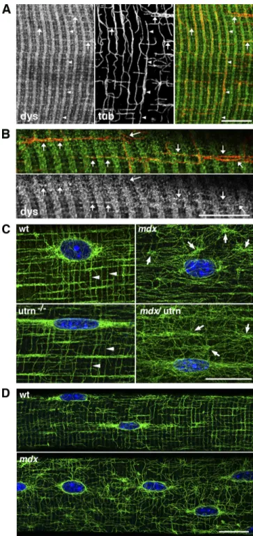
To determine whether dystrophin and MTs localize to similar structures in skeletal muscle, we conducted immunofluorescence analysis on teased extensor digitorum longus muscle fibers co labeled with antidystrophin and anti–tubulin antibodies (Fig. 1, A and B). Dystrophin forms a subsarcolemmal network with transverse components along the I bands and the M line and with longitudinal components (Williams and Bloch, 1999), whereas MTs form a subsarcolemmal lattice, which in fast fibers, has transverse and longitudinal components plus an accumulation of MTs around myonuclei (Ralston et al., 1999). We found that the transverse MTs (Fig. 1 A, arrowheads) weave their course along the I band dystrophin staining for long distances. MTs were also associated with longitudinal lines of dystrophin (Fig. 1 A, ar rows). These data identify domains of the subsarcolemmal cyto skeleton where dystrophin and MTs may interact either directly or indirectly. Next, we examined MT organization in mouse models lacking dystrophin (mdx), dystrophin’s autosomal homo logue utrophin (utrn/), or both dystrophin and utrophin (mdx/utrn/). Consistent with previous results (Percival et al., 2007), loss of dystrophin resulted in MT disorganization with the costameric MTs appearing to be most severely affected (Fig. 1 C) when compared with wild type (wt; Fig. 1 C). Ablation of utro phin had no effect on MT organization (Fig. 1 C), which is likely a result of its very low expression (Rybakova et al., 2002) and restriction to the neuromuscular junction (Ohlendieck et al., 1991). Finally, mdx/utrn/ skeletal muscle exhibited MT dis organization comparable with that of mdx (Fig. 1 C). MT organi zation in 24dold prenecrotic mdx skeletal muscle fibers was also disorganized, whereas agematched wt mice displayed a MT lat tice nearly identical to mature wt mice (Fig. 1 D). Collectively, these results confirm a role for dystrophin in the stabilization and proper organization of costameric MTs independent of muscle necrosis and regeneration.
Figure 1. Dystrophin guides MTs at the surface of the muscle fibers and is necessary for proper MT organization. (A) Isolated muscle fibers from the extensor digitorum longus of 7-wk-old wt mice were costained for dystro-phin (left) and -tubulin (middle). The right panel shows that MTs (red) follow dystrophin (green) bands for long distances both transversely (arrowheads) and longitudinally (arrows). (B) At a higher magnification, dystrophin stain-ing is granular; MTs are studded with dystrophin “dots.” Arrows indicate longitudinal MTs that follow dystrophin. (C) Muscle fibers from the extensor digitorum longus of 7-wk-old wt, mdx, utrn/, and mdx/utrn/ mice were
stained with DM1A anti–-tubulin and Hoechst dye. Both wt and utrn/
fibers show the lattice of transverse and longitudinal MTs characteristic of fast fibers (arrowheads). In mdx and mdx/utrn/ fibers, the regularity of
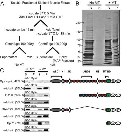
rod domain absent from Dp260. A small amount of Dp260 pel leted in the absence of MTs, but substantially more Dp260 shifted to the pellet fraction when MTs were present (Fig. 3 A). After sub tracting selfpelleting Dp260, Dp260 displayed a concentration
dependent and saturable cosedimentation with a Dp260/
tubulin heterodimer stoichiometry of 1:1.4 and a Kd of 0.66 µM
(Fig. 3 C). As predicted, DysNTermR10 did not cosediment with
MTs up to concentrations approaching 10 µM (Fig. 3, B and C). Next, we assessed how the presence of 1 µM Dp260 affected the tubulin–MT equilibrium in vitro. Dp260 had no significant effect on the fraction of tubulin in the MT fraction when incu bated at room temperature (67.3 ± 0.72% vs. 68.6 ± 1.3%). However, the presence of Dp260 significantly increased the fraction of tubulin retained in the MT pellet (33.6 ± 2.9% vs. 42.2 ± 2.0%) when MTs were induced to depolymerize by incu bating at 4°C. (Fig. 3, D and E). Collectively, these results dem onstrate that dystrophin directly binds and stabilizes MTs from coldinduced depolymerization.
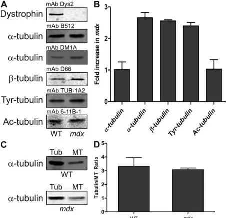
Because misregulation of other MAPs can alter tubulin expression and MT stability (Harada et al., 1994; Takahashi et al., 2003), we investigated how the loss of dystrophin affects the regulation of tubulin expression and the tubulin–MT equi librium in skeletal muscle fibers. Tubulin levels in wt and mdx skeletal muscle extracts were examined by quantitative West ern blot analysis. With mAb B512, we observed no difference Next, we performed a tissuebased MT cosedimentation
assay (Fig. 2 A) to determine whether dystrophin cosedimented with MTs. Under conditions that induced MT depolymeriza tion, virtually no muscle protein pelleted (Fig. 2 B). However, numerous proteins pelleted under MTstabilizing conditions, and this fraction of proteins represents MTs and the MAPs of skeletal muscle (Fig. 2 B). We Western blotted each fraction obtained from the tissue cosedimentation assay performed on wt mice expressing fulllength dystrophin or transgenic mdx
mice expressing Dp260, microdystrophin (R423), or Dp71
(Fig. 2 C, right). Fulllength dystrophin, Dp260, and R423 all pelleted with MTs, whereas Dp 71 did not (Fig. 2 C, left). By comparing the dystrophin domains present or absent in each construct (Fig. 2 C, right) along with each construct’s ability to cosediment with MTs, we suggest that spectrinlike repeat 24 through the first third of the WW domain encodes a novel MTbinding domain.
To test for a direct interaction between dystrophin and MTs, we performed MT cosedimentation using two purified recombi nant dystrophin constructs and purified tubulin. The two recombi nant constructs used were Dp260, which encodes from spectrinlike repeat 10 through the carboxy terminus of dystrophin, including the proposed MTbinding domain, and DysNTermR10 (Rybakova
et al., 2006), which encodes the aminoterminal, tandem CH actinbinding domain and spectrinlike repeats 1–10 of the middle
in tubulin expression in mdx skeletal muscle (Fig. 4, A and B). Because levels of and tubulin are coregulated (Gonzalez Garay and Cabral, 1995), we investigated tubulin levels to determine whether tubulin is upregulated in mdx skeletal muscle. Tubulin expression was elevated 2.5fold in mdx skeletal muscle (Fig. 4, A and B), suggesting that expression of both and tubulin is increased in mdx skeletal muscle. Thus, we conclude that mAb DM1A is able to recognize a pop ulation of tubulin not detected by mAb B512. To examine MT stability in mdx skeletal muscle, we analyzed levels of tyrosinated tubulin, a marker of dynamic MTs (Gundersen et al., 1984, 1987), and acetylated tubulin, a marker of long lived MTs (Bulinski and Gundersen, 1991). The levels of tyro sinated tubulin were increased 2.5fold in mdx extracts (Fig. 4, A and B), whereas the levels of acetylated tubulin were not (Fig. 4, A and B). The loss of dystrophin’s MTstabilizing ability may explain why acetylated tubulin was not more abundant in mdx skeletal muscle extracts, but alterations in the tubulin–MT equilibrium could also explain the lack of more stable MTs. Therefore, we examined the tubulin–MT equilib rium in wt and mdx skeletal muscles and found that the loss of dystrophin did not affect the equilibrium (Fig. 4, C and D). Collectively, these results show that tubulins are misregulated in dystrophindeficient skeletal muscle without affecting the tubulin–MT equilibrium. The loss of dystrophin’s MTstabilizing ability likely explains why there are not more stabilized MTs even in the presence of more tubulin dimer in dystrophindeficient skeletal muscle.
An indirect link between dystrophin and MTs mediated by ankyrinB was recently shown to be important for proper trafficking of dystrophin and dystroglycan to the sarcolemma (Ayalon et al., 2008). However, costameric MTs are disorga nized in mdx skeletal muscle even in the presence of properly localized ankyrinB (Ayalon et al., 2008). Because the MT and ankyrinB–binding domains of dystrophin do not overlap (Fig. 5), our results and previous results suggest that dystrophin interacts with MTs in vivo through two distinct mechanisms. We propose that ankyrinB delivers dystrophin to the sarcolemma depen dent on MTs and that dystrophin and ankyrinB collaborate to stabilize and organize MTs in skeletal muscle.
As with other cytolinkers, the ability to bind multiple com ponents of the filamentous cytoskeleton likely allows dystrophin to protect the sarcolemma from mechanically induced damage. One highly truncated microdystrophin construct (R423) is very effective in restoring function in the dystrophindeficient mdx mouse (Harper et al., 2002). Interestingly, the R423 micro dystrophin contains all sequences required for interaction with the three cytoskeletal filament systems: the aminoterminal tan dem CH domain, which binds actin (Way et al., 1992) and cyto keratin filaments (Stone et al., 2005), the spectrinlike repeat 3 and the cysteinerich regions, which are necessary for synemin intermediate filament binding (Bhosle et al., 2006), and the MTbinding domain. In contrast, Dp260 lacks the cytokeratin filament–binding domain and portions of the synemin and actin binding domains, which likely alters the binding affinities to both actin and synemin filaments and may explain why transgenic overexpression of Dp260 only partially alleviates the mdx in tubulin expression between wt and mdx skeletal muscle
extracts (Fig. 4, A and B), which was consistent with what we (Prins et al., 2008) and others (Barton et al., 2002) reported previously. However, mAb DM1A showed an 2.5fold increase
pathophysiology. For example, disorganized MTs are also asso ciated with Golgi mislocalization (Percival et al., 2007), which in combination, would likely lead to impaired trafficking of membranebound proteins and may explain the decreased levels of dystroglycan and the sarcoglycans at the sarcolemma of mdx skeletal muscle (Ohlendieck and Campbell, 1991). Because no MT knockout mouse has been generated, the exact function of MTs in skeletal muscle remains unknown. However, the impor tance of MTs in skeletal muscle biology is illustrated by the muscle weakness and increased levels of serum creatine kinase associ ated with colchicine toxicity in human patients (Boomershine, 2002; Caglar et al., 2003; Wilbur and Makowsky, 2004; Altman et al., 2007). Therefore, it is possible that derangement of the MT cytoskeleton contributes to some of the phenotypes associ ated with dystrophin deficiency.
Materials and methods
Mice
Control C57BL/6 and mdx mice were initially obtained from The Jackson Laboratory. The utrn/ and mdx/utrn/ mice were provided by D. Lowe
(University of Minnesota, Minneapolis, MN). Mdx mice transgenically ex-pressing Dp260 and R4-R23 were provided by J. Chamberlain (Univer-sity of Washington, Seattle, WA), and the Dp71 line was provided by J. Rafael-Fortney (Ohio State University, Columbus, OH). All animals were housed and treated following guidelines set by the University of Minnesota Institutional Animal Care and Use Committee.
Antibodies
The mAbs to -tubulin (B512), -tubulin (D66), tyrosinated tubulin (TUB 1-A2), and acetylated tubulin (6-11B-1) were purchased from Sigma-Aldrich. The mAb to -tubulin (DM1A) was purchased from Abcam. The mAb to dystrophin (Dys2) was purchased from Novacastra. The polyclonal antibody to dystrophin (Rb2) was described previously (Rybakova et al., 1996). Infrared dye–conjugated anti–mouse secondary antibodies were purchased from LI-COR Biosciences.
phenotype (Warner et al., 2002). Because Dp116 harbors only the MTbinding domain and one of two syneminbinding sequences, an inability to bind cytokeratin filaments and actin filaments likely explains why transgenic overexpression of Dp116 fails to rescue the mdx phenotype (Judge et al., 2006). Finally, the mild muscle phenotypes of cytoactin (Sonnemann et al., 2006) or keratin 19
knockout mice (Stone et al., 2007) may be explained by dystro phin’s linkage with the remaining components of the cortical cyto skeleton. Collectively, these results support the hypothesis that dystrophin must bind all three components of the cellular cyto skeleton to function properly in skeletal muscle.
Although the dystrophin–MT interaction fits well with the structural/organization functions previously ascribed to dystro phin, the importance of MTs in trafficking of proteins, vesicles, organelles, and mRNAs (for review see Hirokawa and Noda, 2008; Gennerich and Vale, 2009) also suggests how MT disrup tion in mdx skeletal muscle could contribute to the dystrophic
Figure 4. Mdx mice exhibit tubulin misregulation in the
absence of an altered tubulin–MT equilibrium. (A) Repre-sentative Western blots of tubulin levels in skeletal muscle extracts from wt and mdx mice. (B) Quantification of tubu-lin levels from three wt and three mdx extracts. (C) Repre-sentative Western blots of tubulin (tub) and MT fractions of skeletal muscle extracts from wt and mdx mice using mAb DM1A. (D) Quantification of tubulin–MT equilibrium from five wt and four mdx tibialis anterior muscles. Loss of dystrophin does not affect the tubulin–MT equilibrium. Error bars represent mean ± SEM.
30 min at 4°C. The supernatant and pellet fractions were prepared and quantified as described in the previous paragraph.
Western blot analysis and quantification
Western blot analysis and quantification from three wt and three mdx skel-etal muscle extracts were performed as described previously (Prins et al., 2008). In brief, 25 µg of skeletal muscle extract was subjected to SDS-PAGE and transferred to nitrocellulose membranes, which were washed/ blocked in a 5% milk solution in PBS for 1 h. The membranes were incu-bated overnight with primary antibody at room temperature. The primary antibodies and dilutions used were mAb Dys2 (1:50), mAb B512 (1:250), mAb DM1A (1:250; Sigma-Aldrich), mAb D66 (1:100), and mAb 6-11B-1 (1:100). Membranes were washed two times for 10 min in 5% milk solu-tion at room temperature, incubated with infrared dye–conjugated second-ary antibody (1:10,000) for 30 min at room temperature, and the membranes were washed in a 0.5% Tween solution in PBS two times for 10 min. Western blots were imaged and quantified with an infrared imag-ing system (Odyssey; LI-COR Biosciences). The Coomassie blue–stained posttransfer gel was analyzed densitometrically using UVP software and served as the loading control.
In vivo tubulin–MT equilibrium assay
The tibialis anterior was dissected, immediately placed in 1 ml of MT stabi-lization buffer (1% Triton X-100, 50% glycerol, 5% DMSO, 10 mM Na2HPO4, 0.5 mM EGTA, and 0.5 mM MgSO4), and homogenized with 10 strokes in a homogenizer. The resulting homogenate was centrifuged at 100,000 g for 30 min at 25°C. The soluble portion (tubulin containing) was saved for analysis, whereas the pelleted portion (MT fraction) was re-suspended in 1 mL of 1% SDS buffer then boiled for 10 min. The pellet fraction was centrifuged at 13,000 g for 10 min, and the soluble portion was saved for analysis. 25 µl of the tubulin and MT fraction was analyzed and quantified via Western blot using mAb DM1A on the infrared imaging system (Odyssey).
Statistical analysis
All data are presented as mean ± SEM. Comparison between groups was performed using a t test with significance defined as P ≤ 0.05.
We would like to thank Dr. Kevin Sonnemann for generating the Dp260 baculovirus expression construct and Dr. Sonnemann and Thomas Cheever for their helpful discussions.
This work was supported by the National Institutes of Health Training Program in Muscle Research (AR007612), a grant from the National Institutes of Health (AR042423), and Gregory Marzolf Muscular Dystrophy Fellow-ships to J.L. Humston and K.W. Prins. K.W. Prins is a member of the Medical Scientist Training Program at the University of Minnesota. E. Ralston, V. Tate, and A. Mehta were supported by the National Institutes of Health Intramural Research Program.
Submitted: 11 May 2009 Accepted: 6 July 2009
References
Altman, A., M. SzyperKravitz, and Y. Shoenfeld. 2007. Colchicineinduced rhabdomyolysis. Clin. Rheumatol. 26:2197–2199.
Andra, K., H. Lassmann, R. Bittner, S. Shorny, R. Fassler, F. Propst, and G. Wiche. 1997. Targeted inactivation of plectin reveals essential function in main taining the integrity of skin, muscle, and heart cytoarchitecture. Genes Dev. 11:3143–3156.
Ayalon, G., J.Q. Davis, P.B. Scotland, and V. Bennett. 2008. An ankyrinbased mech anism for functional organization of dystrophin and dystroglycan. Cell. 135:1189–1200.
Barton, E.R., L. Morris, A. Musaro, N. Rosenthal, and H.L. Sweeney. 2002. Musclespecific expression of insulinlike growth factor I counters mus cle decline in mdx mice. J. Cell Biol. 157:137–148.
Bhosle, R.C., D.E. Michele, K.P. Campbell, Z. Li, and R.M. Robson. 2006. Interactions of intermediate filament protein synemin with dystrophin and utrophin. Biochem. Biophys. Res. Commun. 346:768–777.
Boomershine, K.H. 2002. Colchicineinduced rhabdomyolysis. Ann. Pharmacother. 36:824–826.
Bulinski, J.C., and G.G. Gundersen. 1991. Stabilization of posttranslational modifica tion of microtubules during cellular morphogenesis. Bioessays. 13:285–293. Caglar, K., Z. Odabasi, M. Safali, M. Yenicesu, and A. Vural. 2003. Colchicine
induced myopathy with myotonia in a patient with chronic renal failure. Clin. Neurol. Neurosurg. 105:274–276.
Immunofluorescence analysis
To analyze the MT lattice in dystrophic animal models, hindlegs of wt (3 and 8 wk), mdx (3, 5, and 8 wk), utrn/ (8–10 wk), and mdx:utrn/ (3, 5, and 8 wk) mice were skinned, cut as close as possible to the body, and fixed at room temperature for 2 h with 4% para-formaldehyde in phos-phate buffer. They were stored in phosphos-phate buffer until the extensor digito-rum longus muscle was dissected and separated with fine forceps into mostly single fibers. These were transferred to a 24-well tissue culture plate and incubated with mouse on mouse (MOM)–blocking buffer (Vector Labo-ratories) for 2 h at room temperature. Blocking buffer and every subse-quent buffer for incubation or washing contained 0.04% saponin for permeabilization and 0.05% sodium azide. Fibers were incubated over-night with mouse antitubulin (DM1A, 1:500; or B512, 1:4,000) in MOM diluent, washed three times for 20 min, and stained with 1:500 dilutions of Alexa Fluor 488 anti–mouse and Alexa Fluor 568 anti–rabbit secondary antibodies (Invitrogen) in MOM diluent for 2 h at room temperature. After three 20-min washes, one of which contained the nuclear stain Hoechst 33342 (Sigma-Aldrich) at 2 µg/ml, fibers were mounted onto a glass slide in a drop of Vectashield (Vector Laboratories). Confocal images were cap-tured with a 63× NA 1.4 oil immersion lens on a TCS SP5 confocal micro-scope (Leica) in the Light Imaging Section of the National Institute of Arthritis and Musculoskeletal and Skin Diseases. Gain and laser power set-tings were adjusted to avoid saturation and use the whole linear range of fluorescence intensity. Unless specified, the parameters were adjusted for each new fiber imaged. The raw TIF images were transferred to a com-puter (Macintosh G5; Apple), opened in Photoshop (CS2; Adobe), assem-bled into montages, and adjusted for brightness when needed. The final illustrations give a faithful representation of the collected images.
Tissue MT cosedimentation assay
Tissue-based cosedimentation was performed as described previously (Hughes et al., 2008) with the following exception. The starting material was 200 mg of frozen skeletal muscle that was pulverized in a mortar and pestle cooled with liquid nitrogen then added to MT buffer (1% Triton X-100, 50 mM Hepes, 50 mM KCl, 1 mM MgCl2, 1 mM EGTA, 0.75 mM benz-amidine, 0.1 mM PMSF, 0.6 µg/ml pepstatin A, 0.5 µg/ml aprotinin, 0.5 µg/ml leupeptin, iodoacetamide, and E64). The extracts were incu-bated for 1 h at 4°C and centrifuged at 100,000 g for 40 min at 25°C. 1 mM of both GTP and DTT was added to the soluble fraction of the extract and incubated at 37°C for 5 min. The extract was split into two fractions, one that was incubated on ice for 15 min, and 20 µM taxol was added to the other and incubated at 37°C for 15 min. 300 µl of each fraction was lay-ered onto a cushion buffer (MT buffer plus 40% sucrose) and centrifuged at 100,000 g for 30 min at 25°C. The supernatant was removed, and the pellet fraction was resuspended in a Laemmli sample buffer.
Protein purification
A cDNA-encoding Flag-tagged Dp260 (Warner et al., 2002) provided by J. Chamberlain was cloned into pFASTbac1 to generate a recombinant baculovirus expression vector using previously described methods (Rybakova et al., 2002). Dp260 and dystrophin Nterm-R10 were expressed and purified using the baculovirus expression system and anti-Flag M2 affinity chroma-tography, respectively, as previously described (Rybakova et al., 2002), except the proteins were dialyzed into MT buffer without Triton X-100. Pro-tein concentration was determined using A280 using Nanodrop software with an extinction coefficient of 272,495 M1 cm1 for Dp260 and 221,115 M1 cm1 for DysN-R10 as predicted by the Expert Protein Analy-sis System proteomics server (Swiss Institute of Bioinformatics).
MT cosedimentation analysis
MT cosedimentation assay was performed as described by the manufactur-er’s instructions (Cytoskeleton, Inc). In brief, increasing amounts of purified protein were added to preformed MTs then centrifuged at 100,000 g for 30 min. The amount of free and bound protein was determined densito-metrically from Coomassie blue–stained gels of the supernatant and pel-leted fractions using imaging system software (UVP). The fraction of protein pelleting in the absence of MTs was subtracted from each data point. The resultant data from three independent experiments were fitted to a hyper-bolic binding equation using nonlinear regression analysis on Prism soft-ware (GraphPad Softsoft-ware, Inc.).
Cold-induced depolymerization assay
Suzuki, A., M. Yoshida, H. Yamamoto, and E. Ozawa. 1992. Glycoprotein binding site of dystrophin is confined to the cysteinerich domain and the first half of the carboxyterminal domain. FEBS Lett. 308:154–160. Takahashi, M., H. Shiraishi, Y. Ishibashi, K.L. Blade, P.J. McDermott, D.R.
Menick, D. Kuppuswamy, and G. Cooper IV. 2003. Phenotypic conse quences of beta1tubulin expression and MAP4 decoration of micro tubules in adult cardiocytes. Am. J. Physiol. Heart Circ. Physiol. 285:H2072–H2083.
Warner, L.E., C. DelloRusso, R.W. Crawford, I.N. Rybakova, J.R. Patel, J.M. Ervasti, and J.S. Chamberlain. 2002. Expression of Dp260 in muscle teth ers the actin cytoskeleton to the dystrophinglycoprotein complex and partially prevents dystrophy. Hum. Mol. Genet. 11:1095–1105.
Way, M., B. Pope, R.A. Cross, J. KendrickJones, and A.G. Weeds. 1992. Expression of the Nterminal domain of dystrophin in E. coli and demon stration of binding to Factin. FEBS Lett. 301:243–245.
Wilbur, K., and M. Makowsky. 2004. Colchicine myotoxicity: case reports and literature review. Pharmacotherapy. 24:1784–1792.
Williams, M.W., and R.J. Bloch. 1999. Differential distribution of dystrophin and betaspectrin at the sarcolemma of fast twitch skeletal muscle fibers. J. Muscle Res. Cell Motil. 20:383–393.
Fuchs, E., and I. Karakesisoglou. 2001. Bridging cytoskeletal intersections. Genes Dev. 15:1–14.
Gennerich, A., and R.D. Vale. 2009. Walking the walk: how kinesin and dynein coordinate their steps. Curr. Opin. Cell Biol. 21:59–67.
GonzalezGaray, M.L., and F. Cabral. 1995. Overexpression of an epitope tagged betatubulin in Chinese hamster ovary cells causes an increase in endogenous alphatubulin synthesis. Cell Motil. Cytoskeleton. 31:259–272.
Gundersen, G.G., M.H. Kalnoski, and J.C. Bulinski. 1984. Distinct populations of microtubules: tyrosinated and nontyrosinated alpha tubulin are distrib uted differently in vivo. Cell. 38:779–789.
Gundersen, G.G., S. Khawaja, and J.C. Bulinski. 1987. Postpolymerization de tyrosination of alphatubulin: a mechanism for subcellular differentiation of microtubules. J. Cell Biol. 105:251–264.
Guo, L., L. Degenstein, J. Dowling, Q.C. Yu, R. Wollmann, B. Perman, and E. Fuchs. 1995. Gene targeting of BPAG1: abnormalities in mechanical strength and cell migration in stratified epithelia and neurologic degener ation. Cell. 81:233–243.
Harada, A., K. Oguchi, S. Okabe, J. Kuno, S. Terada, T. Ohshima, R. Sato Yoshitake, Y. Takei, T. Noda, and N. Hirokawa. 1994. Altered microtubule organization in smallcalibre axons of mice lacking tau protein. Nature. 369:488–491.
Harper, S.Q., M.A. Hauser, C. DelloRusso, D. Duan, R.W. Crawford, S.F. Phelps, H.A. Harper, A.S. Robinson, J.F. Engelhardt, S.V. Brooks, and J.S. Chamberlain. 2002. Modular flexibility of dystrophin: implications for gene therapy of Duchenne muscular dystrophy. Nat. Med. 8:253–261. Hirokawa, N., and Y. Noda. 2008. Intracellular transport and kinesin superfamily pro
teins, KIFs: structure, function, and dynamics. Physiol. Rev. 88:1089–1118.
Hoffman, E.P., R.H. Brown Jr., and L.M. Kunkel. 1987. Dystrophin: the protein product of the Duchenne muscular dystrophy locus. Cell. 51:919–928.
Hughes, J.R., A.M. Meireles, K.H. Fisher, A. Garcia, P.R. Antrobus, A. Wainman, N. Zitzmann, C. Deane, H. Ohkura, and J.G. Wakefield. 2008. A micro tubule interactome: complexes with roles in cell cycle and mitosis. PLoS Biol. 6:e98.
Judge, L.M., M. Haraguchiln, and J.S. Chamberlain. 2006. Dissecting the signal ing and mechanical functions of the dystrophinglycoprotein complex. J. Cell Sci. 119:1537–1546.
Leung, C.L., K.J. Green, and R.K. Liem. 2002. Plakins: a family of versatile cytolinker proteins. Trends Cell Biol. 12:37–45.
Ohlendieck, K., and K.P. Campbell. 1991. Dystrophinassociated proteins are greatly reduced in skeletal muscle from mdx mice. J. Cell Biol. 115:1685–1694.
Ohlendieck, K., J.M. Ervasti, K. Matsumura, S.D. Kahl, C.J. Leveille, and K.P. Campbell. 1991. Dystrophinrelated protein is localized to neuromuscu lar junctions of adult skeletal muscle. Neuron. 7:499–508.
Percival, J.M., P. Gregorevic, G.L. Odom, G.B. Banks, J.S. Chamberlain, and S.C. Froehner. 2007. rAAV6microdystrophin rescues aberrant Golgi complex organization in mdx skeletal muscles. Traffic. 8:1424–1439. Petrof, B.J., J.B. Shrager, H.H. Stedman, A.M. Kelly, and H.L. Sweeney. 1993.
Dystrophin protects the sarcolemma from stresses developed during mus cle contraction. Proc. Natl. Acad. Sci. USA. 90:3710–3714.
Prins, K.W., D.A. Lowe, and J.M. Ervasti. 2008. Skeletal musclespecific abla tion of gamma(cyto)actin does not exacerbate the mdx phenotype. PLoS ONE. 3:e2419.
Ralston, E., Z. Lu, and T. Ploug. 1999. The organization of the Golgi complex and microtubules in skeletal muscle is fiber typedependent. J. Neurosci. 19:10694–10705.
Rybakova, I.N., K.J. Amann, and J.M. Ervasti. 1996. A new model for the inter action of dystrophin with Factin. J. Cell Biol. 135:661–672.
Rybakova, I.N., J.R. Patel, K.E. Davies, P.D. Yurchenco, and J.M. Ervasti. 2002. Utrophin binds laterally along actin filaments and can couple costameric actin with sarcolemma when overexpressed in dystrophindeficient mus cle. Mol. Biol. Cell. 13:1512–1521.
Rybakova, I.N., J.L. Humston, K.J. Sonnemann, and J.M. Ervasti. 2006. Dystrophin and utrophin bind actin through distinct modes of contact. J. Biol. Chem. 281:9996–10001.
Sonnemann, K.J., D.P. Fitzsimons, J.R. Patel, Y. Liu, M.F. Schneider, R.L. Moss, and J.M. Ervasti. 2006. Cytoplasmic gammaactin is not required for skeletal muscle development but its absence leads to a progressive myop athy. Dev. Cell. 11:387–397.
Stone, M.R., A. O’Neill, D. Catino, and R.J. Bloch. 2005. Specific interaction of the actinbinding domain of dystrophin with intermediate filaments con taining keratin 19. Mol. Biol. Cell. 16:4280–4293.