and Treatment Outcomes in Children: A Systematic
Review
Sander Brons
1*, Machteld E. van Beusichem
1, Ewald M. Bronkhorst
2, Jos M. Draaisma
3, Stefaan J. Berge´
4,
Jan G. Schols
1, Anne Marie Kuijpers-Jagtman
11Department of Orthodontics and Craniofacial Biology, Radboud University Nijmegen Medical Centre, Nijmegen, The Netherlands,2Department of Preventive and Curative Dentistry, Radboud University Nijmegen Medical Centre, Nijmegen, The Netherlands,3Department of Pediatrics, Radboud University Nijmegen Medical Centre, Nijmegen, The Netherlands,4Department of Oral and Maxillofacial Surgery, Radboud University Nijmegen Medical Centre, Nijmegen, The Netherlands
Abstract
Context:
Longitudinal assessment of cranial dimensions of growing children provides healthcare professionals with
information about normal and deviating growth as well as treatment outcome.
Objective:
To give an overview of soft tissue–based methods for quantitative longitudinal assessment of cranial dimensions
in children until age 6 years and to assess the reliability of these methods in studies with good methodological quality.
Data source:
PubMed, EMBASE, Cochrane Library, Web of Science, Scopus, and CINAHL were searched. A manual search was
performed to check for additional relevant studies.
Study selection:
Primary publications on facial growth and treatment outcomes in children younger than age 6 years were
included.
Data extraction:
Independent data extraction was performed by two observers. A quality assessment instrument was used
to determine methodological quality. Methods used in studies with good methodological quality were assessed for
reliability expressed as the magnitude of the measurement error and the correlation coefficient between repeated
measurements.
Results:
In total, 165 studies were included, forming three groups of methods: head circumference anthropometry, direct
anthropometry, and 2D photography and 3D imaging techniques (surface laser scanning and stereophotogrammetry). In
general, the measurement error was below 2 mm, and correlation coefficients were very good.
Conclusion:
Various methods for measuring cranial dimensions have shown to be reliable. Stereophotogrammetry is the
most versatile method for quantitative longitudinal assessment of cranial dimensions and shapes in children. However,
direct anthropometry continues to be the best method for routine clinical assessments of linear cranial dimensions in
growing children until age 6 years.
Citation:Brons S, van Beusichem ME, Bronkhorst EM, Draaisma JM, Berge´ SJ, et al. (2014) Methods to Quantify Soft Tissue–Based Cranial Growth and Treatment Outcomes in Children: A Systematic Review. PLoS ONE 9(2): e89602. doi:10.1371/journal.pone.0089602
Editor:Samuel J Lin, Harvard Medical School, United States of America
ReceivedJune 4, 2013;AcceptedJanuary 23, 2014;PublishedFebruary 27, 2014
Copyright:ß2014 Brons et al. This is an open-access article distributed under the terms of the Creative Commons Attribution License, which permits unrestricted use, distribution, and reproduction in any medium, provided the original author and source are credited.
Funding:This work was supported by a grant from the Dutch Technology Foundation to STW (grant number 10315). The funders had no role in study design, data collection and analysis, decision to publish, or preparation of the manuscript.
Competing Interests:The authors have declared that no competing interests exist. * E-mail: [email protected]
Introduction
Longitudinal assessment of cranial dimensions of growing
children provides healthcare professionals with information about
normal and deviating growth as well as treatment outcome, for
example in cases of deformational plagiocephaly and
craniosyn-ostosis [1,2]. Accurate quantitative evaluation of cranial
dimen-sions by comparison of an individual patient to normative values
can provide insight into an underlying pathologic process or create
a basis for treatment planning, as in cases of autism and
hydrocephalus [3,4].
stereophotogrammetry [15–17] have become available for
de-scribing and comparing craniofacial dimensions and shapes,
making a diagnosis or planning treatment, and evaluating growth
and treatment outcomes.
In an earlier systematic review, we described various methods
for quantitative evaluation of facial dimensions in children up to
age 6 years for a variety of purposes [18]. This study describes the
methods for quantitative evaluation of cranial dimensions. Its aims
are to 1) give an overview of soft tissue–based methods for
quantitative longitudinal assessment of cranial dimensions in
children up to age 6 years; 2) assess the methodological quality of
the studies using such approaches; and 3) assess the reliability of
these methods applied in studies with good methodological quality.
Methods
Protocol and registration
Inclusion criteria and methods of analysis were specified in
advance and documented in a protocol. A registration number is
not available for this review since PROSPERO [19] was still in
development when it was performed.
Eligibility criteria
Eligible for inclusion were primary publications reporting on 1)
soft tissue–based evaluation of the head and face; 2) children under
age 6 years at the start of the study; 3) quantitative changes; and 4)
longitudinal studies.
Excluded were publications describing 1) skeletal changes, 2)
fetal growth, 3) animal studies, or 4) cross-sectional studies or
featuring 5) case reports, reviews, and letters. No restrictions for
language, publication date, and publication status were imposed.
Information resources
Studies were identified by searching electronic databases. The
search was applied to PubMed (from 1948), EMBASE Excerpta
Medica (from 1980), Cochrane Library (from 1993), Web of
Science (from 1945), Scopus (from 2004), and CINAHL (from
1982). The last search was run on October 1, 2012. In addition,
we manually searched the reference lists of included studies for
potentially eligible studies. Digital full-text publications were
retrieved from licensed digital publishers, and paper publications
were retrieved from the library. In cases in which the full-text
publication could not be retrieved, authors were requested by
e-mail to provide the article. The gray literature was not searched.
Search strategy
The search strategy was developed and databases selected with
the help of a senior librarian specialized in health sciences.
Databases selected were PubMed, EMBASE, Excerpta Medica,
Cochrane Library, Web of Science, Scopus, and CINAHL.
Medical Subject Headings and free-text words were used for the
search strategy of PubMed (Table 1). The search strategies for the
other databases were directly derived from the former. The last
search was performed on October 1, 2012.
The search strategy focused on four categories of terms, as
follows: (1) terms to search for the population of interest (
i.e
.,
babies, infants, and pre-school children), for which a selection of
the appropriate terms from the ‘Child’ search strategy was made to
sort out citations not reporting on children between 0 and 6 years
of age [20]; (2) terms to search for growth and methods for
quantitative evaluation (
i.e.
, growth, anthropometrics, and imaging
techniques); (3) terms to search for the anatomic region of interest
(
i.e.
, face and head); and (4) terms to search for the longitudinal
aspect (
i.e.
, cohort and follow-up studies).
Study selection
First, studies were independently screened on title and abstract
by two reviewers (SB and MB) in a standardized manner. In an
additional step, disagreements between reviewers were resolved by
discussion and consensus. Second, full-text assessments for
eligibility were independently performed by the same two
reviewers in a standardized manner. Again, in an additional step,
disagreements were resolved by discussion and consensus. Results
of both first and second independently performed assessments of
eligibility were analyzed to assess inter-rater reliability. Third, the
first author performed a manual search of the reference lists of the
included studies. Finally, all included studies were categorized as
describing facial or cranial evaluation of growth and treatment
outcome. The plane connecting the glabella with the left and right
euryon arbitrarily separates the cranium from the face.
Measure-ments on or above this plane were considered to be cranial and
those below this plane to be facial. Studies describing cranial
evaluation of growth and treatment are included in this review.
Quality assessment
Study quality was assessed by the quality assessment instrument
(QAI) for clinical trials used by Gordon et al. (Table 2) [21]. This
instrument includes an assessment of study bias. A checkmark was
made when a criterion was fulfilled. Depending on study design,
quality assessment was performed on a maximum of 15 criteria. In
case criteria were not applicable to a certain study design, fewer
than 15 criteria were scored. Study quality is expressed as the
percentage of criteria fulfilled in relation to the total number of
applicable criteria.
The score per study is calculated as a percentage by dividing the
number of checkmarks by the number of applicable criteria and
multiplying by 100. Studies were grouped according to similarity
of methods for measurement of cranial growth or treatment
outcome. A mean quality score for each group of methods was
calculated. Arbitrarily, a cut-off of 60% or higher was graded as
good quality and below 60% as poor quality. To assess the
inter-rater reliability of the assessment of study quality, 19 randomly
selected studies were scored by two reviewers (SB and AK).
Data extraction
Methods used in studies with good methodological quality were
assessed for reliability expressed as the magnitude of the
measurement error and the correlation coefficient between
repeated measurements.
Statistics
Cohen’s kappa statistics were used to assess the inter-rater
agreement for the process of study selection and for each criterion
of the QAI. According to Landis and Koch, the level of inter-rater
agreement is very good if the value of K is 0.81–1.00, good if K is
0.61–0.80, moderate if K is 0.41–0.60, fair if K is 0.21–0.40, and
poor if K is
#
0.20 [22].
Analysis of variance and non-parametric Kruskal-Wallis tests
were performed to evaluate differences in mean scores between
groups of methods. SPSS version 21.0 was used for analysis.
Results
Study selection
The search of PubMed, EMBASE, Cochrane Library, Web of
Science, Scopus, and CINAHL provided a total of 7027 citations,
and the manual search provided 198 citations. After adjusting for
duplicates, 5599 citations remained for screening of title and
abstract. Of these, 4490 studies were discarded because they did
not meet the eligibility criteria so that a total of 1109 studies
remained for full-text assessment. Of these, 897 studies were
excluded for different reasons, and 195 were discarded because the
full-text publication could not be retrieved. We ultimately
identified 212 studies that met the inclusion criteria. The last step
in the inclusion process divided the studies into those on facial
evaluation (n = 47) and studies on cranial evaluation (n = 165). A
total of 188 studies originated from the electronic databases; the
remaining 24 studies originated from the additional manual search
of the references of the included studies. Figure 1 shows the
PRISMA flow diagram, and Checklist S1 shows the PRISMA
checklist [23]. The current systematic review is restricted to studies
on cranial evaluation of growth and treatment outcomes in
Table 1.
Search strategy PubMed.
Search strategy PubMed
(‘‘Face’’[Mesh:noexp] OR face[TiAb] OR facial[TiAb] OR craniofacial[TiAb] OR OR OR born* craniomaxillofacial[TiAB] OR maxillofacial[TiAb] OR dentofacial[TiAb] OR ‘‘Facies’’[Mesh] facies[TiAb] OR ‘‘Head’’[Mesh:noexp] OR head[TiAb]) AND (‘‘Growth and
Development’’[Mesh:noexp] OR ‘‘Growth’’[Mesh:noexp] OR ‘‘growth and development’’[Sh] growth[TiAb] OR ‘‘Anthropometry’’[Mesh:noexp] OR anthropometr*[TiAb] OR
‘‘cephalometry’’[Mesh] OR cephalometr*[TiAb] OR ‘‘imaging, three-dimensional’’[MeSH Terms] OR ‘‘three-dimensional imaging’’[TiAb] OR ‘‘3d imaging’’[TiAb] OR
‘‘Photogrammetry’’[Mesh] OR photogrammetry[TiAb] OR ‘‘Tomography, X Ray
Computed’’[Mesh] OR ‘‘Tomography, X Ray Computed’’[TiAb] OR ‘‘Lasers’’[Mesh:noexp] OR laser[TiAb] OR ‘‘Magnetic Resonance Imaging’’[Mesh:noexp] OR ‘‘magnetic resonance imaging’’[TiAb] OR MRI[TiAb]) AND (infant OR infan* OR newborn OR newborn* OR new OR baby OR baby* OR babies OR neonat* OR perinat* OR postnat* OR toddler* OR kindergar* OR preschool* OR pre school) AND (‘‘Cohort Studies’’[Mesh] OR ((cohort[TiAb] OR longitudinal[TiAb] OR followup[TiAb] OR follow up*[TiAb]) AND (study[TiAb] OR studies[TiAb])))
doi:10.1371/journal.pone.0089602.t001
Table 2.
Quality assessment instrument [21].
I. Study design (7 )
A. Objective—objective clearly formulated ( ) B. Sample size—considered adequate ( )
C. Sample size—estimated before collection of data ( ) D. Selection criteria—clearly described ( )
E. Baseline characteristics—similar baseline characteristics ( ) F. Timing—prospective ( )
G. Randomization—stated ( ) II. Study measurements (3 )
H. Measurement method—appropriate to the objective ( ) I. Blind measurement—blinding ( )
J. Reliability—adequate level of agreement ( ) III. Statistical analysis (5 )
K. Dropouts—dropouts included in data analysis ( ) L. Statistical analysis—appropriate for data ( ) M. Confounders—confounders included in analysis ( ) N. Statistical significance level—p value stated ( ) O. Confidence intervals provided ( )
children; a systematic review of studies on facial evaluation of
growth and treatment outcomes in children is described in a
separate publication [17];
Of the 165 included studies, 136 studies used direct
anthro-pometry for head circumference, 19 studies used other direct
cranial anthropometry approaches, 3 studies used 2D
photogra-phy, and 7 studies used 3D imaging techniques (5
stereophoto-grammetry and 2 surface laser scanning).
Study quality assessment
Inter-rater reliability values for all 15 criteria of the QAI were
between kappa 0.19 and 1.00; 11 out of 15 criteria had a kappa of
0.50 or higher. Inter-rater agreement on criteria E (similar
baseline characteristics), I (blind measurement), and K (dropouts
included in data analysis) was below 0.20.
Assessment of methodological quality of all reviewed studies
resulted in scores ranging from 20% to 100%. A total of 118
studies qualified as good based on a methodological quality score
equal to or above 60%. Score summaries of studies with good
methodological quality using direct anthropometry for head
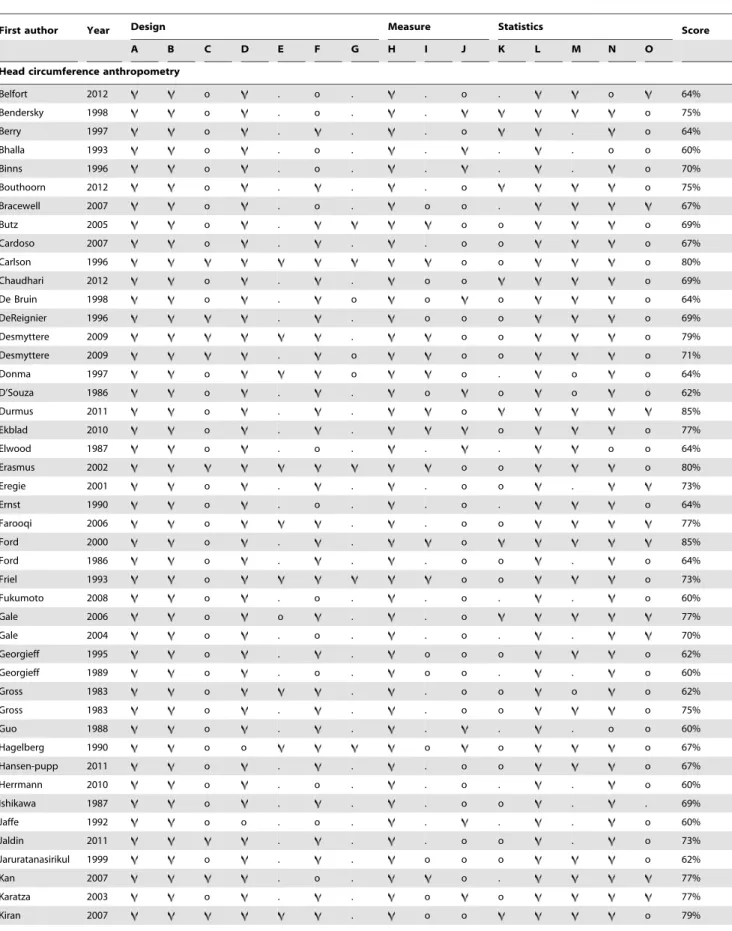
Table 3.
Methodological quality scores of studies using direct anthropometry for head circumference reporting on soft tissue–
based quantitative longitudinal assessment of cranial dimensions in children until age 6 years with a score equal to or above 60%
(n = 95) [24–118].
First author Year Design Measure Statistics Score
A B C D E F G H I J K L M N O
Head circumference anthropometry
Belfort 2012 o . o . . o . o 64%
Bendersky 1998 o . o . . o 75%
Berry 1997 o . . . o . o 64%
Bhalla 1993 o . o . . . . o o 60%
Binns 1996 o . o . . . . o 70%
Bouthoorn 2012 o . . . o o 75%
Bracewell 2007 o . o . o o . 67%
Butz 2005 o . o o o 69%
Cardoso 2007 o . . . o o o 67%
Carlson 1996 o o o 80%
Chaudhari 2012 o . . o o o 69%
De Bruin 1998 o . o o o o 64%
DeReignier 1996 . . o o o o 69%
Desmyttere 2009 . o o o 79%
Desmyttere 2009 . o o o o 71%
Donma 1997 o o o . o o 64%
D’Souza 1986 o . . o o o o 62%
Durmus 2011 o . . o 85%
Ekblad 2010 o . . o o 77%
Elwood 1987 o . o . . . o o 64%
Erasmus 2002 o o o 80%
Eregie 2001 o . . . o o . 73%
Ernst 1990 o . o . . o . o 64%
Farooqi 2006 o . . o o 77%
Ford 2000 o . . o 85%
Ford 1986 o . . . o o . o 64%
Friel 1993 o o o o 73%
Fukumoto 2008 o . o . . o . . o 60%
Gale 2006 o o . . o 77%
Gale 2004 o . o . . o . . 70%
Georgieff 1995 o . . o o o o 62%
Georgieff 1989 o . o . o o . . o 60%
Gross 1983 o . . o o o o 62%
Gross 1983 o . . . o o o 75%
Guo 1988 o . . . o o 60%
Hagelberg 1990 o o o o o 67%
Hansen-pupp 2011 o . . . o o o 67%
Herrmann 2010 o . o . . o . . o 60%
Ishikawa 1987 o . . . o o . . 69%
Jaffe 1992 o o . o . . . . o 60%
Jaldin 2011 . . . o o . o 73%
Jaruratanasirikul 1999 o . . o o o o 62%
Kan 2007 . o . o . 77%
Karatza 2003 o . . o o 77%
Table 3.
Cont.
First author Year Design Measure Statistics Score
A B C D E F G H I J K L M N O
Head circumference anthropometry
Kitchen 1992 o . o . o o . 67%
Koo 2006 o o 87%
Lainhart 1997 o . . . o . o 75%
Lasekan 2011 o o o o 73%
Lasekan 2006 o o 87%
Lira 2009 o . o . . o 75%
Lucas 2001 o o 87%
Maguire 2008 o o 87%
Makrides 2000 o 93%
Makrides 1999 o o 87%
Mamabolo 2004 o . o . . o . . o 60%
Marks 1979 o . o . . o . . o 60%
Maserei 2007 o o 87%
Mathur 2009 o . . . o o o 62%
McCowan 1999 o . o . . o . . 70%
McLeod 2011 o . . . o o . o 64%
Mercuri 2000 o . o . o . . o 64%
Meyer-Marcotty 2012 o . . o o o o 62%
Moore 1995 o . . . o o o 62%
Moye 1993 o . . o o o 69%
Nelson 1997 o . . . o o o 62%
Ochiai 2008 o . . . o o o 73%
Olivan 2003 o . . . o o . o 64%
Oliveira 2007 o . . . o o o 67%
Padilla 2010 . . o o 85%
Paul 2008 . . . o o . o 72%
Peng 2005 o . o o o 71%
Piemontese 2011 o o 87%
Polberger 1999 o o o o 73%
Rijken 2007 o . . . o 77%
Roberfroid 2012 o o 87%
Roche 1987 o o . o . . o . o 60%
Rodriguez Garcia 2003 o . . . o . . o o 60%
Ross 2012 o . . . o o o 67%
Rothenberg 1999 o . o . . . 82%
Saliba 1990 o . . o o o 69%
Sawada 2010 o . . . o o . o 64%
Schaefer 1994 o . . . o o o 62%
Sharma 2011 o o . . . o o . 64%
Shaw 1999 o o o o 76%
Sheth 1995 o . o . . o . o o 60%
Shortland 1998 o o o o o 76%
Tan 2008 o o o o 73%
Tinoco 2009 . o . . o . . o 70%
Touwslager 2008 o . o . . o . . o 60%
Vaidya 2008 100%
circumference [24–118] are shown in Table 3 (n = 95); those for
other direct anthropometry [119–132] are shown in Table 4
(n = 14); and those for indirect 2D and 3D imaging techniques
[133–141] are shown in Table 5 (n = 9).
Analysis of variance (p = 0.14) and Kruskal-Wallis test (p = 0.16)
revealed no statistically significant difference for methodological
quality, expressed as a percentage between groups of methods.
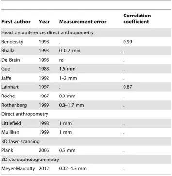
Reliability
Scores for reliability of methods for soft tissue–based
quantita-tive longitudinal assessment are shown in Table 6. Only 12 of the
118 studies with good methodological quality report data for the
reliability of the method to quantify cranial dimensions; 8 studies
used
direct
anthropometry
for
head
circumference
[25,27,35,58,63,71,100,103], two used other kinds of direct
anthropometry [126,127], one study used 3D laser scanning
[136], and one used 3D stereophotogrammetry [139].
Regarding direct anthropometry for head circumference, 5
studies with good methodological quality reported a measurement
error equal to or below 1.7 mm. Two studies with good
methodological quality using direct anthropometry reported
correlation coefficients between repeated measurements of 0.87
and 0.99 and were both qualified as very good. Regarding other
kinds of direct anthropometry, two studies with good
methodo-logical quality reported a measurement error of 1 mm.
Studies of good methodological quality using 2D photography
and reporting the measurement error or correlation coefficients
were not identified among the included studies. One study with
good methodological quality using 3D laser scanning reported a
measurement error of 0.5 mm, and another using 3D
stereo-photogrammetry reported a measurement error of 0.02–4.3 mm.
Discussion
Summary of evidence
The objectives of this systematic review were to 1) give an
overview of soft tissue–based methods for quantitative longitudinal
assessment of cranial dimensions in children until age 6 years; 2)
assess the methodological quality of the studies using such
approaches; and 3) assess reliability of these quantitative
measurement methods used in studies with good methodological
quality.
In the literature, various terms to describe measurement error
exist. Some studies use accuracy to describe landmark
identifica-tion error, which in turn may consist of operator error, capture
error, and registration error [142]. More often in the literature,
reliability is used to describe landmark identification error of a
method. Reliability can be expressed by the measurement error or
a
correlation
coefficient
between
repeated
measurements
[9,143,144] and represents the ability of observers to make a
consistent analysis. In this systematic review, reliability in studies
with good methodological quality was assessed and expressed by
duplicate measurement errors and correlation coefficients between
repeated measurements. Direct measurement of head
circumfer-ence is the most often used method for soft tissue–based evaluation
of cranial growth and treatment outcomes. The use of growth
charts in the clinical assessment of growing infants and children
and in pediatric nutritional screening and epidemiologic
assess-ments has already been recommended for decades [27]. For this
purpose, length and weight also are recorded in many countries
from birth onwards. Direct anthropometry for head circumference
seems to be a generally accepted method for most researchers
because only 8 out of 95 studies with good methodological quality
reported on its reliability. Measurement errors varied from 0.2 to
1.7 mm, and correlation coefficients were very good. Other kinds
of direct anthropometry yielded a measurement error of 1 mm.
Normal growth of head circumference shows an increase for mean
head circumference from 34–36 cm at birth to 51–52 by age 6
years [145]. The measurement errors for head circumference
anthropometry presented in this review are within 1% of the
values of normal growth, which seems to be negligible. Direct
anthropometry is a reliable and cheap method to study linear
dimensions and has been regarded as the gold standard for many
decades, but it requires patient cooperation and precludes
archiving [146].
Photographic techniques, on the other hand, can capture
images for data storage. The most common imaging technique is
2D photography, which has the advantages of being safe, relatively
cheap, and user friendly. However, because none of the included
studies reported a measurement error or correlation coefficient, its
reliability for evaluation of cranial growth and treatment outcome
is uncertain.
Various 3D imaging techniques have been recently introduced
and were applied in 6 out of 165 studies included in this systematic
review. Only two studies with good methodological quality
reported the measurement error (Table 6). The measurement
error in one study using 3D laser scanning was 0.5 mm; in one
study using 3D stereophotogrammetry, it was 0.02 mm for head
width, 0.04 mm for head circumference, and 4.3 mm for vertex
height. Therefore, 3D imaging might be a reliable method to
quantify cranial dimensions. Reliability of measurements from 3D
imaging seems to be more related to the exact anatomical region
of interest than to the method itself.
When reviewing the literature for this study, we found only six
included studies with good methodological quality using 3D
imaging to quantify soft tissue–based cranial growth and treatment
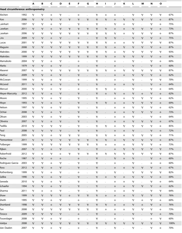
Table 3.
Cont.
First author Year Design Measure Statistics Score
A B C D E F G H I J K L M N O
Head circumference anthropometry
Whitehouse 2010 o . . . o o o 67%
Wood 2003 o . . . o o 75%
Zabaneh 2011 o . . . o o o 67%
= fulfilled the methodological criteria satisfactorily. o = did not fulfill the methodological criteria. . = not applicable.
outcome. The explanation is that only recently have techniques
become available to capture the full 360
u
image needed to study
cranial dimensions. Most studies using 3D imaging concern facial
growth and treatment outcome because this technique has been
available for two decades. Therefore, it is expected that within the
next decade, more studies using 3D imaging of cranial dimensions
will be published. Advantages of these 3D techniques are
millisecond fast image capture, archival capabilities, a
good-resolution color representation, and no exposure to ionizing
radiation. Furthermore, assessment of linear dimensions and
cranial size and shape can be made three-dimensionally. These are
major advantages compared to more simplistic analyses performed
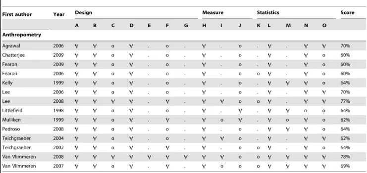
Table 4.
Methodological quality scores of studies using direct anthropometry reporting on soft tissue–based quantitative
longitudinal assessment of cranial dimensions in children until age 6 years with a score equal to or above 60% (n = 14) [119–132].
First author Year Design Measure Statistics Score
A B C D E F G H I J K L M N O
Anthropometry
Agrawal 2006 o . o . . o . . 70%
Chatterjee 2009 o . o . . o . . o 60%
Fearon 2009 o . o . . o . . o 60%
Fearon 2006 o . o . . o o . o 60%
Kelly 1999 o . o . . o . o 64%
Lee 2006 o . o . . o . . 70%
Lee 2008 . . o o . 77%
Littlefield 1998 o . o . . . o o 64%
Mulliken 1999 o . . o . o o 62%
Pedroso 2008 o . o . . o . o 64%
Teichgraeber 2004 o . o . o . . 62%
Teichgraeber 2002 o . . . o o . o 64%
Van Vlimmeren 2008 o o 78%
Van Vlimmeren 2007 o . . o o o 69%
= fulfilled the methodological criteria satisfactorily. o = did not fulfill the methodological criteria. . = not applicable.
doi:10.1371/journal.pone.0089602.t004
Table 5.
Methodological quality scores of studies using indirect 2D and 3D imaging techniques reporting on soft tissue–based
quantitative longitudinal assessment of cranial dimensions in children until age 6 years with a score equal to or above 60% (n = 9)
[133–141].
First author Year Design Measure Statistics Score
A B C D E F G H I J K L M N O
2D photography
Hutchinson 2010 o o o o 73%
Hutchinson 2004 o . . . o o 75%
Hutchison 2011 o . . . o o o 67%
3D surface laser scanning
Plank 2006 o . o o o . o o 62%
3D stereophotogrammetry
Collet 2012 o . . 92%
Lipira 2010 o o . o o . o 62%
Meyer-Marcotty 2012 o . . o o o o 62%
Schaaf 2010 o . . . o o . o 64%
Toma 2010 o . o . . o . o 64%
= fulfilled the methodological criteria satisfactorily. o = did not fulfill the methodological criteria. . = not applicable.
with direct anthropometry. A shortcoming of 3D imaging is its
inability to capture surface morphology in the presence of cranial
hair. The reliability of 3D imaging techniques for soft tissue–based
evaluation of cranial growth and treatment outcomes needs to be
investigated further.
Limitations
Failure to identify all relevant reports for a systematic review is
likely to result in bias [147]. For this reason, highly sensitive search
strategies were developed with the help of a senior librarian
specialized in health sciences for a combination of both narrow
and broad health science databases.
The process of study selection was performed in an independent
standardized manner by two reviewers to prevent unjustified
exclusion of eligible studies. The manual search of the reference
lists of the included studies was performed by only one reviewer.
Possibly eligible studies could have been missed in this stage of the
selection process. However, because only approximately one out of
ten studies was retrieved by the manual search, this omission might
be negligible. Furthermore, every effort was made to contact the
authors by email in cases where online access was not permitted or
the journal was not available in the library. Nevertheless, failure to
retrieve full-text publications of possibly eligible studies (n = 195)
was inevitable.
The instrument used to assess methodological quality was
adapted from Lagrave`re et al. [148] who developed a
methodo-logical quality checklist to assess study design, study
measure-ments, and statistical analysis in clinical trials. Since the
introduction the checklist has been modified and used by Gordon
et al. [21] and Van Vlijmen et al. [149]. Scientific use of the
checklist has been accepted because the criteria check for generally
accepted reasons for bias, despite a lack of validation of the QAI.
The majority of inter-rater disagreements arose in the assessment
of applicability of criteria E, I, and K to certain studies (similar
baseline characteristics, blind measurement, and dropouts
includ-ed in data analysis, respectively). This greater frequency can be
explained by the absence of adequate instructions of this QAI
together with the presence of a wide variety of study designs.
Therefore, raters should test this QAI thoroughly and obtain
consensus before scoring. In the literature, no single tool is an
obvious candidate for assessment of methodological quality of
non-randomized studies [150]. Attempts to validate QAIs like the
Newcastle-Ottowa [151] scale or the Jadad scale [152] produce
highly arbitrary results and cannot demonstrate significant effects
on quality scores [153,154]. There is a need for a validated QAI
that is preferably applicable to a wide range of study designs.
Conclusions
Direct anthropometrical measurement of head circumference in
growing children below age 6 years is a reliable method for
assessing cranial dimensions. The non-invasive 3D surface laser
scanning and 3D-stereophogrammetry techniques can assess size
and shape three-dimensionally. However, their reliability for
assessing cranial dimensions needs to be investigated further.
Supporting Information
Checklist S1
PRISMA checklist.
(DOCX)
Acknowledgments
We thank senior health sciences librarian Elmie Peters for supporting the development of search strategies.
Author Contributions
Conceived and designed the experiments: S. Brons AK. Performed the experiments: S. Brons MvB AK. Analyzed the data: S. Brons MvB. Contributed reagents/materials/analysis tools: S. Brons EB. Wrote the paper: S. Brons. Intellectual contribution: JD JS S. Berge.
References
1. McGarry A, Dixon MT, Greig RJ, Hamilton DR, Sexton S, et al. (2008) Head shape measurement standards and cranial orthoses in the treatment of infants with deformational plagiocephaly. Dev Med Child Neurol 50: 568–576. 2. Chan FC, Kawamoto HK, Federico C, Bradley JP (2013) Soft-tissue volumetric
changes following monobloc distraction procedure: analysis using digital three-dimensional photogrammetry system (3dMD). J Craniofac Surg 24: 416–420. 3. Morhardt DR, Barrow W, Jaworski M, Accardo PJ (2013) Head Circumfer-ence in Young Children With Autism: The Impact of Different Head Circumference Charts. J Child Neurol.
4. Boros CA, Spence D, Blaser S, Silverman ED (2007) Hydrocephalus and macrocephaly: new manifestations of neonatal lupus erythematosus. Arthritis Rheum 57: 261–266.
5. Farkas LG (1994) Anthropometry of the head and face; kj, editor. New York: Raven Press. 405 p.
6. Bartzela TN, Katsaros C, Bronkhorst EM, Rizell S, Halazonetis D, et al. (2011) A two-centre study on facial morphology in patients with complete bilateral cleft lip and palate at nine years of age. Int J Oral Maxillofac Surg In press. 7. Broadbent BH (1931) A new X-ray technique and its application to
orthodontia. Angle Orthod 1: 45–66.
8. Nollet PJ, Katsaros C, Huyskens RW, Borstlap WA, Bronkhorst EM, et al. (2008) Cephalometric evaluation of long-term craniofacial development in unilateral cleft lip and palate patients treated with delayed hard palate closure. Int J Oral Maxillofac Surg 37: 123–130.
9. Farkas LG, Bryson W, Klotz J (1980) Is photogrammetry of the face reliable? Plast Reconstr Surg 66: 346–355.
10. Davis JP, Valentine T, Davis RE (2010) Computer assisted photo-anthropo-metric analyses of full-face and profile facial images. Forensic Sci Int 200: 165– 176.
Table 6.
Reliability of methods for soft tissue–based
quantitative longitudinal assessment of cranial dimensions in
children until age 6 years in studies with good
methodological quality (methodological quality score equal
to or above 60%).
First author Year Measurement error
Correlation coefficient
Head circumference, direct anthropometry
Bendersky 1998 . 0.99
Bhalla 1993 0–0.2 mm .
De Bruin 1998 ns .
Guo 1988 1.6 mm .
Jaffe 1992 1–2 mm .
Lainhart 1997 . 0.87
Roche 1987 0.9 mm .
Rothenberg 1999 0.8–1.7 mm .
Direct anthropometry
Littlefield 1998 1 mm .
Mulliken 1999 1 mm .
3D laser scanning
Plank 2006 0.5 mm .
3D stereophotogrammetry
Meyer-Marcotty 2012 0.02–4.3 mm . . = not reported.
ns = not significant.
doi:10.1371/journal.pone.0089602.t006
11. Cevidanes LH, Alhadidi A, Paniagua B, Styner M, Ludlow J, et al. (2011) Three-dimensional quantification of mandibular asymmetry through cone-beam computerized tomography. Oral Surg Oral Med Oral Pathol Oral Radiol Endod In press.
12. Nada RM, Maal TJ, Breuning KH, Berge SJ, Mostafa YA, et al. (2011) Accuracy and reproducibility of voxel based superimposition of cone beam computed tomography models on the anterior cranial base and the zygomatic arches. PLoS One 6: e16520.
13. Djordjevic J, Toma AM, Zhurov AI, Richmond S (2011) Three-dimensional quantification of facial symmetry in adolescents using laser surface scanning. Eur J Orthod.
14. Toma AM, Zhurov A, Playle R, Richmond S (2008) A three-dimensional look for facial differences between males and females in a British-Caucasian sample aged 151/2 years old. Orthod Craniofac Res 11: 180–185.
15. Kau CH, Kamel SG, Wilson J, Wong ME (2011) New method for analysis of facial growth in a pediatric reconstructed mandible. Am J Orthod Dentofacial Orthop 139: e285–290.
16. Maal TJ, van Loon B, Plooij JM, Rangel F, Ettema AM, et al. (2010) Registration of 3-dimensional facial photographs for clinical use. J Oral Maxillofac Surg 68: 2391–2401.
17. van Loon B, van Heerbeek N, Maal TJ, Borstlap WA, Ingels KJ, et al. (2011) Postoperative volume increase of facial soft tissue after percutaneous versus endonasal osteotomy technique in rhinoplasty using 3D stereophotogramme-try. Rhinology 49: 121–126.
18. Brons S, van Beusichem ME, Bronkhorst EM, Draaisma J, Berge SJ, et al. (2012) Methods to quantify soft-tissue based facial growth and treatment outcomes in children: a systematic review. PLoS One 7: e41898.
19. PROSPERO: International prospective register of systematic reviews. 20. Boluyt N, Tjosvold L, Lefebvre C, Klassen TP, Offringa M (2008) Usefulness of
systematic review search strategies in finding child health systematic reviews in MEDLINE. Arch Pediatr Adolesc Med 162: 111–116.
21. Gordon JM, Rosenblatt M, Witmans M, Carey JP, Heo G, et al. (2009) Rapid palatal expansion effects on nasal airway dimensions as measured by acoustic rhinometry. A systematic review. Angle Orthod 79: 1000–1007.
22. Landis JR, Koch GG (1977) The measurement of observer agreement for categorial data. Biometrics 33: 159–174.
23. The PRISMA Statement website. Available: http://www.prisma-statement. org. Accessed 2013 May 9.
24. Belfort MB, Rifas-Shiman SL, Sullivan T, Collins CT, McPhee AJ, et al. (2011) Infant growth before and after term: effects on neurodevelopment in preterm infants. Pediatrics 128: e899-906.
25. Bendersky M, Koons A, Lewis M, Hegyi T (1998) Developmental implications of head growth following intracranial hemorrhage Clinical Pediatrics 37 469– 476
26. Berry MA, Conrod H, Usher RH (1997) Growth of very premature infants fed intravenous hyperalimentation and calcium-supplemented formula Pediatrics 100 647–653.
27. Bhalla AK, Walia BN (1993) Percentile growth charts for head circumference in Punjabi infants Indian Pediatr 30 41–46.
28. Binns HJ, Senturia YD, LeBailly S, Donovan M, Christoffel KK (1996) Growth of Chicago-area infants, 1985 through 1987. Not what the reference curves predict. Pediatric Practice Research Group. Arch Pediatr Adolesc Med 150: 842–849.
29. Bouthoorn SH, van Lenthe FJ, Hokken-Koelega AC, Moll HA, Tiemeier H, et al. (2012) Head circumference of infants born to mothers with different educational levels; the Generation R Study. PLoS One 7: e39798. 30. Bracewell MA, Hennessy EM, Wolke D, Marlow N (2008) The EPICure study:
growth and blood pressure at 6 years of age following extremely preterm birth Arch Dis Child Fetal Neonatal Ed 93 F108–114.
31. Butz AM, Pulsifer MB, Belcher HME, Leppert M, Donithan M, et al. (2005) Infant head growth and cognitive status at 36 months in children with In-Utero drug exposure Journal of Child & Adolescent Substance Abuse 14 15–39. 32. Cardoso LE, Falcao MC (2007) Nutritional assessment of very low birth weight
infants: relationships between anthropometric and biochemical parameters Nutr Hosp 22322–329.
33. Carlson SE, Werkman SH, Tolley EA (1996) Effect of long-chain n-3 fatty acid supplementation on visual acuity and growth of preterm infants with and without bronchopulmonary dysplasia American Journal of Clinical Nutrition 63 687–697.
34. Chaudhari S, Otiv M, Khairnar B, Pandit A, Hoge M, et al. (2012) Pune low birth weight study, growth from birth to adulthood. Indian Pediatr 49: 727– 732.
35. de Bruin NC, Degenhart HJ, Gal S, Westerterp KR, Stijnen T, et al. (1998) Energy utilization and growth in breast-fed and formula-fed infants measured prospectively during the first year of life Am J Clin Nutr 67885–896. 36. deRegnier RA, Guilbert TW, Mills MM, Georgieff MK (1996) Growth failure
and altered body composition are established by one month of age in infants with bronchopulmonary dysplasia J Nutr 126 168–175.
37. Desmyttere S, Bonduelle M, Nekkebroeck J, Roelants M, Liebaers I, et al. (2009) Growth and health outcome of 102 2-year-old children conceived after preimplantation genetic diagnosis or screening Early Hum Dev 85 755–759. 38. Desmyttere S, De Schepper J, Nekkebroeck J, De Vos A, De Rycke M, et al.
(2009) Two-year auxological and medical outcome of singletons born after
embryo biopsy applied in preimplantation genetic diagnosis or preimplantation genetic screening Hum Reprod 24 470–476.
39. Donma MM, Donma O (1997) The influence of feeding patterns on head circumference among Turkish infants during the first 6 months of life. Brain Dev 19: 393–397.
40. D’Souza SW, Gowland M, Richards B, Cadman J, Mellor V, et al. (1986) Head size, brain growth, and lateral ventricles in very low birthweight infants Arch Dis Child 61 1090–1095.
41. Durmus B, Kruithof CJ, Gillman MH, Willemsen SP, Hofman A, et al. (2011) Parental smoking during pregnancy, early growth, and risk of obesity in preschool children: the Generation R Study. Am J Clin Nutr 94: 164–171. 42. Ekblad M, Korkeila J, Parkkola R, Lapinleimu H, Haataja L, et al. (2010)
Maternal smoking during pregnancy and regional brain volumes in preterm infants J Pediatr 156 185–190 e181.
43. Elwood PC, Sweetnam PM, Gray OP, Davies DP, Wood PD (1987) Growth of children from 0-5 years: with special reference to mother’s smoking in pregnancy Ann Hum Biol 14543–557.
44. Erasmus HD, Ludwig-Auser HM, Paterson PG, Sun D, Sankaran K (2002) Enhanced weight gain in preterm infants receiving lactase-treated feeds: a randomized, double-blind, controlled trial J Pediatr 141 532–537.
45. Eregie CO (2001) Exclusive breastfeeding and infant growth studies: reference standards for head circumference, length and mid-arm circumference/head circumference ratio for the first 6 months of life J Trop Pediatr 47 329–334. 46. Ernst JA, Bull MJ, Rickard KA, Brady MS, Lemons JA (1990) Growth
outcome and feeding practices of the very low birth weight infant (less than 1500 grams) within the first year of life J Pediatr 117 S156–166.
47. Farooqi A, Hagglof B, Sedin G, Gothefors L, Serenius F (2006) Growth in 10-to 12-year-old children born at 23 10-to 25 weeks’ gestation in the 1990s: a Swedish national prospective follow-up study Pediatrics 118 e1452–1465. 48. Ford G, Rickards A, Kitchen WH, Ryan MM, Lissenden JV (1986)
Relationship of growth and psychoneurologic status of 2-year-old children of birthweight 500-999 g Early Hum Dev 13 329–337.
49. Ford GW, Doyle LW, Davis NM, Callanan C (2000) Very low birth weight and growth into adolescence Arch Pediatr Adolesc Med 154 778–784. 50. Friel JK, Andrews WL, Matthew JD, Long DR, Cornel AM, et al. (1993) Zinc
Supplementation in Very-Low-Birth-Weight Infants Journal of Pediatric Gastroenterology and Nutrition 17 97–104.
51. Fukumoto A, Hashimoto T, Ito H, Nishimura M, Tsuda Y, et al. (2008) Growth of head circumference in autistic infants during the first year of life. J Autism Dev Disord 38: 411–418.
52. Gale CR, O’Callaghan FJ, Bredow M, Martyn CN (2006) The influence of head growth in fetal life, infancy, and childhood on intelligence at the ages of 4 and 8 years Pediatrics 118 1486–1492.
53. Gale CR, O’Callaghan FJ, Godfrey KM, Law CM, Martyn CN (2004) Critical periods of brain growth and cognitive function in children Brain 127 321–329. 54. Georgieff MK, Hoffman JS, Pereira GR, Bernbaum J, Hoffman-Williamson M (1985) Effect of neonatal caloric deprivation on head growth and 1-year developmental status in preterm infants. J Pediatr 107: 581–587.
55. Georgieff MK, Mills MM, Zempel CE, Chang PN (1989) Catch-up growth, muscle and fat accretion, and body proportionality of infants one year after newborn intensive care J Pediatr 114 288–292.
56. Gross SJ, Eckerman CO (1983) Normative early head growth in very-low-birth-weight infants J Pediatr 103 946–949.
57. Gross SJ, Oehler JM, Eckerman CO (1983) Head growth and developmental outcome in very low-birth-weight infants Pediatrics 71 70–75.
58. Guo S, Roche AF, Moore WM (1988) Reference data for head circumference and 1-month increments from 1 to 12 months of age J Pediatr 113 490–494. 59. Hagelberg S, Lindblad BS, Persson B (1990) Amino acid levels in the critically ill preterm infant given mother’s milk fortified with protein from human or cow’s milk. Acta Paediatr Scand 79: 1163–1174.
60. Hansen-Pupp I, Lofqvist C, Polberger S, Niklasson A, Fellman V, et al. (2011) Influence of insulin-like growth factor I and nutrition during phases of postnatal growth in very preterm infants. Pediatr Res 69: 448–453.
61. Herrmann KR, Herrmann KR (2010) Early parenteral nutrition and successful postnatal growth of premature infants Nutr Clin Pract 25 69–75.
62. Ishikawa T, Furuyama M, Ishikawa M, Ogawa J, Wada Y (1987) Growth in head circumference from birth to fifteen years of age in Japan Acta Paediatr Scand 76 824–828.
63. Jaffe M, Tal Y, Hadad B, Tirosh E, Tamir A (1992) Variability in head circumference growth rate during the first 2 years of life Pediatrics 90 190–192. 64. Jaldin MM, Pinheiro FS, Dos Santos AM, Muniz NC, Brito LMO (2011) Head circumference growth of exclusively breastfed infants during the first six months of life. Rev Paul Pediatr 29: 509–514.
65. Jaruratanasirikul S, Chanvitan P, Janjindamai W, Ritsmitchai S (1999) Growth patterns of low-birth-weight infants: 2-year longitudinal study. J Med Assoc Thai 82: 325–331.
66. Kan E, Roberts G, Anderson PJ, Doyle LW (2008) The association of growth impairment with neurodevelopmental outcome at eight years of age in very preterm children Early Hum Dev 84 409–416.
67. Karatza AA, Varvarigou A, Beratis NG (2003) Growth up to 2 years in relationship to maternal smoking during pregnancy Clin Pediatr (Phila) 42 533–541.
69. Kitchen WH, Doyle LW, Ford GW, Callanan C, Rickards AL, et al. (1992) Very low birth weight and growth to age 8 years. II: Head dimensions and intelligence Am J Dis Child 14646–50.
70. Koo WWK, Hockman EM (2006) Posthospital discharge feeding for preterm infants: effects of standard compared with enriched milk formula on growth, bone mass, and body composition American Journal of Clinical Nutrition 84 1357–1364.
71. Lainhart JE, Piven J, Wzorek M, Landa R, Santangelo SL, et al. (1997) Macrocephaly in children and adults with autism. J Am Acad Child Adolesc Psychiatry. United States. pp. 282–290.
72. Lasekan JB, Jacobs J, Reisinger KS, Montalto MB, Frantz MP, et al. (2011) Lactose-free milk protein-based infant formula: impact on growth and gastrointestinal tolerance in infants. Clin Pediatr (Phila) 50: 330–337. 73. Lasekan JB, Koo WW, Walters J, Neylan M, Luebbers S (2006) Growth,
tolerance and biochemical measures in healthy infants fed a partially hydrolyzed rice protein-based formula: a randomized, blinded, prospective trial J Am Coll Nutr 25 12–19.
74. Lira PI, Eickmann SH, Lima MC, Amorim RJ, Emond AM, et al. (2010) Early head growth: relation with IQ at 8 years and determinants in term infants of low and appropriate birthweight Dev Med Child Neurol 52 40–46. 75. Lucas A, Fewtrell MS, Morley R, Singhal A, Abbott RA, et al. (2001)
Randomized trial of nutrient-enriched formula versus standard formula for postdischarge preterm infants Pediatrics 108 703–711.
76. Maguire CM, Veen S, Sprij AJ, Le Cessie S, Wit JM, et al. (2008) Effects of basic developmental care on neonatal morbidity, neuromotor development, and growth at term age of infants who were born at,32 weeks Pediatrics 121 e239–245.
77. Makrides M, Neumann MA, Jeffrey B, Lien EL, Gibson RA (2000) A randomized trial of different ratios of linoleic to alpha-linolenic acid in the diet of term infants: effects on visual function and growth American Journal of Clinical Nutrition71 120–129.
78. Makrides M, Neumann MA, Simmer K, Gibson RA (1999) Dietary long-chain polyunsaturated fatty acids do not influence growth of term infants: a randomized clinical trial Pediatrics 104 468–475.
79. Mamabolo RL, Alberts M, Mbenyane GX, Steyn NP, Nthangeni NG, et al. (2004) Feeding practices and growth of infants from birth to 12 months in the central region of the Limpopo Province of South Africa Nutrition 20 327–333. 80. Marks KH, Maisels MJ, Moore E, Gifford K, Friedman Z (1979) Head growth
in sick premature infants—a longitudinal study J Pediatr 94 282–285. 81. Masarei AG, Wade A, Mars M, Sommerlad BC, Sell D (2007) A randomized
control trial investigating the effect of presurgical orthopedics on feeding in infants with cleft lip and/or palate Cleft Palate-Craniofacial Journal 44 182– 193.
82. Mathur NB, Dhingra D (2009) Perceived breast milk insufficiency in mothers of neonates hospitalized in neonatal intensive care unit Indian J Pediatr 76 1003– 1006.
83. McCowan L, Harding J, Barker S, Ford C (1999) Perinatal predictors of growth at six months in small for gestational age babies Early Human Development 56 205–216.
84. McLeod G, Simmer K, Benninger H, Mitoulas L, Doherty D, et al. (2011) Preterm infants with chronic lung disease: are protein and energy intakes after discharge sufficient for optimal growth? J Paediatr Child Health 47: 127–133. 85. Mercuri E, Ricci D, Cowan FM, Lessing D, Frisone MF, et al. (2000) Head growth in infants with hypoxic-ischemic encephalopathy: correlation with neonatal magnetic resonance imaging Pediatrics 106 235–243.
86. Meyer-Marcotty P, Bohm H, Linz C, Kunz F, Keil N, et al. (2012) Head orthesis therapy in infants with unilateral positional plagiocephaly: an interdisciplinary approach to broadening the range of orthodontic treatment. J Orofac Orthop 73: 151–165.
87. Moore WM, Bannister RP, Ward BS, Hillier VF, Bamford FN (1995) Fetal and postnatal growth to age 2 years by mother’s country of birth Early Hum Dev 42 111–121.
88. Moye J, Jr., Rich KC, Kalish LA, Sheon AR, Diaz C, et al. (1996) Natural history of somatic growth in infants born to women infected by human immunodeficiency virus. Women and Infants Transmission Study Group J Pediatr 128 58–69.
89. Nelson KG, Goldenberg RL, Hoffman HJ, Cliver SP (1997) Growth and development during the first year in a cohort of low income term-born American children Acta Obstet Gynecol Scand Suppl 165 87–92.
90. Ochiai M, Nakayama H, Sato K, Iida K, Hikino S, et al. (2008) Head circumference and long-term outcome in small-for-gestational age infants J Perinat Med 36 341–347.
91. Olivan G (2003) Catch-up growth assessment in long-term physically neglected and emotionally abused preschool age male children Child Abuse Negl 27 103– 108.
92. Oliveira MG, Silveira RC, Procianoy RS (2008) Growth of very low birth weight infants at 12 months corrected age in southern Brazil J Trop Pediatr 54 36–42.
93. Padilla N, Perapoch J, Carrascosa A, Acosta-Rojas R, Botet F, et al. (2010) Twelve-month neurodevelopmental outcome in preterm infants with and without intrauterine growth restriction Acta Paediatr
94. Paul B, Saha I, Dasgupta A, Chaudhuri RN (2008) A study on catch up growth among low birth weight infants in an urban slum of Kolkata Indian J Public Health 52 16–20.
95. Peng Y, Huang B, Biro F, Feng L, Guo Z, et al. (2005) Outcome of low birthweight in China: a 16-year longitudinal study Acta Paediatr 94 843–849. 96. Piemontese P, Gianni ML, Braegger CP, Chirico G, Gruber C, et al. (2011) Tolerance and safety evaluation in a large cohort of healthy infants fed an innovative prebiotic formula: a randomized controlled trial. PLoS One 6: e28010.
97. Polberger S, Raiha NC, Juvonen P, Moro GE, Minoli I, et al. (1999) Individualized protein fortification of human milk for preterm infants: comparison of ultrafiltrated human milk protein and a bovine whey fortifier J Pediatr Gastroenterol Nutr 29 332–338.
98. Rijken M, Wit JM, Le Cessie S, Veen S (2007) The effect of perinatal risk factors on growth in very preterm infants at 2 years of age: the Leiden Follow-Up Project on Prematurity Early Hum Dev 83 527–534.
99. Roberfroid D, Huybregts L, Lanou H, Ouedraogo L, Henry MC, et al. (2012) Impact of prenatal multiple micronutrients on survival and growth during infancy: a randomized controlled trial. Am J Clin Nutr 95: 916–924. 100. Roche AF, Mukherjee D, Guo SM, Moore WM (1987) Head circumference
reference data: birth to 18 years. Pediatrics 79: 706–712.
101. Rodriguez Garcia J, Bosch Gimenez VM, Alonso Garcia MA, Borrajo Guadarrama E, Perez Flores D (2003) Longitudinal study of the growth of preterm newborn infants] An Pediatr (Barc) 58 241–251.
102. Ross GS, Krauss AN, Perlman JM (2012) Physical growth and cognitive abilities in concordant versus discordant birth weight twins at three years old. Early Hum Dev 88: 753–756.
103. Rothenberg SJ, Schnaas L, Perroni E, Hernandez RM, Martinez S, et al. (1999) Pre- and postnatal lead effect on head circumference: A case for critical periods Neurotoxicology and Teratology 21 1–11.
104. Saliba E, Bertrand P, Gold F, Vaillant MC, Laugier J (1990) Area of lateral ventricles measured on cranial ultrasonography in preterm infants: reference range Arch Dis Child 65 1029–1032.
105. Sawada A, Ikeda H, Kimura-Ohba S, Matsuzawa S, Awaya T, et al. (2010) Head growth evaluation in early childhood, from the Japan Children’s Study Pediatrics International 52 343–346.
106. Schaefer F, Burgard P, Batzler U, Rupp A, Schmidt H, et al. (1994) Growth and skeletal maturation in children with phenylketonuria Acta Paediatr 83 534–541.
107. Sharma PK, Sankar MJ, Sapra S, Saxena R, Karthikeyan CV, et al. (2011) Growth and neurosensory outcomes of preterm very low birth weight infants at 18 months of corrected age. Indian J Pediatr 78: 1485–1490.
108. Shaw WC, Bannister RP, Roberts CT (1999) Assisted feeding is more reliable for infants with clefts—a randomized trial Cleft Palate Craniofac J 36 262–268. 109. Sheth RD, Mullett MD, Bodensteiner JB, Hobbs GR (1995) Longitudinal head growth in developmentally normal preterm infants Arch Pediatr Adolesc Med 149 1358–1361.
110. Shortland GJ, Walter JH, Stroud C, Fleming PJ, Speidel BD, et al. (1998) Randomised controlled trial of L-carnitine as a nutritional supplement in preterm infants Arch Dis Child Fetal Neonatal Ed 78 F185–188.
111. Tan M, Abernethy L, Cooke R (2008) Improving head growth in preterm infants—a randomised controlled trial II: MRI and developmental outcomes in the first year. Arch Dis Child Fetal Neonatal Ed. England. pp. F342–346. 112. Tinoco SMB, Sichieri R, Setta CL, Moura AS, Carmo MGTD (2009) N-3
polyunsaturated fatty acids in milk is associate to weight gain and growth in premature infants Lipids in Health and Disease 8
113. Touwslager RN, Gerver WJ, Mulder AL, Jansen AJ, de Bruin R (2008) Longitudinal growth during the first years of life: what is normal? Horm Res 70 273–277.
114. Vaidya A, Saville N, Shrestha BP, Costello AM, Manandhar DS, et al. (2008) Effects of antenatal multiple micronutrient supplementation on children’s weight and size at 2 years of age in Nepal: follow-up of a double-blind randomised controlled trial Lancet 371 492–499.
115. van Daalen E, Swinkels SH, Dietz C, van Engeland H, Buitelaar JK (2007) Body length and head growth in the first year of life in autism Pediatr Neurol 37 324–330.
116. Whitehouse AJ, Maybery MT, Hart R, Sloboda DM, Stanley FJ, et al. (2010) Free testosterone levels in umbilical-cord blood predict infant head circumfer-ence in females Dev Med Child Neurol 52e73–77.
117. Wood NS, Costeloe K, Gibson AT, Hennessy EM, Marlow N, et al. (2003) The EPICure study: growth and associated problems in children born at 25 weeks of gestational age or less Arch Dis Child Fetal Neonatal Ed 88 F492–500. 118. Zabaneh R, Smith LM, LaGasse LL, Derauf C, Newman E, et al. (2012) The
effects of prenatal methamphetamine exposure on childhood growth patterns from birth to 3 years of age. Am J Perinatol 29: 203–210.
119. Agrawal D, Steinbok P, Cochrane DD (2006) Long-term anthropometric outcomes following surgery for isolated sagittal craniosynostosis J Neurosurg 105 357–360.
120. Chatterjee JS, Mahmoud M, Karthikeyan S, Duncan C, Dover MS, et al. (2009) Referral pattern and surgical outcome of sagittal synostosis. J Plast Reconstr Aesthet Surg. Netherlands. pp. 211–215.
121. Fearon JA, Ruotolo RA, Kolar JC (2009) Single sutural craniosynostoses: surgical outcomes and long-term growth Plast Reconstr Surg 123 635–642. 122. Fearon JA, McLaughlin EB, Kolar JC (2006) Sagittal craniosynostosis: surgical
123. Kelly KM, Littlefield TR, Pomatto JK, Ripley CE, Beals SP, et al. (1999) Importance of early recognition and treatment of deformational plagiocephaly with orthotic cranioplasty Cleft Palate-Craniofacial Journal 36 127–130. 124. Lee WT, Richards K, Redhed J, Papay FA (2006) A pneumatic orthotic cranial
molding helmet for correcting positional plagiocephaly J Craniofac Surg 17 139–144.
125. Lee RP, Teichgraeber JF, Baumgartner JE, Waller AL, English JD, et al. (2008) Long-term treatment effectiveness of molding helmet therapy in the correction of posterior deformational plagiocephaly: a five-year follow-up Cleft Palate-Craniofacial Journal 45 240–245.
126. Littlefield TR, Beals SP, Manwaring KH, Pomatto JK, Joganic EF, et al. (1998) Treatment of craniofacial asymmetry with dynamic orthotic cranioplasty Journal of Craniofacial Surgery 9 11–17.
127. Mulliken JB, Vander Woude DL, Hansen M, LaBrie RA, Scott RM (1999) Analysis of posterior plagiocephaly: deformational versus synostotic Plast Reconstr Surg 103 371–380.
128. Pedroso SF, Rotta N, Quintal A, Giordani G (2008) Evolution of anterior fontanel size in normal infants in the first year of life Journal of Child Neurology 23 1419–1423.
129. Teichgraeber JF, Seymour-Dempsey K, Baumgartner JE, Xia JJ, Waller AL, et al. (2004) Molding helmet therapy in the treatment of brachycephaly and plagiocephaly. J Craniofac Surg 15: 118–123.
130. Teichgraeber JF, Ault JK, Baumgartner J, Waller A, Messersmith M, et al. (2002) Deformational posterior plagiocephaly: diagnosis and treatment Cleft Palate Craniofac J 39: 582–586.
131. van Vlimmeren LA, van der Graaf Y, Boere-Boonekamp MM, L’Hoir MP, Helders PJ, et al. (2008) Effect of pediatric physical therapy on deformational plagiocephaly in children with positional preference: a randomized controlled trial. Arch Pediatr Adolesc Med 162: 712–718.
132. van Vlimmeren LA, van der Graaf Y, Boere-Boonekamp MM, L’Hoir MP, Helders PJ, et al. (2007) Risk factors for deformational plagiocephaly at birth and at 7 weeks of age: a prospective cohort study Pediatrics 119: e408–418. 133. Hutchison BL, Stewart AW, De Chalain TB, Mitchell EA (2010) A randomized
controlled trial of positioning treatments in infants with positional head shape deformities Acta Paediatrica, International Journal of Paediatrics 99 1556– 1560.
134. Hutchison BL, Hutchison LAD, Thompson JMD, Mitchell EA (2004) Plagiocephaly and brachycephaly in the first two years of life: A prospective cohort study Pediatrics 114 970–980
135. Hutchison BL, Stewart AW, Mitchell EA (2011) Deformational plagiocephaly: a follow-up of head shape, parental concern and neurodevelopment at ages 3 and 4 years. Arch Dis Child 96: 85–90.
136. Plank LH, Giavedoni B, Lombardo JR, Geil MD, Reisner A (2006) Comparison of infant head shape changes in deformational plagiocephaly following treatment with a cranial remolding orthosis using a noninvasive laser shape digitizer. J Craniofac Surg 17: 1084–1091.
137. Collett BR, Heike CL, Atmosukarto I, Starr JR, Cunningham ML, et al. (2012) Longitudinal, three-dimensional analysis of head shape in children with and without deformational plagiocephaly or brachycephaly. J Pediatr 160: 673–678 e671.
138. Lipira AB, Gordon S, Darvann TA, Hermann NV, Van Pelt AE, et al. (2010) Helmet versus active repositioning for plagiocephaly: a three-dimensional analysis. Pediatrics 126: e936–945.
139. Meyer-Marcotty P, Bo¨hm H, Linz C, Kunz F, Keil N, et al. (2012) Head orthosis therapy in infants with unilateral positional plagiocephaly: an interdisciplinary approach to broadening the range of orthodontic treatment. J Orofac Orthop 73 (2): 151–65
140. Schaaf H, Malik CY, Streckbein P, Pons-Kuehnemann J, Howaldt HP, et al. (2010) Three-dimensional photographic analysis of outcome after helmet treatment of a nonsynostotic cranial deformity. J Craniofac Surg 21: 1677– 1682.
141. Toma R, Greensmith AL, Meara JG, Da Costa AC, Ellis LA, et al. (2010) Quantitative morphometric outcomes following the melbourne method of total vault remodeling for scaphocephaly Journal of Craniofacial Surgery 21: 637– 643
142. Ayoub A, Garrahy A, Hood C, White J, Bock M, et al. (2003) Validation of a vision-based, three-dimensional facial imaging system. Cleft Palate Craniofac J 40: 523–529.
143. Wong JY, Oh AK, Ohta E, Hunt AT, Rogers GF, et al. (2008) Validity and reliability of craniofacial anthropometric measurement of 3D digital photo-grammetric images. Cleft Palate Craniofac J 45: 232–239.
144. Swennen GR, Grimaldi H, Berten JL, Kramer FJ, Dempf R, et al. (2004) Reliability and validity of a modified lateral cephalometric analysis for evaluation of craniofacial morphology and growth in patients with clefts. J Craniofac Surg 15: 399–412; discussion 413–394.
145. Rollins JD, Collins JS, Holden KR (2010) United States head circumference growth reference charts: birth to 21 years. J Pediatr 156: 907–913, 913 e901– 902.
146. Aldridge K, Boyadjiev SA, Capone GT, DeLeon VB, Richtsmeier JT (2005) Precision and error of three-dimensional phenotypic measures acquired from 3dMD photogrammetric images. Am J Med Genet A 138A: 247–253. 147. Robinson KA, Dickersin K (2002) Development of a highly sensitive search
strategy for the retrieval of reports of controlled trials using PubMed. Int J Epidemiol 31: 150–153.
148. Lagravere MO, Major PW, Flores-Mir C (2005) Long-term skeletal changes with rapid maxillary expansion: a systematic review. Angle Orthod 75: 1046– 1052.
149. Van Vlijmen OJ, Kuijpers MA, Berge´ SJ, Schols JG, Maal TJ, et al. (2012) Evidence supporting the use of cone-beam computed tomography in orthodontics. J Am Dent Assoc 143 (3): 241–52
150. Sanderson S, Tatt ID, Higgins JP (2007) Tools for assessing quality and susceptibility to bias in observational studies in epidemiology: a systematic review and annotated bibliography. Int J Epidemiol 36: 666–676.
151. The Newcastle-Ottawa Scale website. Available: http://www.ohri.ca/ programs/clinical_epidemiology/oxford.asp. Accessed 2013 May 9. 152. Jadad AR, Moore RA, Carroll D, Jenkinson C, Reynolds DJ, et al. (1996)
Assessing the quality of reports of randomized clinical trials: is blinding necessary? Control Clin Trials 17: 1–12.
153. Stang A (2010) Critical evaluation of the Newcastle-Ottawa scale for the assessment of the quality of nonrandomized studies in meta-analyses. Eur J Epidemiol 25: 603–605.
![Table 2. Quality assessment instrument [21].](https://thumb-eu.123doks.com/thumbv2/123dok_br/18289308.346428/3.918.93.847.132.447/table-quality-assessment-instrument.webp)