https://doi.org/10.1590/0004-282X20180131
ARTICLE
Pseudotumor cerebri in childhood and
adolescence: data from a specialized service
Pseudotumor cerebral na infância e adolescência: dados de um serviço especializado
Gabriela G. M. Balbi
1, Sandro L. Matas
2, Claudio A. Len
1, Melissa M. Fraga
1, Iggor O. Sousa
1, Maria Teresa Terreri
1Pseudotumor cerebri, or benign intracranial
hyperten-sion, is a syndrome that presents with clinical features of
ele-vated intracranial pressure without radiological evidence of
an intracranial mass, infection, vascular abnormality,
hydro-cephalus or changes in the level of consciousness
1,2,3,4,5.
The incidence of pseudotumor cerebri in general pop
-ulation is 1:100,000. It is rare in childhood. The incidence
increases between the ages of 12 and 15 years, and 60% of
the children who develop the syndrome are over 10 years
of age
2,6,7,8.
The pathogenesis of pseudotumor cerebri is still
unknown; some hypotheses include decreased absorption of
cerebrospinal fluid (CSF) associated with vascular resistance
in the sinus
2,7,9,10,11.
Several conditions are associated with pseudotumor cere
-bri (secondary pseudotumor), such as systemic diseases and
drug exposure. The term idiopathic intracranial hypertension
is used when the cause of this condition is not found
12,13,14.
There are few studies in the literature, most of which are
case reports, describing secondary pseudotumor syndrome
in Pediatrics. Our objective was to report all cases of children
and adolescents diagnosed with pseudotumor cerebri, with
or without rheumatic disease, who were followed by the
pedi-atric rheumatologists and neurologists of our hospital.
1Universidade Federal de São Paulo, Departamento de Pediatria, Unidade de Reumatologia Pediátrica, São Paulo SP, Brasil;
2Universidade Federal de São Paulo, Departamento de Líquido Cefalorraquidiano, São Paulo SP, Brasil.
Correspondence: Gabriela G. M. Balbi; Unidade de Reumatologia Pediátrica da UNIFESP; Rua Borges Lagoa, 802; 04021-001 São Paulo SP, Brasil; E-mail: [email protected]
Conflict of interest: There is no conflict of interest to declare.
Received 24 February 2018; Received in final form 27 May 2018; Accepted 15 August 2018.
ABSTRACT
Objective:
To report cases of children and adolescents diagnosed with pseudotumor cerebri associated or not with rheumatic disease.
Methods:
This was a retrospective study based on medical reports of 29 patients, up to 18 years of age and diagnosed with pseudotumor
cerebri, followed up in the Pediatric Rheumatology and Neurology outpatient clinics of a tertiary hospital, until December 2016.
Results:
Among the 29 patients diagnosed with pseudotumor cerebri, 51.7% were girls and the mean age at the disease onset was 12.3 years.
In 18 patients (62%) where an etiology was found, four were associated with a rheumatic disease. The most common symptom was
headache (69%) and acetazolamide was the most used medication (69%). Two patients developed blindness and 10 are still being
followed up.
Conclusion:
Although rare, pseudotumor cerebri should be considered in children with headaches, especially in patients
with rheumatic disease.
Keywords:
Pseudotumor cerebri; headache; rheumatic diseases; childhood.
RESUMO
Relatar os casos de crianças e adolescentes com diagnóstico de pseudotumor cerebral com ou sem doença reumática.
Métodos:
Estudo
retrospectivo através de revisão de prontuários, 29 pacientes com idade até 18 anos e diagnóstico de pseudotumor, atendidos nos
ambulatórios de Reumatologia Pediátrica e Neurologia de um hospital terciário, registrados até dezembro de 2016.
Resultados:
Dentre
os 29 pacientes com diagnóstico de pseudotumor cerebral, 51,7% eram meninas. A média de idade de aparecimento dos sintomas foi de
12,3 anos. Em relação à etiologia do pseudotumor cerebral, em 18 pacientes (62%) foi possível identificar uma causa, sendo o diagnóstico
de doença reumática associada em quatro desses casos. Cefaléia foi o sintoma mais frequente (69%), e a medicação mais utilizada
foi a acetazolamida (69%). Dois pacientes evoluíram para cegueira e 10 ainda se encontram em seguimento ambulatorial.
Conclusão:
Concluímos que, apesar de raro, o diagnóstico de pseudotumor cerebral deve ser considerado em crianças com cefaleia, principalmente
nos pacientes com doença reumática.
METHODS
This was a retrospective cohort study evaluating 29
patients, up to 18 years of age, with the diagnosis of
pseu-dotumor cerebri according to the criteria of Dandy et al.
15,16modified by Rangwala and Liu
17. Patients were selected
from the Pediatric Rheumatology and Neurology outpatient
clinics of the Federal University of São Paulo, in Brazil, until
December 2016. The following data were analyzed: demo
-graphics, pseudotumor etiology, clinical features, treatment
and outcome. All patients underwent diagnostic spinal
manometry with opening pressure equivalent to or greater
than 25 mm of water
17.
RESULTS
Of the 29 patients, 51.7% (15 patients) were girls. The
mean age of symptom onset was 12.3 ± 4.3 years. The mean
age at first evaluation was 15.4 ± 4.4 years.
Regarding the etiology of the pseudotumor cerebri,
11 (37.9%) patients were diagnosed with idiopathic pseu
-dotumor and 18 patients (62.1%) had secondary pseudotu
-mors. In four of these latter patients (13.8%) a rheumatic
disease was identified. Among these, two had juvenile der
-matomyositis undergoing oral glucocorticoid withdrawal.
One patient had Henoch-Schönlein purpura, and was also
receiving glucocorticoids. The fourth patient presented
with antiphospholipid antibody syndrome. Other causes
were obesity/overweight (three patients), use of drugs such
as tacrolimus and growth hormone (two), renal transplan
-tation (two), cavernous sinus thrombosis (two), hypervi
-taminosis A (one), Bardet-Biedl syndrome (one), cranial
trauma (one), immune thrombocytopenic purpura (one),
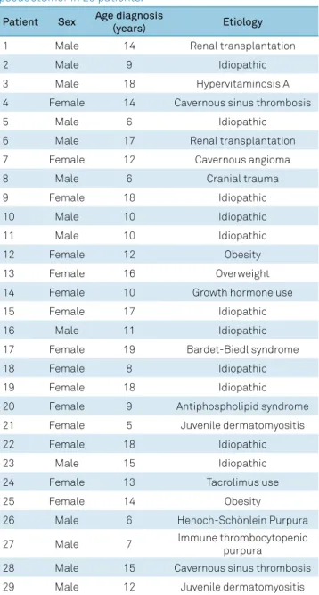
and cavernous angioma (one). Table 1 shows the demo
-graphic characteristics and etiology of the pseudotumor
cerebri patients.
The most frequent symptoms and signs were: papilledema
and headaches in 20 patients (69.0%), decreased visual acu
-ity in 16 (55.2%), and nausea and vomiting in seven (24.1%).
Headache occurred in 13 patients with secondary
pseu-dotumor and in seven patients with primary pseupseu-dotumor.
The most frequent characteristics of the headaches were:
holocranial location with nuchal irradiation, continuous,
worse at night and in the mornings and in some cases
associ-ated with nausea and vomiting.
Table 2 shows the signs and symptoms presented by
each patient.
Imaging studies were performed on all patients. The find
-ings included: empty sella, prominent gyri and sulci, and
optic nerve sheath edema.
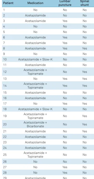
The most commonly-used medication was acetazol
-amide as monotherapy in 20 (69.0%) patients and in combi
-nation with topiramate in four (13.8%) patients. Nine (31.0%)
patients did not use any drug treatment. Table 3 shows the
treatment for each patient.
Regarding the outcome, there was a resolution of the
condition in 27 patients. Two patients developed blindness
(partial/total) due to pseudotumor cerebri and there were
no deaths. The median of time until clinical resolution was
120 days (ranging from 14 to 1,800 days) and 10 patients are
still being followed up as outpatients.
DISCUSSION
Pseudotumor cerebri is a rare condition in childhood
and adolescence. Association with other entities and
prog-nosis differ from the adult presentation due to lower rates
of chronicity and recurrence
8,18. In our study, we observed a
Table 1.
Demographic characteristics and etiology of cerebral
pseudotumor in 29 patients.
Patient
Sex
Age diagnosis
(years)
Etiology
1
Male
14
Renal transplantation
2
Male
9
Idiopathic
3
Male
18
Hypervitaminosis A
4
Female
14
Cavernous sinus thrombosis
5
Male
6
Idiopathic
6
Male
17
Renal transplantation
7
Female
12
Cavernous angioma
8
Male
6
Cranial trauma
9
Female
18
Idiopathic
10
Male
10
Idiopathic
11
Male
10
Idiopathic
12
Female
12
Obesity
13
Female
16
Overweight
14
Female
10
Growth hormone use
15
Female
17
Idiopathic
16
Male
11
Idiopathic
17
Female
19
Bardet-Biedl syndrome
18
Female
8
Idiopathic
19
Female
18
Idiopathic
20
Female
9
Antiphospholipid syndrome
21
Female
5
Juvenile dermatomyositis
22
Female
18
Idiopathic
23
Male
15
Idiopathic
24
Female
13
Tacrolimus use
25
Female
14
Obesity
26
Male
6
Henoch-Schönlein Purpura
27
Male
7
Immune thrombocytopenic
purpura
28
Male
15
Cavernous sinus thrombosis
predominance of secondary disease with an association with
rheumatic diseases in about a quarter of the identifiable eti
-ologies. We found a positive outcome in most cases, with the
exception of visual sequelae in two patients.
The mean age of onset of the pseudotumor cerebri was
approximately 12 years and the youngest patient was five
years old. This finding is consistent with Babikian et al.,
who reported that approximately 60% of pediatric patients
with pseudotumor cerebri were 10 years of age or older
19.
Disease frequency increases with age and peaks in
adoles-cence
3. In our study, sex did not influence the frequency of
the disease
8,20.
Clinical criteria are well established. We used Dandy’s
15criteria, modified by Friedman and adapted for children by
Rangwala and Liu
16. The definition of normal spinal manom
-etry is still controversial. Several authors postulate that open
-ing pressure is related to the age group and to the presence
or absence of papilledema
2,17,21. Headache relief after lumbar
puncture is an alert for the diagnosis of pseudotumor
22.
The most frequent signs and symptoms in our study
were papilledema and headaches, followed by visual loss.
Most authors describe headache as the most common
symptom (61% to 94% of cases)
18. In the study by Tibussek
et al., headache was described as chronic, daily, or
mimick-ing acute migrane
23.
In our study, four patients were diagnosed with
pseudo-tumor secondary to rheumatic diseases; glucocorticoid was
used in three of these patients (two in tapering doses); and
there was one case related to antiphospholipid syndrome. The
study by Sussman et al.
24showed that prothrombotic events
play an important role in the pathogenesis of pseudotumor.
The presence of antiphospholipid antibodies was observed
Table 2.
Signs and symptoms presented by 29 patients with
cerebral pseudotumor.
Patient
Headache
Nausea/vomiting
Visual
loss
Papilledema
1
No
No
No
No
2
Yes
No
Yes
Yes
3
Yes
No
Yes
Yes
4
No
No
No
No
5
No
No
No
No
6
Yes
No
Yes
Yes
7
Yes
Yes
Yes
Yes
8
Yes
No
Yes
No
9
Yes
v
Yes
Yes
10
Yes
Yes
Yes
Yes
11
No
Yes
No
Yes
12
Yes
Yes
No
Yes
13
Yes
No
Yes
Yes
14
Yes
No
No
Yes
15
Yes
No
Yes
No
16
No
No
No
Yes
17
Yes
No
Yes
No
18
No
No
No
Yes
19
Yes
Yes
No
Yes
20
Yes
No
Yes
Yes
21
Yes
No
No
Yes
22
Yes
No
No
No
23
No
No
Yes
Yes
24
Yes
No
Yes
Yes
25
Yes
No
Yes
Yes
26
No
No
No
No
27
No
No
Yes
Yes
28
Yes
Yes
No
No
29
Yes
Yes
Yes
Yes
Table 3.
Treatment of 29 patients with cerebral pseudotumor.
Patient
Medication
Lumbar
puncture
Spinal
shunt
1
No
No
No
2
Acetazolamide
No
No
3
Acetazolamide
Yes
No
4
No
No
No
5
No
No
No
6
Acetazolamide
Yes
No
7
Acetazolamide
Yes
No
8
Acetazolamide
Yes
Yes
9
No
No
No
10
Acetazolamide + Slow-K
No
No
11
Acetazolamide
No
No
12
Acetazolamide +
Topiramate
No
Yes
13
No
Yes
Yes
14
Acetazolamide +
Topiramate
Yes
Yes
15
Acetazolamide
No
No
16
Acetazolamide
No
No
17
No
Yes
Yes
18
Acetazolamide + Slow-K
No
No
19
Acetazolamide +
Topiramate
No
Yes
20
Acetazolamide +
Bicarbonate
No
Yes
21
Acetazolamide
No
Yes
22
Acetazolamide
No
No
23
Acetazolamide
No
No
24
Acetazolamide
No
No
25
Acetazolamide +
Topiramate
No
No
26
No
No
No
27
No
Yes
No
28
No
Yes
No
in 32% of the patients
24.
Leker and Steiner’s study
25described
the association of pseudotumor cerebri and the presence of
anticardiolipin in six of 14 patients (43%), suggesting anticar
-diolipin as a risk factor for a thrombotic cause of
pseudotu-mor cerebri. The presence of these antibodies was assessed
only in the patient with antiphospholipid syndrome.
The role of glucocorticoid tapering in triggering pseu
-dotumor has been described. Although the pathogenesis is
unknown, patients with onset of headaches after
glucocor-ticoid discontinuation should be evaluated for intracranial
hypertension with eye fundoscopy, CSF examination and
imaging
25. However, glucocorticoids are not recommended
for the treatment of children with chronic pseudotumor
cere-bri because of their adverse effects, such as weight gain and
rebound of intracranial hypertension during periods of
medi-cation tapering
25,26.
Conditions of hypercoagulability, such as in Behçet’s dis
-ease and systemic lupus erythematosus, may lead to dural
sinus thrombosis and pseudotumor
22. There are no descrip
-tions in the literature of pseudotumor associated with
juvenile dermatomyositis or Henoch-Schönlein purpura
in childhood. Therefore, we believe that the true cause of
pseudotumor in our patients may have been glucocorticoid
tapering, since all patients were receiving this medication
in progressively smaller doses. Case reports of patients with
Cushing’s syndrome have shown that treating hypercorti
-solism with drugs such as ketoconazole could trigger pseu
-dotumor cerebri
27.
One of the most frequent causes of pseudotumor in adult
patients is obesity
9,10. However, only three patients in our
group were obese.
Similar to the literature, the first-choice medication was
acetazolamide (20 patients), followed by topiramate. Both
drugs reduce the production of CSF. Acetazolamide decreases
the severity of headaches, reduces the risk of papilledema
and stabilizes visual function
19. Current knowledge shows
no benefit in multiple relief lumbar punctures due to uncer
-tain results, technical difficulties, need for sedation and rapid
reestablishment of previous CSF levels. Some authors recom
-mend the use of glucocorticoids to control CSF production,
but their use is restricted to cases of severe headache, severe
papilledema and very high intracranial pressure
18,25,26. A
spi-nal shunt was performed in seven patients due to failure of
clinical treatment. Nine patients did not receive medication
because their headaches improved after lumbar puncture.
Papilledema in childhood usually disappears after three
to six months of treatment, although in some cases it may
last longer and lead to atrophy of the optic nerve
18. Visual
loss at onset was reported in 6% to 20% of pediatric cases,
although loss of the visual field may occur in up to 91% of
these patients
18. We observed visual loss in two patients.
Studies show that children have an increased risk of perma
-nent visual loss due to papilledema
12.
Among the positive points of our study, we emphasize the
description of pseudotumor cerebri associated with rare
sys-temic conditions in pediatric patients, such as in rheumatic
diseases. This is a rare and entity that can lead to permanent
damage. This study was a pioneer in reporting cases of pseu
-dotumor cerebri associated with juvenile dermatomyositis
and Henoch-Schönlein purpura, although the most-likely
cause in these patients was glucocorticoid withdrawal.
Since this was a retrospective study, it was impossible
to detail the data, such as doses and glucocorticoid
reduc-tion. In addition, there was a small number of patients and,
in some of them, the determination of antiphospholipid
anti-bodies was not performed.
Although rare, pseudotumor cerebri is a clinically severe
syndrome that can cause permanent visual loss in children if
not promptly diagnosed. Rheumatic disorders are important
causes of this syndrome.
References
1. Galgano MA, Deshaies EM. An update on the management of pseudotumor cerebri. Clin Neurol Neurosurg. 2013 Mar;115(3):252-9. https://doi.org/10.1016/j.clineuro.2012.11.018
2. Per H, Canpolat M, Gümüş H, Poyrazoğlu HG, Yıkılmaz A, Karaküçük S et al. Clinical spectrum of the pseudotumor cerebri in children: etiological, clinical features, treatment and prognosis. Brain Dev. 2013 Jun;35(6):561-8.
https://doi.org/10.1016/j.braindev.2012.08.008
3. Soiberman U, Stolovitch C, Balcer LJ, Regenbogen M, Constantini S, Kesler A. Idiopathic intracranial hypertension in children: visual outcome and risk of recurrence. Childs Nerv Syst. 2011 Nov;27(11):1913-8. https://doi.org/10.1007/s00381-011-1470-5
4. Patiroglu T, Ozcan A, Karakukcu M, Ozdemir MA, Poyrazoglu G, Canpolat M et al. Mycophenolate mofetil-induced pseudotumor cerebri in a boy with autoimmune lymphoproliferative disease. Childs Nerv Syst. 2011 May;27(5):853-5. https://doi.org/10.1007/s00381-011-1402-4
5. Costa KMAH, Almeida JB, Félix RHM, Silva Júnior MF. Pseudotumor cerebral associado ao uso de ciclosporina após transplante renal. J Bras Nefrol. 2010;32(1):138-41. https://doi.org/10.1590/S0101-28002010000100022
6. Spennato P, Ruggiero C, Parlato RS, Buonocore MC, Varone A, Cianciulli E et al. Pseudotumor cerebri. Childs Nerv Syst. 2011 Feb;27(2):215-35. https://doi.org/10.1007/s00381-010-1268-x
7. Digre KB. Idiopathic intracranial hypertension headache. Curr Pain Headache Rep. 2002 Jun;6(3):217-25. https://doi.org/10.1007/s11916-002-0038-1
8. Ko MW, Liu GT. Pediatric idiopathic intracranial hypertension (pseudotumor cerebri). Horm Res Paediatr. 2010;74(6):381-9. https://doi.org/10.1159/000321180
10. Pearce J. From pseudotumor cerebri to idiopathic intracranial hypertension. Pract Neurol. 2009;9(6):353-6. https://doi.org/10.1136/jnnp.2009.194837
11. Killer HE, Jaggi GP, Miller NR, Huber AR, Landolt H, Mironov A et al. Cerebrospinal fluid dynamics between the basal cisterns and the subarachnoid space of the optic nerve in patients with papilloedema. Br J Ophthalmol. 2011 Jun;95(6):822-7. https://doi.org/10.1136/bjo.2010.189324
12. Phillips PH. Pediatric pseudotumor cerebri. Int Ophthalmol Clin. 2012;52(3):51-9. https://doi.org/10.1097/IIO.0b013e31825a12f6
13. Hacifazlioglu Eldes N, Yilmaz Y. Pseudotumour cerebri in children: etiological, clinical features and treatment modalities. Eur J Paediatr Neurol. 2012 Jul;16(4):349-55. https://doi.org/10.1016/j.ejpn.2011.09.002
14. Distelmaier F, Sengler U, Messing-Juenger M, Assmann B, Mayatepek E, Rosenbaum T. Pseudotumor cerebri as an important differential diagnosis of papilledema in children. Brain Dev. 2006 Apr;28(3):190-5. https://doi.org/10.1016/j.braindev.2005.07.003
15. Dandy WE. Intracranial pressure without brain tumor: diagnosis and treatment. Ann Surg. 1937 Oct;106(4):492-513. https://doi.org/10.1097/00000658-193710000-00002
16. Rangwala LM, Liu GT. Pediatric idiopathic intracranial hypertension. Surv Ophthalmol. 2007 Nov-Dec;52(6):597-617. https://doi.org/10.1016/j.survophthal.2007.08.018
17. Acheson JF. Idiopathic intracranial hypertension and visual function. Br Med Bull. 2006;79-80(1):233-44. https://doi.org/10.1093/bmb/ldl019
18. Incecik F, Hergüner MO, Altunbaşak S. Evaluation of sixteen children with pseudotumor cerebri. Turk J Pediatr. 2011 Jan-Feb;53(1):55-8.
19. Babikian P, Corbett J, Bell W. Idiopathic intracranial hypertension in children: the Iowa experience. J Child Neurol. 1994 Apr;9(2):144-9. https://doi.org/10.1177/088307389400900208
20. Standridge SM. Idiopathic intracranial hypertension in children: a review and algorithm. Pediatr Neurol. 2010 Dec;43(6):377-90. https://doi.org/10.1016/j.pediatrneurol.2010.08.001
21. Friedman D, Liu G, Digre K. Revised diagnostic criteria for the pseudotumor cerebri syndrome in adults and children. 2013 Sep;81(13):1159-65. https://doi.org/10.1212/WNL.0b013e3182a55f17
22. Değerliyurt A, Teber S, Karakaya G, Güven A, Şeker ED, Arhan EP et al. Pseudotumor cerebri/idiopathic intracranial hypertension in children: an experience of a tertiary care hospital. Brain Dev. 2014 Sep;36(8):690-9. https://doi.org/10.1016/j.braindev.2013.09.007
23. Tibussek D, Schneider DT, Vandemeulebroecke N, Turowski B, Messing-Juenger M, Willems PH et al. Clinical spectrum of the pseudotumor cerebri complex in children. Childs Nerv Syst. 2010 Mar;26(3):313-21. https://doi.org/10.1007/s00381-009-1018-0
24. Sussman J, Leach M, Greaves M, Malia R, Davies-Jones GA. Potentially prothrombotic abnormalities of coagulation in benign intracranial hypertension. J Neurol Neurosurg Psychiatry. 1997 Mar;62(3):229-33. https://doi.org/10.1136/jnnp.62.3.229
25. Leker RR, Steiner I. Anticardiolipin antibodies are frequently present in patients with idiopathic intracranial hypertension. Arch Neurol. 1998 Jun;55(6):817-20. https://doi.org/10.1001/archneur.55.6.817
26. Jindal M, Hiam L, Raman A, Rejali D. Idiopathic intracranial hypertension in otolaryngology. Eur Arch Otorhinolaryngol. 2009 Jun;266(6):803-6. https://doi.org/10.1007/s00405-009-0973-0