ORIGINAL ARTICLE
Abstract
Background: Gastroesophageal reflux disease (GERD) prevalence in patients with sulfur mustard (SM)-induced bronchiolitis obliterans (BO) is higher than exposed cases with mild lung injuries. In this study, we aimed to evaluate the prevalence of microaspirations using nuclear scintiscan among BO patients with SM exposure.
Methods: This was a prospective cross-sectional study conducted on patients with SM-induced BO and pulmonary symptom exacerbation referred to the Baqiyatallah Hospital, Tehran, Iran during the year 2009. Following the endoscopy-based diagnosis of GERD by a gastroenterologist, anti-reflux medications were withdrawn for 72 hours and then the patients underwent nuclear scintigraphy scan following 12 hour ingestion of fat containing food as radionuclide dinner. High resolution computed tomography (HRCT), spirometry and bronchoscopy were also performed for all patients.
Results:In this study, 39 patients (94.9% men) with mean (± SD) age of 45.1 ± 6.2 years were enrolled. The most common clinical complaints of the patients were thick sputum (97.4%) and dyspepsia (94.7%), followed by chest tightness (89.7%), nocturnal cough (82.1%), and nocturnal dyspnea (66.7%). In HRCT, air-trapping was the most common pulmonary finding (92.1%). In spirometry, mean (±SD) FEV1 and FEV1/FVC were 52.7 ± 22.4% and 70.4 ± 13.9%, respectively. In bronchoscopy, the most common finding was airway remodeling (62.2%), followed by false vocal cord hypertrophy (24.3%). In scintigraphic imaging, only 1 patient had a remarkable finding, in whom, the radionuclide material was seen in the pharynx (proximal GERD), but did not produce marked microaspiration of gastric substances into the airways.
Conclusion:Although previous reports demonstrated high prevalence of GERD and microaspiration in patients with SM-induced BO, we did not find remarkable evidence for microaspiration in scintiscan in patients included in this study.
Keywords:Bronchiolitis Obliterans; Gastroesophageal Reflux; Mustard Gas; Poisoning; Radionuclide Imaging
Pulmonary Scintiscan Findings in Sulfur Mustard Injured
Patients Suspected for Gastroesophageal Reflux; a Descriptive
Cross-Sectional Study
may occur after acute lung injury following sulfur mustard (SM) exposure; however, other lung disorders such as bronchomalasia, bronchitis, or respiratory distress syndrome were also reported in SM-injured patients (7-9).
Previous studies have reported that GER prevalence in patients with SM-induced BO is higher than exposed cases with mild lung injuries (10,11). Furthermore, it has been proven that GER can worsen the severity of BO in the SM-exposed cases (10). Although the traditional GER medications can improve the pulmonary symptoms in SM-injured patients (12), some patients do not show considerable GER symptoms. In this respect, it is well documented that some GER markers are found in bronchoalveolar lavage fluid in undiagnosed cases for GER (13,14). Therefore, it seems that GERD can be a contributing factor for respiratory disorders.
Although most of the routine methods are reliable for
___________
Micro and macro- aspirations following gastro-esophageal reflux (GER) can trigger airway hyperresponsivness (AHR) and worsen other pulmonary diseases such as bronchiolitis or chronic obstructive pulmonary disease (COPD) (1,2). Nocturnal microaspiration is a common problem in patients with pulmonary diseases resistant to treatment such as asthma, chronic cough, idiopathic pulmonary fibrosis, and posterior laryngitis. Gastroesophageal reflux disease (GERD) is one of the most common disorders in developing and developed countries. Reflux esophagitis prevalence is up to 80% among the pulmonary patients (3,4). It has been shown that there is a strong association between the incidence of bronchiolitis obliterans (BO) and GER (5). BO is the most challenging respiratory disease in patients with toxic agent exposure and allograft rejection after lung transplant (6). BO
INTRODUCTION
*
Correspondence to: Prof. Mostafa Ghanei; MD. Chemical Injuries Research Center, Baqiyatallah University of Medical Sciences, Mollasadra St, Vanak Sq, Tehran 1435116471, Iran.
Tel/Fax: +98 21 8860 0067, E-mail: mghaneister@gmail.com
ALI GHAZVINI1, ASHRAF KARBASI2, AMIN SABURI1,3, RASOUL ALIANNEJAD1, MOSTAFA GHANEI1*
1
Chemical Injuries Research Center, Baqiyatallah University of Medical Sciences, Tehran, Iran 2
Baqiyatallah Research Center of Gastroenterology and Hepatology, Baqiyatallah University of Medical Sciences, Tehran, Iran 3
Birjand Atherosclerosis and Coronary Artery Research Center, Birjand University of Medical Sciences, Birjand, Iran
macroaspiration detection, there is no efficient and reliable diagnostic method for microaspiration as an invisible harmful factor in pulmonary patients (15). Assessing the sputum’s pepsin, bile, and pH level has been suggested as indices for the diagnosis of microaspiration, but it seems that nuclear scintiscan is more efficient (16-20). In this study, we aimed to evaluate the prevalence of microaspirations using nuclear scintiscan for the first time among SM injured patients with BO.
This was a prospective cross-sectional study conducted on patients with SM-induced BO and pulmonary symptom exacerbation referred to the Baqiyatallah Hospital affiliated to the Baqiyatallah University of Medical Sciences (Tehran, Iran) during the year 2009. Patients with following criteria were included in the study: diagnosis of BO, no history of other pulmonary diseases such as pneumonia (based on a pulmonologist assessment), non-smokers, and previous history of single exposure to SM (patients who had contact with SM in one occasion for a short period based on the documents of the Iranian organization for survivors of chemical warfare exposure). Upper gastrointestinal (GI) endoscopy was done by a gastroenterologist to confirm the GERD diagnosis and to rule out other GI disorders.
All patients gave their written informed consent before starting experiments. Anti-reflux medications were stopped for 72 hours, and then the patients underwent nuclear scintigraphy scan after 12 hours of ingestion of fat-containing food with radionuclide (300 mg fatty soap labeled with 185 MBq 99mTc sulfur colloid) at dinner. Scans using nuclear scintigraphy were performed and interpreted by a nuclear medicine specialist. In addition, high resolution computed tomography (HRCT) and bronchoscopy were done in Baqiyatallah University Hospital, for all patients.
Severity of pulmonary disease (dyspnea) was scaled according to American Thoracic Society (ATS) guidelines (21). Spirometric parameters such as forced expiratory volume in the first second (FEV1) and the ratio of FEV1 to forced volume capacity (FVC), that is, FEV1/FVC ratio were measured with a Vmax 20 Spirometer (Chest co., Italy). The best of three maneuvers was selected and expressed as a percentage of the predicted and absolute value.
This study was approved by the Scientific and Ethic Board of Baqiyatallah University of Medical Sciences, Tehran, Iran. The Chemical Injury Research Center, Baqiyatallah University of Medical Sciences, Tehran, supported this research financially and no extra-routine charge was imposed to patients. Data were analyzed using SPSS software. Results are expressed with frequency and percentage for qualitative variables and with mean and standard deviation (SD) for quantitative variables.
In this study, 39 patients (94.9% men) with mean (± SD) age of 45.1 ± 6.2 years were enrolled.
The most common clinical complaints of the patients were thick sputum (97.4%) and dyspepsia (94.7%), followed by ___________
chest tightness (89.7%), nocturnal cough (82.1%), nocturnal dyspnea (66.7%), and hemoptysis (41%). The severity of dyspnea in patients was mild in 10 patients (25.6%), moderate in 22 patients (56.4%), and severe in 6 patients (15.4%) according to ATS criteria. The baseline spirometric findings are shown in table 1. The mean (±SD) of FEV1 and FEV1/FVC were 52.7 ± 22.4% and 70.4 ± 13.9%, respectively.
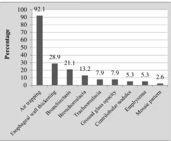
In HRCT, air-trapping was the most common pulmonary finding (92.1%) followed by bronchiectasia (21.1%) and bronchomalacia (13.2%) (Figure 1).
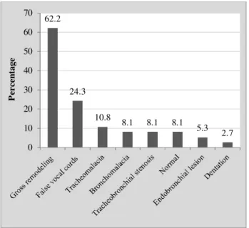
In bronchoscopy, the most common finding was airway remodeling (62.2%), followed by false vocal cord hypertrophy (24.3%) (Figure 2).
The most common endoscopic findings were erosive gastritis (63.2%), followed by reflux esophagitis grade A (50%) and hiatal herniation (31.6%) (Figure 3).
METHODS
RESULTS
Table 1. Spirometric findings of patients with sulfur mustard induced bronchiolitis obliterans (n = 39)
Min Max
Mean ± SD Variable
17 118
52.7 ± 22.4 FEV1
41 92
70.4 ± 13.9 FEV1/FVC
29 114
59.4 ± 20.0 FVC
51 213
92.9 ± 38.1 DLCO
7 120
39.4 ± 30.3 MEF
70 174
103.1 ± 36.1 FRC
18 92
49.6 ± 18.2 IC
82 543
190.8 ± 93.7 RV
FEV1: Forced expiratory volume in first second,FVC: Forced vital capacity, DLCO: Diffusing capacity of the lungs for carbon monoxide,MEF: Mean expiratory flow,FRC: Functional residual capacity,IC: Inspiratory capacity,RV: Residual volume
Figure 1. HRCT findings of patients with sulfur mustard induced bronchiolitis obliterans (n = 39)
92.1
28.9 21.1
13.2
7.9 7.9 5.3 5.3 2.6 0
10 20 30 40 50 60 70 80 90 100
P
e
r
c
en
ta
g
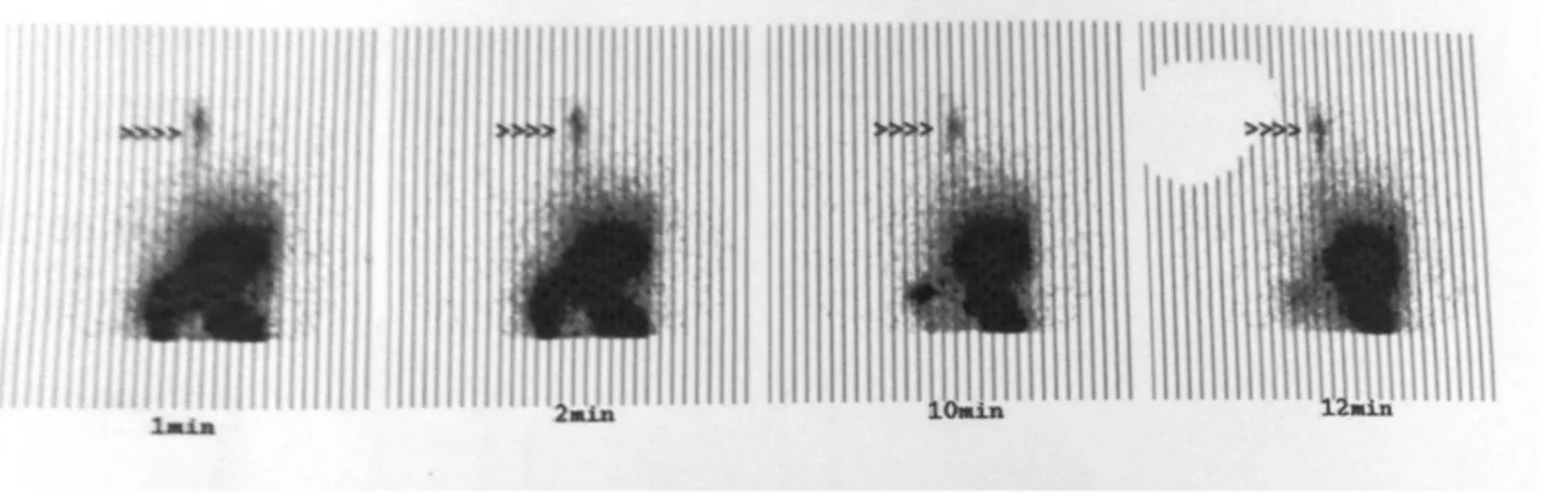
In scintigraphic imaging, only 1 patient had a remarkable finding. In this patient, the radionuclide material was seen in the pharynx (proximal GERD), but did not produce marked microaspiration of gastric substances into the airways.
In this descriptive cross-sectional study on SM-exposed patients, we found that thick sputum was the most common clinical finding followed by dyspepsia and chest tightness. The mean of FEV1 and FEV1/FVC were lower than normal ___________
levels. In HRCT scan, air-trapping was the most common finding. Only 1 patient had remarkable finding in scintiscan, which could not provide enough evidence for analysis between positive and negative groups in terms of clinical variables. This study extended our knowledge about the GERD in patients with SM exposure. In a study by Aliannejad et al (14), bile acids in bronchoalveolar lavage (BAL) fluid as a marker for microaspiration among SM-injured patients were found in 84% of them, though pepsin and trypsin (other microaspiration biomarkers) were detected in BAL fluid of all patients. They finally concluded that in addition to the high prevalence of GER in BO patients after exposure to SM, GERper se might be an aggregating factor for BO exacerbation.
There is a growing concern about relationship between GERD and pulmonary diseases. Nowadays, it has been approved that micro and macro gastroesophageal reflux can induce chronic lung diseases such as asthma, COPD, and even pulmonary fibrosis, but there are some doubts and controversies about this relationship (2). It is known that aspiration of gastroesophageal fluids; either micro or macroaspiration, is notorious for pathogenesis of reactive airway diseases. It was demonstrated that gastroesophageal microaspiration into the airways, which is generally associated with limited or no GI symptoms, has a significant role on producing chronic cough (2). However, microaspiration is often neglected in patients with pulmonary symptoms. Macroaspiration, on the other hand, raises more remarkable GI symptoms and patients with such problem seek for treatment for it. Hence, macroaspiration may be less effective in progression of GER-induced pulmonary diseases due to the fact it is treated at earlier stages.
It should be determined which component of the GI juice such as bile acids, pepsin, gastric acid, etc. is more dangerous for lungs. The low effectiveness of current acid suppressive therapies, even powerful proton pump inhibitors (PPIs), on extra-esophageal symptoms of GERD may suggest that non-acid reflux can exert symptoms through mechanisms beyond excessive acid production (2,22). In this respect, Tutuian et al demonstrated that in GER patients treated with anti-acid suppressive medications such as PPIs, non-acid reflux could induce a lot of symptoms such as cough (23).
It is a matter of debate whether GERD induces lung diseases or it only accelerates progression of these disorders. There are so many trials to respond to this question. It has been reported that adding PPIs even in high doses, compared with placebo, improves neither symptoms nor lung function of patients with asthma, but was associated with more adverse events such as higher incidence of respiratory infections (24,25). However, it has been shown that using a PPI, omeprazole, in addition to a dopamine antagonist, domperidone, for asthmatic adults with GER is beneficial by reducing asthma symptoms, and improving pulmonary function (26). In general, because of low adverse effects of PPIs or anti-acid medications, it is better to use them in patients with refractory symptoms of pulmonary diseases.
There is still no concrete agreement over the best method for GERD diagnosis in patients with pulmonary diseases. Sputum or salivary pepsinogen and pepsin levels can be ___________
Figure 2.Bronchospcopic findings of patients with sulfur mustard induced bronchiolitis obliterans (n = 39)
Figure 3. Endospcopic findings of patients with sulfur mustard induced bronchiolitis obliterans (n = 39)
62.2
24.3
10.8
8.1 8.1 8.1 5.3
2.7 0
10 20 30 40 50 60 70
P
e
r
c
e
n
ta
g
e
63.2
50
31.6
18.4 18.4
6.9
0 10 20 30 40 50 60 70
P
e
rc
en
ta
g
e
measured for this purpose (27,28). In addition, substance P and neurokinin A are increased in response to inflammatory process at the respiratory epithelium (29). Although scintigraphy has been recommended for GER detection in patients who are presented with chronic cough and pulmonary diseases (28), in this study, we found discouraging evidence over the utility of this diagnostic method for GER. In this regard, in our study on SM-injured patients with BO, although GER was detected with endoscopic findings in the majority of cases, only one patient had remarkable findings in scintigraphy. These results can be attributed to shorter duration between nucleotide food ingestion and performing scintiscan (30). It seems that using other methods (such as pH metry) would be helpful to achieve more accurate diagnosis of GER due to non acid components such as bile and pepsin components (2).
Finally, many scientists argued over the causative role of GERD for pulmonary disorders. There has been evidence approving the similarity of both airway and esophagus innervations (2,31). Hence, as reflux may stimulate vagal reflex arc, cough and/or bronchospasm consequently might occur (32-34). In contrast, patients with constrictive pulmonary diseases are susceptible to GERD because of the treatments they receive, i.e. muscle relaxant effect of beta blockers or corticosteroids on lower esophageal sphincter.
One of the limitations of this study was lack of a control group, though it was a cross-sectional and descriptive study on sulfur mustard induced BO patients, and further studies are better to be conducted in case control design. In addition, we need studies which evaluate microaspiration treatment efficacy on GERD extra-pulmonary manifestations to confirm the role of any degree of reflux in GERD pathogenesis in lung tissue. In addition, there are no methods for following up treatment efficacy and it can be proposed for future studies” (2).
Although previous reports demonstrated high prevalence ________
of GERD and microaspiration (using measurement of pepsin) in patients with SM-induced BO, we did not find remarkable evidence for microaspiration in scintiscan in patients included in this study.
Authors would like to thank staff of Baqiyatallah Hospital and all patients who kindly cooperated in this study.
Conflict of interest:None to be declared.
Funding and support: This research was supported by Baqiyatallah University of Medical Sciences, Tehran, Iran.
REFERENCES
1. Sontag SJ. Gastroesophageal reflux disease and asthma. J Clin Gastroenterol 2000;30:S9-30.
2. Ghanei M, A S. Gastro-Esophageal Reflux and Pulmonary Medicine; Where Are We and What Should We Do? J Pulmonar Respirat Med 2012;2:107.
3. Emilsson OI, Bengtsson A, Franklin KA, Toren K, Benediktsdottir B, Farkhooy A, et al. Nocturnal gastroesophageal reflux, asthma and symptoms of obstructive sleep apnoea: a longitudinal, general population study. Eur Respir J 2013;41:1347-54.
4. Blake K, Teague WG. Gastroesophageal reflux disease and childhood asthma. Curr Opin Pulm Med 2013;19:24-9. 5. Hartwig MG, Davis RD. Gastroesophageal reflux
disease-induced aspiration injury following lung transplantation. Curr Opin Organ Transplant 2012;17:474-8.
6. Ghanei M, Harandi AA. Long term consequences from exposure to sulfur mustard: a review. Inhal Toxicol 2007;19:451-6.
7. Saber H, Saburi A, Ghanei M. Clinical and paraclinical guidelines for management of sulfur mustard induced bronchiolitis obliterans; from bench to bedside. Inhal Toxicol 2012;24:900-6.
8. Panahi Y, Poursaleh Z, Amini-Harandi A, Saburi A, Shohrati M, Ghanei M. Study on Effectiveness of Low Dose Theophylline as Add-on to Inhaled Corticosteroid for Patients with Sulfur Mustard Induced Bronchiolitis. Asia Pac J Med Toxicol 2013;2:126-30.
CONCLUSION
REFERENCES
ACKNOWLEDGEMENT
Figure 4. Pulmonary scinitigraphic imaging of the index case (Pointer shows reflux of the tracer to the pharynx from the stomach)
9. Amini M, Oghabian Z. Late-onset Radiologic Findings of Respiratory System Following Sulfur Mustard Exposure. Asia Pac J Med Toxicol 2013;2:58-62.
10. Ghanei M, Hosseini AR, Arabbaferani Z, Shahkarami E. Evaluation of chronic cough in chemical chronic bronchitis patients. Environ Toxicol Pharmacol 2005;20:6-10.
11. Ghanei M, Khedmat H, Mardi F, Hosseini A. Distal esophagitis in patients with mustard-gas induced chronic cough. Dis Esophagus 2006;19:285-8.
12. Hartwig MG, Appel JZ, Davis RD. Antireflux surgery in the setting of lung transplantation: strategies for treating gastroesophageal reflux disease in a high-risk population. Thorac Surg Clin 2005;15:417-27.
13. Patra S, Singh V, Chandra J, Kumar P, Tripathi M. Diagnostic modalities for gastro-esophageal reflux in infantile wheezers. J Trop Pediatr 2011;57:99-103.
14. Aliannejad R, Hashemi-Bajgani S-M, Karbasi A, Jafari M, Aslani J, Salehi M, et al. GERD related micro-aspiration in chronic mustard-induced pulmonary disorder. J Res Med Sci 2012;17:777-81.
15. Decalmer S, Stovold R, Houghton LA, Pearson J, Ward C, Kelsall A, et al. Chronic cough: relationship between microaspiration, gastroesophageal reflux, and cough frequency. Chest 2012;142:958-64.
16. Ervine E, McMaster C, McCallion W, Shields MD. Pepsin measured in induced sputum--a test for pulmonary aspiration in children? J Pediatr Surg 2009;44:1938-41.
17. Farhath S, He Z, Nakhla T, Saslow J, Soundar S, Camacho J, et al. Pepsin, a marker of gastric contents, is increased in tracheal aspirates from preterm infants who develop bronchopulmonary dysplasia. Pediatrics 2008;121:e253-9. 18. Fiorucci S, Distrutti E, Di Matteo F, Brunori P, Santucci L,
Mallozzi E, et al. Circadian variations in gastric acid and pepsin secretion and intragastric bile acid in patients with reflux esophagitis and in healthy controls. Am J Gastroenterol 1995;90:270-6.
19. Fujimoto K, Yamaguchi S, Urushibata K, Koizumi T, Kubo K. Sputum eosinophilia and bronchial responsiveness in patients with chronic non-productive cough responsive to anti-asthma therapy. Respirology 2003;8:168-74.
20. Grabowski M, Kasran A, Seys S, Pauwels A, Medrala W, Dupont L, et al. Pepsin and bile acids in induced sputum of chronic cough patients. Respir Med 2011;105:1257-61. 21. Pierson DJ. Clinical practice guidelines for chronic obstructive
pulmonary disease: a review and comparison of current resources. Respir Care 2006;51:277-88.
22. Tutuian R, Mainie I, Agrawal A, Adams D, Castell DO. Nonacid reflux in patients with chronic cough on acid-suppressive therapy. Chest 2006;130:386-91.
23. Tutuian R, Vela MF, Hill EG, Mainie I, Agrawal A, Castell DO. Characteristics of symptomatic reflux episodes on Acid suppressive therapy. Am J Gastroenterol 2008;103:1090-6. 24. Holbrook JT, Wise RA, Gold BD, Blake K, Brown ED,
Castro M, et al. Lansoprazole for children with poorly controlled asthma: a randomized controlled trial. JAMA 2012;307:373-81.
25. Littner MR, Leung FW, Ballard ED 2nd, Huang B, Samra NK, Lansoprazole Asthma Study Group. Effects of 24 weeks of lansoprazole therapy on asthma symptoms, exacerbations, quality of life, and pulmonary function in adult asthmatic patients with acid reflux symptoms. Chest 2005;128:1128-35. 26. Sharma B, Sharma M, Daga MK, Sachdev GK, Bondi E. Effect of omeprazole and domperidone on adult asthmatics with gastroesophageal reflux. World J Gastroenterol 2007;13:1706-10.
27. Knight J, Lively MO, Johnston N, Dettmar PW, Koufman JA. Sensitive pepsin immunoassay for detection of laryngopharyngeal reflux. Laryngoscope 2005;115:1473-8. 28. Karbasi A, Goosheh H, Aliannejad R, Saber H, Salehi M, Jafari
M, et al. Pepsin and bile acid concentrations in sputum of mustard gas exposed patients. Saudi J Gastroenterol 2013;19:121-5.
29. Patterson RN, Johnston BT, Ardill JE, Heaney LG, McGarvey LP. Increased tachykinin levels in induced sputum from asthmatic and cough patients with acid reflux. Thorax 2007;62:491-5.
30. Ruth M, Carlsson S, Mansson I, Bengtsson U, Sandberg N. Scintigraphic detection of gastro-pulmonary aspiration in patients with respiratory disorders. Clin Physiol 1993;13:19-33. 31. Jadcherla SR. Upstream effect of esophageal distention: effect
on airway. Curr Gastroenterol Rep 2006;8:190-4.
32. Mansfield LE, Hameister HH, Spaulding HS, Smith NJ, Glab N. The role of the vague nerve in airway narrowing caused by intraesophageal hydrochloric acid provocation and esophageal distention. Ann Allergy 1981;47:431-4.
33. Bingol Boz A, Aydn F, Celmeli F, Boz A, Artan R, Gungor F. Does gastroesophageal reflux scintigraphy correlate with clinical findings in children with chronic cough? Nucl Med Commun 2009;30:802-6.