Occult HBV Infection May Be Transmitted
through Close Contact and Manifest as an
Overt Infection
Li-Ping Hu1,2, De-Ping Liu3, Qin-Yan Chen1, Tim J. Harrison4, Xiang He5, Xue-Yan Wang1, Hai Li1, Chao Tan1, Qing-Li Yang1, Kai-Wen Li1,2, Zhong-Liao Fang1*
1Guangxi Zhuang Autonomous Region Center for Disease Prevention and Control, Guangxi Key Laboratory for the Prevention and Control of Viral Hepatitis, Nanning, Guangxi 530028, China,2School of Preclinical Medicine, Guangxi Medical University, 22 ShuangYong Road, Nanning, Guangxi 530021, China,3Chong Zuo Center for Disease Prevention and Control, Lijiang road, Chong Zuo, Guangxi 532200, China,4Division of Medicine, UCL Medical School, London, United Kingdom,5Guangdong Provincial Institute of Public Health, Guangdong Provincial Center for Disease Control and Prevention, Guangzhou 511430, China
*zhongliaofang@hotmail.com
Abstract
The importance of transmission of occult HBV infection (OBI) via transfusion, organ trans-plantation and hemodialysis has been widely recognized. However, data regarding the transmission of OBI through close contact remain limited. In this study, serum samples were obtained from a child and his parents. The child had received the standard vaccination regimen at birth and produced protective antibody. Sera were tested for HBV serological markers. Nested PCR assays were used to detect HBV DNA and the amplicons were cloned and their sequences subjected to phylogenetic analysis. The results showed that both parents had occult infections while the child had an overt infection. Twelve, eleven and nine clones, from the father, mother and son, respectively, were sequenced. Serotypes adrq+, ayw1, ayw and ayr were found in the father and ayw1, adw2 and adwq+ in the mother; adrq+ was the only serotype in son. Genotype B, subgenotype C2 and a recombi-nant were identified in the father and genotype B, subgenotype C5 and three recombirecombi-nants were found in the mother. Subgenotype C2 was the only genotype identified in the child. A phylogenetic tree showed that all of the child’s sequences and most of the father’s sequences clustered together. However, none of mother’s sequences clustered with those of the child. The surface gene from the child and his father had the same amino acid substi-tution pattern (T118K, T123N and G145A). We concluded that the father was the source of the son’s HBV infection, suggesting that occult HBV infection may be transmitted through close contact and manifest as an overt infection.
OPEN ACCESS
Citation:Hu L-P, Liu D-P, Chen Q-Y, Harrison TJ, He X, Wang X-Y, et al. (2015) Occult HBV Infection May Be Transmitted through Close Contact and Manifest as an Overt Infection. PLoS ONE 10(10): e0138552. doi:10.1371/journal.pone.0138552
Editor:Tatsuo Kanda, Chiba University, Graduate School of Medicine, JAPAN
Received:July 14, 2015
Accepted:September 1, 2015
Published:October 12, 2015
Copyright:© 2015 Hu et al. This is an open access article distributed under the terms of theCreative Commons Attribution License, which permits unrestricted use, distribution, and reproduction in any medium, provided the original author and source are credited.
Data Availability Statement:Data are available from
http://dx.doi.org/10.6084/m9.figshare.1554852,http:// dx.doi.org/10.6084/m9.figshare.1554851,http://dx. doi.org/10.6084/m9.figshare.1554850, andhttp://dx. doi.org/10.6084/m9.figshare.1554848.
Funding:This work was supported by the National Natural Science Foundation of China (Grant No. 81260439/H2609) and the Guangxi Science Foundation (Gui Ke Gong 1140003A-37).
Introduction
Persistent infection with hepatitis B virus (HBV) remains a major global public health problem. Infection with hepatitis B virus may lead to a wide spectrum of liver disease that range, in acute infection, from mild, self-limited to fulminant hepatitis and, in persistent infection, from an asymptomatic carrier state to severe chronic hepatitis, cirrhosis and hepatocellular carcinoma. More than two billion people, one third of the world’s population alive today, have been infected with HBV at some time in their lives and around 240 million of them remain infected (chronic HBsAg carriers) [1]. However, these classes do not include all HBV infections; there is another form of HBV infection, occult HBV infection (OBI). This was first reported by Tabor et al. 36 years ago in a case report of HBV infection following blood transfusion with blood from donors positive for anti-HBc only [2]. OBI is defined by the absence of HBsAg despite the presence of HBV DNA in the liver, blood serum, or peripheral blood mononuclear cells, irre-spective of the presence of other hepatitis B viral antibodies and antigens [3].
The prevalence of OBI varies widely across the globe and ranges from 1% to 95% worldwide, depending on the level of endemic disease, the assays used in the studies and the various popu-lations studied [4,5]. However, it is well known that certain groups of patients are at a much higher risk of having occult HBV infection, regardless of the geographical location, such as those with chronic HCV infection and HIV infection, hemodialysis, liver transplant and hepa-tocellular carcinoma (HCC) patients and injection drug users [6]. Occult infection may be reactivated, leading to acute and severe forms of classical hepatitis B. The long-term persistence of the virus in the liver may favor the progression of the chronic liver disease to cirrhosis and HCC [7].
One of the major public health problems of occult HBV infection is the potential for trans-mission. Blood transfusion and liver transplantation remains the major routes of transmission, although the risk of HBV transmission through blood transfusion has decreased following the introduction of sensitive and specific diagnostic assays [8]. In additional, it has also been reported that intrauterine HBV infection is possible in pregnant women who are HBsAg and HBeAg negative [9]. The possibility of horizontal transmission of HBV from individuals with occult infection to close contacts does exist [10]. When occult viruses are transmitted to other individuals, the outcomes in terms of liver disease are the same as those following transmission from overt cases [8].
It has been reported that the prevalence of OBI is higher in HBV endemic areas such as East Asia and lower in low endemic areas such as North America [5]. Guangxi is one of the prov-inces in China with the highest prevalence of persistent HBV infection, affecting 9.2% of the general population [11]. We reported previously that the prevalence of OBI among family members of children from Long An county, Guangxi who were positive for both HBsAg and anti-HBs after vaccination is 11.5% [12], suggesting that occult HBV infection is common in Guangxi. In this study, we provide evidence of transmission of occult HBV from a family con-tact to a child who was vaccinated successfully at birth but became infected overtly.
Materials and Methods
Study population and sample design
The study subjects were a three member family, a boy and his parents. Serum samples were obtained from the three individuals in April, 2015.
the same program but the doses were 30μg, 20μg and 10μg, respectively. He remained nega-tive for all of HBV serological markers after the last dose. He became weakly posinega-tive for anti-HBc in 2004 and positive for both anti-HBe and anti-anti-HBc in 2008. He was vaccinated again in 2009 with one 60μg dose. He was positive for anti-HBc only after vaccination.
The woman is also 44 years old and had married in 1999. She was found to be positive for anti-HBs in 1996. She was immunized with a full course of vaccination (10μg doses of vaccine given at 0, 1 and 6 months) in 2004 when she found to be weakly positive for anti-HBs. She was positive for anti-HBs (298 IU/ml), anti-HBe and anti-HBc in 2008.
The child was born in 2001. He received in time a full course of vaccination (10μg doses of vaccine given at 0, 1 and 6 months). He was anti-HBs positive with a titer of10 IU/L after the last dose. However, he became positive for HBsAg in 2004. He was positive for HBsAg, anti-HBe and anti-HBc in 2007.
All vaccines used above are yeast-derived recombinant hepatitis B vaccine (National Vac-cine and Serum Institute, Beijing, China).
Informed consent in writing was obtained from the parents and that of the child was from the parents in his behalf. The study protocol conforms to the ethical guidelines of the 1975 Declaration of Helsinki and has been approved by the Guangxi Institutional Review Board.
Serological Testing
Sera were tested for HBsAg/anti-HBs, HBeAg/anti-HBe, anti-HBc and anti-hepatitis C virus (HCV) using enzyme immunoassays (Zhong Shan Biological Technology Company, Limited, Guangzhou, China). Alanine aminotransferase (ALT) levels were determined using a Reitman kit (Sichuan Mike Scientific Technology Company, Limited, Chengdu, China).
Nested polymerase chain reaction (PCR) for HBV DNA and nucleotide
sequencing
DNA was extracted from 85μl serum by pronase digestion followed by phenol/chloroform extraction. In order to avoid false positive, two regions of the HBV genome were amplified using nested PCR, from PreS1 to the X gene and a smaller region covering the S gene only.
For PreS1 to the X gene, the first round PCR was carried out in a 50μl reaction using prim-ers LSOB1 (nt 2739–2762, 5'-GGCATTATTTGCATACCCTTTGG-3') and P2 (nt 1823–1806 5'-CCGGAAAGCTTGAGCTCTTCAAAAAGTTGCATGGTGCTGG-3’) [13], with 5 min hot start followed by 30 cycles of 94°C for 30 sec, 50°C for 30 sec, and 72°C for 90 sec. Second round PCR was carried out on 5μl of the first round products in a 50μl reaction using primers LSBI1 (nt 2809–2829, TTGTGGGTCACCATATTCTT-3') and POLSEQ2 (nt1168-1188, 5'-AGCAAACACTTGGCATAGGC-3') and the same amplification protocol as first round.
For the S gene, the first round PCR was carried out in a 50μl reaction using primers MD14 (nt 418–433, 5'-GCGCTGCAGCTATGCCTCATCTTC-3') and HCO2 (nt 761–776, 5'-GCG AAGCTTGCTGTACAGACTTGG-3'), with 5 min hot start followed by 35 cycles of 94°C for 45 sec, 45°C for 45 sec, and 72°C for 120 sec. The second round PCR was carried out on 5μl of the first round products in a 50μl reaction using primersME15(nt 455–470, 5'-GCGCTGCAG CAAGGTATGTTGCCCG-3') and HDO3 (nt 734–748, 5'-GCGAAGCTTCATCATCCATA TAGC-3') with 5 min hot start followed by 30 cycles of 94°C for 45 sec, 55°C for 45 sec, and 72°C for 120 sec.
Biosystems, Foster City, USA) with sequencing primer PSISEQ2F (nt 65–84, 5'-GGCTCCA ATTCCGGAACAGC-3') and POLSEQ2. Meanwhile, PreS1/S2 region of HBV from each sam-ple was sequenced directly without cloning using sequencing primer LSBI1 in The Sangon Bio-tech (Shanghai, China).
Measurement of Viral loads
Serum HBV DNA concentrations were quantified by real time PCR using commercial reagents (Shanghai ZJ Bio-Tech Co., Ltd. (Shanghai, China)) in an ABI Prism 7500 sequence detection system (Applied Biosystems, Foster City, CA, California, USA), using HBV primers and a dual labeled TaqMan probe, as described previously [14].
HBV serotyping
Serotypes were determined according to a single amino acid at the following position: adrq-(122 K + 127P + 134 F + 159 V + 160R +177A +178P), adrq + adrq-(122 K + 127P + 134 F + 159A +160R + 177 V +178P), adw2 (122 K + 127P + 134 F +159A + 160 K + 177 V +178P), adwq+ (122 K + 127 L +134 F + 159A + 160 K + 177 V +178P), ayr (122R + 127P +134 F + 159A + 160R + 177 V +178P), ayw1 (122R +127P + 134 F + 159A + 160 K + 177 V +178P), ayw2 (122R + 127P + 134Y + 159G + 160 K + 177 V +178P), ayw3 (122R + 127 T + 134 F + 159G + 160 K + 177V +178P) [15–17].
HBV genotyping
HBV genotypes were determined using phylogenies reconstructed on the basis of the complete S region (678 nt) of the viruses. The sequences were aligned to 21 HBV sequences of all known genotypes retrieved from GenBank using Clustal W and visually confirmed with the sequence editor BioEdit [18]. The reference sequences were A1_M57663_Philippines, B1_D23677_Ja-pan, B2_AY217358_China, B2_AF121249_Vietnam, B3_AB033555_Sumatra, B4_AB073835_ Vietnam, B5_AB219427_Philippines, B6_DQ463801_Canada, C1_AF458664_China,
C2_AY217371_China, C3_X75656_Polynesia, C4_AB048704_Australia, C5_JN827415_ Thailand, D1_AF280817_China, E_AB091255_Ivory Coast, F_AY090458_Costa Rica, G_ AF160501_USA, H_AY090460_USA, I1_AB231908_Vietnam, I1_FR714504_Longan_China, I2_FJ023664_Laos. Neighbor-Joining trees were reconstructed under the Kimura 2-parameter substitution model with the program MEGA [19]. The reliability of clusters was evaluated using interior branch test with 1000 replicates and the internal nodes with over 95% support were considered reliable.
Sequences that were not determined by phylogenies were genotyped using the NCBI Geno-typing Tool (http://www.ncbi.nlm.nih.gov/projects/genotyping/formpage.cgi).
Identification of MHR mutations and overlapping polymerase mutations
Results
General information
Successful PCR amplification of two regions of the viral genome was achieved for all three indi-viduals. Both parents have OBI, the father is positive for anti-HBc only and the mother is posi-tive for anti-HBs and anti-HBc. The ALT levels of the parents are normal but that of the child is abnormal. The viral load of the father is the highest and that of the mother is the lowest (Table 1). All three individuals are negative for anti-HCV. Complete PreS1/S2 sequences were obtained for the three individuals; 12, 11 and 9 clones were constructed from the father’s, mother’s and son’s amplicons, respectively. The S-gene was sequenced in both directions, cov-ering the entire MHR (GenBank accession number: KT585753-KT585784).
Serotypes in different clone sequence
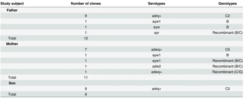
Serotypes adrq+, ayw1, ayw and ayr were predicted from the father’s twelve sequences. Four serotypes, including ayw1, adw2 and adwq+ were predicted from the mother’s sequences. All nine sequences from the son’s sample predicted serotype adrq+ (Table 2). These data suggest that transmission between father and son is possible because they have the same serotype adrq + and the transmission is from father to son because the father has more serotypes. Transmis-sion between the father and mother also is possible because they share serotype: ayw1. How-ever, transmission between the mother and son is not possible for because they do not share any serotype.
Table 1. Serological characteristics of the study subjects.
Samples Ages HBsAg Anti-HBs HBeAg Anti-HBe Anti-HBc Viral loads ALT (IU/ml)
Father 44 - - - - + 8×10⁵IU/ml <40
Mother 44 - + - - + 3.53×10³IU/ml <40
Son 14 + - + - + 5.42×10⁵IU/ml 170
doi:10.1371/journal.pone.0138552.t001
Table 2. Serotypes and genotypes predicted from the sequences from each study subject.
Study subject Number of clones Serotypes Genotypes
Father
9 adrq+ C2
1 ayw1 B
1 ayw B
1 ayr Recombinant (B/C)
Total 12
Mother
7 adwq+ C5
1 ayw1 B
1 ayw1 Recombinant (B/C)
1 adw2 Recombinant (B/C)
1 adwq+ Recombinant (C/G)
Total 11
Son
9 adrq+ C2
Total 9
Genotypes in different clone sequences
Using phylogenetic analysis and the NCBI genotyping analysis, three genotypes, subgenotype C2, genotype B and a recombinant were identified from the father’s sequences. Subgenotype C5, genotype B and three recombinants were identified in the mother. Subgenotype C2 was the only genotype identified in the son (Table 2andFig 1). The phylogenetic tree shows that all of the son’s sequences and most of the father’s sequences cluster together. However, none of the mother’s sequences cluster with her son’s sequences (Fig 1). Clearly, the transmission was from father to son. There is no evidence of transmission between the mother and son. It is also possi-ble for transmission to have occurred between the parents because some of their sequences cluster together, supported by a 77% bootstrap value.
Amino acid substitutions within the
“
a
”
determinant and MHR
Amino acid substitutions in the major hydrophilic region predicted from twelve clones of HBV from the father’s sample include T115I, T116A, S117G, T118K, 123N, Q129L, T131N, M133L, M133S, F134L, G145A and I152V. Half of them are located within“a”determinant (aa 124–
147). Five of the twelve substitutions, including T118K, T123N, T131N, M133L and N 145A, are associated with antibody escape. The T118K and N145A substitutions may result in vaccine escape. T118K and T123N may result in failure to detect of HBsAg. The 123N and 145A muta-tions may result from escape from immunoglobin therapy. Seven clones have the same amino acid substitution pattern as that in the son: T118K, T123N and N145A. Two clones have T118K and N145A substitutions, two other clones have T131N and the final clone has M133L (Fig 2).
Amino acid substitutions in the major hydrophilic region predicted from nine clones of HBV from the son’s sample include L104S, T118K, T123N, S143L and G145A. Only two of them are located within the“a”determinant. Except for L104S, all are escape substitutions. All of the nine clones from the son have the same amino acid substitution pattern (T118K, T123N and G145A) as seen in his father. Furthermore, one of the nine sequences has an amino acid substitution at position 143, which may result in vaccine escape and failure to detect HBsAg (Fig 2).
Amino acid substitution mutations in the major hydrophilic region predicted from eleven clones of HBV from the mother’s sample include D99N, T131N, F161S and V168A. Only the T131N mutation causes detection failure and this mutation could be seen in one clone only (Fig 2).
Clearly, the frequency of amino acid substitution mutations within the“a”determinant and MHR is highest in the father’s sample. The next is that from the son. It is possible that there was transmission between father and son because they have the same amino acid substitution pattern. The transmission was from father to son because the father has a more complex pat-tern of mutations. Transmission between father and mother or mother and son is impossible because they do not share the same amino acid substitution pattern.
Mutations in the preS1/preS2 region
PreS1 contains 357 bases, encoding 119 amino acids and PreS2 contains 165 bases, encoding 55 amino acids. No deletion was found in either PreS1 or PreS2 from the three samples. No point mutation was found in the initiation codon of preS2 in the three samples.
The impact of mutations in the S gene on the overlapping polymerase
region
in the overlapping polymerase in all clones from the son’s sample, including R15L, V23I, T38A, T38K, H55Q, S57F, L72P, L77S, H126Q, V191I and H197R. There are twenty-one amino acid substitutions in the overlapping polymerase in all clones from father’s sample, including F46S, R51K, H55Q, H55R, S57F, P109S, N118T, N124R, Y124H, Q125R, H126Q, 127R, N134D, C136R, N139K, Y141F, S143T, H160R, A211T, S213T and Q215H. Amino acid substitutions in mother include T16I, R41S, V44A,N53S, H55R, W58R, N76D, S81T, V103I, G107E, N121I, I122L, N123D, Q125K, H126Y, N134D, N139H, N139Q, Y158H, I163V, F178L, S185N, V207M, Q215L,Y221F, A222T, I224V, G232R. However, none of these is asso-ciated with drug resistance.
Discussion
The major finding in the study is that the son has one serotype (adrq+) only and this was seen in the father but not the mother. The son also has one genotype (subgenotype C2) only and this could also be seen in the father but not the mother. All sequences from the son clustered with that from father in the phylogenetic tree. All of the sequences from the son have the same
Fig 1. Neighbor-Joining trees.The trees were reconstructed on the basis of the complete S region (678 nt) of the viruses under the Kimura 2-parameter substitution model with the program MEGA [19]. The branch lengths represent the number of substitutions per site. The reliability of clusters was evaluated using the interior branch test with 1000 replicates and the internal nodes with over 95% support are considered reliable.
doi:10.1371/journal.pone.0138552.g001
Fig 2. Frequency and distribution of amino acid substitutions in the MHR of HBsAg from each clone.
amino acid substitution pattern in the S protein as that seen in the father. Furthermore, the son was found to be positive for HBsAg when he was tested prior to entrance to kindergarten, sug-gesting that household contact is the only likely pattern of transmission. These findings provide strong evidence of transmission from father to son. The father shares one serotype and geno-type with the mother but not the amino acid substitution pattern in the S protein. Meanwhile, it also is suggested that the mother became infected outside the family. The strength of the study is that detailed medical records are available for the study subjects, which may provide additional evidence of transmission. The weakness of the study is that we did not test HBsAg with different commercial diagnostic kits, which may provide information about the associa-tion of amino acid substituassocia-tions with detecassocia-tion failure.
The recognized patterns of spread of HBV include perinatal, sexual and parenteral/percuta-neous routes. Routes of parenteral transmission include injection drug use, transfusions and dialysis, acupuncture and tattooing; household contact with infected individuals and working in a health-care setting also are risk factors for horizontal transmission [20,21]. Compared to overt HBV infection, the routes of transmission of occult HBV have been studied less. Most of these studies focused on blood transfusion and liver transplantation because these may trans-mit hepatitis B [8]. In addition, it has also been reported that intrauterine HBV infection is pos-sible in pregnant women with OBI [9].
Occult HBV infection may be common in household contacts of individuals with chronic hepatitis B [22]. A study from India found that sequences from both occult HBV and overt HBV are similar in terms of genotype and surface variants or non-variants (wild-type) and clustered together in the phylogenetic tree. The authors considered the possibility of horizontal transmission of HBV from individuals with occult infection to their contacts [10]. However, this claim is not strong enough because it remains possible that the transmission of HBV was from individuals with overt infection but resulted in occult infection. In our study, with cloning and sequencing, we found that the father has more serotypes and genotypes than that of the son, suggesting that transmission was from father to son.
Immunization with hepatitis B vaccine is the most effective means of preventing acute infec-tion by HBV [23]. However, the titer of vaccine-induced antibody decays exponentially over time, irrespective of the population immunized [24]. It has been claimed that neonatal HBV immunization is efficacious in inducing long-term immunity and cell-mediated immune mem-ory for up to two decades and booster vaccinations are not required [25]. The fact that the child produced protective levels of anti-HBs after immunization but became infected by HBV clearly challenges these findings, suggesting that the monitoring of the level of anti-HBs among vaccinated subjects for booster vaccination is necessary. Our findings also suggested that occult HBV infection may be transmitted through close contact. Therefore, susceptible individuals should be vaccinated against hepatitis B in endemic regions. Nucleic acid testing for OBI is nec-essary for the staff in some occupations, such as nursery teachers, in regions where HBV is endemic.
A study from Taiwan showed that most non-responders among anti-HBc positive subjects apparently had occult HBV infection [31]. However, this finding was not supported by a subse-quent study from Iran [32]. In our study, the father was negative for all HBV serological mark-ers before and after the first two full courses of vaccination. He became weakly positive for anti-HBc many years later. Then, he was immunized for the third time but remained negative for anti-HBs. Clearly, our data support the result from Taiwan and suggest that nucleic acid testing should be considered for non-responders to exclude OBI, especially in regions where HBV is endemic.
The predominant genotype in Guangxi is genotype C, followed by genotypes B and I (a recombinant) [33]. In the study, both parents are infected with genotype B and C and recombi-nants (between genotype B and C). In the future, we will determine whether the recombinant sequences are from genotype B and C in the same person or from outside sources, which may provide more information about the occurrence of recombination.
Acknowledgments
We are indebted to the family providing serum samples and staff members of Chong Zuo Cen-ter for Disease Prevention and Control for their help in handling the sera.
Author Contributions
Conceived and designed the experiments: ZLF TJH DPL. Performed the experiments: LPH QYC HL QLY. Analyzed the data: XH XYW. Contributed reagents/materials/analysis tools: DPL CT KWL. Wrote the paper: ZLF TJH.
References
1. World Health Organization. Hepatitis B Fact sheet N°204. (2014) Available:http://www.who.int/ mediacentre/factsheets/fs204/en/
2. Tabor E, Hoofnagle JH, Smallwood LA, Drucker JA, Pineda-Tamondong GC, Ni LY, et al. Studies of donors who transmit posttransfusion hepatitis. Transfusion. 1979; 19: 725–731. PMID:230620 3. Martinez MC, Kok CC, Baleriola C, Robertson P, Rawlinson WD. Investigation of occult hepatitis B
virus infection in anti-hbc positive patients from a liver clinic. PLoS One. 2015; 10: e0117275. doi:10. 1371/journal.pone.0117275PMID:25763579
4. Gutiérrez-García ML, Fernandez-Rodriguez CM, Lledo-Navarro JL, Buhigas-Garcia I. Prevalence of occult hepatitis B virus infection. World J Gastroenterol. 2011; 17: 1538–42. doi:10.3748/wjg.v17.i12
PMID:21472117
5. Kwak MS, Kim YJ. Occult hepatitis B virus infection. World J Hepatol. 2014; 6: 860–9. doi:10.4254/ wjh.v6.i12.860PMID:25544873
6. Samal J, Kandpal M, Vivekanandan P. Molecular mechanisms underlying occult hepatitis B virus infec-tion. Clin Microbiol Rev. 2012; 25: 142–63. doi:10.1128/CMR.00018-11PMID:22232374
7. Squadrito G, Spinella R, Raimondo G. The clinical significance of occult HBV infection. Ann Gastroen-terol. 2014; 27: 15–19. PMID:24714731
8. Raimondo G, Caccamo G, Filomia R, Pollicino T. Occult HBV infection. Semin Immunopathol. 2013; 35: 39–52. doi:10.1007/s00281-012-0327-7PMID:22829332
9. Gui QD, Yue YF, Li SH, Zhang F. Study on intrauterine infection of hepatitis B virus in pregnant women with hepatitis B surface antigen and hepatitis B e antigen negative. Chinese Journal of Obstetrics and Gynecology. 2005; 40: 99–102.
10. Datta S, Banerjee A, Chandra PK, Chowdhury A, Chakravarty R. Genotype, phylogenetic analysis, and transmission pattern of occult hepatitis B virus (HBV) infection in families of asymptomatic HBsAg carri-ers. J Med Virol. 2006; 78: 53–9. PMID:16299727
12. Fang ZL, Zhuang H, Wang XY, Ge XM, Harrison TJ. Hepatitis B virus genotypes, phylogeny and occult infection in a region with a high incidence of hepatocellular carcinoma in China. World J Gastroenterol. 2004; 10: 3264–8. PMID:15484297
13. Gunther S, Li BC, Miska S, Krüger DH, Meisel H, Will H. A novel method for efficient amplification of whole hepatitis B virus genomes permits rapid functional analysis and reveals deletion mutants in immunosuppressed patients. Journal of Virology. 1991; 69: 5437–44.
14. Fang ZL, Sabin CA, Dong BQ, Wei SC, Chen QY, Fang KX, et al. The association of HBV core pro-moter double mutations (A1762T and G1764A) with viral load differs between HBeAg positive and anti-HBe positive individuals: a longitudinal analysis. J Hepatol. 2009; 50: 273–80. doi:10.1016/j.jhep. 2008.09.014PMID:19070921
15. Courouce-Pauty AM, Plancon A, Soulier JP. Distribution of HBsAg subtype in the world. Vox Sang. 1983; 44:197–211. PMID:6845678
16. Okamoto H, Tsuda F, Sakugawa H, Sastrosoewignjo RI, Imai M, Miyakawa Y, et al. Typing hepatitis B virus by homology in nucleotide sequence: comparison of surface antigen subtypes. J Gen Virol. 1988; 69:2575–2583. PMID:3171552
17. Chen X, Gao J, Ji Z, Zhang W, Zhang L, Xu R, et al. A description of the hepatitis B virus genomic back-ground in a high-prevalence area in China. Virol J. 2014; 11:101. doi:10.1186/1743-422X-11-101
PMID:24884702
18. Hall TA. BioEdit: a user-friendly biological sequence alignment editor and analysis program for Win-dows 95/98/NT. Nucleic Acids Symposium Series. 1999; 41: 95–98.
19. Tamura K, Stecher G, Peterson D, Filipski A, Kumar S. MEGA6: Molecular Evolutionary Genetics Anal-ysis version 6.0. Molecular Biology and Evolution. 2013; 30: 2725–2729. doi:10.1093/molbev/mst197
PMID:24132122
20. Yao GB. Importance of perinatal versus horizontal transmission of hepatitis B virus infection in China. Gut. 1996; 38 Suppl 2, S39–42. PMID:8786052
21. Gerlich WH. Medical virology of hepatitis B: how it began and where we are now. Virol J. 2013; 10:239. doi:10.1186/1743-422X-10-239PMID:23870415
22. Kumar GT, Kazim SN, Kumar M, Hissar S, Chauhan R, Basir SF, et al. Hepatitis B virus genotypes and hepatitis B surface antigen mutations in family contacts of hepatitis B virus infected patients with occult hepatitis B virus infection. J Gastroenterol Hepatol. 2009; 24:588–98. doi:10.1111/j.1440-1746.2008. 05727.xPMID:19207682
23. Zhou YH, Wu C, Zhuang H. Vaccination against hepatitis B: the Chinese experience. Chin Med J (Engl). 2009; 122: 98–102.
24. Jack AD, Hall AJ, Maine N, Mendy M, Whittle HC. What level of hepatitis B antibody is protective? J Infect Dis. 1999; 179: 489–492. PMID:9878036
25. Saffar H, Saffar MJ, Ajami A, Khalilian AR, Shams-Esfandabad K, Mirabi AM. Long-term T-cell-medi-ated immunologic memory to hepatitis B vaccine in young adults following neonatal vaccination. Hepat Mon. 2014; 14: e22223. doi:10.5812/hepatmon.22223PMID:25368659
26. Ireland JH, O'Donnell B, Basuni AA, Kean JD, Wallace LA, Lau GK, et al. Reactivity of 13 in vitro expressed hepatitis B surface antigen variants in 7 commercial diagnostic assays. Hepatology. 2000; 31: 1176–82. PMID:10796895
27. Ly TD, Servant-Delmas A, Bagot S, Gonzalo S, Férey MP, Ebel A, et al. Sensitivities of four new com-mercial hepatitis B virus surface antigen (HBsAg) assays in detection of HBsAg mutant forms. J Clin Microbiol. 2006; 44: 2321–6. PMID:16825343
28. Kfoury Baz EM, Zheng J, Mazuruk K, Van Le A, Peterson DL. Characterization of a novel hepatitis B virus mutant: demonstration of mutation-induced hepatitis B virus surface antigen group specific "a" determinant conformation change and its application in diagnostic assays. Transfus Med. 2001; 11: 355–62. PMID:11696228
29. Cooreman MP, Leroux-Roels G, Paulij WP. Vaccine- and hepatitis B immune globulin-induced escape mutations of hepatitis B virus surface antigen. J Biomed Sci. 2001; 8: 237–47. PMID:11385295 30. Seddigh-Tonekaboni S, Lim WL, Young B, Hou JL, Waters J, Luo KX, et al. Hepatitis B surface antigen
variants in vaccinees, blood donors and an interferon-treated patient. J Viral Hepat. 2001; 8: 154–8. PMID:11264736
31. Yen YH, Chen CH, Wang JH, Lee CM, Changchien CS, Lu SN. Study of hepatitis B (HB) vaccine non-responsiveness among health care workers from an endemic area (Taiwan). Liver Int. 2005; 25: 1162– 8. PMID:16343067
32. Aghasadeghi MR, Banifazl M, Aghakhani A, Eslamifar A, Vahabpour R, Ramezani A. No evidence for occult HBV infection in hepatitis B vaccine non-responders. Iran J Microbiol. 2014; 6: 350–3. PMID:
![Fig 1. Neighbor-Joining trees. The trees were reconstructed on the basis of the complete S region (678 nt) of the viruses under the Kimura 2-parameter substitution model with the program MEGA [19]](https://thumb-eu.123doks.com/thumbv2/123dok_br/18282861.345776/8.918.250.861.533.802/neighbor-joining-reconstructed-complete-viruses-parameter-substitution-program.webp)
