online | memorias.ioc.fiocruz.br
High prevalence of occult hepatitis B virus genotype H infection
among children with clinical hepatitis in west Mexico
Griselda Escobedo-Melendez1,2,3, Arturo Panduro1,3, Nora A Fierro1,3,4, Sonia Roman1,3/+
1Department of Molecular Biology in Medicine 4Immunovirology Unit, Civil Hospital of Guadalajara Fray Antonio Alcalde, Guadalajara, Jalisco, Mexico 2Paediatric Infectious Disease Unit, Department of Paediatric Hematology and Oncology, Viral Hepatitis Clinic,
Civil Hospital of Guadalajara Juan I Menchaca, Guadalajara, Jalisco, Mexico 3Health Sciences University Center, University of Guadalajara, Guadalajara, Jalisco, Mexico
Studies on the prevalence of infection with hepatitis B virus (HBV) among children are scarce in Latin American countries, especially in Mexico. This study was aimed to investigate the prevalence of HBV infection, occult hepatitis B infection (OBI) and HBV genotypes among children with clinical hepatitis. In total, 215 children with clinical hepa-titis were evaluated for HBV infection. HBV serological markers and HBV DNA were analysed. OBI diagnosis and HBV genotyping was performed. HBV infection was found in 11.2% of children with clinical hepatitis. Among these HBV DNA positive-infected children, OBI was identified in 87.5% (n = 21/24) of the cases and 12.5% (n = 3/24) were positive for both HBV DNA and hepatitis B surface antigen. OBI was more frequent among children who had not been vaccinated against hepatitis B (p < 0.05) than in those who had been vaccinated. HBV genotype H was preva-lent in 71% of the children followed by genotype G (8%) and genotype A (4%). In conclusion, OBI is common among Mexican children with clinical hepatitis and is associated with HBV genotype H. The results show the importance of the molecular diagnosis of HBV infection in Mexican paediatric patients with clinical hepatitis and emphasise the necessity of reinforcing hepatitis B vaccination in children.
Key words: occult hepatitis B infection - clinical hepatitis - HBV genotype H - children - vaccination
Viral hepatitis is a leading cause of chronic liver disease and a serious public health problem in children worldwide. Hepatitis B virus (HBV) is one of the most frequent aetiological agents of viral hepatitis in paediat-ric patients who are infected primarily through mother-to-infant transmission (Kelly 2006, Yeung & Roberts 2010, Pol et al. 2011). In children, the age at which pri-mary HBV infection is acquired is relevant because it may lead to chronic liver inflammation in 90% of the cases when acquired at birth and in 50% of those in-fected during childhood. Indeed, this may be of great concern in regions with a high prevalence of HBV infec-tion (Broderick & Jonas 2003, Yen-Hsuan 2011).
Overt HBV infection is diagnosed by the presence of hepatitis B surface antigen (HBsAg) and/or HBV DNA in serum (Lok & McMahon 2009). In contrast, occult hepatitis B infection (OBI) presents low titres of HBV
genomes (≈200 IU/mL) in serum or in liver tissue in
subjects who are negative for HBsAg, but either posi-tive or negaposi-tive for serological markers of past HBV in-fection (Raimondo et al. 2008b, Hollinger & Sood 2010). OBI has significant clinical implications for patients who
doi: 10.1590/0074-0276140058
Financial support: COECYTJAL-Universidad de Guadalajara
(2009-1-06-2009-431) (to AP), CONACYT (Salud-2010-01-139085),
COECYTJAL-Universidad de Guadalajara (5-2010-1-1041) (to SR)
+ Corresponding author: [email protected] Received 18 February 2014
Accepted 28 May 2014
receive transplants or organ donations, blood transfusion or haemodialysis because it increases their risk of becom-ing infected by HBV,which may potentially contribute to the development of chronic liver disease, cirrhosis, acute liver failure (ALF) and hepatocellular carcinoma (Chemin & Trépo 2005, Pollicino et al. 2007, Romero et al. 2011). In the paediatric setting, OBI has been reported among recipients of blood transfusions (Liu et al. 2006),
hepa-titis B-vaccinated children (Mu et al. 2009, Utsumi et al.
2010), patients with haematological diseases and paedi-atric cancer patients (Said et al. 2009) in countries with a high prevalence of HBV infection. In these groups, the prevalence of OBI ranges from 0.6-21%.
HBV infection has distinct clinical outcomes based on the heterogeneous geographical distribution of HBV gen-otypes associated with distinct host populations (Roman et al. 2014). In Mexico, the endemicity of HBV infection is associated with the predominance of HBV genotype H (Panduro et al. 2013, Roman & Panduro 2013), consist-ing of a low HBsAg marker prevalence (< 0.3%) and a contrasting high anti-HBV core antigen (HBc) prevalence (2.6-15.9%) (Roman et al. 2009), a high OBI prevalence (14.2%) (Roman et al. 2010) and clinically low viral loads (VL) (Panduro et al. 2013). In addition, HBV genotypes D and A are reported among acutely or chronically infected adult Mexican patients (Panduro et al. 2013) and HBV
genotype G has been detected among particular popula -tions, such as patients infected with the human immuno-deficiency virus (Roman & Panduro 2013).
and several high-prevalence regions for HBV infection have been identified throughout the country (Roman et al. 2010). This epidaemiological pattern is the common scenario for OBI infection, which may be underestimated due to the immunological response of Mexicans to HBV genotype H (Fierro et al. 2011) or to a lack of sensitivity of the HBsAg assay (Roman et al. 2009). Additionally, OBI
infection ranges from 2.4% among blood donors
(Garcia-Montalvo & Ventura-Zapata 2011) to 14.2% among native Mexican communities located in regions of high ende-micity for HBV infection (Roman et al. 2010).
Previously, we reported the presence of the HBsAg marker in 3.1% of cases among 215 children with clinical hepatitis from western Mexico, along with a prevalence of 13.9% of cases among this group without a known aetio-logical agent (Escobedo-Melendez et al. 2012). However, to date, information regarding the prevalence of OBI and the main HBV genotypes among Mexican paediatric pa-tients with clinical hepatitis is unknown. Therefore, this study was aimed to analyse the clinical and molecular characteristics of HBV infection in children, which may contrast to what has been reported in adults.
SUBJECTS, MATERIALS AND METHODS Study population - In the present study, serum sam-ples from 215 paediatric patients were tested by molecu-lar analysis to confirm the diagnosis of clinical hepa-titis due to HBV infection. We previously reported the serological profile of these patients, who were admitted to the Paediatric Infectious Disease Department of the
Civil Hospital of Guadalajara during a five-year period
(2005-2009) (Escobedo-Melendez et al. 2012). This ter-tiary-level hospital provides medical attention to people from the rural towns and urban cities of western Mexico who have a low income and very limited access to social security hospital insurance.
Clinical hepatitis was defined as hepatomegaly, fe-ver (> 38ºC) and/or jaundice with elevated levels of
se-rum aspartate aminotransferase (AST) (> 38 UI/L) and alanine aminotransferase (ALT) (> 35 UI/L)
(Escobedo-Melendez et al. 2012). Based on their molecular and HBV serological profiles, the patients were diagnosed with HBV infection when they tested positive for HBsAg and/ or HBV DNA. OBI-infected patients were confirmed as positive for HBV infection if testing negative for HBsAg and positive for HBV DNA. HBV DNA detection was based first on the use of a diagnostic set of primers and a subsequent confirmatory PCR that consisted of the use of four sets of primers [1st-round and nested polymerase chain reaction (PCR)] that annealed within four differ-ent regions of the viral genome (Raimondo et al. 2008a). OBI samples were considered positive for HBV DNA when positive for at least three PCR assays (1 diagnostic and 2 confirmatory PCR reactions). Patients with an OBI diagnosis were further classified into OBI-seronegative (HBV DNA+/anti-HBc-) or OBI-seropositive (HBV DNA+/anti-HBc+) groups, as previously described (Rai-mondo et al. 2008a, Hollinger & Sood 2010).
Clinical and demographic data - All patients were evaluated by a trained paediatrician using a structured questionnaire to investigate clinical history and
demo-graphical data (Escobedo-Melendez et al. 2012). This information included age, gender and clinical features attributable to hepatic inflammation such as jaundice, hepatomegaly, nausea, vomiting, fever, abdominal pain, choluria, acholia and ALF. The child’s medical history was registered to establish the time of onset of these clini-cal symptoms in months. Hepatitis A and B vaccinations were verified by the child’s vaccination card. Vaccina-tion was defined as complete if he/she had a two-dose schedule at six and 12 months of age for hepatitis A and a three-dose schedule at two, four and six months of age for hepatitis B. Risk factors known to be associated with HBV infection were investigated in both the children and the children’s parents during the medical visit, as previously described (Sanchez et al. 2007). Hepatitis B infection was also investigated in the children’s parents.
Serum samples and laboratory tests - Blood samples were collected from the 215 children with clinical hepa-titis and stored as serum at -80ºC until use. ALT, AST, direct bilirubin (DB) and albumin levels were measured
in the serum using an enzymatic method (Human, Ger -many) with an automatic analyser. Elevated levels of
se-rum ALT and AST (> 35 UI/L and > 38 UI/L, respective -ly) were considered abnormal. Samples from the HBV DNA+ children were screened to detect HBsAg, anti-HBc IgM and total anti-anti-HBc. HBsAg was analysed using a third-generation microparticle immunoenzymatic assay
[AxSYM HBsAg (V2), Abbott Laboratories, USA] with the AxSYM analyser. Total anti-HBc (IgM and IgG) and
anti-HBc IgM were assessed with an immunoenzymatic assay (MONOLISA Anti-HBc PLUS and anti-HBc IgM
PLUS, Bio-Rad Laboratories, USA) and a PR 3100 TSC
analyser. As previously reported, all serum samples were tested for anti-hepatitis A virus (HAV) and anti-hepatitis C virus (HCV) antibodies to test for the presence of these viruses (Escobedo-Melendez et al. 2012).
OBI diagnosis and genotyping - Diagnostic PCR assay - Briefly, DNA was extracted from a 100-µL ali-quot of serum using a phenol-chloroform protocol, as described previously (Sanchez et al. 2002). All samples were analysed at least twice and in duplicate. The de-tection of HBV DNA was performed by a standardised first-round and nested PCR of the S gene using bidirec-tional primers to amplify a 418-nt and 232-nt fragment, respectively, as previously described (Sanchez et al. 2002) (Table IA). Standard precautions to avoid cross-contamination were exercised throughout both the ex-traction and amplification procedures. A negative serum sample was included in each run to ensure specificity.
TABLE I
Diagnostic, confirmatory and genotyping-polymerase chain reaction (PCR) assays for the molecular analysis of hepatitis B virus (HBV) genome
A: diagnostic assay
PCR primers Primer sequence Nucleotide position (HBV) Fragment size (bp) Tm (C)
S gene First round
DS-7 (sense) 5’-TCCTGCTGGTGGCTCCAGTT-3’ 55-74 418 60
DS-8 (antisense) 5’-CAAACGGGCAACATACCTTG-3’ 474-455 Nested
MS-1 (sense) 5’-GGACCCCTGCTCGTGTTACA-3’ 182-201 232 60
MS2 (antisense) 5’-CAGGATGAAGAGGAA(T/G)ATGA-3’ 415-396
final reaction volume: 25 μL. PCR buffer: 1X MgCl2,2 mM, dNTPs, 75 μM, Taq DNA polymerase 1 IU (Invitrogen™, Life Technologies™). Primers: 0.5 μM each, 5 μL of DNA sample. PCR cycle: 94ºC 3 min, 94ºC 30 s, 60ºC 30 s, 72ºC 30 s, 72ºC 5
min. Forty and 25 cycles, first and nested PCR, respectively. Analytical sensitivity: 1-10 copies HBV DNA (Sanchez et al. 2002). Tm: melting temperature.
B: confirmatory-PCR assay
PCR primers Primer sequence Nucleotide position (HBV) Fragment size (bp) Tm (C)
S gene First round
S1-sense 5’-CATCAGGATTCCTAGGACCCCT-3’ 168-189 290 55
S3-antisense 5’-AGGACAAACGGGCAACATAC-3’ 478-458
Nested
S2-sense 5’-CTTGTTGACAAGAATCCTCACA-3’ 214-235 228 55
S4-antisense 5’-CCAACAAGAAGATGAGGCATA-3’ 442-420
C gene First round
C1-sense 5’-TCACCTCTGCCTAATCATC-3’ 1825-1843 566 55
C3-antisense 5’-GAGGGAGTTCTTCTTCTAGG-3’ 2391-2371
Nested
C2-sense 5’-TTCAAGCCTCCAAGCTGTGCC-3’ 1862-1882 415 68
C4-antisense 5’-AGGAGTGCGAATCCACACTCC-3’ 2277-2267 P gene
First round
P1-sense 5’-CGTCGCAGAAGATCTCAATC-3’ 2420-2439 425 60
P3-antisense 5’-TCTTGTTCCCAAGAATATGGT-3’ 2845-2825 Nested
P2-sense 5´-CCTTGGACTCATAAGGT-3’ 2463-2479 376 55
P4-antisense 5’-TCCCAAGAATATGGTGACCC-3’ 2839-2820
X gene
First round
X1-sense 5’-CGCCAACTTACAAGGCCTTTC-3’ 1100-1120 528 60
X3-antisense 5’-GGCGTTCACGGTGGTCTCCAT-3’ 1628-1608
Nested
X2-sense 5’-CCATACTGCGGAACTCCTAG-3’ 1266-1685 274 55
X4-antisense 5’-CGTAAAGAGAGGTGCGCCCC-3’ 1540-1521
final reaction volume 50 μL. PCR buffer: 1X MgCl2,2 mM, dNTPs, 75 μM, Taq DNA polymerase 1 IU (Invitrogen™, Life Technologies™). Primers: 0.5 μM each, 5 μL of DNA sample. PCR cycle: 94ºC 3 min, 94ºC 30 s, 30 s, 72ºC 30 s, 72ºC 5 min.
Forty and 25 cycles, first and nested PCR, respectively. The analytical sensitivity of OBI primer sets: 1-10 copies HBV-DNA. Tm: melting temperature.
C: HVB genotype H-specific PCR-assay
PCR primers Primer sequence Nucleotide position (HBV) Fragment size (bp) Tm (C)
S gene
H1-sense 5’-CTACAGCATGGGAGCACCTCTCTC(A/C)ACGGC-3´ 2844-2873 279 60
H2-antisense5’-GTGGATC(G/T)GGTGGCGAGGTTGTCAGAATGC-3’ 3123-3098
final reaction volume 25 μL. PCR buffer: 1X MgCl2,1.5 mM, dNTPs, 200 μM, Hot Star Taq polymerase 2.5 IU (Qiagen). Primers: 0.2 μM each, 5 μL of DNA sample. PCR cycle: 95ºC 15 min, 94ºC 1 min, 60ºC 1 min, 72ºC 2 min. Single PCR, 35 cycles. Analytical
HBV/H-specific genotyping PCR assay - We have observed that during the course of an acute HBV geno-type H infection, VL are low, perhaps due to an immune response that rapidly decreases HBsAg in conjunction with a gradual decline in HBV DNA. This leads to a wide window of occult infection that makes detection of HBV DNA by standard procedures difficult. This, to-gether with the fact that serum is not available in large amounts, did not enable the sequencing of these samples. Thus, genotyping of the HBV DNA+ samples was car-ried out by a PCR assay using genotype H-specific prim-ers under previously validated conditions (Roman et al. 2010, Panduro et al. 2013) (Table IC).
For this assay, a full-length HBV DNA genotype H genome that had been previously sequenced was used as a positive control in each run to avoid false-positives. Samples that were positive for the diagnostic and con-firmatory PCR were only then genotyped by this meth-od. Non-typeable samples were designated as probable genotype H in cases that had a low VL, but insufficient serum to reproduce the genotyping.
Samples that were negative for genotype H were sub-jected to bidirectional direct sequencing with the corre-sponding PCR amplification primers using the BigDye Terminator Cycle Sequencing kit (Applied Biosystems,
USA) and a GeneAmp PCRsystem 2700 thermal cycler
(Applied Biosystems). The sequences were determined using an automated fluorescent DNA analyser (ABI 3130, Applied Biosystems); sequence alignment was
performed by MEGA software v.4.0 (megasoftware.net)
and confirmed by visual inspection. The genetic dis-tances were estimated using the six-parameter method and a neighbour-joining phylogenetic tree was built to identify the HBV genotypes.
Statistical analyses - The data for continuous vari-ables are reported as the mean, median and standard de-viation (SD). The clinical data, demographics and risk factors are provided as simple frequencies and
propor-tions. HBV infection, OBI cases and HBV genotypes were analysed by comparing the children’s age groups. Statistical associations of a hepatitis B-positive status were conducted by Student’s t test, the chi-square test or Fisher’s exact test when appropriate. The odds ratio with 95% confidence interval was estimated for risk factors. A p < 0.05 was considered as significant.
Ethics - Informed consent was obtained from the children’s parents at the time of clinical evaluation. The
local Ethical Committee of the Civil Hospital of Gua -dalajara approved the study protocol. The protocol was conducted in accordance with the Declaration of Hel-sinki of 1975, as revised in 1983.
RESULTS
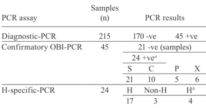
Prevalence of HBV infection in children with clinical hepatitis - Data regarding the results of the HBV DNA-PCR assays among the 215 children are depicted in Table II. From 215 children with clinical hepatitis included in this study, 45 cases (21%) were re-tested by the confirma-tory PCR assay to confirm HBV infection. Among these cases, 24 cases were confirmed positive for HBV DNA, with 87.5% (21/24) being positive for HBV DNA and neg-ative for HBsAg (OBI) and 12.5% (3/24) being positive for both HBV DNA and HBsAg (HBV DNA+/HBsAg+).
Table III shows the demographical and clinical data of the study adjusted by the HBV DNA+ (n = 24) and HBV DNA- children (n = 191). Among the HBV DNA+ patients, 13 were males and 11 were females (age range, 1-15 years; mean, 6.8; SD, 4.1 years). Additionally, values of the
me-dian ALT and AST were 629 UI/L and 461 UI/L, respec -tively, the median DB value was 3.2 mg/dL and the medi-an albumin value was 4.4 mg/dL. Within the HBV DNA+ group, 70% (17/24) had less than six months of clinical symptoms. However, the frequency of HBV DNA+ chil-dren with more than six months of clinical symptoms was significantly higher than in the group of HBV-DNA- chil-dren (30% vs. 7%) (p < 0.001). Among the HBV DNA+ children, 54% were positive for anti-HAV IgM antibody, though none were positive for anti-HCV antibody.
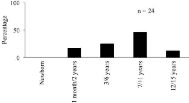
Age group-based analysis of HBV infection - The prevalence of HBV DNA+ children (24 cases) adjusted by age showed an upward trend that was not significant (p = 0.096), beginning at 0% in newborns, 17% among the infancy age group (1 month-2 years), 25% in the preschool-age group (age 3-6 years) and 46% in school children (7-11 years) (Figure). However, a reduction in the prevalence of HBV infection (12%) in the adolescent group (12-15 years) was detected. The HBV DNA- chil-dren with clinical hepatitis who were registered also fol-lowed the same pattern with respect to age distribution. However, the prevalence of HBV DNA- children among the total cases of clinical hepatitis was lower in the in-fancy age group (7%) and the distribution by age group was not significant (p = 0.565) (data not shown).
High prevalence of OBI genotype H patients - Among the HBV DNA+ children, 87.5% (21/24) were diagnosed with OBI. Based on their serological anti-HBc marker profile, 90.5% of the children with OBI were negative for the anti-HBc marker (OBI-seroneg-TABLE II
Polymerase chain reaction (PCR) results for hepatitis B virus (HBV)-DNA positivity among the 215 children
with clinical hepatitis
PCR assay
Samples
(n) PCR results
Diagnostic-PCR 215 170 -ve 45 +ve
Confirmatory OBI-PCR 45 21 -ve (samples) 24 +vea
S C P X
21 10 5 6
H-specific-PCR 24 H Non-H Hb
17 3 4
ative) and only 9.5% were positive (OBI-seropositive). When the HBV DNA+ children were analysed accord-ing to hepatitis B vaccination status, all of the non-vac-cinated children had OBI (p < 0.05) and 93% of them were OBI seronegative (Tables IV, V).
HBV genotype H was the most frequent genotype in the HBV-infected children, appearing in 71% of the
TABLE III
Demographical and clinical data in hepatitis B virus (HBV)-positive and HBV-negative Mexican children with clinical hepatitis from 2005-2009
Subject data HBV-positivea HBV-negativeb pc
n (%) 24 (11.2) 191 (88.8)
-Age (year) [mean ± SD (range)] 6.8 ± 4.1 (1-15) 7.3 ± 3.7 (1-15) 0.584
Sex [female/male (n/%)] 11/13 (46/54) 95/96 (50/50) 0.718
Clinical features [n (%)]
Time of onset of clinical symptoms: </> six months 17/6 (70/30) 169/13 (93/7) 0.0004d
Jaundice 21 (95) 174 (97) 0.560
Hepatomegaly 20 (91) 176 (98) 0.130
Vomiting 17 (74) 145 (81) 0.558
Nauseas 17 (77) 144 (80) 0.884
Fever 16 (73) 129 (72) 0.917
Abdominal pain 15 (68) 127 (71) 0.818
Choluria 13 (60) 110 (61) 0.855
Acholia 6 (27) 53 (29) 0.833
Acute liver failure 1 (4) 5 (3) 0.664
ALT (UI/L) [mean ± SD (range)]
(median)
1,881 ± 5,290 (76-25,823) [629]
1,272 ± 2,426 (40-25,894)
[782] 0.375
AST (UI/L) [mean ± SD (range)]
(median)
670 ± 740 (64-2,650) [461]
835 ± 955 (42-5,011)
[415] 0.453
Direct bilirubin (mg/dL) [mean ± SD (range)] (median)
3.5 ± 3.2 (0.31-12.3) [3.2]
3.8 ± 4.3 (0.31-39)
[3.2] 0.626
Albumin (mg/dL) [mean ± SD (range)] (median)
4.1 ± 1.2 (1.5-6.1) [4.4]
4.5 ± 1.1 (1.8-7.6)
[4.6] 0.222
Anti-HAV IgM positive [n (%)] 13/24 (54) 92/113 (81) 0.630
Anti-HCV positive [n (%)] 0 (0) 2 (1) 0.605
a: positive for HBV DNA in at least two different regions within the HBV genome; b: negative for HBV DNA; c: by t test, chi-square test or Fisher’s exact test; d: p < 0.05; ALT: alanine aminotransferase; AST: aspartate aminotransferase; SD: standard deviation. Hepatitis B infection by age group in the 24 hepatitis B virus (HBV)
DNA-positive children with clinical hepatitis (2005-2009). The data are presented as percentage of each children’s age group who were positive for HBV DNA.
cases, followed by HBV genotype G in 8% of the cases
and HBV genotype A in 4% of the cases. In four cases, the HBV genotype was designated as probable genotype H, as explained in the Subjects, Materials and Methods section (Tables II, IV, V). There was no significant dif-ference related to age, sex or hepatic injury among the
different HBV genotypes (H, G or A). Seventy-three
per cent of the HBV genotype H cases and both cases of
HBV genotype G were not vaccinated against hepatitis
B. In contrast, only the HBV genotype A case had been vaccinated. There was no significant difference between HBV genotypes among the hepatitis B-vaccinated and non-vaccinated groups (p = 0.469) (Table IV).
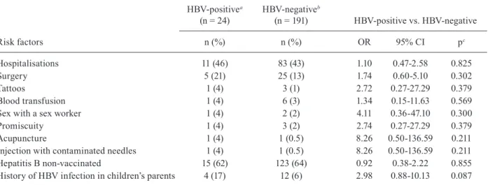
Risk factors in children with HBV infection - History of hospitalisation and surgery were the most common risk factors found in the HBV DNA+ children, at 46% and 21% of the cases, respectively. Other risk factors found in these children with HBV infection were tat-toos (4%), blood transfusion (4%), sex with a sex worker (4%), promiscuity (4%), acupuncture (4%) and inocula-tion with contaminated needles (4%). No male child re-ported sexual relationships with men. Only one of the
HBV genotype G-infected children had a history of
HBV (recombinant vaccine) was not found in 62% of the HBV DNA+ children at two, four and six months of age and none of these children had received hepatitis A vaccination. Additionally, none of the children received hepatitis B vaccination at birth. Regarding the hepati-tis B vaccination status and risk factors, there were no significant differences between HBV DNA+ children vs. HBV DNA- children with clinical hepatitis. A history of hepatitis B infection among the parents of four HBV DNA+ children and in 12 parents among the HBV DNA- children was not significant (p = 0.087) (Table VI).
DISCUSSION
HBV infection is the leading cause of chronic liver disease and a serious public health problem in children throughout the world. Further information regarding the prevalence of HBV infection, circulating genotypes and a reliable OBI diagnosis are required to have a deeper understanding of the role of HBV infection in the devel-opment of liver diseases among paediatric patients.
The prevalence of HBV infection among 215 children with clinical hepatitis was 11.2% after testing for HBV DNA. These data are consistent with the fact that OBI is common among the adult Mexican population (Roman
et al. 2010, Garcia-Montalvo & Ventura-Zapata 2011,
Panduro et al. 2013). In this study, the data support the notion that HBV infection is underestimated when viral DNA is not evaluated. This is particularly relevant be-cause Mexico is classified as a low-prevalence region for HBV infection, but only by serological testing (HBsAg) (Panduro et al. 2011). Within the HBV DNA+ group, 70% (17/24) had less than six months of symptoms based only on clinical criteria. However, the serological, biochemi-cal and molecular findings suggest that these patients were diagnosed at a late phase of an acute HBV infection, concordant with the reduced levels of serum HBV DNA
and undetectable HBsAg, resulting in occult B infection. Therefore, it may be convenient to practice a follow-up clinical evaluation in such patients because most chronic infections in adults are acquired during childhood (Brod-erick & Jonas 2003, Yen-Hsuan 2011).
Most of the HBV-infected children were OBI se-ronegative (negative for anti-HBc). This finding con-trasts with studies that report a high prevalence of OBI seropositivity (positive for anti-HBc) among adults, which may indicate past HBV infection (Raimondo et al. 2008a). However, several studies in children have reported seronegative cases more often than OBI-seropositive cases (Liu et al. 2006, Mu et al. 2009, Said
et al. 2009, Utsumi et al. 2010). This may be explained by
potential differences in the immune response of HBV-infected children and HBV-HBV-infected adults; thus, sero-logical markers that indicate viral elimination may be undetectable (Rehermann 2003, Fierro et al. 2011).
With regard to HBsAg test sensitivity, the samples were tested with the AxSYM MEIA system because it was the most available kit at the time of the study. Re-garding the plausibility that HBsAg mutations in the “a” determinant region may have given false-negatives using these kits, we have reported that mutations in the S and
X regions are rare in OBI genotype H infection (Pan -duro et al. 2013). If S-mutations had been present, they would yield escape mutants with non-detectable HBsAg and higher VL, which are considered “false OBI” (Rai-mondo et al. 2008a). Nonetheless, we observed a high di-versity of the S region, which may be related to the non-detection of HBsAg with the use of commercial kits that have been validated using non-H genotype samples.
Although we cannot discard the presence of specific mutations in some cases, it appears that the main cause of OBI among the Mexican population is due to an effective T cell response with a rapid decline of serum HBsAg,
TABLE IV
Hepatitis B virus (HBV) infection, occult hepatitis B infection (OBI) and HBV genotypes in Mexican children with clinical hepatitis from 2005-2009 according to vaccination status
HBV infection
Total (n = 24)
Hepatitis B vaccinateda
(n = 9)
Hepatitis B non-vaccinated (n = 15)
n (%) n (%) n (%) pb
HBV DNA+/HBsAg+ 3 (12.5) 3 (33) 0 (0) 0.0415c
OBI (HBV DNA+/HBsAg-) 21 (87.5) 6 (67) 15 (100)
-OBI-seropositive (anti-HBc+) 2/21 (9.5) 1/6 (17) 1/15 (7) 0.500
OBI-seronegative (anti-HBc-) 19/21 (90.5) 5/6 (83) 14/15 (93)
-HBV-genotypes
Genotype H 17 (71) 6 (67) 11 (73) 0.469
Genotype G 2 (8) 0 (0) 2 (13)
-Genotype A 1 (4) 1 (11) 0 (0)
-Probable genotype Hd 4 (17) 2 (22) 2 (13)
resulting in HBsAg negativity and a low VL (Fierro et al. 2011). On-going studies in our laboratory have repeatedly encountered low prevalence or null detection of HBsAg and positivity for HBV-DNA, even with commercial kits with higher sensitivity. In addition, with regard to the lack of sequence verification to preclude environmental contamination, we exerted additional precautions using a PCR confirmatory test, as recommended, before PCR genotyping (Raimondo et al. 2008a).
In accordance with the fact that the major circulat-ing HBV genotype is H among distinct Mexican popu-lations (Panduro et al. 2013, Roman & Panduro 2013), in the present study, HBV genotype H was predominant in both OBI and non-OBI infection, followed by HBV
genotypes G and A among the OBI patients, but these
differences were not significant. However, the trend of low VL among HBV genotype H-infected patients in Mexico is most likely to be associated with the finding of OBI, which requires further investigation. Interestingly,
the children infected with HBV genotype G had a his -tory of blood transfusions, but lacked a paternal his-tory of sexual relationships with men. HBV infection with
genotype G has been reported not only in patients who
engage in homosexual relationships with men (Chudy et al. 2006, Sanchez et al. 2007, Dao et al. 2011), but also among blood donors and haemodialysis patients (Mora et al. 2011, Sayan & Dogan 2012). Because HBV
geno-type G infection may occur as OBI, the use of serologi -cal probes may be unsuitable for diagnosis (Zaaijer et al. 2011). Moreover, given that this genotype can cause liver fibrosis in immunocompromised individuals (Chudy et al. 2006) and in chimeric mice carrying human hepa-tocytes (Sugiyama et al. 2007), nucleic acid testing for
HBV genotype G and clinical follow-up of infected pa -tients may be required.
In 2000, the National Ministry of Health declared mandatory HBV vaccination for all children under five. A three-dose schedule at two, four and six months of age with the DPTw-HB/Hib pentavalent vaccine (diph-theria, tetanus and pertussis, whole-cell-hepatitis B/ haemophilus influenza type-b and recombinant HBsAg) was indicated, but was not effectively given until mid-2001. In 2007, the recombinant antihepatitis B vaccine was indicated at birth and at two and six months of age,
TABLE V
Clinical and molecular description by case of the hepatitis B virus (HBV)-infected Mexican children with clinical hepatitis from 2005-2009
Case Age Sex ALT
(UI/L) (mg/dL)DB ALF
Hepatitis B vaccinea
[doses (n)]
Anti-HAV
IgM HBsAg
Anti-HBc total
Anti-HBc IgM
HBV genotype
1 10 M 328 1.6 No 3 - + - - H
2 5 F 224 0.4 No 3 + + - - NG
3 6 M 689 4.3 No 3 - + - - NG
4 2 M 88 0.3 No 3 - - + - H
5 4 F 629 1.4 No 3 + - - + H
6 2 F 205 3.9 No 3 + - - - H
7 3 F 1,982 3.2 No 3 + - - - H
8 4 M 680 4.1 No 3 + - - - H
9 6 F 1,041 3.2 No 0 - - - - H
10 6 M 210 3.5 No 0 - - - - H
11 7 F 334 2.9 No 0 + - - - H
12 7 M 2,877 12.3 No 0 - - - - H
13 7 M 3,389 4.4 No 0 + - - - H
14 8 M 1,110 1.7 No 0 + - - - H
15 11 M 171 1.3 No 0 + - - - H
16 11 F 74 1.8 No 0 + - - - H
17 14 F 76 5.2 No 0 - - - - H
18 14 M 220 0.8 No 0 + - - - H
19 15 F 664 1.1 No 0 + - - - H
20 5 M 620 2.9 No 0 + - - - NG
21 5 M 107 0.7 No 0 - - - - NG
22 1 F 25,823 11.0 Yes 0 - - - - G
23 9 M 805 0.4 No 0 - - - - G
24 2 F 913 8.1 No 3 - - - - A
a:a child with a complete three-dose schedule at two, four and six months of age; ALF: acute liver failure; ALT: alanine amin-otransferase; DB: direct bilirubin; F: female; HAV: hepatitis A virus; HBc: hepatitis B core antibody; HBsAg: hepatitis B surface
TABLE VI
Frequency of risk factors related to hepatitis B virus (HBV) infection in HBV-infected Mexican children with hepatitis from 2005-2009
Risk factors
HBV-positivea
(n = 24)
HBV-negativeb
(n = 191) HBV-positive vs. HBV-negative
n (%) n (%) OR 95% CI pc
Hospitalisations 11 (46) 83 (43) 1.10 0.47-2.58 0.825
Surgery 5 (21) 25 (13) 1.74 0.60-5.10 0.302
Tattoos 1 (4) 3 (1) 2.72 0.27-27.29 0.379
Blood transfusion 1 (4) 6 (3) 1.34 0.15-11.63 0.569
Sex with a sex worker 1 (4) 2 (2) 4.11 0.36-47.10 0.300
Promiscuity 1 (4) 3 (2) 2.74 0.27-27.29 0.379
Acupuncture 1 (4) 1 (0.5) 8.26 0.50-136.59 0.211
Injection with contaminated needles 1 (4) 1 (0.5) 8.26 0.50-136.59 0.211
Hepatitis B non-vaccinated 15 (62) 123 (64) 0.92 0.38-2.22 0.855
History of HBV infection in children’s parents 4 (17) 12 (6) 2.98 0.88-10.13 0.087
a: positive for HBV DNA by two sets of primers within the HBV genome; b: negative for HBV DNA by two sets of primers within the HBV genome; c: by chi-square test or Fisher’s exact test; CI: confidence interval; OR: odds ratio.
even though it only became effective in mid-2008. No-tably, among the 24 cases of HBV DNA+ children, 62% of them were not vaccinated against hepatitis B. In all of these cases, the children had OBI and 93% of them were OBI seronegative. A possible reason why these children missed their vaccination schedule may be explained by the age range of the 15 non-vaccinated children (1-15 years) at the time of the study. Those nine or older were born before the establishment of the mandatory univer-sal vaccination programme that included the DPTw-HB/ Hib vaccine. Those of nine years or younger (n = 9) may have not received the latest monovalent vaccine at birth because this 2007 vaccination scheme did not become effective until mid-2008-2009. Another possibility may be a low level of education among the children’s parents, combined with several of the HBV-related risk factors that hinder a successful follow-up of vaccination care. These observations suggest that without a widespread hepatitis B vaccination programme, a large number of hepatitis B cases will occur, including OBI cases.
In contrast, the nine HBV DNA+ children, three of which were HBsAg-positive, reported a complete sched-ule of vaccination. These children received the recombi-nant hepatitis B vaccine (included in the DPTw-HB/Hib vaccine) at two, four and six months of age. Although the immunogenicity and efficacy of this hepatitis B vac-cine was reported after three doses (Santos et al. 2002), this schedule might not protect against HBV infection acquired at birth.
The lack of hepatitis B vaccination among HBV DNA+ infected children and their parents indicates the necessity of focusing on thorough hepatitis B vaccina-tion campaigns among high-risk children as well as in the general population. Vigilance strategies are required to ensure that hepatitis B vaccination occurs at birth (day 0) in Mexican children to avoid the dissemination of
HBV infection and its clinical complications (CID 2012). Regarding the HAV, Mexico is a region of high ende-micity for this infection because 80% of children below the age of 10 and 90% of adults in general are positive
for anti-HAV IgG antibodies (Panduro et al. 2011). Thus,
it is not surprising that in this study, 54% of the HBV DNA+ children, with most being OBI, had anti-HAV IgM antibodies. This fact also coincides with the finding that none of them had been vaccinated. Therefore, HAV-HBV co-infections among OBI cases may be more com-mon than expected; moreover, they are not easily dis-criminated from each other by clinical evaluation only. The high prevalence of OBI found in this study suggests that nucleic acid testing for HBV may be a more objec-tive diagnostic tool due to the misconception among most physicians that HAV is the only virus responsible for hepatitis in children. Both HAV and HAV-HBV co-infections can lead to ALF with high mortality (Squires et al. 2006);hence, vaccination against hepatitis A in children should be reinforced (AAP/CID 2007).
With regard to HBV-related risk factors, history of HBV infection in the children’s parents was found to be more frequent among the HBV DNA+ children than the HBV DNA- children (17% vs. 6%). This finding is rel-evant because the main routes of transmission in chil-dren are either vertical or horizontal in their first years of life. However, in this study, vertical transmission was not confirmed due to the lack of serum samples from the children’s parents. Therefore, case-control studies related to risk factors for HBV infection and vertical transmission are required to assess their impact on the transmission of HBV in paediatric clinical settings.
sub-stantial in paediatric patients and the aetiological diag-nosis has relevant clinical implications. HBV genotype H predominates in Mexico and is associated with OBI in children. The high prevalence of OBI among hepatitis B-non-vaccinated children observed in this study em-phasises the importance of HBV vaccination in young children in Mexico.
ACKNOWLEDGEMENTS
To Dr Montserrat Maldonado-Gonzalez, for her help -ful contributions during the field study, and to Dr Bertha
Garcia-Armenta and Dr Roberto Hinojosa-Ibarra, for their
medical assistance.
REFERENCES
AAP/CID - American Academy of Pediatrics/Committee on Infec-tious Diseases 2007. Hepatitis A vaccine recommendations.
Pe-diatrics 120:189-199.
Broderick AL, Jonas MM 2003. Hepatitis B in children. Semin Liver
Dis 23: 59-68.
Chemin I, Trépo C 2005. Clinical impact of occult HBV infections.
J Clin Virol 34 (Suppl.): S15-S21.
Chudy M, Schmidt M, Czudai V, Scheiblauer H, Nick S, Mosebach
M, Hourfar MK, Seifried E, Roth WK, Grünelt E, Nübling CM 2006. Hepatitis B virus genotype G monoinfection and its trans -mission by blood components. Hepatology 44:99-107.
CID - Committee on Infectious Diseases 2012. Recommended
child-hood and adolescent immunizations schedules - United States,
2012. Pediatrics 129:385-386.
Dao DY, Balko J, Attar N, Neak E, Yuan HJ, Lee WM, Jain MK 2011.
Hepatitis B virus genotype G: prevalence and impact in patients
co-infected with human immunodeficiency virus. J Med Virol 83: 1551-1558.
Escobedo-Melendez G, Fierro NA, Roman S, Maldonado-Gonzalez
M, Zepeda-Carrillo E, Panduro A 2012. Prevalence of hepatitis A, B and C serological markers in children from western Mexico.
Ann Hepatol 11:194-201.
Fierro NA, Roman S, Realpe M, Hernandez-Nazara Z, Zepeda-Car-rillo EA, Panduro A 2011. Multiple cytokine expression profiles reveal immune-based differences in occult hepatitis B genotype H-infected Mexican Nahua patients. Mem Inst Oswaldo Cruz 106:1007-1013.
Garcia-Montalvo BM, Ventura-Zapata LP 2011. Molecular and sero -logical characterization of occult hepatitis B infection in blood donors from Mexico. Ann Hepatol 10:133-141.
Hollinger FB, Sood G 2010. Occult hepatitis B virus infection: a co -vert operation. J Viral Hepat 17:1-15.
Kelly D 2006. Viral hepatitis B and C in children. J R Soc Med 99: 353-357.
Liu CJ, Lo SC, Kao JH, Tseng PT, Lai MY, Ni YH, Yeh SH, Chen PJ, Chen DS 2006. Transmission of occult hepatitis B virus by transfusion to adult and pediatric recipients in Taiwan. J Hepatol 44: 39-46.
Lok ASF, McMahon BJ 2009. Chronic hepatitis B: update 2009. AASLD practice guideline update. Hepatology 50: 1-36.
Mora MVA, Romano CM, Gomes-Gouvea MS, Gutierrez MF, Bo -telho L, Carrilho FJ, Pinho JR 2001. Molecular characterization of the hepatitis B virus genotypes in Colombia: a Bayesian infer-ence on the genotype F. Infect Genet Evol 11: 103-108.
Mu SC, Lin YM, Jow GM, Chen BF 2009. Occult hepatitis B virus
infection in hepatitis B vaccinated children in Taiwan. J Hepatol 50:264-272.
Panduro A, Escobedo-Melendez G, Fierro NA, Ruiz-Madrigal B,
Zepeda-Carrillo E, Roman S 2011. Epidemiology of viral hepati-tis in Mexico. Salud Publica Mex 53 (Suppl.): S37-S45.
Panduro A, Maldonado-Gonzalez M, Fierro NA, Roman S 2013. Dis -tribution of hepatitis B virus genotypes F and H in Mexico and Central America. Antivir Ther 18:475-484.
Pol S, Corouge M, Fontaine H 2011. Hepatitis virus infection and pregnancy. Clin Res Hepatol Gastroenterol 35: 6218-6222.
Pollicino T, Raffa G, Constantino L, Lisa A, Campello C, Squadri
-to G, Levrero M, Raimondo G 2007. Molecular and functional
analysis of occult hepatitis B virus isolates from patients with hepatocelular carcinoma. Hepatology 45: 277-285.
Raimondo G, Allain JP, Brunetto MR, Buendia MA, Chen DS, Co
-lombo M, Craxi A, Donato F, Ferrari C, Gaeta GB, Gerlich WH, Levrero M, Locarnini S, Michalak T, Mondelli MU, Pawlotsky
JM, Pollicino T, Prati D, Puoti M, Samuel D, Shouval D, Smedile
A, Squadrito G, Trepo C, Villa E, Will H, Zanetti AR, Zoulim F
2008a. Statements from the Taormina expert meeting on occult hepatitis B virus infection. J Hepatol 49: 652-657.
Raimondo G, Navarra G, Mondello S, Costantino L, Colloredo L, Colloredo G, Cucinotta E, Di Vita G, Scisca C, Squadrito G, Pol -licino T 2008b. Occult hepatitis B virus in liver tissue of indi-viduals without hepatitis disease. J Hepatol 48:743-746.
Rehermann B 2003. Immune responses in hepatitis B virus infection.
Semin Liver Dis 23:21-37.
Roman S, Jose-Abrego A, Fierro NA, Escobedo-Melendez G, Ojeda-Granados C, Martinez-Lopez E, Panduro A 2014. Hepatitis B
virus infection in Latin America: a genomic medicine approach.
World J Gastroenterol 20: 7181-7196.
Roman S, Panduro A 2013. HBV endemicity in Mexico is
associ-ated with HBV genotypes H and G. World J Gastroenterol19: 5446-5453.
Roman S, Panduro A, Aguilar-Gutierrez Y, Maldonado M,
Vazquez-Vandyck M, Martinez-Lopez E, Ruiz-Madrigal B, Hernandez-Nazara Z 2009. A low steady HBsAg seroprevalence is associated with a low incidence of HBV-related liver cirrhosis and hepato-cellular carcinoma in Mexico: a systematic review. Hepatol Int 3:343-355.
Roman S, Tanaka Y, Khan A, Kurbanov F, Kato H, Mizokami M, Panduro A 2010. Occult hepatitis B in the genotype H-infected Nahuas and Huichol native Mexican population. J Med Virol 82: 1527-1536.
Romero M, Madejon A, Fernandez-Rodriguez, Garcia-Samaniego J
2011. Clinical significance of occult hepatitis B virus infection.
World J Gastroenterol 17:1549-1552.
Said ZN, El-Sayed MH, El-Bishbishi IA, El-Fouhil DF, Abdel-Rheem SE, El-Abedin MZ, Salama II 2009. High prevalence of occult hepatitis B in hepatitis C-infected Egyptian children with haema-tological disorders and malignancies. Liver Int 29: 518-524.
Sanchez LV, Maldonado M, Bastidas-Ramirez BE, Norder H,
Pan-duro A 2002. Genotypes and S-gene variability of Mexican hepa -titis B virus strain. J Med Virol 68:24-32.
Sanchez LV, Tanaka Y, Maldonado M, Mizokami M, Panduro A 2007. Difference of hepatitis B virus genotype distribution in two groups of Mexican patients with different risk factors.
Santos JI, Martin A, De Leon T, Rivera L, Gaitan ME, Del Rio E, Oselka G, Cervantes Y, Rubio P, Clemens SA, de Mendonça JS
2002. DPTw-HB and HiB primary and booster vaccination: com-bined versus separate administration to Latin American children.
Vaccine 20:1887-1893.
Sayan M, Dogan C 2012. Hepatitis B virus genotype G infection in
a Turkish patient undergoing hemodialysis therapy. Hepat Mon 12: 118-121.
Squires Jr RH, Shneider BL, Bucuvalas J, Alonso E, Sokol RJ, Narke-wicz MR, Dhawan A, Rosenthal P, Rodriguez-Baez N, Murray KF,
Horslen S, Martin MG, Lopez MJ, Soriano H, McGuire BM, Jonas
MM, Yazigi N, Shepherd RW, Schwarz K, Lobritto S, Thomas DW, Lavine JE, Karpen S, Ng V, Kelly D, Simonds N, Hynan LS 2006. Acute liver failure in children: the first 348 patients in the pediatric acute liver failure study group. J Pediatr 148:652-658.
Sugiyama M, Tanaka Y, Sakamoto Y, Maruyama I, Shimada T,
Taka-hashi S, Shirai T, Kato H, Nagao M, Miyakawa Y, Mizokami M 2007. Early dynamics of hepatitis B virus in chimeric mice car-rying human hepatocytes monoinfected or coinfected with
geno-type G. Hepatology 45:929-937.
Utsumi T, Yano Y, Lusida MI, Amin M, Soetjipto, Hotta H, Hayashi
Y 2010. Serologic and molecular characteristics of hepatitis B virus among school children in East Java, Indonesia. Am J Trop
Med Hyg 83: 189-193.
Yen-Hsuan N 2011. Natural history of hepatitis B virus infection: pediatric perspective. J Gastroenterol 46: 1-8.
Yeung LT, Roberts EA 2010. Current issues in the management of paediatric viral hepatitis. Liver Int 30: 5-18.
Zaaijer HL, Boot HJ, van Swieten P, Koppelman MH, Cuypers HTM 2011. HBsAg-negative mono-infection with hepatitis B virus