e-ultrasonography.org Ultrasonography 33(4), October 2014
303
Papillary thyroid microcarcinoma in a
thyroid pyramidal lobe
Tae Kwun Ha1, Dong Wook Kim2, Ha Kyoung Park1, Soo Jin Jung3
Departments of 1General Surgery, 2Radiology, and 3Pathology, Busan Paik Hospital, Inje University College of Medicine, Busan, Korea
http://dx.doi.org/10.14366/usg.14026 pISSN: 2288-5919 • eISSN: 2288-5943
Ultrasonography 2014;33:303-306
We report an extremely rare case of papillary thyroid microcarcinoma (PTMC) in the thyroid pyramidal lobe (TPL). A 48-year-old woman underwent ultrasound-guided ine-needle aspiration for a small thyroid nodule in the right lobe in local clinic, and it revealed a malignant cytology. On preoperative ultrasonography for tumor staging in our hospital, another small suspiciously malignant hypoechoic nodule was detected in the left TPL. Total thyroidectomy and central nodal dissection were performed. Histopathology confirmed PTMCs in the left TPL and both thyroid lobes. Ultrasonography for TPL should be required for complete evaluation of possible multifocality of thyroid malignancy.
Keywords: Thyroid nodule; Pyramidal lobe; Papillary thyroid microcarcinoma; Ultrasonography
Received: June 11, 2014 Revised: July 4, 2014 Accepted: July 13, 2014
Correspondence to:
Dong Wook Kim, MD, Department of Radiology, Busan Paik Hospital, Inje University College of Medicine, 75 Bokji-ro, Busanjin-gu, Busan 614-735, Korea
Tel. +82-51-890-6549 Fax. +82-51-896-1085 E-mail: [email protected]
CASE REPORT
This is an Open Access article distributed under the terms of the Creative Commons Attribution Non-Commercial License (http://creativecommons.org/ licenses/by-nc/3.0/) which permits unrestricted non-commercial use, distribution, and reproduction in any medium, provided the original work is properly cited.
Copyright © 2014 Korean Society of Ultrasound in Medicine (KSUM)
How to cite this article:
Ha TK, Kim DW, Park HK, Jung SJ. Papillary thyroid microcarcinoma in a thyroid pyramidal lobe. Ultrasonography. 2014 Oct;33(4):303-306.
Introduction
The third lobe of the thyroid gland is known as the thyroid pyramidal lobe (TPL). Based on autopsy- and surgery-based studies, its prevalence ranges from 45% to 75% [1-3]. Complete removal of the thyroid tissue is generally a mandatory component of total thyroidectomy [1]. Most TPL can be fully excised during thyroid surgery, but TPLs that are located especially high in the neck, may escape notice [1,4]. A missed TPL may present as scintigraphic residue after total thyroidectomy, and radioisotope therapy may be required for any remnant thyroid tissue [1,5].
Thyroid ultrasonography (US) has been widely used in the evaluation of thyroid nodules, resulting in the establishment of certain characteristics that indicate benign and malignant thyroid nodules [6]. In general, US indings suggestive of a malignant thyroid nodule include marked hypoechogenicity, spiculated margin, microcalciications, and a taller-than-wide shape [6,7]. In the literature, only a few reports regarding malignant tumors arising from the TPL have been found [8,9]. To the best of our knowledge, however, US detection of malignant tumors arising from the TPL has not been reported. Herein, we report a case of primary papillary thyroid microcarcinoma (PTMC) in the TPL, which was detected on preoperative US.
Case Report
Tae Kwun Ha, et al.
304
Ultrasonography 33(4), October 2014 e-ultrasonography.orgsuspicious malignant thyroid nodule in the right lobe in local clinic, and it revealed a malignant cytology. The patient showed normal values on a thyroid function test (T3, 104.4 ng/dL; free T4, 1.17 ng/dL; thyroid-stimulating hormone, 3.61 mIU/L), and no specific abnormalities were detected on a physical examination of the neck. The patient had previously undergone a left partial thyroidectomy at another hospital, because of a palpable thyroid mass that had the histological features of nodular hyperplasia. Subsequent to the partial thyroidectomy, the patient had not received any clinical or US follow-up at our hospital.
A preoperative thyroid US revealed two suspicious small malignant thyroid nodules in the left TPL (4.1 mm in the longest dimension) and right mid-lobe (3.6 mm in the longest dimension), a small hypoechoic nodule in the left mid-lobe (about 2.0 mm), and there was no grossly deined metastatic lymph nodes (Fig. 1A-C). Because of known cytologically determined, malignant thyroid
nodule in the right lobe, she did not undergo an ultrasound-guided ine-needle aspiration biopsy for a suspicious small thyroid nodule in the left TPL. Total thyroidectomy and central compartment neck dissection were performed without complications. Histopathologic investigation conirmed two PTMCs in the left TPL and right thyroid lobe (Fig. 1D, E), which corresponded to the US indings. The small hypoechoic nodule that was detected in the left thyroid lobe was conirmed to be a PTMC (Fig. 1F). No nodal metastases were found in the central neck. Chronic lymphocytic thyroiditis was evident in the entire thyroid tissue. The patient was discharged after surgery without radioiodine therapy. Follow-ups are regularly conducted with the patient (most recent follow-up: September 2013), and she is receiving hormone therapy for thyroid-stimulating hormone suppression without any complications.
A B
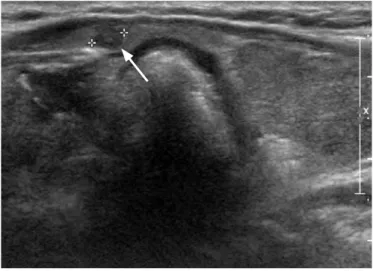
Fig. 1. Papillary thyroid microcarcinoma in the left thyroid pyramidal lobe of a 48-year-old woman.
Gray-scale ultrasonograms of thyroid gland (A, transverse; B, longitudinal) reveal a small hypoechoic nodule in the left thyroid pyramidal lobe (arrow, 2.6×2.7×4.1 mm). C. Transverse gray-scale ultrasonogram shows a small suspiciously malignant thyroid nodule with taller-than-wide shape and hypoechogenicity in right thyroid lobe (arrow, 2.4×2.6×3.6 mm) (known malignant cytology in local clinic).
Papillary thyroid microcarcinoma in pyramidal lobe
e-ultrasonography.org Ultrasonography 33(4), October 2014
305
Discussion
The thyroid gland is one of the largest endocrine gland, and comprises two lateral lobes that are connected along the median line by an isthmus, as well as the TPL, which extends superiorly from the isthmus of the thyroid gland [1,2]. The TPL appears to be related to the distal portion of the thyroglossal duct, an embryological structure, which initially develops along the migratory path of the thyroid gland and usually disappears during later development [3]. Most autopsy- or surgery-based studies have reported that a TPL is present in more than 50% of the general population [1-3], but a study using computed tomography has demonstrated that the prevalence of TPL is 44.6% in South Korea [10]. To date, no US-based studies of TPL prevalence have been published.
Malignant tumors arising from TPL are rare. Two cases of
malignant nodules in the TPL have been described in the literature [8,9]. The first was a follicular thyroid carcinoma, which was surgically resected because of an increase in the size of the palpable cervical mass [8]. The second was a recurrent papillary thyroid carcinoma after total thyroidectomy [9]. In this report, we have described US and histopathological findings of primary PTMC in the left TPL and associated multifocal PTMCs in both thyroid lobes, which were incidentally detected on thyroid US.
Multifocality is a relatively common feature of PTMC; the prevalence of multifocality in papillary thyroid carcinomas ranges from 10% to 62% [11]. The TPL can be the origin or recurrent site of papillary thyroid carcinomas. Besides its role as a primary and secondary site of thyroid malignancy, the TPL is clinically interesting because of its importance to Graves disease and the efficacy of radioactive iodine therapy. Particularly, hyperthyroidism can recur
D E
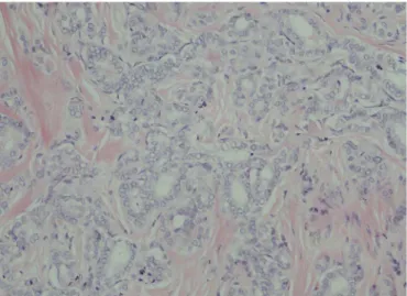
Fig. 1.D. Gross specimen of thyroid pyramidal lobe shows a small ill-defined infiltrating mass lesion (arrow). E. Histological investigation reveals that papillary thyroid microcarcinoma in the thyroid pyramidal lobe, composed of neoplastic cells with papillary structures and intervening sclerotic stroma (H&E, ×200). F. Schematic drawing of the location of surgically conirmed papillary thyroid microcarcinomas (black circles) in the thyroid gland.
F
Tae Kwun Ha, et al.
306
Ultrasonography 33(4), October 2014 e-ultrasonography.orgafter surgical removal of thyroid tissue if the TPL is not itself excised. Furthermore, remnant TPL can absorb most 131I and thereby nullify
the beneit of postoperative radioactive iodine therapy [5].
In summary, we have reported the successful surgical resection of primary PTMC in the TPL and associated multifocal PTMCs in both thyroid lobes. US for TPL should be included for complete evaluation of possible multifocality of thyroid malignancy.
ORCID: Tae Kwun Ha: http://orcid.org/0000-0001-7980-3700; Dong Wook Kim: 9826-1326; Ha Kyoung Park: http://orcid.org/0000-0002-7610-8590; Soo Jin Jung: http://orcid.org/0000-0002-9139-701X
Conlicts of Interest
No potential conlict of interest relevant to this article was reported.
References
1. Braun EM, Windisch G, Wolf G, Hausleitner L, Anderhuber F. The pyramidal lobe: clinical anatomy and its importance in thyroid surgery. Surg Radiol Anat 2007;29:21-27.
2. Prakash, Rajini T, Ramachandran A, Savalgi GB, Venkata SP, Mokhasi V. Variations in the anatomy of the thyroid gland: clinical implications of a cadaver study. Anat Sci Int 2012;87:45-49. 3. Mohebati A, Shaha AR. Anatomy of thyroid and parathyroid glands
and neurovascular relations. Clin Anat 2012;25:19-31.
4. Geraci G, Pisello F, Li Volsi F, Modica G, Sciume C. The importance of pyramidal lobe in thyroid surgery. G Chir 2008;29:479-482. 5. Pacini F, Schlumberger M, Harmer C, Berg GG, Cohen O, Duntas
L, et al. Post-surgical use of radioiodine (131I) in patients with papillary and follicular thyroid cancer and the issue of remnant ablation: a consensus report. Eur J Endocrinol 2005;153:651-659. 6. Moon WJ, Baek JH, Jung SL, Kim DW, Kim EK, Kim JY, et al.
Ultrasonography and the ultrasound-based management of thyroid nodules: consensus statement and recommendations. Korean J Radiol 2011;12:1-14.
7. Kim DW, Park JS, In HS, Choo HJ, Ryu JH, Jung SJ. Ultrasound-based diagnostic classiication for solid and partially cystic thyroid nodules. AJNR Am J Neuroradiol 2012;33:1144-1149.
8. Ogawa C, Kammori M, Onose H, Yamada E, Shimizu K, Yamada T. Follicular carcinoma arising from the pyramidal lobe of the thyroid. J Nippon Med Sch 2009;76:169-172.
9. Lee YS, Kim KJ, Kim BW, Chang HS, Park CS. Recurrence of papillary thyroid carcinoma in a remnant pyramidal lobe. ANZ J Surg 2011;81:304.
10. Kim DW, Jung SL, Baek JH, Kim J, Ryu JH, Na DG, et al. The prevalence and features of thyroid pyramidal lobe, accessory thyroid, and ectopic thyroid as assessed by computed tomography: a multicenter study. Thyroid 2013;23:84-91.