Cop
yright
© ABE&M t
odos os dir
eit
os r
eser
vados
.
Thyroid nodules in acromegaly
Nódulos tiroideanos na acromegalia
Amelia Rogozinski1, Alejandra Furioso1, Patricia Glikman1, Marcelo Junco1, Rosa Laudi2, Adriana Reyes1, Alicia Lowenstein1
ABSTRACT
Objective: We made a prospective study evaluating the prevalence of thyroid nodular disease in acromegalic patients. Subjects and methods: Thyroid ultrasound and ultrasound-guided ine needle aspiration biopsy were performed when nodules were detected. Nodules were characterized by cytology and histopathology. Results: We found high prevalence of nodular thyroid disorder, 23/34 (67%) in acromegalic patients. High risk and malignant cytology were signiicantly higher in acromegalic patients than in our non-acromegalic population (25% vs. 9%). Differentiated thyroid carcinoma was present in 11% of the acromegalic patients. Con-clusions: We strongly recommend periodic thyroid evaluation by ultrasound in patients with acromegaly. Fine needle aspiration biopsy should be performed in nodules larger than 10 mm, and in all suspicious nodules, regardless of the size. Arq Bras Endocrinol Metab. 2012;56(5):300-4
Keywords
Acromegaly; thyroid nodules; thyroid ultrasonography; ine needle aspiration biopsy; thyroid carcinoma
RESUMO
Objetivo: Realizamos um estudo prospectivo avaliando a prevalência de patologia nodular tireói dea em 34 pacientes acromegálicos. Sujeitos e métodos: Avaliamos os pacientes com ecograia tireóidea e punção biópsia com agulha ina quando se detectavam nódulos. Resul-tados: Encontramos uma alta prevalência de patologia nodular tireóidea 23/34 (67%) em acro-megálicos. A citologia tireóidea de alto risco e maligna foi signiicativamente mais elevada em pacientes acromegálicos que em uma população não acromegálica (25% vs. 9%). O grupo
acromegálico apresentou carcinoma diferenciado de tireoides em 11%. Conclusões: Recomen-damos fortemente a ecograia periódica tireóidea em pacientes acromegálicos. Uma punção biópsia aspirativa com agulha ina deve ser realizada em presença de nódulos tireóideos maio-res que 10 mm e daqueles com critérios ecográicos suspeitos de malignidade, independente-mente do tamanho deles. Arq Bras Endocrinol Metab. 2012;56(5):300-4
Descritores
Acromegalia; nódulos tireóideos; ecograia tireóidea; punção biópsia aspirativa tireóidea; carcinoma tireóideo
1 División Endocrinología,
Hospital J. M. Ramos Mejía, Buenos Aires, Argentina
2 Anatomia Patológica,
Hospital J. M. Ramos Mejía, Buenos Aires, Argentina
Correspondence to:
Amelia Rogozinski División Endocrinología, Hospital J. M. Ramos Mejía, Urquiza 609, 1221, CABA, Argentina [email protected]
Received on Jan/19/2012 Accepted on Jun/19/2012
INTRODUCTION
A
cromegaly is a chronic and multisystemic disease, with an estimated prevalence of 40-70 cases/mil-lion (1-2). Chronic GH hypersecretion is caused by be-nign pituitary adenoma in most of the cases (3).It is established that untreated acromegaly is as-sociated with reduced life expectancy. Several studies have demonstrated a 2-3 fold increase in mortality due to cardiovascular, respiratory, and metabolic disease. These patients may have a greater risk of developing neoplastic disease as a consequence of their high GH and IGF-I levels (4). The prevalence of this association
is still controversial, as most studies have been uncon-trolled (5). An increased incidence of colorectal, breast, prostate, and hematological malignancies (6) has also been described in acromegalic patients.
Cop
yright
© ABE&M t
odos os dir
eit
os r
eser
vados
.
Most of the reports are retrospective and do not have selection criteria.
We present a prospective study that evaluated thyroid nodules in 34 consecutive acromegalic patients.
SUBJECTS AND METHODS
We studied 34 consecutive acromegalic patients (12 men and 22 women), 25-80 years old. Acromegaly was diagnosed according to clinical data, OGTT-GH test and IGF-I serum levels.
Acromegalic patients were at different disease sta-tus at the irst appointment. Some patients were enrol-led without any treatment (n = 11), and others with previous treatments, such as surgery, radiotherapy, or somatostatin analogs (n = 23). Despite of these treat-ments, 33 out of 34 patients had active disease.
Serum IGF-I was measured using an IRMA assay after acid-ethanol extraction (DSL, Webster, TX). IGF--I data were analyzed following logarithmically trans-formed IGF-I values. Data were converted into Stan-dard Deviation Scores (SDS) using reference values, according to the following formula: (x-average x)/SD, where x = log IGF-I of the patient, average x = mean log IGF-I at the corresponding age and sex, and SD = standard deviation from the mean. Active disease was deined by clinical features and IGF-I serum concen-trations over 2 SDS. Duration of the disease was esti-mated taking into account medical history, clinical, and laboratory data.
GH, TSH, Free T4 (FT4), and anti-thyroperoxida-se antibodies (TPOAb.) were measured using an im-munochemiluminescent automated assay (IMMULI-TE, Siemens). Reference ranges in our laboratory were (0.5-4.0) µUI/mL for TSH, (0.9-2.0) ng/dL for FT4, and < 20 UI/mL for ATPO.
All patients were evaluated by thyroid ultrasound scan Philips Envisor HD Ultrasound Machine (US)for the presence of diffuse, uninodular, and multinodular goiter (9). Ultrasound-guided ine needle aspiration biopsies (FNAB) of the thyroid were performed in no-dules larger than 1 cm in diameter. When malignancy was suspected by known US criteria (hypoechogenicity compared to the normal thyroid parenchyma, increa-sed intranodular vascularity, irregular iniltrative mar-gins, presence of microcalciications, absent halo, and a shape taller than the width measured in the transverse dimension), FNAB was performed, no matter the size of the nodule. Cytological results were classiied in one of four categories, according to the Bethesda system
(10), unsatisfactory (Bethesda I), benign (Bethesda II), high risk (Bethesda III, IV and V), and malignant (Be-thesda VI). Patients with suspicious (Be(Be-thesda V) and malignant (Bethesda VI) cytology were sent to surgery. As a control group, we considered cytology re-sults of 8,532 FNAB performed in our Thyroid Unit bet ween October 1994 and February 2010. These patients were clinically evaluated in order to exclude pituitary disease. This group consisted of 92% females and the distribution of age was: 0.7% under 20 years old, 13.5%, between 20 and 40 years old, 47.9% betwe-en 40 and 60 years old, and 37.8% over 60 years old (range: 14-87 years old).
This study was submitted and approved by the Ethics Committee of our hospital.
Statistics: Unpaired t test, Multiple Regression test, and Fisher’s Exact test were performed using GraphPad Instat software (version 3.06).
RESULTS
Clinical data of the patients
We studied 34 consecutive patients (Table 1), 22 wo-men and 12 wo-men, median age of 55 years old (range 25-80). Patients with and without goiter had similar median age (Unpaired t test).
Active acromegaly was diagnosed in 33 patients, with IGF-I SDS median = 5.4, range = (2.1-10.1). In all active acromegalic patients, GH serum levels after
Table 1. Clinical data
Patients without goiter
Patients with goiter
Diffuse Uninodular Polynodular
No. of patients 5 6 11 12
Age (years)
Median 42 47 58 58
Range (25-60) (38-70) (26-80) (31-73)
Gender (n)
Female 2 2 8 10
Male 3 4 3 2
Disease activity 5/5 6/6 10/11 12/12
IGF-I (SDS)
Median 6.6 5.1 5.3 5.3
Range (5.8-10.1) (2.5-6.7) (1.4-9.1) (2.5-8.6)
Evolution (years)
Median 5 6 11 12
Cop
yright
© ABE&M t
odos os dir
eit
os r
eser
vados
.
OGTT were higher than 1 ng/mL. Goiter was present in 85% (29/34) of the acromegalic patients: 69% were women, 50% of them with polynodular goiter, and 40% with uninodular goiter. Diffuse goiter was predominan-tly seen in men (44.4%). Higher incidence of thyroid nodules was seen in females (18/22) vs. men (5/12)
(Fisher’s Exact test, p = 0.0256).
Except for one patient with history of Graves’ di-sease, none had previous thyroid disease.
Euthyroidism was present in 32/34 acromegalic patients; 2/34 patients had secondary hypothyroidism. One patient who was euthyroid at the irst appoint-ment, developed hyperthyroidism associated to thyro-tropinoma 6 months later (TSH = 1.77 µUI/mL and FT4 = 2.69 ng/dL). There were no signiicant diffe-rences in TSH or FT4 levels between patients with or without goiter (Unpaired t test). Median and (range) were: 1.22 (0.26 -3.40) vs. 1.10 (1.03-2.60) µUI/mL for TSH, and 1.27 (0.74-1.60) vs. 1.20 (1.10-1.70) ng/dL for FT4, respectively. Only 3/34 patients had positive TPOAb levels.
The presence of goiter in our acromegalic popula-tion did not show any associapopula-tion with their age, gen-der, or disease history (Multiple Regression test). Ho-wever, patients with nodular goiter had a trend toward a longer history of acromegaly.
Thyroid nodular ultrasound scan
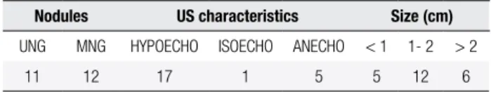
Thyroid nodules were detected by US in 23/34 (67%) patients. Nodules were mostly hypoechoic (17/23), and 18/23 were larger than 10 mm in diameter. US characteristics in multinodular goiter were based only on the largest nodule (Table 2).
13 patients. Two nodules showed high risk cytology: the irst with follicular proliferation (Bethesda III) with a 7-mm diameter nodule, and the other one suspicious for malignancy (Bethesda V) with a 12-mm diameter nodule. Malignant cytology (Bethesda VI) was found in two nodules: one of them of 14 mm, and the other with 8 mm in diameter. Only one sample was unsatis-factory (Bethesda I).
In summary, high risk and malignant cytology oc-curred in 22.2% cases of acromegaly. However, in our previously studied non-acromegalic population of 8,532 FNAB, this result was equal to 9.0%
FNAB of a cervical node was also performed in a fe-male patient who did not present nodular goiter. Meta-static follicular carcinoma was diagnosed by cytology.
Table 3. FNAB, thyroid nodular cytology
Cytology
Non-diagnostic Benign
Follicular proliferation
Suspicious for malignacy
Malignant
n = 18 1 13 1 1 2
Table 2. US of nodular goiter
Nodules US characteristics Size (cm)
UNG MNG HYPOECHO ISOECHO ANECHO < 1 1- 2 > 2
11 12 17 1 5 5 12 6
UNG: uninodular goiter; MNG: multinodular goiter; HYPOECHO: hypoechoic; ISOECHO: isoechoic; ANECHO: anechoic.
Thyroid nodular cytology
In our Thyroid Unit, with a large experience in 8,532 FNAB, the distribution of thyroid cytology was: 2% malignancy (Bethesda VI), 7% high risk (Bethesda III, IV, V), 63% benign (Bethesda II), 3% cysts, and 15% unsatisfactory (Bethesda I).
In this study, FNAB was performed in 18 patients (Table 3). Benign cytology (Bethesda II) was found in
Surgery
Five patients underwent thyroid surgery. Histology results conirmed one case of colloid goiter, three of papillary carcinoma, and one of follicular carcinoma. We diagnosed four differentiated thyroid carcinomas in 34 consecutive acromegalic patients. All these patients were females.
Follicular carcinoma was diagnosed by FNAB in a cervical adenopathy, without the inding of the pri-mitive thyroid tumor at surgery. It should be empha-sized that detection of the primary neoplasm may require extremely careful microscopic study, with embedding of the entire thyroid gland, and cutting of blocks at various levels (11).
The remaining cases had neither lymphadenopa-thy nor extra thyroidal extensions.
Three of the four patients diagnosed with diffe-rentiated thyroid carcinoma were free of disease two years after the surgery. The fourth patient recently un-derwent surgery. In the irst control, persistence of the disease was not observed.
DISCUSSION
Cop
yright
© ABE&M t
odos os dir
eit
os r
eser
vados
.
comorbidities, and increased mortality (4). Neoplasms are often found, but their incidence is controversial (6).
The association between acromegaly and goiter has long been recognized. However, the evaluation of thyroid disorders in the presence of GH and IGF-I ex-cess has yielded conlicting results, since the prevalence of thyroid disease in acromegaly ranged from 25% to 92% in different series (7-14). These discrepancies may be partially explained by differences in how goiter was diagnosed: palpation, imaging studies, such as US, or evaluation of iodine intake in the area.
In our study, we performed US in all of the acro-megalic patients to evaluate thyroid size and nodular pathology in a known iodine-suficient area (15).
Gasperi and cols. (7) reported, in an Italian multi--center study of 258 acromegalic patients, that 78% had thyroid disease. In the control group (non-functioning adenomas or prolactinomas), they found that 27% of the patients had thyroid disease. The prevalence was higher in acromegalic patients with nodular, diffuse nontoxic goiter, and toxic nodular goiter.
Chronic elevation of GH and IGF-I levels could be responsible for benign nodules and tumors in this dise-ase. IGF-I acts by means of two mechanisms: increasing proliferation and reducing apoptosis. It was demonstra-ted that IGF-I increases proliferation of porcine thyroid cells and FRTL-5 rat thyroid cells, and potentiates TSH-mediated thyroid cell proliferation (4-16). In physiological conditions, thyroid follicular cells express IGF-I receptors. Local IGF-I is suggested by the detec-tion of its producdetec-tion in follicular and papillary cell li-nes (17,18). In surgical thyroid specimens, tumoral im-munoreactive IGF-I levels were greater than in normal tissue (4,19). Thus, bioactivity of IGF-I tissue in vivo
does not result only from IGF-I and IGFBP3 serum levels, but also from its local production, which adds to an autocrine-paracrine effect (12).
In our study, serum IGF-I levels were not signiicant-ly associated with the presence of goiter. We performed this study in a prospective and consecutive format in 34 acromegalic patients, focusing on the thyroid nodular disease. We indicated thyroid US at the irst appoint-ment in all 34 consecutive acromegalic patients. We found goiter in 85% of the patients, and thyroid nodules in 67% of them, similar to literature indings (7).
Patients with and without goiter had similar median age. Higher incidence of thyroid nodules was seen in females (p = 0.0256). The chance of developing thy-roid nodules increased with the duration of the disease
(14). Although we found a trend toward a longer his-tory of acromegaly in our patients with nodular goiter, this association was not statistically signiicant.
Acromegaly treatment inhibits peripheral thyroid hormone deiodination, decreasing T3 and increasing rT3 (20). It is tempting to speculate that the con-trol of the disease could determine changes in the evolution of the thyroid nodules. Most of our pa-tients with nodular goiter had active acromegaly when they were enrolled. Thus, we cannot know if changes in the activity of the disease are responsible for differences in the evolution of thyroid disease.
We studied the evolution of thyroid nodules of 15 acromegalic patients whose IGF-I serum levels were decreased by different treatments. We com-pared these results with the evolution of nodular goiter in 45 euthyroid non-acromegalic patients af-ter 12 months of routine follow-up by US. We did not ind any signiicant difference between both groups (data not shown).
Most studies focusing on cancer incidence and/or prevalence do not provide conclusive evidence of in-creased cancer risk (21), but most of them were retro-spective and uncontrolled (6). We found an increased incidence of thyroid cancer in our acromegalic patients, which was not observed in previous studies. The pro-spective and controlled design of our study could be the explanation of this difference.
Since acromegaly is a rare disease, it is dificult to have enough patients for statistical analysis at a single center. In this study we found that high risk and ma-lignant cytology was 22.2% in acromegalic patients, whereas it was 9% in our previously studied non-acro-megalic patients.
In the report by Gasperi and cols. (7), FNAB of thyroid nodules was performed in 62 acromegalic pa-tients. Seven nodules were suspicious and patients were submitted to thyroid surgery, which found papillary thyroid carcinoma in three of them.
We diagnosed three papillary carcinomas and one follicular carcinoma in this study. The prevalence of thyroid carcinoma was 11% in our acromegalic patients.
The presence of men in our acromegalic group was different from our control group (35.3 vs. 8%). Male
gender is considered a risk factor for thyroid carcinoma. However, in our population of acromegalic patients, all carcinomas were found in females.
Cop
yright
© ABE&M t
odos os dir
eit
os r
eser
vados
.
Baris and cols. (22) reported a standardized incidence ratio of 3.7 (95% CI 1.8-10.9) for thyroid carcinoma in one of the largest analyses of 1,634 acromegalic patients. Kurimoto and cols. (23) found thyroid carcinoma in 4/83 patients (4.8%). Gullu and cols. (24) described thy-roid cancer as the most common malignancy associated with acromegaly: the incidence rate of thyroid cancer was 5% for all patients, and 8% in patients who had a nodule in the thyroid gland. Balkany and Cushing (25) reported that thyroid tumors are predominantly papillary and oc-casionally aggressive, as conirmed by the rare occurrence of multifocal tumors and by low mortality rates (1,26).
In our experience, three out of four patients were monitored for more than 2 years after thyroid surgery. They were free from the disease. The fourth one under-went recent surgery, and up to now has not shown any evidence of persistent disease.
We are aware that our study has evaluated a limited number of acromegalic patients (n = 34). This could have distorted our statistical analysis and could be the reason of the lack of association between age, gender, or disease history with the prevalence of thyroid nodu-lar disease. However, we considered it was important to communicate that, in this prospective study of 34 consecutive acromegalic patients, we found:
– high prevalence of nodular thyroid pathology (67%);
– malignant and high risk cytology higher than in our previously studied population of non-acro-megalic patients (25% vs. 9%).
We strongly recommend periodic ultrasound eva-luation of the thyroid gland in patients with acrome-galy, based on the high incidence of differentiated thyroid carcinoma found in our group. Fine needle as-piration biopsy should be performed according to cur-rent criteria (27) in nodules larger than 10 mm, and in all suspicious nodules, regardless of the size.
Disclosure: no potential conlict of interest relevant to this article was reported.
REFERENCES
1. Nabarro JDN. Acromegaly. Clin Endocrinol (Oxf). 1987;26:481-512. 2. Vasilev V, Daly A, Zacharieva S, Beckers A. Management of
acro-megaly. F1000 Med Rep. 2010;2:54.
3. Melmed S. Acromegaly. N Engl J Med. 1990;322:966-77. 4. Colao A, Ferone D, Marzullo P, Lombardi G. Systemic
complica-tions of acromegaly: epidemiology, pathogenesis, and manage-ment. Endocr Rev. 2004;25(1):102-52.
5. Siegel G, Tomer Y. Is there an association between acromegaly and thyroid carcinoma? A critical review of the literature. Endocr Res. 2005;31(1):51-8.
6. Loeper S, Ezzat S. Acromegaly: re-thinking the cancer risk. Rev Endocr Metab Disord. 2008;9(1):41-58.
7. Gasperi M, Martino E, Manetti L, Arosio M, Porretti S, Faglia G, et al. Acromegaly Study Group of the Italian Society of Endocri-nology. Prevalence of thyroid diseases in patients with acrome-galy: results of an Italian multi-center study. J Endocrinol Invest. 2002;25(3):240-5.
8. Orme SM, McNally RJ, Cartwright RA, Belchetz PE. Mortality and cancer incidence in acromegaly: a retrospective cohort study. United Kingdom Acromegaly Study Group. J Clin Endocrinol Me-tab. 1998;83(8):2730-4.
9. Middleton WD, Kurtz AB, Hertzberg BS. Ecografía. Madrid: Mar-ban, 2005.
10. Cibas ES, Ali SZ. The Bethesda System for Reporting Thyroid Cytopathology. Thyroid. 2009;19:1159-65.
11. Klinck GH, Winship T. Occult sclerosing carcinoma of the thyroid. Cancer. 1955;8(4):701-6.
12. Ayuk J, Sheppard MC. Does acromegaly enhance mortality? Rev Endocr Metab Disord. 2008;9(1):33-9. Review
13. Kasagi K, Shimatsu A, Miyamoto S, Misaki T, Sakahara H, Konishi J. Goiter associated with acromegaly: sonographic and scintigra-phic indings of the thyroid gland. Thyroid. 1999;9(8):791-6. 14. Cheung NW, Boyages SC. The thyroid gland in acromegaly: an
ultrasonographic study. Clin Endocrinol (Oxf). 1997;46(5):545-9. 15. Gruñeiro-Papendieck L, Chiesa A, Mendez V, Bengolea S, Prieto L.
Neonatal TSH levels as an index of iodine suficiency: differences related to time of screening sampling and methodology. Horm Res. 2004;62(6):272-6.
16. Tramontano D, Cushing GW, Moses AC, Ingbar SH. Insulin-like growth factor-I stimulates the growth of rat thyroid cells in culture and synergizes the stimulation of DNA synthesis induced by TSH and Graves’-IgG. Endocrinology. 1986;119:940-2.
17. Tode B, Serio M, Rotella CM, Galli G, Franceschelli F, Tanini A, et al. Insulin-like growth factor-I: autocrine secretion by human thyroid follicular cells in primary culture. J Clin Endocrinol Me-tab. 1989;69:639-47.
18. Onoda N, Ohmura E, Tsushima T, Ohba Y, Emoto N, Isozaki O, et al. Autocrine role of insulin-like growth factor (IGF)-I in a human thyroid cancer cell line. Eur J Cancer. 1992;28A:1904-9.
19. Minuto F, Barreca A, Del Monte P, Cariola G, Torre GC, Giordano G. Immunoreactive insulin-like growth factor I (IGF-I) and IGF-I--binding protein content in human thyroid tissue. J Clin Endocri-nol Metab. 1989;68:621-6.
20. Feldt-Rasmussen U. Interactions between growth hormone and the thyroid gland with special reference to biochemical diagno-sis. Curr Med Chem. 2007;14(26):2783-8. Review
21. Melmed S. Acromegaly and cancer: not a problem? J Clin Endo-crinol Metab. 2001;86(7):2929-34.
22. Baris D, Gridley G, Ron E, Weiderpass E, Mellemkjaer L, Ekbom A, et al. Acromegaly and cancer risk: a cohort study in Sweden and Denmark. Cancer Causes Control. 2002;13:395-400.
23. Kurimoto M, Fukuda I, Hizuka N, Takano K. The prevalence of be-nign and malignant tumors in patients with acromegaly at a sin-gle institute. Endocr J. 2008;55(1):67-71.
24. Gullu BE, Celik O, Gazioglu N, Kadioglu P. Thyroid cancer is the most common cancer associated with acromegaly. Pituitary. 2010;13(3):242-8.
25. Balkany C, Cushing GW. An association between acromegaly and thyroid carcinoma. Thyroid. 1995;5:47-50.
26. Barzilay J, Heatley GJ, Cushing GW. Benign and malignant tumors in patients with acromegaly. Arch Intern Med. 1991;151:1629-32. 27. Revised American Thyroid Association management guidelines