Vitamin D Prevents
Hypoxia/Reoxygenation-Induced Blood-Brain Barrier Disruption via
Vitamin D Receptor-Mediated NF-
k
B
Signaling Pathways
Soonmi Won, Iqbal Sayeed*, Bethany L. Peterson, Bushra Wali, Jared S. Kahn, Donald G. Stein
Department of Emergency Medicine Brain Research Laboratory, Emory University, Atlanta, Georgia, United States of America
*isayeed@emory.edu
Abstract
Maintaining blood-brain barrier integrity and minimizing neuronal injury are critical compo-nents of any therapeutic intervention following ischemic stroke. However, a low level of vita-min D hormone is a risk factor for many vascular diseases including stroke. The
neuroprotective effects of 1,25(OH)2D3 (vitamin D) after ischemic stroke have been stud-ied, but it is not known whether it prevents ischemic injury to brain endothelial cells, a key component of the neurovascular unit. We analyzed the effect of 1,25(OH)2D3on brain
endo-thelial cell barrier integrity and tight junction proteins after hypoxia/reoxygenation in a mouse brain endothelial cell culture model that closely mimics many of the features of the blood-brain barrierin vitro. Following hypoxic injury in bEnd.3 cells, 1,25(OH)2D3treatment
prevented the decrease in barrier function as measured by transendothelial electrical resis-tance and permeability of FITC-dextran (40 kDa), the decrease in the expression of the tight junction proteins zonula occludin-1, claudin-5, and occludin, the activation of NF—kB, and the increase in matrix metalloproteinase-9 expression. These responses were blocked when the interaction of 1,25(OH) )2D3with the vitamin D receptor (VDR) was inhibited by
pyridoxal 5’-phosphate treatment. Our findings show a direct, VDR-mediated, protective ef-fect of 1,25(OH) )2D3against ischemic injury-induced blood-brain barrier dysfunction in
ce-rebral endothelial cells.
Introduction
Cerebrovascular disorders including ischemic stroke are among the main causes of death and disability in industrialized countries. A large number of studies have shown that cerebral ische-mia-reperfusion injury causes structural and functional breakdown of the blood–brain barrier (BBB), resulting in increased BBB permeability [1], and the extent of disruption is directly cor-related with the severity and duration of the insult [2]. BBB breakdown not only facilitates
OPEN ACCESS
Citation:Won S, Sayeed I, Peterson BL, Wali B, Kahn JS, Stein DG (2015) Vitamin D Prevents Hypoxia/Reoxygenation-Induced Blood-Brain Barrier Disruption via Vitamin D Receptor-Mediated NF-kB Signaling Pathways. PLoS ONE 10(3): e0122821. doi:10.1371/journal.pone.0122821
Academic Editor:Robyn Klein, Washington University, UNITED STATES
Received:November 21, 2014
Accepted:February 21, 2015
Published:March 27, 2015
Copyright:© 2015 Won et al. This is an open access article distributed under the terms of the Creative Commons Attribution License, which permits unrestricted use, distribution, and reproduction in any medium, provided the original author and source are credited.
Data Availability Statement:All relevant data are within the paper.
brain edema and hemorrhage, but has also been linked to an increase in the expression of vari-ous cytokines and chemokines, predisposing the brain to a secondary cascade of ischemic inju-ry. Following a brain injury, rapid protection against BBB disruption would be a critical component of any therapeutic intervention to minimize secondary neuronal injury.
The BBB regulates the transport of molecules to the central nervous system and restricts permeability across brain endothelium [3]. Tight junction (TJ) proteins are the most promi-nent feature of brain endothelium for maintaining BBB integrity and a critical compopromi-nent of the paracellular pathway, which is vulnerable to ischemic injury [4]. Many studies usingin vitroBBB models have shown that hypoxia and hypoxia/reoxygenation (H/R) induce an in-crease in BBB permeability and/or TJ disturbance [5]. Factors leading to BBB disruption during and after stroke include alteration in intracellular calcium, production of vascular endothelial growth factor (VEGF), and increased production of nitric oxide and reactive oxygen species (ROS) [6]. ROS contribute to brain injury by inflicting damage on proteins, lipids, and nucleic acids, as well as by activating a number of redox-sensitive signaling pathways. Many laborato-ries have shown that the production of ROS influences BBB permeability through the upregula-tion of cell-disrupting enzymes and protein complexes such as matrix metalloproteinases (MMPs), nuclear factor kappa B (NF-kB), VEGF and activator protein 1 (AP-1), among others [7,8].
Vitamin D is a steroid hormone synthesized in human skin from 7-dehydrocholesterol by UV light, and is primarily metabolized in the liver and then in the kidney into calcitriol (1,25 (OH)2D3). Calcitriol is the most biologically active metabolite of vitamin D and an FDA-ap-proved molecule whose neuroprotective and neurotrophic actions are being increasingly recog-nized, and it may prove to be a candidate intervention for stroke. Calcitriol exerts steroid-like effects throughout the body, directly affecting the expression of multiple genes [9] through the nuclear vitamin D receptor (VDR). While 1,25(OH)2D3has classically been associated with systemic calcium homeostasis, there is now substantial evidence that it is also a potent modula-tor of the immune system that can regulate inflammation, neuromuscular function, cell-cycle control, and neuroprotective functions by regulating neurotrophin production [10]. Low levels of 25-hydroxyvitamin D3 (25(OH)D) and 1,25-dihydroxyvitamin D (1,25(OH)2D) are inde-pendently predictive for fatal strokes, while 1,25(OH)2D3deficiency exacerbates experimental stroke injury in rats [11]. Although the neuroprotective effects of 1,25(OH)2D3after ischemic stroke are well documented, less is known about the effects of vitamin D and its underlying mechanisms on brain endothelial cells after hypoxic injury.
We do know that the actions of 1,25(OH)2D3are mediated primarily by the VDR, a member of the nuclear receptor family [12]. Vitamin D has been shown to exert its immuno-modulato-ry activity on both mononuclear and polynuclear cell lines through its effects on the VDR [13]. The important role of the VDR in mediating the effect of vitamin D has been demonstrated in VDR-knockout mice, in which cardiomyocytes developed cardiac hypertrophy [14] in the face of this depletion.
Using bEnd.3 cells as anin vitroBBB model, we investigated the effects of 1,25(OH)2D3and the role of the VDR in BBB permeability, cerebral endothelial cell death, and increased produc-tion of ROS-induced NF-kB—MMP-2/9 expression after H/R. Previous studies have found brain endothelial cells to be a suitablein vitroBBB model for studying TJ protein and gene ex-pression, sucrose permeability, transendothelial electrical resistance (TEER), and solute trans-port [15–18]. This model allowed us to study the mechanisms of action of 1,25(OH)2D3by directly testing the effects of the VDR antagonist pyridoxal 5’-phosphate (P5P) (the active form of vitamin B), which inin vivotesting could have confounding mechanistic effects inde-pendent of the VDR [19,20]. We hypothesized that 1,25(OH)2D3blocks NF-kB-mediated hyp-oxia-induced BBB disruption and cerebral endothelial cell death by binding to the VDR, and
this beneficial effect is blocked by P5P. Our findings in this study show that the hormonal form of vitamin D can play an important role repairing BBB damaged by stroke.
Materials and Methods
Endothelial cell culture
An immortalized mouse brain endothelial cell line (bEnd.3) was purchased from American Type Culture Collection (ATCC, Manassas, VA) and cultured in Dulbecco’s modified Eagle medium (DMEM; ATCC) supplemented with 10% fetal bovine serum (ATCC). Cell cultures were incubated at 37°C in a 5/95% mixture of CO2 and atmospheric air, and the medium was replaced every 2 days (see below for measurement details).
Drug treatment
Cells were exposed to various concentrations (0, 5, 20, or 200 nmol/L) of 1,25(OH)2D3 (Sigma-Aldrich, St Louis, MO) for 24 h before H/R. In all cases, the treatments continued throughout the H/R period. 1,25(OH)2D3was dissolved in ethanol (Sigma) and further dilutions were made with culture medium. To investigate the specificity of the functional effects of 1,25 (OH)2D3, we blocked the VDR with the VDR antagonist P5P (1 mM), administering it for 24 h before H/R and continuously over the H/R period with or without 1,25(OH)2D3.
Normoxia and H/R study
Normoxic cells were transferred into a serum-free medium of glucose-containing (4.5 g/L) phenol red-free DMEM. To measure BBB permeability without endothelial cell death under is-chemic conditionsin vitro, bEnd.3 cells were exposed to 6-h hypoxia and 3-h reoxygenation as previously described [5]. To measure and quantify cerebral endothelial cell death, bEnd.3 cells were subjected to 16-h hypoxia and 10-min reoxygenation. Based on our preliminary paramet-ric testing, we determined that this prolonged hypoxia was necessary for the cell viability assay. In brief, confluent bEnd.3 cells were subjected to an ischemic injury by transferring cultures to glucose-free medium (DMEM without glucose) pre-equilibrated with 95% N2and 5% CO2. The oxygen concentration was0.1% as monitored by an oxygen analyzer (Biospherix, Red-field, NY). Reoxygenation was initiated by adding glucose-containing (4.5 g/L) phenol red-free DMEM for 3h at 37° C in 95% air and 5% CO2.
BBB permeability assay
Monolayer bEnd.3 cells were seeded on the luminal side of the filter (0.4μm pore size; Corning,
Lowell, MA) coated with fibronectin (5μg/mL) in 12-well plates. TEER was measured with a
millicell ERS-2 volt/ohm meter (Millipore, Billarica, MA). TEERs of cell-free fibronectin-coat-ed filters were subtractfibronectin-coat-ed from the measurfibronectin-coat-ed TEERs shown asOx cm2as previously described [5]. Paracellular BBB permeability with TEER measurement was confirmed using FITC-dex-tran fluorescein. FITC-dexFITC-dex-tran (40μL, 1 mg/ml final concentration) (Sigma) was filled to the
insert before 3-h reoxygenation. FiftyμL of medium was collected from the lower chamber.
The aliquots were diluted to 1 ml with 1x PBS. We transferred 100μL of each diluted sample
into 96-well black plates and measured the fluorescent content for FITC-dextran at 492/520 nm absorption/emission wavelengths.
Immunofluorescence staining
cells were fixed in 4% paraformaldehyde for 10 min at RT. After permeabilization with 0.5% Triton X-100 or 0.1% saponin followed by blocking with 5% bovine albumin (BSA), cells were incubated with anti-ZO-1, anti-claudin-5, anti-occludin (1:200; Invitrogen), or NF-kB p65 (1:200; Cell Signaling, Danvers, MA) in PBS with 5% BSA at 4° C overnight and followed by Alexa fluor 488 conjugated anti-rabbit or Alexa fluor 594 conjugated anti-mouse IgG (1:400; Molecular Probes, Eugene, OR) in 5% BSA for 1h at RT. The cell nuclei were counterstained with DAPI.
Western blot analysis
Endothelial (bEnd.3) cell homogenate 8–12% was separated in SDS-PAGE gels, transferred to a polyvinylidene difluoride (PVDF) membrane, and incubated with primary antibodies against claudin-5 (1:1000; Invitrogen), occludin (1:1000; Invitrogen), phospho-IkB (1:1000; Cell Sig-naling), MMP2/9 (1;1000; Abcam, Cambridge, MA), andβ-actin (1:3000; Sigma). Enhanced chemiluminescence (Denville Scientific, Metuchen, NJ) was used for visualization. Intensity of the bands was measured using Image Gauge 4.0 (FUJI Film, Tokyo, Japan).
Reactive oxygen species assays
Fluorescence detection of mitochondrial superoxide and hydrogen peroxide was measured using the fluorogenic probe MitoSOX Red (Invitrogen). MitoSOX Red selectively enters mito-chondria within living cells. When oxidized by superoxide anions in the mitomito-chondria, the dye emits red fluorescence. After 60-min hypoxia, cells were switched to reoxygenation conditions and incubated for 20 min at 37° C with MitoSOX Red (5μmol/L). Cells were then washed with
Hanks’Balanced Salt Solution to remove excess dye. DAPI staining was used to confirm the lo-calization of nucleus to cells. The intensity of MitoSOX Red fluorescence was quantified by ImageJ 1.45s software (NIH). H2O2was measured using the HRP-linked Amplex Red (Invitro-gen) fluorometric assay. The cell media was reacted with working solution (100μmol/L
Amplex Red reagent and 2 U/mL HRP in a 1X reaction buffer) in 24-well plates and incubated at 37° C for 20 min. Absorbance of the color reaction was determined spectrophotometrically (BioTek, Winooski, VT) at Ex530/Em590. The results were expressed as fluorescence per mg protein.
MTT assay
For cell viability determination, bEnd.3 cells (2 x 10⁵cells/mL) were seeded in 96-well plates and subjected to16-h hypoxia and 10 min reoxygenation. Medium containing 5 mg/ml 3-(4,5-dimethylthiazol-2-yl)-2,5-diphenyltetrazolium bromide (MTT) was then added to the cells for a final concentration of 0.5 mg/ml and incubated at 37° C for 4 h. The medium was as-pirated, and the formazan product was solubilized with dimethyl sulfoxide. The absorbance of the sample was read directly in the wells at a wavelength of 570 nm. Data were normalized to respective controls and represented as percent viability compared to controls.
Statistical analysis
Results
1,25(OH)
2D
3prevents BBB disruption via the VDR
To test the effects of 1,25(OH)2D3on BBB disruption without direct loss of endothelial cells after H/R, we first measured TEER, an important parameter of paracellular BBB permeability in cell cultures. We found that TEER values statistically significantly decreased after 6-h hypox-ia/3-h reoxygenation compared with normoxia (Fig. 1). Next we examined the dose-response in TEER values after H/R in bEnd.3 cells pre-treated with 1,25(OH)2D3at concentrations of 0, 5, 20, or 200 nM for 24 h before H/R. We found a statistically significant dose-dependent in-crease, and identified the optimal effective dose of 100 nM (Fig. 1A).
To confirm the effect of 1,25(OH)2D3on BBB permeability using TEER measurement, we added 40 kDa FITC-dextran to the upper compartment of a Transwell after 6-h hypoxia and measured the level of dextran across the BBB in the lower chamber after 3-h reoxygenation. The level of 40kDa FITC-dextran was statistically significantly increased after H/R, and blocked by treatment with 1,25(OH)2D3(100 nM) (p<0.001,Fig. 1C).
Treatment with P5P blocked the protective effect of 1,25(OH)2D3on H/R-induced reduc-tion of TEER values and on the H/R-induced increase of FITC-dextran across the BBB in bEnd.3 cells (p<0.01,Fig. 1B and C), suggesting that the effect of 1,25(OH)2D3is VDR-mediated.
The protective effects of 1,25(OH)
2D
3on TJ alterations were blocked by
P5P
Studies have shown that TJ integrity is related to TEER inin vitroBBB models [21]. We investi-gated whether the effect of 1,25(OH)2D3on BBB permeability without cerebral endothelial cell death was associated with the TJ proteins ZO-1, claudin-5, or occludin and whether any such effect was mediated through the VDR in H/R-mediated BBB injury. Immunofluorescence staining in bEnd.3 cell monolayers showed that the immunoreactivity of claudin-5, occludin and ZO-1 was decreased and intermittently stained in the cell-to-cell borders in H/R compared with the intact proteins in normoxia. These decreases were prevented with 1,25(OH)2D3 treat-ment. Further, the protective effects of 1,25(OH)2D3on these TJs were blocked by treatment with the VDR receptor antagonist P5P (Fig. 2A). Western blotting showed that claudin-5 and occludin levels decreased significantly(p<0.001) in bEnd.3 cells after H/R, and this decrease was prevented by 1,25(OH)2D3treatment(p<0.01,Fig. 2A and B). Again, the effect of 1,25 (OH)2D3was blocked by P5P treatment (p<0.001,Fig. 2B). We used an MTT assay to test the effect of 1,25(OH)2D3on cell viability after H/R. Cell viability was decreased by H/R (60%,
p<0.001), 1,25(OH)2D3treatment dose-dependently blocked the decrease (p<0.001,
Fig. 2C), and P5P treatment cancelled that effect (p<0.001,Fig. 2C).
Effects of 1,25(OH)
2D
3on ROS
To determine whether the protective effect of 1,25(OH)2D3on BBB damage occurs through in-hibition of ROS generation after H/R, we measured mitochondrial superoxide level and intra-cellular hydrogen peroxide after H/R using MitoSOX Red fluorescence. As shown inFig. 3, H/ R caused a marked increase in MitoSOX Red fluorescence in vehicle-treated cells compared with normoxia. However, this effect was substantially attenuated (58%) in cells pretreated with 100 nmol/L 1,25(OH)2D3. In contrast, when cells were treated with P5P in addition to 1,25 (OH)2D3, the protective effect of 1,25(OH)2D3was substantially decreased.
other ROS. H2O2generation in bEnd.3 cells was measured by Amplex Red/HRP assay
(Fig. 3C). Hypoxia induced a statistically significant increase of H2O2production compared
with normoxia. Treatment with 1,25(OH)2D3elicited statistically significant(p<0.01) Fig 1. Effects of 1,25(OH)2D3on transendothelial electrical resistance (TEER) in bEnd.3 cells under hypoxia/reoxygenation (H/R). (A)TEER was decreased by H/R. The decrease was significantly prevented by pretreatment with 1,25(OH)2D3(Vit D) at 20, 50, 100, and 200 nM. Values are represented as mean±s.e.m.(B-C)Pretreatment with vitamin D receptor antagonist pyridoxal 5’-phosphate (P5P) decreased the effect of Vit D (with an observed maximum protective dose of 100 nM) on H/R-induced TEER reduction and 40 kDa FITC-dextran increase in bEnd.3 cell monolayers. Values shown are mean±s.e.m. (n = 3) of a representative experiment. Similar results were obtained from three independent experiments;*p<0.05,**p<0.01, and ***p<0.001.
inhibition of H2O2production in bEnd.3 cells compared to hypoxia alone, whereas treatment with P5P and 1,25(OH)2D3statistically significantly(p<0.001) increased the Amplex Red/ HRP signals compared to the 1,25(OH)2D3probe (Fig. 3C). These data demonstrate that 1,25 (OH)2D3can reduce the production of ROS in a VDR-dependent manner.
Effects of 1,25(OH)
2D
3on NF-kB activation
We assessed the effect of 1,25(OH)2D3on the activation of NF-kB and the role of the VDR in that effect. We treated bEnd.3 cells with 1,25(OH)2D3or 1,25(OH)2D3with P5P under hypox-ia. We used immunocytochemistry to assess the translocation of p65 after hypoxhypox-ia. As shown
inFig. 4A, innormoxic cells, p65 staining resided primarily in the cytoplasm, whereas hypoxia
induced the translocation of the p65 subunit of NF-kB into the nucleus. Treatment with 1,25 (OH)2D3inhibited this translocation and P5P with 1,25(OH)2D3eliminated that effect
(Fig. 4A). We also measured the phosphorylation of IkB after hypoxia with Western blotting
and found that induced phosphorylation of IkB was statistically significantly(p<0.01) inhib-ited by treatment with 1,25(OH)2D3,and P5P blocked that effect(p<0.05) (Fig. 4B). These findings demonstrate that the inhibitory effect of 1,25(OH)2D3on hypoxia-induced NF-kB ac-tivation is VDR-mediated.
Effects of 1,25(OH)
2D
3on MMP-2/9
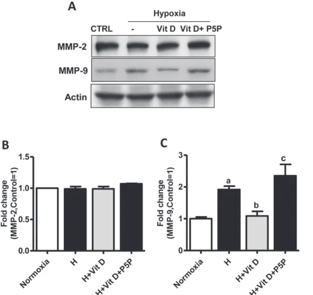
We tested the possibility that hypoxic injury could affect MMP-2/9 expression in bEnd.3 cells and observed a statistically significant(p<0.05) increase in the expression of MMP-9 but not of MMP-2 after hypoxia. We then evaluated the effect of 1,25(OH)2D3on MMP-9 expression in bEnd.3 cells. As shown inFig. 5, 1,25(OH)2D3statistically significantly(p<0.01) decreased MMP-9 expression compared to hypoxia, and P5P treatment blocked this effect(p<0.001). We take these results to indicate that MMP-9 is the major gelatinase contributing to BBB dis-ruption after H/R in bEnd.3 cells, and the inhibitory effect of 1,25(OH)2D3on MMP-9 expres-sion is VDR-mediated.
Discussion
Several lines of evidence show that vitamin D hormone has neuroprotective effects following ischemic brain injury [11], but the role of vitamin D and the VDR in H/R-induced BBB perme-ability has not yet been investigated. In the present study, we found that 1,25(OH)2D3 attenuat-ed H/R-inducattenuat-ed TJ loss and therefore membrane permeability by blocking the increasattenuat-ed production of ROS, NF-kB activation, and MMP-9 expression. These protective effects of 1,25 (OH)2D3were blocked by treatment with the VDR antagonist P5P. The genomic action of 1,25 (OH)2D3, is mediated through the interaction of the VDR with vitamin D response elements (VDRES) from the target genes [22].
To investigate the effect of 1,25(OH)2D3 on BBB function after H/R, we monitored TEER and FITC-dextran permeation, the typical and widely used markers for measuringin vitro
Fig 2. 1,25(OH)2D3effects on tight junction protein expression and cell viability after hypoxia/reoxygenation in bEnd.3 cells. (A)Representative double immunofluorescence staining of ZO-1 (green), claudin-5 (green), occludin (red), and 4’,6-Diamidino-2-phenylindole (DAPI, blue) under hypoxia-reoxygenation (H/R) in the presence or absence of 100 nmol/L 1,25(OH)2D3with or without vitamin D receptor antagonist pyridoxal 5 phosphate pyridoxal 5’ -phosphate (P5P) in brain endothelial cells.(B)Immunoblot images and quantitative data showing the expression of occludin and claudin-5 after H/R in bEnd.3 cell monolayers. H/R-induced loss of occludin and claudin-5 compared with normoxia was prevented by pretreatment with 1,25(OH)2D3. These effects were blocked by P5P treatment. The density of proteins in normoxia was used as a standard (arbitrary unit) to compare the relative density of the other groups. Values shown are mean±s.e.m. (n = 3). Similar results were obtained from three independent experiments.(C)Representative data from MTT assay showing cell viability after 16 h hypoxia/10 min reperfusion in b.End3 cells. The reduced cell viability after hypoxia was prevented by pretreatment with 1,25(OH)2D3. These effects were blocked by P5P treatment. Values shown are mean±s.e.m. (n = 16).*p<0.05,**p<0.01, and***p<0.001
Fig 3. Effects of 1,25(OH)2D3on hypoxia/reoxygenation-induced mitochondrial superoxide production and hydrogen peroxide in bEnd.3 cells. (A) Mitochondrial superoxide production in live cells was measured by fluorescence microscopy using MitoSOX Red dye. Representative fluorescence images show localization of MitoSOX Red fluorescence and DAPI fluorescence. Scale bar 50μm.(B)MitoSOX Red fluorescence per cell was quantified using ImageJ software. Image data from 51–60 cells per treatment condition were averaged (n = 3).(C)Extracellular H2O2production with the use of Amplex Red (n = 4). Data are expressed as mean±s.e.m;*p<0.05,**p<0.01, and***p<0.001.
functionality of BBB TJs and permeability, respectively, in bEnd.3 cells after 6-h hypoxia and 3-h reoxygenation [5,23–25]. In our study, TEER values dropped and FITC-dextran perme-ation across the BBB increased. We found that adding 1,25(OH)2D3 prevented the decrease of TEER and the increase of 40 kDa FITC-dextran. P5P blocked these effects (Fig. 1B and C), sug-gesting that they are VDR-mediated. To the best of our knowledge the present study is the first to show the role of the VDR in modulating BBB permeability. Our results suggest that VDR modulation can be targeted as a possible therapeutic intervention for restoring BBB permeabil-ity following ischemic insult.
Fig 4. Effect of 1,25(OH)2D3 on NF-kB activation in bEnd.3 cells after hypoxia. (A)Representative immunofluorescence images show the translocation of the p65 subunit of NF-κB (green) with DAPI (blue) nuclear counterstaining. bEnd.3 cells were treated with 1,25(OH)2D3(100 nmol/L), or 1,25(OH)2D3plus vitamin D receptor antagonist pyridoxal 5’-phosphate (P5P; 1 mM) under hypoxia/reoxygenation. Scale bar 50μm. 1,25(OH)2D3prevented the hypoxia-induced translocation of the p65 subunit of NF-kB into the nucleus, which was blocked by P5P treatment.(B)Protein levels of phosphorylated IkBαin bEnd.3 cells as determined using Western blotting and imaging analysis. Representative Western blots for IkBα. The density of proteins in normoxia was used as a standard (arbitrary unit) to compare the relative density of the other groups. Values shown are mean±s.e.m. (n = 3) of a representative experiment. Similar results were obtained from three independent experiments;*p<0.05,**p<0.01, and***p<0.001.
TJs play an important role in preserving BBB integrity under various pathological conditions.
A number of studies have indicated that hypoxia and H/R lead to disruption of BBB TJs in
in vitromodels of the BBB [26]. We and others have previously shown that claudin-5 and occludin expression is highly susceptible during ischemic events and also correlates with TEER changes in brain endothelial cells [5,27]. The literature also reports a reduced level of ZO-1 fol-lowing hypoxic exposure [28], a finding consistent with our current ZO expression results as measured by immunocytochemistry (Fig. 2A). The observed TJ alterations in our experiment may have been caused by the loss of ZO-1. Because cytoplasmic ZO-1 is linked with actin fila-ments and the transmembrane protein claudin-5 [29], impairment of the actin filaments may lead to a loss or alteration of ZO-1 and claudin-5. As shown inFig. 2, the loss of TJ proteins after H/R in bEnd.3 cells was prevented by 1,25(OH)2D3treatment, and blocking the VDR by Fig 5. Effects of 1,25(OH)2D3on MMP-2/9 in bEnd.3 cells.Cells were pretreated with 1,25(OH)2D3at 100 nM or 1,25(OH)2D3with vitamin D receptor antagonist pyridoxal-5-phosphate (1mM) for 24 h.(A)Representative Western blots for MMP-2/9.(B-C)The density of proteins in normoxia was used as a standard (arbitrary unit) to compare the relative density of the other groups. Values shown are mean±s.e.m. Similar results were obtained from three independent experiments; a, different from normoxia;*p<0.05,**p<0.01, and***p<0.001.
P5P diminished the beneficial effect of 1,25(OH)2D3. These results can be taken to suggest that this VDR-mediated effect of 1,25(OH)2D3is associated with stabilization of the TJ complex.
It has been suggested that BBB disruption can start with focal damage to the vascular wall which then results in endothelial cell apoptosis caused by plasma proteins such as CD8+ T lym-phocytes, granzyme B and amyloid-β. Endothelial apoptosis is also induced by cytokines in-cluding TGF-β1, platelets and ROSin vitro[30,31]. Here we found that H/R in bEnd.3 cells induced a marked decrease in cell viability (apoptosis), and 1,25(OH)2D3treatment prevented this effect through its actions on the VDR (Fig. 2C).
Several hypotheses have been proposed to explain the molecular mechanisms of BBB dys-function through regulation of TJ proteins in hypoxic injury, including ROS generation, oxida-tive stress, upregulation of MMPs, cytokines and VEGF, and activation of Rho kinase and myosin light chain kinase [32,33]. ROS-inducing oxidative stress is a cellular signaling media-tor for various molecules such as glutamate [34], cytokines, and growth factors released from activated glia or microglia [34,35], which in turn have been associated with activation of NF-kB [36]. These findings led us to investigate the inhibitory effect of 1,25(OH)2D3on H/R–induced mitochondrial superoxide and intracellular hydrogen peroxide. It has been proposed that mito-chondrial ROS are a major factor in BBB impairment in ischemia followed by reperfusion [37]. Here, we found that brain endothelial cells exposed to H/R display elevated ROS production and this is decreased by 1,25(OH)2D3treatment (Fig. 3). Treatment with P5P and 1,25 (OH)2D3blocks that beneficial effect.
Hypoxia-induced activation of NF-kB observed in various cell types is known to be mediat-ed through the phosphorylation of IkB [38]. It has been suggested that the intracellular ROS level regulates NF-kB activity, but the molecular mechanism involved remains unclear [39]. In most unstimulated cells, the inactive p65 component of NF-kB is retained as dimers in the cy-toplasm that are bound to IkB proteins. After stimulation, IkB proteins are phosphorylated and targeted for degradationviathe proteasome pathway, allowing the translocation of the p65 component of NF-kB to the nucleus. Phosphorylation of IkB in Ser32 and Ser36 of the N-ter-minal leads to ubiquitination-dependent proteolysis and results in the translocation of NF-kB to the nucleus, followed by the activation of specific target genes [38,40]. Here we demonstrate that 1,25(OH)2D3prevents the translocation of p65 into the nucleus and the phosphorylation of IkB after H/R in bEnd.3 cells, and that the effect of 1,25(OH)2D3on reduction of NF-kB ac-tivity is mediated through the VDR (Fig. 4). Our results are in line with the data of Tse et al. showing reduced NF-kB transcriptional activity after 1,25(OH)2D3treatment in VDR-positive MCF-7 breast cancer cells [41]. Our data indicate that the VDR is required for 1,25(OH)2D3 action on NF-kB, an observation supported by Janjetovic et al., who found that 20-hydroxy-vi-tamin D3did not affect IkBαmRNA levels in keratinocytes lacking the VDR [42].
the cell media used in the Liu et al. study. MMP-2 is responsible for early BBB disruption, while MMP-9 is involved in brain injury at relatively late stroke stagesin vivoandin vitro
[43,46]. Based on the results reported here, we think that the increased membrane permeability after 6-h hypoxia/3-h reoxygenation is caused by increased MMP-9 expression rather than by membrane disruption.
The effects of 1,25(OH)2D3on MMP-2/9 expression in bEnd.3 cells after hypoxic injury have not been previously reported. Treatment with 1,25(OH)2D3prevented the hypoxia-in-duced increase of MMP-9 expression. Interestingly, blocking of the VDR with P5P eliminated this effect (Fig. 5). This inhibitory effect of 1,25(OH)2D3on MMP-9 expression after hypoxia may be explained by the inhibiting action of 1,25(OH)2D3on ROS production and
NF-kB activation.
Increased vascularization or vascular protection following ischemic/reperfusion injury may be critical to mediate functional recovery, with endothelial cells being the primary cell type re-sponsible for angiogenesis [47]. While the beneficial role of angiogenesis in stroke is recognized [48], there is considerable evidence from the cancer literature suggesting that high doses of 1,25-(OH)2D3 and its analogs inhibit cellular proliferation and angiogenesis and stimulate the apoptotic process [49]. While inhibition of tumor invasiveness and angiogenesis by vitamin D is reported to be mediated by inhibition of MMPs, VEGF, and parathyroid hormone-related protein [50], vitamin D has also been reported to stimulate angiogenesis in endothelial colony-forming cells by increasing VEGF expression and pro-MMP-2 activity [51]. VEGF activation has been shown to be beneficial at a later stage of stroke by enhancing angiogenesis [52,53], but in the acute stage, it is also known to increase microvascular permeability, causing increased edema and hemorrhage [53]. Our study shows that 1,25(OH)2D3protects against ischemic in-jury-induced BBB dysfunction in cerebral endothelial cells by protecting them from ROS and proinflammatory cytokines. The differential effects of vitamin D in cancerous and normal tis-sue merits further investigation.
In stroke, early disruption of the BBB appears to be a major risk factor for hemorrhagic complications and edema formation induced by treatment with tPA, currently the only FDA-approved stroke drug [1,5,54]. Overall efficacy for tPA reperfusion therapy could be increased and risk-to-benefit ratio reduced by preventing endothelial cell injury and BBB breakdown as-sociated with tPA treatment. Although a more detailedin vivostudy in stroke models with vita-min D is necessary, ourin vitroresults suggest to us that the neurovascular protective effects of 1,25(OH)2D3treatment may prove an important adjunct strategy to improve the safety and ef-ficacy of thrombolytic therapies such as tPA.
Fig 6. Schematic presentation of a possible mechanism of 1,25(OH)2D3action on hypoxia-induced blood-brain barrier (BBB) disruption.Following hypoxic injury, an increase in intracellular reactive oxygen species (ROS) causes mitochondrial dysfunction, which further leads to intracellular oxidative stress (1). As a result, damaged mitochondria activate nuclear factor kappa B (NF-κB) signaling pathways, causing IkB proteosomal degradationviaphosphorylation of IkBα(2), which leads to proteolysis and the translocation of NF-κB to the nucleus (3). The attachment of NF-κB subunits to its DNA binding site modulates MMP-9 transcription, which leads to increased MMP-9 expression (4). Increased expression of MMP-9 mediates BBB disruption (low TEER values) through degradation of tight junction proteins such as ZO-1, occludin, and claudin-5 (5). 1,25(OH)2D3blocks the signaling cascade by binding to the vitamin D receptor (VDR) (6) and preventing the translocation of p65 into the nucleus and the phosphorylation of IkB (7). The VDR antagonist pyridoxal 5’-phosphate blocks the effect of 1,25(OH)2D3on BBB markers (8).
Author Contributions
Conceived and designed the experiments: IS SW. Performed the experiments: SW JSK BW. Analyzed the data: IS SW BLP. Contributed reagents/materials/analysis tools: IS DGS. Wrote the paper: IS SW DGS.
References
1. Latour LL, Kang DW, Ezzeddine MA, Chalela JA, Warach S. Early blood-brain barrier disruption in human focal brain ischemia. Ann Neurol. 2004; 56: 468–477. PMID:15389899
2. Chen B, Friedman B, Cheng Q, Tsai P, Schim E, Kleinfeld D, et al. Severe blood-brain barrier disruption and surrounding tissue injury. Stroke. 2009; 40: e666–674. doi:10.1161/STROKEAHA.109.551341 PMID:19893002
3. Hawkins BT, Davis TP. The blood-brain barrier/neurovascular unit in health and disease. Pharmacol Rev. 2005; 57: 173–185. PMID:15914466
4. Sandoval KE, Witt KA. Blood-brain barrier tight junction permeability and ischemic stroke. Neurobiol Dis. 2008; 32: 200–219. doi:10.1016/j.nbd.2008.08.005PMID:18790057
5. Won S, Lee JH, Wali B, Stein DG, Sayeed I. Progesterone attenuates hemorrhagic transformation after delayed tPA treatment in an experimental model of stroke in rats: involvement of the VEGF-MMP path-way. J Cereb Blood Flow Metab. 2014; 34: 72–80. doi:10.1038/jcbfm.2013.163PMID:24045404 6. Al Ahmad A, Gassmann M, Ogunshola OO. Involvement of oxidative stress in hypoxia-induced
blood-brain barrier breakdown. Microvasc Res. 2012; 84: 222–225. doi:10.1016/j.mvr.2012.05.008PMID: 22668821
7. Pun PB, Lu J, Moochhala S. Involvement of ROS in BBB dysfunction. Free Radic Res. 2009; 43: 348–
364. doi:10.1080/10715760902751902PMID:19241241
8. Wang LF, Li X, Gao YB, Wang SM, Zhao L, Dong J, et al. Activation of VEGF/Flk-1-ERK Pathway In-duced Blood-Brain Barrier Injury After Microwave Exposure. Mol Neurobiol. 2014.
9. Eelen G, Verlinden L, van Camp M, van Hummelen P, Marchal K, de Moor B, et al. The effects of 1alpha,25-dihydroxyvitamin D3 on the expression of DNA replication genes. J Bone Miner Res. 2004; 19: 133–146. PMID:14753745
10. Peterlik M, Cross HS. Vitamin D and calcium insufficiency-related chronic diseases: molecular and cel-lular pathophysiology. Eur J Clin Nutr. 2009; 63: 1377–1386. doi:10.1038/ejcn.2009.105PMID: 19724293
11. Balden R, Selvamani A, Sohrabji F. Vitamin D deficiency exacerbates experimental stroke injury and dysregulates ischemia-induced inflammation in adult rats. Endocrinology. 2012; 153: 2420–2435. doi: 10.1210/en.2011-1783PMID:22408173
12. Garcion E, Wion-Barbot N, Montero-Menei CN, Berger F, Wion D. New clues about vitamin D functions in the nervous system. Trends Endocrinol Metab. 2002; 13: 100–105. PMID:11893522
13. Gombart AF. The vitamin D-antimicrobial peptide pathway and its role in protection against infection. Future Microbiol. 2009; 4: 1151–1165. doi:10.2217/fmb.09.87PMID:19895218
14. Chen S, Law CS, Grigsby CL, Olsen K, Hong TT, Zhang Y, et al. Cardiomyocyte-specific deletion of the vitamin D receptor gene results in cardiac hypertrophy. Circulation. 2011; 124: 1838–1847. doi:10. 1161/CIRCULATIONAHA.111.032680PMID:21947295
15. Yuan W, Li G, Gil ES, Lowe TL, Fu BM. Effect of surface charge of immortalized mouse cerebral endo-thelial cell monolayer on transport of charged solutes. Ann Biomed Eng. 2010; 38: 1463–1472. doi:10. 1007/s10439-010-9920-xPMID:20087768
16. Brown RC, Morris AP, O'Neil RG. Tight junction protein expression and barrier properties of immortal-ized mouse brain microvessel endothelial cells. Brain Res. 2007; 1130: 17–30. PMID:17169347 17. Nicolazzo JA, Charman SA, Charman WN. Methods to assess drug permeability across the
blood-brain barrier. J Pharm Pharmacol. 2006; 58: 281–293. PMID:16536894
18. Naik P, Cucullo L. In vitro blood-brain barrier models: current and perspective technologies. J Pharm Sci. 2012; 101: 1337–1354. doi:10.1002/jps.23022PMID:22213383
19. Hwang IK, Yoo KY, Kim do H, Lee BH, Kwon YG, Won MH. Time course of changes in pyridoxal 5'-phosphate (vitamin B6 active form) and its neuroprotection in experimental ischemic damage. Exp Neu-rol. 2007; 206: 114–125. PMID:17531224
21. Watson PM, Paterson JC, Thom G, Ginman U, Lundquist S, Webster CI. Modelling the endothelial blood-CNS barriers: a method for the production of robust in vitro models of the rat blood-brain barrier and blood-spinal cord barrier. BMC Neurosci. 2013; 14: 59. doi:10.1186/1471-2202-14-59PMID: 23773766
22. Patel SR, Ke HQ, Vanholder R, Koenig RJ, Hsu CH. Inhibition of calcitriol receptor binding to vitamin D response elements by uremic toxins. J Clin Invest. 1995; 96: 50–59. PMID:7615822
23. Takata F, Dohgu S, Yamauchi A, Matsumoto J, Machida T, Fujishita K, et al. In vitro blood-brain barrier models using brain capillary endothelial cells isolated from neonatal and adult rats retain age-related barrier properties. PLoS One. 2013; 8: e55166. doi:10.1371/journal.pone.0055166PMID:23383092 24. Deli MA, Abraham CS, Kataoka Y, Niwa M. Permeability studies on in vitro blood-brain barrier models:
physiology, pathology, and pharmacology. Cell Mol Neurobiol. 2005; 25: 59–127. PMID:15962509 25. Jiao H, Wang Z, Liu Y, Wang P, Xue Y. Specific role of tight junction proteins claudin-5, occludin, and
ZO-1 of the blood-brain barrier in a focal cerebral ischemic insult. J Mol Neurosci. 2011; 44: 130–139. doi:10.1007/s12031-011-9496-4PMID:21318404
26. Mark KS, Davis TP. Cerebral microvascular changes in permeability and tight junctions induced by hyp-oxia-reoxygenation. Am J Physiol Heart Circ Physiol. 2002; 282: H1485–1494. PMID:11893586 27. Li G, Simon MJ, Cancel LM, Shi ZD, Ji X, Tarbell JM, et al. Permeability of endothelial and astrocyte
co-cultures: in vitro blood-brain barrier models for drug delivery studies. Ann Biomed Eng. 2010; 38: 2499–2511. doi:10.1007/s10439-010-0023-5PMID:20361260
28. Fischer S, Wobben M, Marti HH, Renz D, Schaper W. Hypoxia-induced hyperpermeability in brain microvessel endothelial cells involves VEGF-mediated changes in the expression of zonula occludens-1. Microvasc Res. 2002; 63: 70–80. PMID:11749074
29. Zlokovic BV. The blood-brain barrier in health and chronic neurodegenerative disorders. Neuron. 2008; 57: 178–201. doi:10.1016/j.neuron.2008.01.003PMID:18215617
30. Kyaw T, Winship A, Tay C, Kanellakis P, Hosseini H, Cao A, et al. Cytotoxic and proinflammatory CD8+ T lymphocytes promote development of vulnerable atherosclerotic plaques in apoE-deficient mice. Cir-culation. 2013; 127: 1028–1039. doi:10.1161/CIRCULATIONAHA.112.001347PMID:23395974 31. Kuckleburg CJ, Tiwari R, Czuprynski CJ. Endothelial cell apoptosis induced by bacteria-activated
plate-lets requires caspase-8 and -9 and generation of reactive oxygen species. Thromb Haemost. 2008; 99: 363–372. doi:10.1160/TH07-07-0474PMID:18278187
32. Kuhlmann CR, Zehendner CM, Gerigk M, Closhen D, Bender B, Friedl P, et al. MK801 blocks hypoxic blood-brain-barrier disruption and leukocyte adhesion. Neurosci Lett. 2009; 449: 168–172. doi:10. 1016/j.neulet.2008.10.096PMID:18996441
33. Engelhardt S, Patkar S, Ogunshola OO. Cell-specific blood-brain barrier regulation in health and dis-ease: a focus on hypoxia. Br J Pharmacol. 2014; 171: 1210–1230. doi:10.1111/bph.12489PMID: 24641185
34. Fatma N, Kubo E, Sen M, Agarwal N, Thoreson WB, Camras CB, et al. Peroxiredoxin 6 delivery attenu-ates TNF-alpha-and glutamate-induced retinal ganglion cell death by limiting ROS levels and maintain-ing Ca2+ homeostasis. Brain Res. 2008; 1233: 63–78. doi:10.1016/j.brainres.2008.07.076PMID: 18694738
35. Fatma N, Kubo E, Takamura Y, Ishihara K, Garcia C, Beebe DC, et al. Loss of NF-kappaB control and repression of Prdx6 gene transcription by reactive oxygen species-driven SMAD3-mediated transform-ing growth factor beta signaltransform-ing. J Biol Chem. 2009; 284: 22758–22772. doi:10.1074/jbc.M109. 016071PMID:19553668
36. Qin L, Crews FT. NADPH oxidase and reactive oxygen species contribute to alcohol-induced microglial activation and neurodegeneration. J Neuroinflammation. 2012; 9: 5. doi:10.1186/1742-2094-9-5 PMID:22240163
37. Itoh Y, Takaoka R, Ohira M, Abe T, Tanahashi N, Suzuki N. Reactive oxygen species generated by mi-tochondrial injury in human brain microvessel endothelial cells. Clin Hemorheol Microcirc. 2006; 34: 163–168. PMID:16543632
38. Koong AC, Chen EY, Giaccia AJ. Hypoxia causes the activation of nuclear factor kappa B through the phosphorylation of I kappa B alpha on tyrosine residues. Cancer Res. 1994; 54: 1425–1430. PMID: 8137243
39. Sen CK, Packer L. Antioxidant and redox regulation of gene transcription. FASEB J. 1996; 10: 709–
720. PMID:8635688
41. Tse AK, Zhu GY, Wan CK, Shen XL, Yu ZL, Fong WF. 1alpha,25-Dihydroxyvitamin D3 inhibits tran-scriptional potential of nuclear factor kappa B in breast cancer cells. Mol Immunol. 2010; 47: 1728–
1738. doi:10.1016/j.molimm.2010.03.004PMID:20371119
42. Janjetovic Z, Zmijewski MA, Tuckey RC, DeLeon DA, Nguyen MN, Pfeffer LM, et al. 20-Hydroxychole-calciferol, product of vitamin D3 hydroxylation by P450scc, decreases NF-kappaB activity by increasing IkappaB alpha levels in human keratinocytes. PLoS One. 2009; 4: e5988. doi:10.1371/journal.pone. 0005988PMID:19543524
43. Liu J, Jin X, Liu KJ, Liu W. Matrix metalloproteinase-2-mediated occludin degradation and caveolin-1-mediated claudin-5 redistribution contribute to blood-brain barrier damage in early ischemic stroke stage. J Neurosci. 2012; 32: 3044–3057. doi:10.1523/JNEUROSCI.6409-11.2012PMID:22378877 44. Yang MZ, Mun CH, Choi YJ, Baik JH, Park KA, Lee WT, et al. Agmatine inhibits matrix
metalloprotei-nase-9 via endothelial nitric oxide synthase in cerebral endothelial cells. Neurol Res. 2007; 29: 749–
754. PMID:17588309
45. McColl BW, Rothwell NJ, Allan SM. Systemic inflammation alters the kinetics of cerebrovascular tight junction disruption after experimental stroke in mice. J Neurosci. 2008; 28: 9451–9462. doi:10.1523/ JNEUROSCI.2674-08.2008PMID:18799677
46. Rosenberg GA, Estrada EY, Dencoff JE. Matrix metalloproteinases and TIMPs are associated with blood-brain barrier opening after reperfusion in rat brain. Stroke. 1998; 29: 2189–2195. PMID: 9756602
47. Sunderkotter C, Steinbrink K, Goebeler M, Bhardwaj R, Sorg C. Macrophages and angiogenesis. J Leukoc Biol. 1994; 55: 410–422. PMID:7509844
48. Navaratna D, Guo S, Arai K, Lo EH. Mechanisms and targets for angiogenic therapy after stroke. Cell Adh Migr. 2009; 3: 216–223. PMID:19363301
49. Beer TM, Eilers KM, Garzotto M, Egorin MJ, Lowe BA, Henner WD. Weekly high-dose calcitriol and docetaxel in metastatic androgen-independent prostate cancer. J Clin Oncol. 2003; 21: 123–128. PMID:12506180
50. Nakagawa K, Sasaki Y, Kato S, Kubodera N, Okano T. 22-Oxa-1alpha,25-dihydroxyvitamin D3 inhibits metastasis and angiogenesis in lung cancer. Carcinogenesis. 2005; 26: 1044–1054. PMID:15718253 51. Grundmann M, Haidar M, Placzko S, Niendorf R, Darashchonak N, Hubel CA, et al. Vitamin D improves
the angiogenic properties of endothelial progenitor cells. Am J Physiol Cell Physiol. 2012; 303: C954–
962. doi:10.1152/ajpcell.00030.2012PMID:22932684
52. Zhang ZG, Zhang L, Tsang W, Soltanian-Zadeh H, Morris D, Zhang R, et al. Correlation of VEGF and angiopoietin expression with disruption of blood-brain barrier and angiogenesis after focal cerebral is-chemia. J Cereb Blood Flow Metab. 2002; 22: 379–392. PMID:11919509
53. van Bruggen N, Thibodeaux H, Palmer JT, Lee WP, Fu L, Cairns B, et al. VEGF antagonism reduces edema formation and tissue damage after ischemia/reperfusion injury in the mouse brain. J Clin Invest. 1999; 104: 1613–1620. PMID:10587525