From the Department of Gastroenterology, Hospital das Clínicas, Faculty of Medicine, University of São Paulo and Division of Pathology, Adolfo Lutz Institute - São Paulo/ SP, Brazil.
E-mail: [email protected] Received for publication on
November 05, 2003.
ORIGINAL RESEARCH
P53 AND RB TUMOR SUPPRESSOR GENE
ALTERATIONS IN GASTRIC CANCER
Rejane Mattar, Suely Nonogaki, Cleonice Silva, Venancio Alves, and Joaquim J. Gama-Rodrigues
MATTAR R et al. p53 and Rb tumor suppressor gene alterations in gastric cancer. Rev. Hosp. Clín. Fac. Med. S. Paulo 59(4):172-180, 2004.
Inactivation of tumor suppressor genes has been frequently observed in gastric carcinogenesis. Our purpose was to study the involvement of p53, APC, DCC, and Rb genes in gastric carcinoma.
METHOD: Loss of heterozygosity of the p53, APC, DCC and Rb genes was studied in 22 gastric cancer tissues using polymerase chain reaction; single-strand conformation polymorphism of the p53 gene exons 5-6 and exons 7-8 was studied using 35S-dATP, and p53 expression was detected using a histological immunoperoxidase method with an anti-p53 clone.
RESULTS AND DISCUSSION: No loss of heterozygosity was observed in any of these tumor suppressor genes;
homozygous deletion was detected in the Rb gene in 23% (3/13) of the cases of intestinal-type gastric carcinoma. Eighteen (81.8%) cases showed band mobility shifts in exons 5-6 and/or 7-8 of the p53 gene. The presence of the p53 protein was positive in gastric cancer cells in 14 cases (63.6%). Normal gastric mucosa showed negative staining for p53; thus, the immunoreactivity was likely to represent mutant forms. The correlation of band mobility shift and the immunoreactivity to anti-p53 was not significant (P = .90). There was no correlation of gene alterations with the disease severity.
CONCLUSIONS: The inactivation of Rb and p53 genes is involved in gastric carcinogenesis in our environment. Loss of the Rb gene observed only in the intestinal-type gastric cancer should be further evaluated in association with Helicobacter pylori infection. The p53 gene was affected in both intestinal and diffuse histological types of gastric cancer.
KEY WORDS: Gastric cancer. APC. DCC. Rb. p53.
Gastric cancer is a heterogeneous pathology, being classified by Laurén (1965)1 into 2 general subtypes:
intes-tinal (differentiated) and diffuse (un-differentiated). Intestinal-type gastric cancer may or may not be preceded by preneoplastic lesions; it is more preva-lent in older individuals and in certain geographical areas where there is a high incidence of gastric carcinoma, such as in Japan.2
The differences between these two histological subtypes occur also at the molecular level, suggesting different genetic pathways. The intestinal-type
gastric carcinoma presents tumor sup-pressor gene alterations similar to colorectal tumors and distinct from dif-fuse-type gastric cancer.3
An accumulation of multiple ge-netic and epigege-netic alterations of oncogenes, tumor suppressor genes, DNA repair genes, cell cycle
regula-tors, cell adhesion molecules, and growth factor/receptor systems are in-volved throughout the course of the multistep conversion of normal epithe-lial cells to clinical gastric cancer.4
In gastric cancer, p53 gene
altera-tions have been observed in both his-tological subtypes,5 being correlated
with node-positive cancer,6,7 depth of
tumor invasion,8 and poor survival.9,10
The p53 protein is a transcriptional factor that arrests the cell cycle in the G1 phase when DNA is damaged11 by
PCNA.12,13 Thus, damaged DNA can
not replicate, allowing time for the re-pair system to act.11 If this system fails,
p53 induces apoptosis by transactivation of the bax gene.14
The loss of heterozygosity (LOH) and the loss of expression of the DCC
gene have been more frequently found in the intestinal-type gastric cancer 15-17 and have often been encountered
(35.3%) in gastric cancer in stage III and IV. Thus, in gastric cancer, LOH of the DCC gene was a late event
associ-ated with malignant progression.15,18
The DCC gene, located on the long
arm of chromosome 18,19 encodes a
netrin-1 receptor component with func-tions in cell migration,20 G2/M cell
cy-cle arrest, and apoptosis.21
The mutated APC germline gene on
chromosome 5q21 is responsible for the inheritance of familial adenomatous polyposis; in addition, it was somatically altered in sporadic colorectal cancer patients.22 In gastric
cancer, the incidence of allelic deletions of APC was significantly higher in the
intestinal phenotypes than in the dif-fuse phenotypes.23 Furthermore, APC
down-regulates the proto-oncogene β -catenin that is critical for intercellular adhesion and has linked colorectal car-cinogenesis to the Wnt-signal transduc-tion pathway.24 Increased β-catenin
mRNA levels were significantly more frequent in intestinal-type gastric can-cers as compared with the diffuse-type gastric cancers. APC gene mutations
found in these cases of intestinal-type gastric cancer were associated with the increase of β-catenin mRNA levels.25
Loss of the retinoblastoma (Rb)
gene has been associated with esophageal tumorigenesis,26 and at a
lower rate to gastric cancer develop-ment.17 However, more recent reports
have shown that Helicobacter pylori
(H. pylori) infection generated gastric
cancer through p53-Rb
tumor-suppres-sor system mutation and telomerase re-activation.27 The Rb gene encodes a
nuclear protein that acts as a cell cy-cle control checkpoint at the G1 phase.28
The purpose of our study was to fur-ther analyze the involvement of p53, APC, DCC, and Rb tumor suppressor
genes in gastric carcinoma cases.
MATERIALS AND METHOD
Gastric cancer tissues and corre-sponding leukocytes were obtained from 22 patients after surgical treat-ment during the period of 1996 to 1997 at the Hospital of Clinics, De-partment of Gastroenterology, and were immediately frozen in liquid ni-trogen and stored prior to DNA extrac-tion. The average age of the patients (18 men, 4 women) was 63.4 + 14.3 years. The tumors were classified as in-testinal and diffuse types according to Láuren1; 13 cases were of the intesti-nal type and 9 cases were of the dif-fuse type. The TNM stage grouping was performed according to the crite-ria of the Japanese Classification of Gastric Carcinoma.29
DNA extraction
DNA was extracted from the thawed cancer tissue and peripheral leucocytes using a phenol-chloroform method30 and stored at 4 °C until use.
Polymerase Chain Reaction (PCR)
One microgram or 300 ng of ge-nomic DNA was used as a template in a reaction volume of 50 uL,
contain-ing 50 pmol of each primer (Table 1), 200 uM of each dNTP and 2.5 U of Taq DNA polymerase (Gibco BRL,
Gaithersburg, MD, USA). The PCR was performed in a 2400 GeneAmp PCR system (Perkin Elmer, Branchburg, NJ, USA). Amplification was performed for 35 cycles at an annealing temperature of 68 °C for APC,31 62 °C for p53,32
55 °C for DCC,31 and 57 °C for Rb
-D13S270.33 The amplification
condi-tions for Rb intron1 were 40 cycles in
2 steps (94 °C for 1 min and 50 °C for 1 min), followed by 1 extension step at 72 °C for 5 min; inclusion of 10% dimethyl sulfoxide (DMSO) was nec-essary for generating the 180 bp frag-ment.34 A second primer pair that spans
the same locus was used in the cases where the PCR product was absent from the carcinoma DNA template when compared with a strong product from the paired constitutional DNA template.
Restriction fragment length poly-morphism analysis
Products of the PCR (~45 uL) were
digested with 60 U of MspI (DCC)
(Stratagene, La Jolla, CA, USA) and
RsaI (APC) (Stratagene, La Jolla, CA,
USA), 15 U of BamHI (Rb) (Stratagene,
La Jolla, CA, USA), and 8 U of AccII
(p53) (Amersham Life Science,
Cleve-land, OH, USA) at 37 °C overnight. The DNA fragments were separated by electrophoresis on 4% low-melting-point agarose gels. For VNTR, Rb
(D13S270), and after MspI digestion,
PCR products were separated on 3% low-melting-point agarose gels.
PCR Analysis of Single-Strand Con-formation Polymorphism
Genomic DNA (300 - 500 ng) was used as a template in a reaction volume of 25 µL containing 50 pmol of each primer (Table 1), 200 µM of deoxynucleotide triphosphate (dNTP),
35S-dATP (0.5 µL), and 2.5 U of Taq
DNA polymerase (Amersham Biosciences). Exons 5-6 and 7-8 of the
p53 gene were amplified in 35 cycles
10 mM EDTA and were rediluted 1:2 in sequencing stop solution (95% formamide, 20 mM EDTA, 0.05% bromophenol blue, and 0.05% xylene cyanol); they were then heated at 90 °C for 5 min, chilled on ice, and loaded onto a nondenaturing polyacrylamide gel (6% acrylamide, 10% glycerol, 1x TBE). Electrophoresis was carried out at 8 W at room temperature. After 4 hours of mi-gration for exons 5-6 and 7 to 9 hours of migration for exons 7-8, the gels were dried and subjected to autoradiography using Kodak T-Mat G/RA film at –80 °C with an intensifying screen. One sample of amplification product from blood was run together with tumor DNA amplifica-tion products. The condiamplifica-tions have been previously described, with modifica-tions.35,36 The primers that were used for
PCR were those according to Tohdo et al. 1993.37
LSAB-immunoperoxidase
Sequential sections of 3 µm from formalin-fixed, paraffin-embedded samples were placed on slides previ-ously treated with 3-aminopropyltriethoxy-silane (Sigma, A-3648, USA). After deparaffinization in xylene and rehydration in alcohol, antigen retrieval was performed with
10 mM citric acid pH 6.0 in a pressure cooker for 4 min. Endogenous peroxi-dase activity was blocked with 6% H2O2. Incubation with 1:100 monoclonal antibody anti-p53 clone DO-7 (Dako, M7001, Denmark) in 1% bovine serum albumin-phosphate buff-ered solution, was performed for 16 h at 4 °C. The slides were then incubated for 30 min at 37 °C with secondary biotinylated goat anti-mouse/rabbit Ig, followed by incubation for 30 min at 37 °C with the complex, streptavidin and biotinylated peroxidase (Dako, K492, Denmark). Slides were devel-oped using 3,3’-diaminobenzidine tetrahydrochloride (Sigma, D-5637, USA), 6% H2O2, and dimethyl sul-phoxide and counterstained with Harris’ hematoxylin. The samples were observed with optic microscope by 2 observers (SN and VAFA) and were scored as previously described by Harvey38; estimated proportion and
in-tensity scores were added to obtain a total score that ranged from 0.2 to 8. A score greater than 2 was used to de-fine p53 positivity.
Statistics
The statistic analysis was per-formed with the chi-square and Fisher
exact tests using the SPSS 7.5 for Win-dows, student version. A P value of
<.05 was considered statistically sig-nificant.
RESULTS
The results of the analysis of PCR products for loss of heterozygosity (LOH) at the Rb, APC, DCC, and p53
loci on 22 gastric cancer tissues (13 of intestinal type and 9 of diffuse type) matched with corresponding periph-eral leukocytes are listed in Table 2. Heterozygosity was found for APC in
13 of 21 (61.9%) cases, for DCC in 13
of 22 (59%) cases, for p53 in 11of 21
(52.4%) cases, and for Rb in 7 of 19
(36.8%) cases. Those cases that had no amplification in leukocyte and tumor DNA for unknown reasons were ex-cluded from this analysis. No LOH was observed in these tumor suppressor genes. Nonetheless, no PCR product was obtained for the tumor DNA when compared to the normal DNA at the
DCC gene in 1 patient (CA.6) and at
the Rb gene in 3 patients (CA.6,
CA.11, CA.22), suggesting ho-mozygous deletion.
To confirm homozygous deletion at the DCC and Rb genes, a second pair Table 1 - Primer sets used in polymerase chain reaction analysis for loss of heterozygosity (PCR-LOH) and polymerase chain reaction analysis for single-strand conformation polymorphism (PCR-SSCP) analysis.
Priming region Amplicon size (bp) Polymorphism type Primer Sequences
APC exon 11 133 Rsa I 5’-GGACTACAGGCCATTGCAGAA-3’
5’-GGCTACATCTCCAAAAGTCAA-3’
p53 exon 4 259 AccII 5’-AATGGATGATTTGATGCTGTCCC-3’
5’-CGTGCAAGTCACAGACTTGGC-3’
DCC* 210-150 VNTR 5’-GATGACATTTTCCCTCTAG-3’
5’-GTGGTTATTGCCTTGAAAAG-3’
DCC 3 9 6 MspI 5’-TGCACCATGCTGAAGATTGT-3’
5’-AGTACAACACAAGGTATGTG-3’
Rb intron 1 180 BamHI 5’-CAGGACAGCGGCCCGGAG-3’
5’-CTGCAGACGCTCCGCCGT-3’
Rb D13S270* 104-80 5’- AGTGCCTGGGTATGAACGTG-3’
5’- CTGGAAATGCCTTGGAAGGA-3’
p53 exons 5-6 411 5’-GGAATTCCTCTTCCTACAGTACTCC-3’
5’-GGAATTCAGTTGCAAACCAGACCTCA-3’
p53 exons 7-8 677 5’-GGAATTCTCCTAGGTTGGCTCTGAC-3’
of primers that span the same locus was used, a variable number of the tandem repeat markers (VNTR) was used for the DCC gene, and a microsatellite
marker (D13S270) was used for the Rb
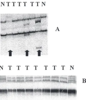
gene. Homozygous deletion was con-firmed only at the Rb gene (Figure 1).
These 3 cases of the intestinal-type gastric carcinomas (3/13—23%) with homozygous deletion at Rb gene were
classified as stage Ia, stage II, and stage IIIb, respectively.
Since p53 gene alterations are very
frequent in gastric cancer and no al-lelic loss was detected, we decided to search for mutations using PCR analy-sis for single-strand conformation polymorphism (PCR-SSCP) and p53 protein overexpression using LSAB-immunoperoxidase. Eighteen (81.8%) cases of both histological types showed mobility shifts in exons 5-6 and/or 7-8 of the p53 gene (Figure 2).
The p53 protein expression was
posi-tive in gastric cancer cells in 14 cases (63.6%). There was no significant cor-relation of band mobility shift and
im-munoreactivity to anti-p53 (P = .90).
The staining of p53 was never ob-served in the normal gastric mucosa adjacent to the tumor tissue; thus, p53 protein immunoreactivity likely indi-cated mutant forms of the p53 gene.
The correlation of band mobility shifts and immunoreactivity was not ob-served in 9 cases. In 7 cases, a mobil-ity shift was detected in exons 7-8 with negative immunoreactivity to anti-p53, 4 were 0 + 0 = 0 and 3 cases were 1p + 1i = 2. The immunoreactiv-ity to anti-p53 was positive in 2 cases (2p + 3i = 5; 2p + 1i = 3) with a nega-tive band mobility shift.
The statistical analysis showed that there was no correlation of sex, age, histology, or severity of disease with mutation and with the immunoreactiv-ity to anti-p53.
DISCUSSION
To understand the molecular events of gastric carcinogenesis in our
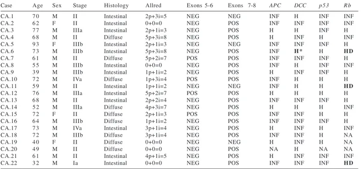
envi-Table 2 - p53 protein immunoreactivity to anti-p53, band mobility shift by single strand conformation polymorphism (SSCP) in exons 5-6 and exons 7-8, and analysis of loss of heterozygosity at APC, DCC, p53, and Rb loci in gastric cancer.
Case Age Sex Stage Histology Allred Exons 5-6 Exons 7-8 APC DCC p 5 3 R b
CA.1 7 0 M II Intestinal 2p+3i=5 NEG NEG INF H INF INF
CA.2 6 2 F II Intestinal 0+0=0 NEG POS INF INF INF INF
CA.3 7 7 M IIIa Intestinal 2p+1i=3 NEG POS H H INF H
CA.4 6 8 M II Diffuse 5p+3i=8 NEG POS H INF H INF
CA.5 9 3 F IIIb Intestinal 2p+1i=3 NEG NEG INF INF INF H
CA.6 7 3 M IIIb Intestinal 5p+3i=8 NEG POS INF H * H HD
CA.7 6 1 M II Diffuse 5p+2i=7 POS POS INF INF INF H
CA.8 5 5 M IIIb Intestinal 0+0=0 NEG POS INF H INF INF
CA.9 3 9 M IIIb Intestinal 1p+1i=2 NEG POS H INF INF H
CA.10 7 2 M IVa Diffuse 1p+3i=4 POS POS INF H H H
CA.11 5 9 M II Intestinal 1p+1i=2 NEG NEG INF H H HD
CA.12 7 6 M IIIa Intestinal 5p+2i=7 POS POS H H H H
CA.13 6 8 M II Intestinal 2p+2i=4 NEG POS INF INF INF H
CA.14 5 2 M IIIa Diffuse 4p+3i=7 NEG POS H H H INF
CA.15 7 2 F II Diffuse 2p+1i=3 POS POS INF INF H H
CA.16 6 4 M IIIb Diffuse 1p+1i=2 NEG POS INF INF INF H
CA.17 7 3 M IVa Intestinal 3p+1i=4 NEG POS H INF H INF
CA.18 7 2 M IIIb Diffuse 3p+1i=4 NEG POS INF INF H NA
CA.19 4 0 F II Diffuse 0+0=0 NEG NEG H INF H NA
CA.20 4 9 M II Diffuse 0+0=0 NEG POS NA H NA NA
CA.21 6 1 M II Intestinal 4p+1i=5 NEG POS H INF INF INF
CA.22 3 2 M Ia Intestinal 0+0=0 NEG POS INF INF INF HD
POS = band mobility shift positive; NEG = no band mobility shift; INF = informative case with no LOH; H = homozygous (no informative); HD = homozygous deletion; NA = no amplification in leukocyte and tumor DNA; H* = homozygous for VNTR marker.
Figure 1 - Homozygous deletion at Rb tumor suppressor genes in gastric cancer. DNA band sizes are indicated in numbers of base pairs. N, leukocytes DNA; T, tumor DNA. (A) Cleavage of 180 bp PCR products by BamHI
ronment, the DNA of 22 gastric tumors paired with corresponding DNA of pe-ripheral leukocytes were studied for loss of heterogeneity (LOH) of the p53, APC, DCC, and Rb genes. These tumor
suppressor genes were chosen for this study based on previous reports show-ing that their inactivations have a role in gastric carcinogenesis.
Loss of heterogeneity at the APC
locus was detected in 87% of primary gastric carcinomas in both intestinal and diffuse types in both early and ad-vanced stages.39 Therefore, LOH at APC was considered one of the most
prevalent genetic alterations in human gastric carcinoma, and it occurred at an early stage of the carcinogenesis and was not a prognostic factor. Moreover, Sano et al.40 had observed LOH on
chromosome 5q where the APC gene
is located in 60% of early well-differ-entiated carcinoma, but not in poorly differentiated carcinoma.
In our study, no LOH was detected at the APC locus. Other authors17,41
studying the same polymorphic site found a low incidence (27% - 30%) of LOH in gastric cancer. Also in Africa,
where the frequency of gastric cancer is low as it is in Brazil, only 1 patient presented LOH in the 5q region.42
Fur-thermore, in gastric cancer patients from north-central Italy, no intragenic mutations were found in APC codons
686 through 1693, and allelic loss was detected in loci near APC.43
These authors argued that epide-miologic studies have not observed a higher risk of gastric cancer in patients with inherited familial adenomatous polyposis. Fundic gland polyps are the most common gastric lesion in famil-ial adenomatous polyposis and are generally believed to have little or no potential for malignant transformation in the population at large. The devel-opment of high-grade dysplasia or gas-tric adenocarcinoma associated with diffuse fundic gland polyposis was de-scribed in a few cases of familial adenomatous polyposis.44 Gastric-type
adenomas were less likely to show high-grade dysplasia and adenocarci-noma and were found in 10 patients with familial adenomatous polyposis.45
High frequencies of allelic dele-tions affecting the DCC locus have
been previously described to occur in 30%17 to 60%16 of cases of both types
of gastric cancer15 and more often in
advanced (50%) than in early (14.3%) disease. Only 1 case in the present study (CA.6) showed what seemed to be homozygous deletion at the DCC
gene by RFLP; however, with another pair of primers, a region of the DCC
gene was amplified. Thus, the possibil-ity of homozygous deletion was ex-cluded. Since the frequency of DCC
loss is higher in advanced disease and more frequent in the intestinal type, we were expecting that at least in the 5 cases that had advanced intestinal-type gastric cancer and were informa-tive for DCC gene, loss would be
found.
Homozygous deletion at the Rb
gene was detected in 3 (3/13–23%) cases of the intestinal-type gastric can-cer, which seems to be an early event, since 2 of these patients had initial dis-ease (stage Ia and stage II). The inacti-vation of the Rb gene by mutation or
loss has been considered an important genetic alteration in esophageal car-cinogenesis.26, 31 Our data suggest that Rb gene inactivation may be involved
in the development and/or progression of intestinal-type gastric cancer.
Homozygous deletions are thought to be the result of 2 events: the loss of a larger chromosomal region and the independent loss of a considerably smaller area.46 In previous reports,
ho-mozygous deletion was described in a variety of cancers as a mechanism of total gene inactivation, and the pres-ence of at least 1 tumor suppressor gene within the deleted region was suggested.47-50
A higher prevalence (30%) of Rb
-LOH in both histological types of gas-tric cancer was found by other au-thors.17 A more recent report51 showed
that the mRNA levels of Rb and p53
in gastric cancer tissues were both sig-nificantly lower than were those in their noncancerous tissue samples.
These studies indicate that the sup-pression of both Rb and p53 may be
associated with the tumorigenesis of the stomach.
Loss of heterozygosity on chromo-some 17p (p53 locus) and mutation of
the p53 gene have been observed in
more than 60% of gastric carcinomas, regardless of the histological type,40
and have been correlated with short survival times.9 Surprising, no LOH at
the p53 gene was observed in our
cases. One possible explanation for the fact that we did not find LOH at p53
gene may be that this gene may be al-tered through another mechanism, such as point mutation. To search for
p53 gene mutations, we decided to use
PCR-SSCP and to correlate with immu-noreactivity to anti-p53. The half-life of wild-type p53 is short, around 20 to
30 minutes; consequently, p53 protein
expression usually is negative in nor-mal tissues. Mutations render the p53 protein to be a more stable compound with a longer half-life (1.5 - 7 h).52
Thus, p53 protein overexpression is
likely to represent mutant forms.53
In the literature, the authors usually employ 32P-dCTP for PCR-SSCP35,36;
however, 32P is a high-energy β
parti-cle emitter, so we decided to try the low-energy β particle emitter, 35
S-dATP. The bands were easily visual-ized in dried gels exposed at –80º C for
at least 3 days. Thus, the use of 35
S-dATP was more advantageous than 32
P-dATP, since it lowered the risk for the personnel of the lab.
Following PCR-SSCP, band mobil-ity shifts were observed mainly in exons 7-8 (86.36%) that encompasses codons 225 to 326. Other authors54
studying p53 in gastric cancer also
found a predominance of mutations in exons 7-8 (70%).
Non-missense mutations or frameshifts could explain negative im-munoreactivity with band mobility shifts in 7 cases. Moreover, analyzing the Allred criteria only 4 were truly negative 0+0=0, and 3 cases were 1p+1i=2 (considered negative). Allred 2 should be better evaluated and, per-haps, considered as positive for p53
expression as determined by immuno-histochemistry. Seta et al., 199854
found a p53 mutation in 2 cases of
gastric cancer with less than 25% of cells positive (+) by immunohisto-chemistry. Assuming Allred 2 to be positive for p53, the agreement of im-munohistochemistry and PCR-SSCP would be 72.7%.
In 2 cases that had immunoreactiv-ity to anti-p53 and most probably harbored mutation in the p53 gene,
PCR-SSCP was unable to detect mu-tation. A discrepancy between results by PCR-SSCP and DNA sequencing
has been reported in the literature. In MKN-7 and MKN-28 gastric cancer cell lines, p53 gene mutations were not
detected by PCR-SSCP36; however,
point mutations at codons 278 and 251, respectively, were found by cDNA sequencing.53
In conclusion, the inactivation of
p53 is involved in the development
and/or progression of both diffuse and intestinal types of gastric cancer. How-ever, loss of the Rb gene plays role
only in the intestinal-type gastric can-cer progression. It has been suggested that H. pylori infection initiates
gas-tric cancer through p53-Rb
tumor-sup-pressor system mutation and telomerase reactivation.27 Even though
the incidence of H. pylori in the
nor-mal population of Brazil is high (80%), the association of H. pylori infection
and gastric cancer has been observed exclusively with the intestinal type (P
= .008)55. Further studies are needed to
demonstrate the direct involvement of
H. pylori in p53 and Rb gene
inacti-vation in our cases of gastric cancer.
ACKNOWLEDGEMENTS
The authors thank Ana Maria Alexandrino for doing PCR-LOH of the cases.
RESUMO
MATTAR R e col. Alterações dos genes supressores tumorais p53 e Rb no
câncer gástrico. Rev. Hosp. Clín. Fac. Med. S. Paulo 59(4):172-180, 2004.
A inativação de genes supressores tumorais tem sido freqüentemente ob-servada na carcinogênese gástrica. O
nosso objetivo foi estudar o envol-vimento dos genes p53, APC, DCC e Rb no câncer gástrico.
MÉTODO: Vinte e dois casos de câncer gástrico foram estudados por PCR-LOH (reação de polimerase em cadeia- perda de alelo heterozigoto) dos genes p53, APC, DCC e Rb; e por
PCR-SSCP (reação de polimerase em
cadeia- polimorfismo de conformação de cadeia única) dos exons 5-6 e exons 7-8 do gene p53, empregando 35
S-dATP e expressão de p53 por imunoperoxidase com monoclonal anti-p53.
homozigótica foi observada no gene
Rb em 23% (3/13) dos casos de
cân-cer gástrico do tipo intestinal. Desvio de motilidade de banda nos exons 5-6 e/ou exons 7-8, indicando mutação do gene p53 foi encontrada em 18 casos
(81.8%). A expressão de p53 foi posi-tiva nas células de câncer gástrico em 14 casos (63.6%). A mucosa gástrica normal não corou com anti-p53,
por-tanto, a reatividade imune deve repre-sentar formas mutantes. A correlação de desvio de motilidade de banda e expressão imune de p53 não foi significante (p=0.90). Não houve cor-relação entre as alterações genéticas e a extensão da doença.
CONCLUSÃO: A inativação dos genes p53 e Rb tem papel na
carci-nogênese gástrica no nosso meio. A
perda do gene Rb observada apenas no
câncer gástrico do tipo intestinal deve ser avaliada posteriormente em associ-ação com infecção pelo Helicobacter pylori. O gene p53 estava afetado em
ambos os tipos histopatológicos.
UNITERMOS: Câncer gástrico.
p53. APC. DCC. Rb.
REFERENCES
1. Laurén P. The two histological main types of gastric carcinoma: diffuse and so-called intestinal-type. Acta Pathol Microbiol Scand 1965;64:31-49.
2. Hirohashi S, Sugimura T. Genetic alterations in human gastric cancer. Cancer Cells 1991;3:49-52.
3. Tahara E. Molecular mechanism of stomach carcinogenesis. J Cancer Res Clin Oncol 1993;119:265-72.
4. Yasui W, Yokozaki H, Fujimoto J, Naka K, Kuniyasu H, Tahara E. Genetic and epigenetic alterations in multistep carcinogenesis of the stomach. J. Gastroenterol 2000;35:111-5.
5. Lee HS, Lee HK, Kim HS, Yang HK, Kim WH. Tumour suppressor gene expression correlates with gastric cancer prognosis. J Pathol 2003;200:39-46.
6. Maehara Y, Tomoda M, Hasuda S, Kabashima A, Tokunaga E, Kakeji Y, et al. Prognostic value of p53 protein expression for patients with gastric cancer- a multivariate analysis. Br J Cancer 1999;79:1255-61.
7. Baldus SE, Schneider PM, Monig SP, Zirbes TK, Fromm S, Meyer W, et al. p21/waf1/cip1 in gastric cancer: associations with histopathological subtypes, lymphonodal metastasis, prognosis and p53 status. Scand J Gastroenterol 2001;36:975-80. 8. Kataoka M, Okabayashi T, Johira H, Nakatani S, Nakashima A,
Takeda A, et al. Aberration of p53 and DCC in gastric and colorectal cancer. Oncol Rep 2000; 7:99-103.
9. Lee WJ, Shun CT, Hong RL, Wu MS, Chang KJ, Chen KM. Overexpression of p53 predicts shorter survival in diffuse type-gastric cancer. Br J Surg 1998;85:1138-42.
10. Kubicka S, Claas C, Staab S, Kuhnel F, Zender L, Trautwein C, et al. p53 mutation pattern and expression of c-erbB2 and c-met in gastric cancer: relation to histological subtypes, Helicobacter pylori infection, and prognosis. Dig Dis Sci 2002;47:114-21. 11. Kastan MB, Onyekwere O, Sidransky D, Vogelstein B, Craig RW. Participation of p53 protein in the cellular response to DNA damage. Cancer Res 1991;51:6304-11.
12. Chen J, Jackson PK, Kirschner MW, Dutta A. Separate domains of p21 involved in the inhibition of Cdk kinase and PCNA. Nature 1995;374:386-8.
13. Waga S, Hannon GJ, Beach D, Stillman B. The p21 inhibitor of cyclin-dependent kinases controls DNA replication by interaction with PCNA. Nature 1994;369:574-8.
14. Miyashita T, Reed JC. Tumor suppressor p53 is a direct transcriptional activator of the human bax gene. Cell 1995;80:293-9.
15. Fang DC, Jass JR,Wang DX. Loss of heterozygosity and loss of expression of the DCC gene in gastric cancer. J Clin Pathol 1998;51:593-6.
16. Uchino S, Tsuda H, Noguchi M, Yokota J, Terada M, Saito T, et al. Frequent loss of heterozygosity at the DCC locus in gastric cancer. Cancer Res 1992;52:3099-102.
17. Cho JH, Noguchi M, Ochiai A, Hirohashi S. Loss of heterozygosity of multiple tumor suppressor genes in human gastric cancers by polymerase chain reaction. Lab Invest 1996;74:835- 41. 18. Candusso ME, Luinetti O, Villani L, Alberizzi P, Klersy C, Fiocca
R, et al. Loss of heterozygosity at 18q21 region in gastric cancer involves a number of cancer-related genes and correlates with stage and histology, but lacks independent prognostic value. J Pathol 2002;197:44-50.
19. Fearon ER, Cho KR, Nigro JM, Kern SE, Simons JW, Ruppert JM, et al. Identification of a chromosome 18q gene that is altered in colorectal cancers. Science 1990;247:49-56. 20. Cooper HM, Gad JM, Keeling SL. The Deleted in Colorectal
Cancer netrin guidance system: a molecular strategy for neuronal navigation. Clin Exp Pharmacol Physiol 1999;26:749-51.
21. Chen YQ, Hsieh JT, Yao F, Fang B, Pong RC, Cipriano SC, et al. Induction of apoptosis and G2/M cell cycle arrest by DCC. Oncogene 1999;18:2747-54.
23. Wu LB, Kushima R, Borchard F, Molsberger G, Hattori T. Intramucosal carcinomas of the stomach: phenotypic expression and loss of heterozygosity at microsatellites linked to the APC gene. Pathol Res Pract 1998;194:405-11. 24. Kolligs FT, Bommer G, Goke B. Wnt/beta-catenin/tcf signaling: a
critical pathway in gastrointestinal tumorigenesis. Digestion 2002;66:131-44.
25. Ebert MP, Fei G, Kahmann S, Muller O, Yu J, Sung JJ, et al. Increased beta-catenin mRNA levels and mutational alterations of the APC and beta-catenin gene are present in intestinal-type gastric cancer. Carcinogenesis 2002;23:87-91.
26. Xing EP, Yang GY, Wang LD, Shi ST, Yang CS. Loss of heterozygosity of the Rb gene correlates with pRb protein expression and associates with p53 alteration in human esophageal cancer. Clin Cancer Res 1999;5:1231-40. 27. Lan J, Xiong YY, Lin YX, Wang BC, Gong LL, Xu HS, et al.
Helicobacter pylori infection generated gastric cancer through p53-Rb tumor-suppressor system mutation and telomerase reactivation. World J Gastroenterol 2003 ;9:54-8.
28. Strobeck MW, Knudsen KE, Fribourg AF, DeCristofaro MF, Weissman BE, Imbalzano AN, et al. BRG-1 is required for RB-mediated cell cycle arrest. Proc Natl Acad Sci USA 2000;97:7748-53.
29. Japanese Research Society for Gastric Cancer - Japanese classification of gastric carcinoma. Tokyo: Kanehara & Co. Ltda., 1995.
30. Sambrook J, Fritsch EF, Maniatis T. Isolation of high-molecular-weight DNA from mammalian cells. In:Molecular Cloning- A Laboratory Manual, 2nd ed. New York:Cold Spring Harbor
Laboratory, 1989. p. 9.14-9.19
31. Huang Y, Boynton RF, Blount PL, Silverstein RJ, Yin J, Tong Y, et al. Loss of heterozygosity involves multiple tumor suppressor genes in human esophageal cancers. Cancer Res 1992;52:6525-30.
32. de la Calle-Martin O, Fabregat V, Romero M, Soler J, Vives J, Yague J. AccII polymorphism of the p53 gene. Nucleic Acids Res 1990;18:4963.
33. Goto A, Kanda H, Ishikawa Y, Matsumoto S, Kawaguchi N, Machinami R, et al. Association of loss of heterozygosity at the p53 locus with chemoresistance in osteosarcomas. Jpn J Cancer Res 1998;89:539-47.
34. Bookstein R, Lai CC, To H, Lee WH. PCR-based detection of a polymorphic BamHI site in intron 1of the human retinoblastoma (RB) gene. Nucleic Acids Res 1990;18:1666. 35. Mazars R, Pujol P, Maudelonde T, Jeanteur P, Theillet C. p53 mutations in ovarian cancer: a late event? Oncogene 1991;6:1685-190.
36. Yamada Y, Yoshida T, Hayashi K, Sekiya T, Yokota J, Hirohashi S, et al. p53 gene mutations in gastric cancer metastases and in gastric cancer cell lines derived from metastases. Cancer Res 1991;51:5800-05.
37. Tohdo H, Yokozaki H, Haruma K, Kajiyama G, Tahara E. p53 gene mutations in gastric adenomas. Virchows Arch B Cell Pathol 1993;63:191-5.
38. Harvey JM, Clark GM, Osborne CK, Allred DC. Estrogen receptor status by immunohistochemistry is superior to the ligand-binding assay for predicting response to adjuvant endocrine therapy in breast cancer. J Clin Oncol 1999;17:1474-81. 39. Sanz-Ortega J, Sanz-Esponera J, Caldes T, Gomez de la Concha
E, Sobel ME, Merino MJ. LOH at the APC/MCC gene (5q21) in gastric cancer and preneoplastic lesions. Prognostic implications. Pathol Res Pract 1996;192:1206-10.
40. Sano T, Tsujino T, Yoshida K, Nakayama H, Haruma K, Ito H, et al. Frequent loss of heterozygosity on chromosomes 1q, 5q, and 17p in human gastric carcinomas. Cancer Res 1991; 51:2926-31.
41. Semba S, Yokozaki H, Yamamoto S, Yasui W, Tahara E. Microsatellite instability in precancerous lesions and adenocarcinomas of the stomach. Cancer 1996;77:1620-27. 42. Chetty R, Naidoo R, Tarin M, Sitti C. Chromosome 2p, 3p, 5q and
18q status in sporadic gastric cancer. Pathology 2002;34:275-81.
43. Powell SM, Cummings OW, Mullen JA, Asghar A, Fuga G, Piva P, et al. Characterization of the APC gene in sporadic gastric adenocarcinomas. Oncogene 1996;12:1953-9.
44. Hofgartner WT, Thorp M, Ramus MW, Delorefice G, Chey WY, Ryan CK, et al. Gastric adenocarcinoma associated with fundic gland polyps in a patient with attenuated familial adenomatous polyposis. Am J Gastroenterol 1999;94:2275-81.
45. Abraham SC, Montgomery EA, Singh VK, Yardley JH, Wu TT. Gastric adenomas: intestinal-type and gastric-type adenomas differ in the risk of adenocarcinoma and presence of background mucosal pathology. Am J Surg Pathol 2002;26:1276-85.
46. Hahn SA, Schutte M, Hoque AT, Moskaluk CA, da Costa LT, Rozenblum E, et al. DPC4, a candidate tumor-suppressor gene at 18q21.1. Science 1996;271:350-3.
47. Hatta Y, Hirama T, Miller CW, Yamada Y, Tomonaga M, Koeffler HP. Homozygous deletions of the p15 (MTS2) and p16 (CDKN2/MTS1) genes in adult T-cell leukemia. Blood 1995;85:2699-704.
48. Hahn SA, Hoque AT, Moskaluk CA, da Costa LT, Schutte M, Rozenblum E, et al. Homozygous deletion map at 18q21.1 in pancreatic cancer. Cancer Res 1996;56:490-4.
49. Sundaresan V, Chung G, Heppell-Parton A, Xiong J, Grundy C, Roberts I, et al. Homozygous deletions at 3p12 in breast and lung cancer. Oncogene 1998;17:1723-9.
50. Chandramohan SI, Shuster M, Bockmühl U, Thakker N, Shah P, Toomes C, et al. Frequent allelic loss and homozygous deletion in chromosome band 8p23 in oral cancer. Int J Cancer 1999;80:25-31.
51. Chen YJ, Shih LS, Chen YM. Quantitative analysis of CDKN2, p53 and retinoblastoma mRNA in human gastric carcinoma. Int J Oncol 1998;13:249-54.
53. Mattar R, Yokozaki H, Yasui W, Ito H, Tahara E. p53 gene mutations in gastric cancer cell lines. Oncol. (Life Sci. Adv.) 1992;11:7-12.
54. Seta T, Imazeki F, Yokosuka O, Saisho H, Suzuki T, Koide Y, et al. Expression of p53 and p21waf1/cip1 proteins in gastric and esophageal cancers: comparison with mutations of the p53 gene. Dig Dis Sci 1998;43(2):279-89.
55. Mattar R, Silva FM, Birbojm I, Zilberstein B, Gama-Rodrigues JJ. Helicobacter pylori infection: a risk factor for gastric cancer? a serological study. Proceedings of the 5th International Gastric